chronic exertional compartment syndrome: diagnosis & treatment · • never close fascia —...
TRANSCRIPT
Chronic Exertional Compartment Syndrome:
Diagnosis & Treatment
Mark R. Hutchinson, M.D. Mary Lloyd Ireland, M.D. Kenneth B. Tepper, M.D. John A. LeBlanc, D.O.
AAOS • 2003

Chronic Leg Pain: The Diagnostic Dilemma•Exercise related leg pain is common among athletes
•Most common in runners
•“Shin splints” are a wastebasket term that does not specify diagnosis or guide treatment and should be discouraged © 2002 Mark R. Hutchinson, M.D.
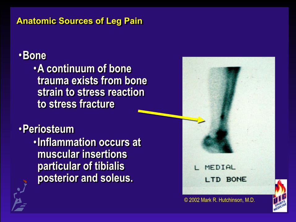
Anatomic Sources of Leg Pain
•Bone •A continuum of bone trauma exists from bone strain to stress reaction to stress fracture
•Periosteum • Inflammation occurs at muscular insertions particular of tibialis posterior and soleus.
© 2002 Mark R. Hutchinson, M.D.
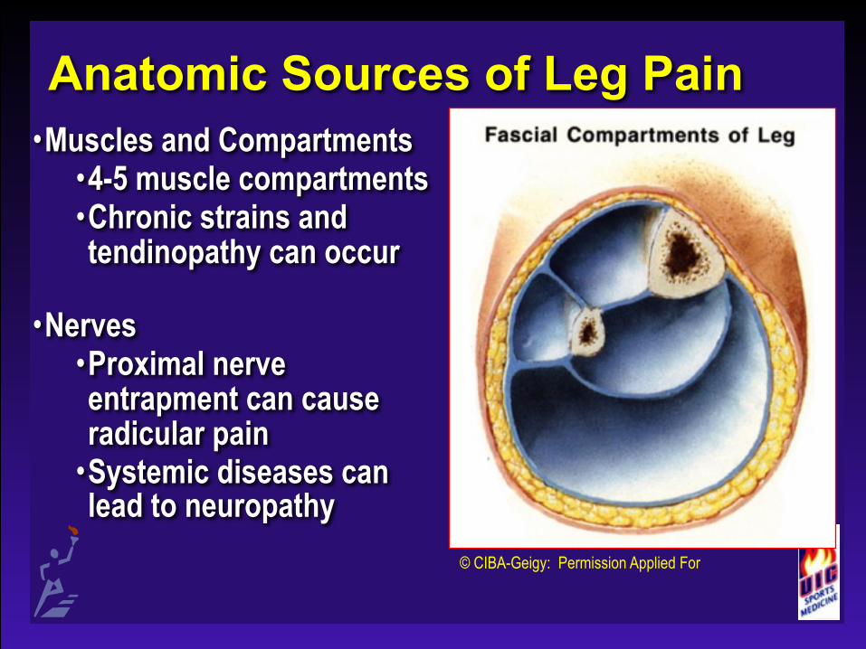
Anatomic Sources of Leg Pain•Muscles and Compartments
•4-5 muscle compartments •Chronic strains and tendinopathy can occur
•Nerves •Proximal nerve entrapment can cause radicular pain
•Systemic diseases can lead to neuropathy
© CIBA-Geigy: Permission Applied For

Anatomic Sources of Leg Pain
•Arteries and Veins •Atherosclerosis can lead to claudication
•Venous phlebitis or thrombosis can occur
•Popliteal artery entrapment and arterial endofibrosis has been described in younger population.
© CIBA-Geigy: Permission Applied For

Differential Dx of Chronic Leg Pain in Athletes• CECS • Muscle herniation • Stress fractures • Medial tibial periostitis
(shin splints) • Chronic muscle strain • Popliteal artery
entrapment • Referred from spine
© 2002 Mark R. Hutchinson, M.D.

Don’t forget the Zebras•Osteosarcoma /Tumors •Trauma /Abuse •Infection including TB, syphilis, bacterial, fungal
•Metabolic •Rickets, Hyperparathyroidism, Sarcoid, Sickle cell, Pagets, etc
© 2002 Mark R. Hutchinson, M.D.

Chronic Exertional Compartment Syndrome
“CECS is an effort induced pathologic elevation of tissue pressures within an osteofascial envelope that results in debilitating pain and neurologic symptoms.”
Leversedge, Am J Sports Med, 2002
© 2002 Mark R. Hutchinson, M.D.
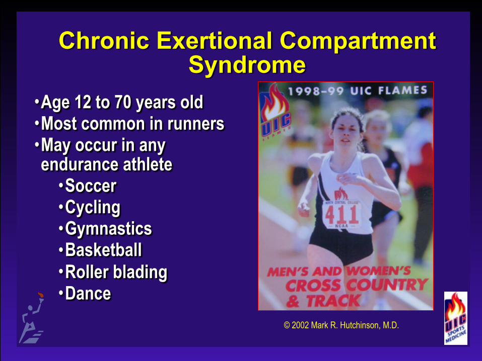
Chronic Exertional Compartment Syndrome
•Age 12 to 70 years old •Most common in runners •May occur in any endurance athlete
•Soccer •Cycling •Gymnastics •Basketball •Roller blading •Dance
© 2002 Mark R. Hutchinson, M.D.

Compartment Syndromes•Traumatic
• Secondary to fracture, crush, and reperfusion injuries
• Surgical emergency• Skin and fascia may both
contribute to compartmental restriction and increased pressure
• Non-physiologic swelling secondary to trauma
• Exertional• Consistently exercise-
induced• Generally endurance athletes• No pain at rest, pain
consistently relieved with cessation of sport
• Attributed to restriction of muscle swelling secondary to tight fascial compartments
• Diagnosed with pre and post exercise pressure measurements

History and Physical:Clinical Pearls in Athletic Leg Pain
•Pain with initial impact •Stress fracture •Periostitis •Muscle strains and tendinitis
•Focal bone pain •Stress fracture
•Diffuse medial bone pain •Medial tibial periostitis
•Focal muscle pain •Strain or Hernia
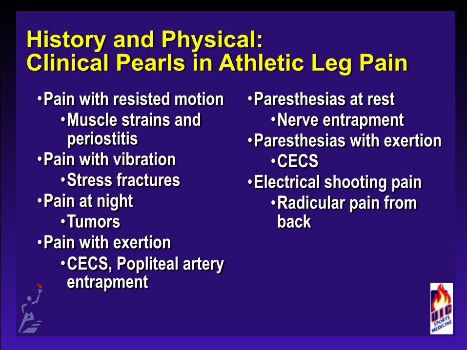
History and Physical:Clinical Pearls in Athletic Leg Pain
•Pain with resisted motion •Muscle strains and periostitis
•Pain with vibration •Stress fractures
•Pain at night •Tumors
•Pain with exertion •CECS, Popliteal artery entrapment
•Paresthesias at rest •Nerve entrapment
•Paresthesias with exertion •CECS
•Electrical shooting pain •Radicular pain from back
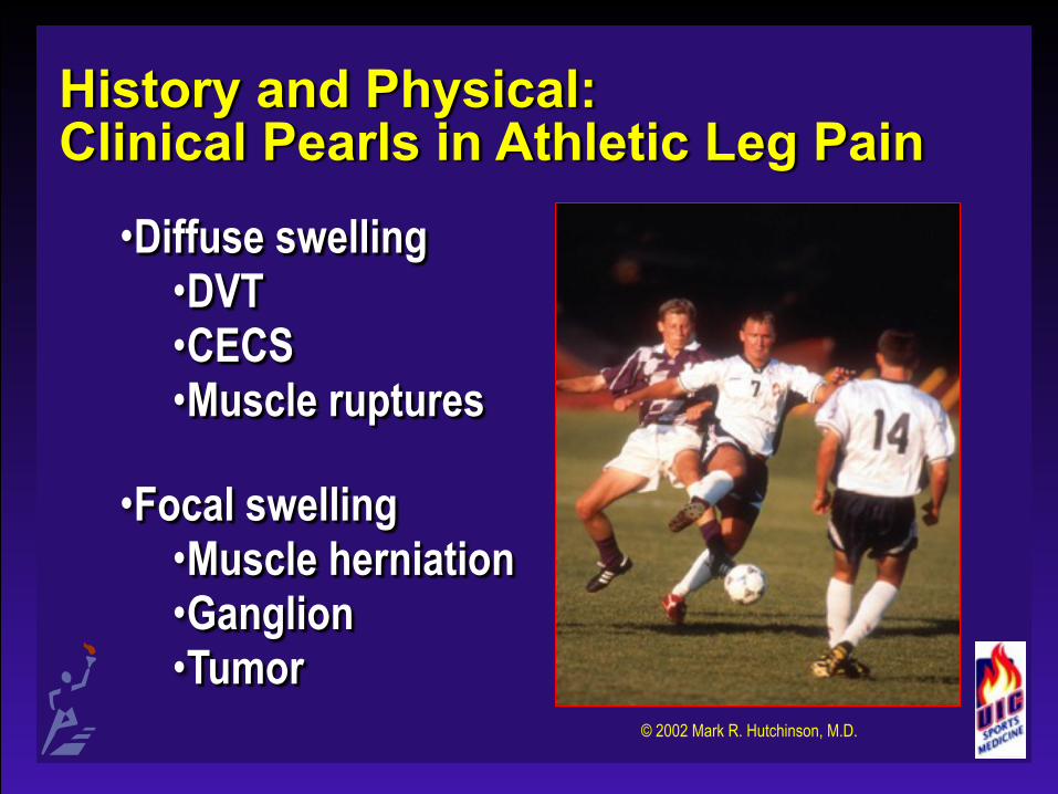
History and Physical:Clinical Pearls in Athletic Leg Pain
•Diffuse swelling •DVT •CECS •Muscle ruptures
•Focal swelling •Muscle herniation •Ganglion •Tumor
© 2002 Mark R. Hutchinson, M.D.
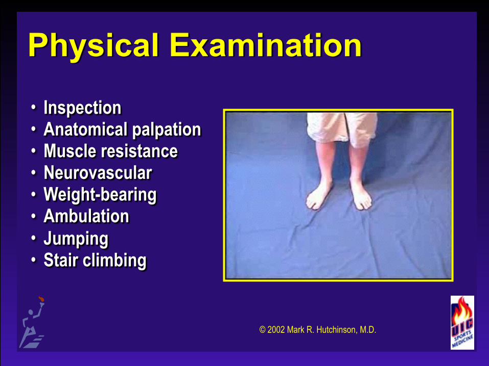
Physical Examination• Inspection • Anatomical palpation • Muscle resistance • Neurovascular • Weight-bearing • Ambulation • Jumping • Stair climbing
© 2002 Mark R. Hutchinson, M.D.
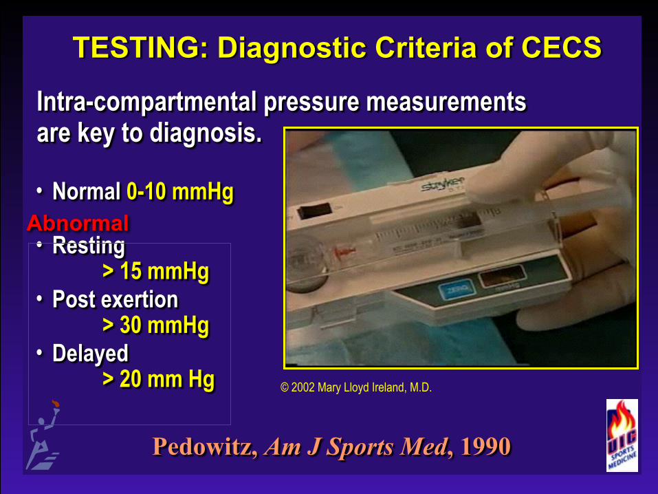
TESTING: Diagnostic Criteria of CECS
Intra-compartmental pressure measurements are key to diagnosis.
• Normal 0-10 mmHg
• Resting > 15 mmHg
• Post exertion > 30 mmHg
• Delayed > 20 mm Hg
Pedowitz, Am J Sports Med, 1990
© 2002 Mary Lloyd Ireland, M.D.
Abnormal

80% of CECS involve the anterior or lateral compartments (Cross-section just above middle of leg)
Flexor digitorum longus m.
TibialisPosterior m.
© CIBA-Geigy: Permission Applied For
Gastrocnemius m.(medial head)
Flexor hallucis longus m.
Soleus m.
Gastrocnemius m.(lateral head)
Extensor digitorum longus m.
Peroneus longus m.
Peroneusbrevis m.
Davey J, Rorabeck C, and Fowler P. The tibialis posterior muscle compartment. Am J Sports Med 12(5):391-397, 1984.

Nerves (Cross-section just above middle of leg)
SuperficialPeroneal n.
Anterior tibial a. and v. and deep peroneal n.
Posterior tibial a. and vv. and tibial n.
Lateral suralCutaneous n.
Medial suralCutaneous n.
© CIBA-Geigy: Permission Applied For
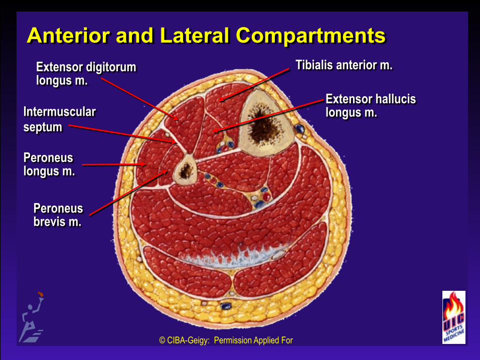
Anterior and Lateral Compartments
Intermuscularseptum
© CIBA-Geigy: Permission Applied For
Tibialis anterior m.
Extensor hallucis longus m.
Extensor digitorum longus m.
Peroneus longus m.
Peroneusbrevis m.

Posterior Compartments: Deep and Superficial
TibialisPosterior m.
© CIBA-Geigy: Permission Applied For
Flexor digitorum longus m.
Gastrocnemius m.(medial head)
Flexor hallucis longus m.
Soleus m.
Gastrocnemius m.(lateral head)
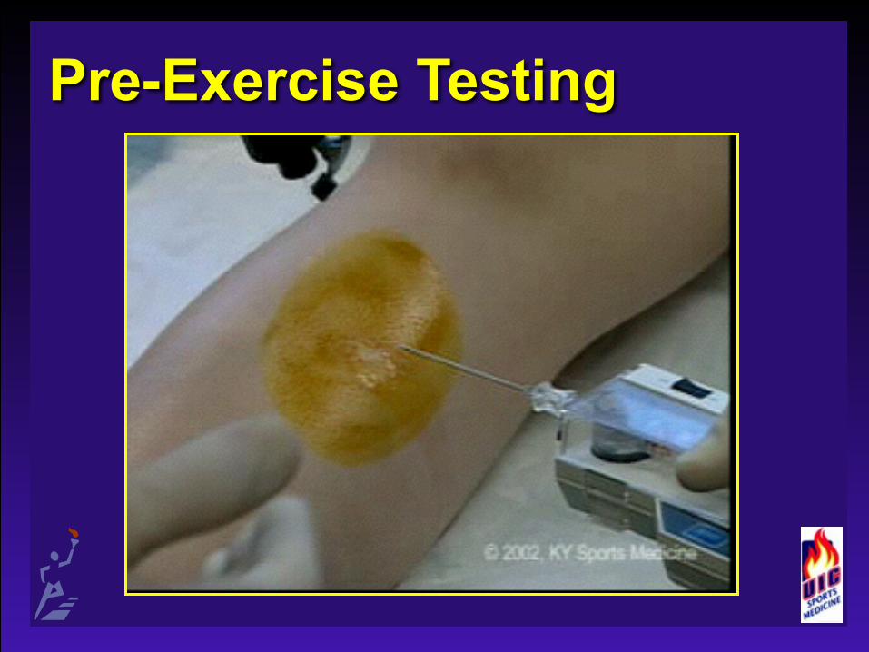
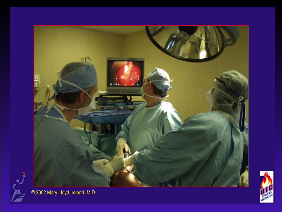
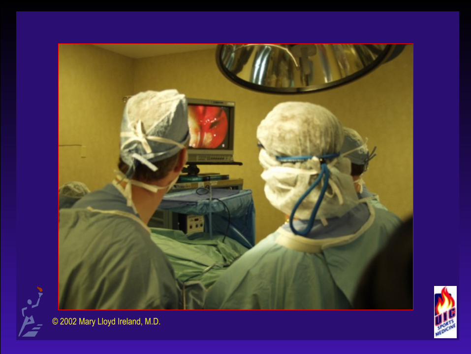
Pre-Exercise Testing

Exercise and Post-Exercise Testing

TESTING: Diagnostic Criteria of CECS
Intracompartmental pressure measurements are key to diagnosis.
Pedowitz, Am J Sports Med, 1990
Normal 0-10 mmHg Resting > 15 mmHg Post exertion > 30mmHg Delayed > 20 mm Hg
ABNORMAL

Treatment Options for CECS• NSAIDS (-) • Massage (-) • Rest (±) • Stretching and strengthening (-) • Modalities (-) • Shoe and surface modification (±) • Fascial release (+)
You must rule out associated factors or diagnoses.
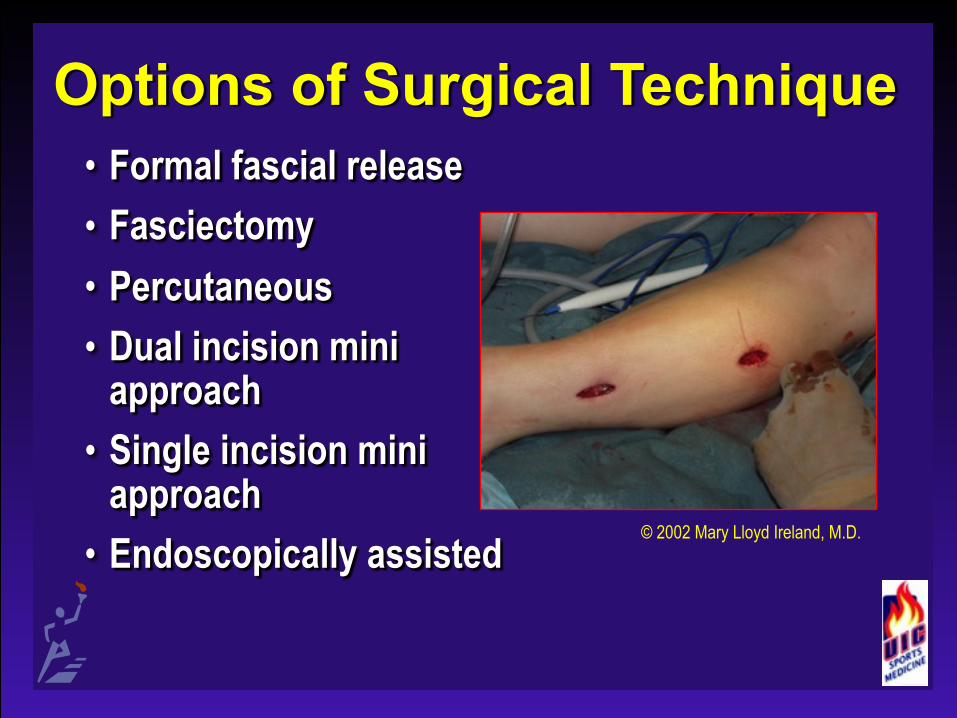
Options of Surgical Technique• Formal fascial release • Fasciectomy • Percutaneous • Dual incision mini
approach • Single incision mini
approach • Endoscopically assisted
© 2002 Mary Lloyd Ireland, M.D.
Development of Endoscopic-Assisted Fascial Releases for CECS
• First described as technique for leg releases as case report in 1999
• Oto et al., Arthroscopy, 1999 • First described as forearm release in vitro study in
1999 • Havig & Leversedge, J Hand Surg, 1999
• First description of 2-incision endoscopic technique in cadaveric study in 2002
• Leversedge et al, Am J Sports Med, 2002
Development of Endoscopic-Assisted Fascial Releases for CECS at UIC
• In 1996, we performed our first endoscopically-assisted fascial release on a young athlete in an aesthetically demanding sport.
• Briner, Hutchinson et al, ACSM Annual Meeting, 1998 • Subsequent embalmed and fresh cadaveric studies
identified the risk and efficacy of the procedure. • Hutchinson MR, Bederka B. AOSSM Annual Meeting,
2000

Results of Anatomic Studies10 cadaveric legs – endoscopic technique 6 cadaveric legs – percutaneous via minimal single incision
• Length of release • Anterior: 210 ± 28 mm • Lateral: 171 ± 27 mm • Sup posterior 189 ± 23 mm • Deep posterior 154 ± 28 mm © 2002 Mark R. Hutchinson, M.D.
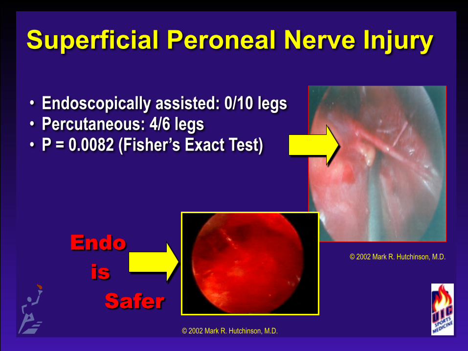
Superficial Peroneal Nerve Injury
• Endoscopically assisted: 0/10 legs • Percutaneous: 4/6 legs • P = 0.0082 (Fisher’s Exact Test)
Endo is Safer
© 2002 Mark R. Hutchinson, M.D.
© 2002 Mark R. Hutchinson, M.D.
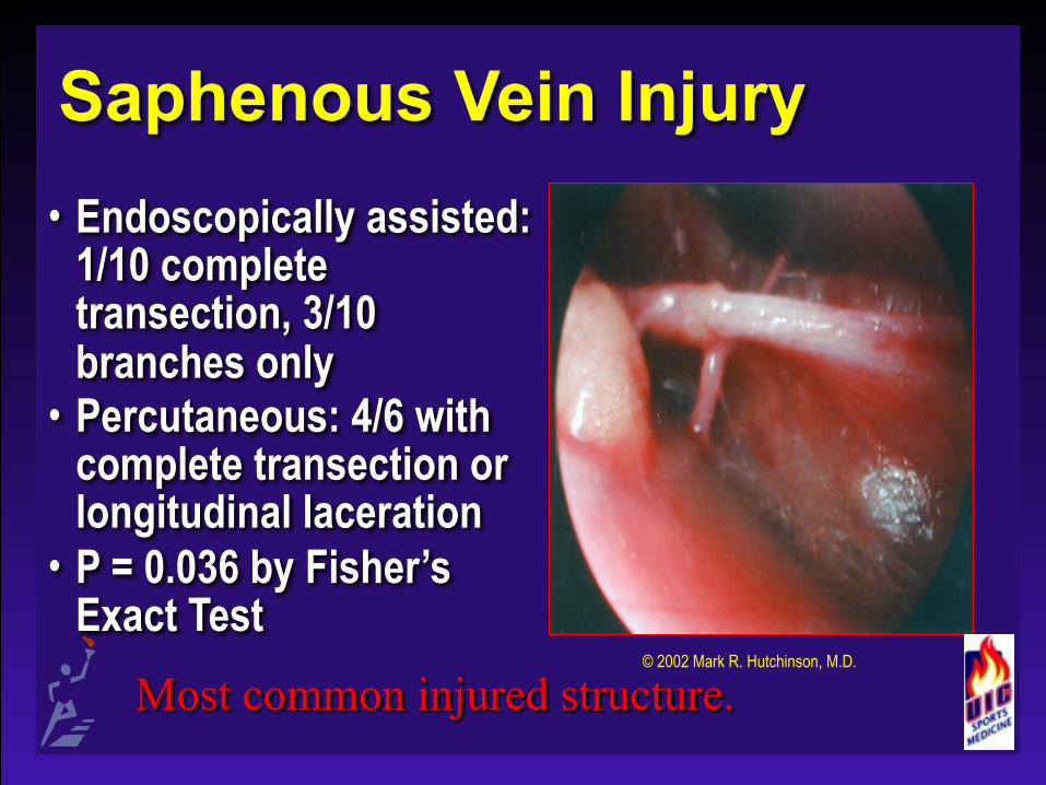
Saphenous Vein Injury• Endoscopically assisted:
1/10 complete transection, 3/10 branches only
• Percutaneous: 4/6 with complete transection or longitudinal laceration
• P = 0.036 by Fisher’s Exact Test
Most common injured structure.© 2002 Mark R. Hutchinson, M.D.
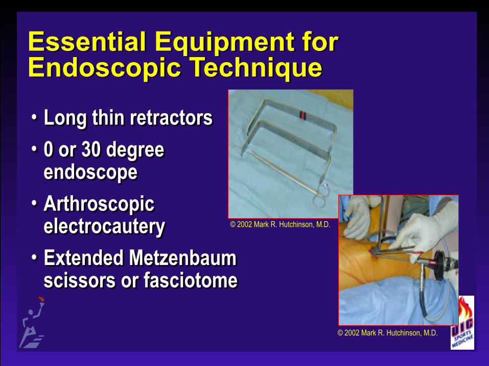
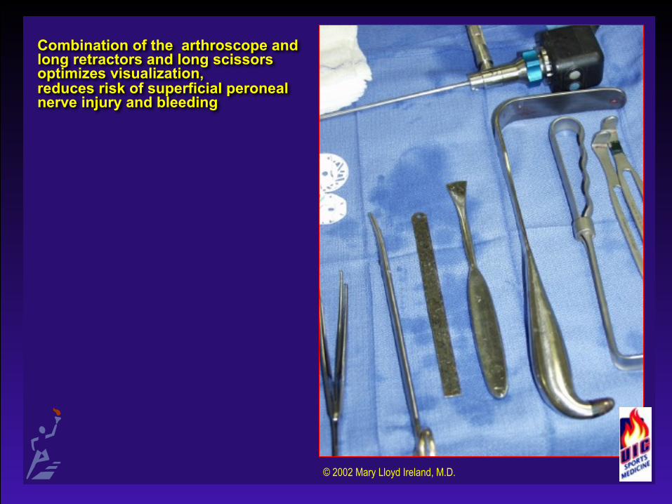
Essential Equipment for Endoscopic Technique• Long thin retractors • 0 or 30 degree
endoscope • Arthroscopic
electrocautery • Extended Metzenbaum
scissors or fasciotome
© 2002 Mark R. Hutchinson, M.D.
© 2002 Mark R. Hutchinson, M.D.

Endoscopically Assisted One-Incision Fascial Release
© 2002 Mark R. Hutchinson, M.D.
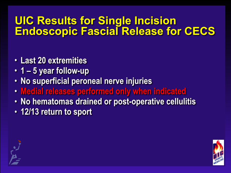
UIC Results for Single Incision Endoscopic Fascial Release for CECS
• Last 20 extremities • 1 – 5 year follow-up • No superficial peroneal nerve injuries • Medial releases performed only when indicated • No hematomas drained or post-operative cellulitis • 12/13 return to sport
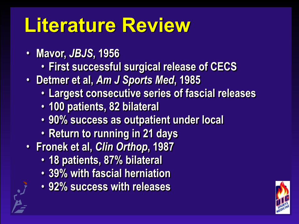
Literature Review• Mavor, JBJS, 1956
• First successful surgical release of CECS • Detmer et al, Am J Sports Med, 1985
• Largest consecutive series of fascial releases • 100 patients, 82 bilateral • 90% success as outpatient under local • Return to running in 21 days
• Fronek et al, Clin Orthop, 1987 • 18 patients, 87% bilateral • 39% with fascial herniation • 92% success with releases
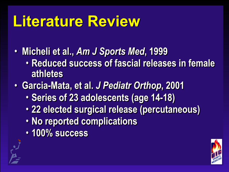
Literature Review
• Micheli et al., Am J Sports Med, 1999 • Reduced success of fascial releases in female
athletes • Garcia-Mata, et al. J Pediatr Orthop, 2001
• Series of 23 adolescents (age 14-18) • 22 elected surgical release (percutaneous) • No reported complications • 100% success
KQ27435Surgery.mpg
#27435
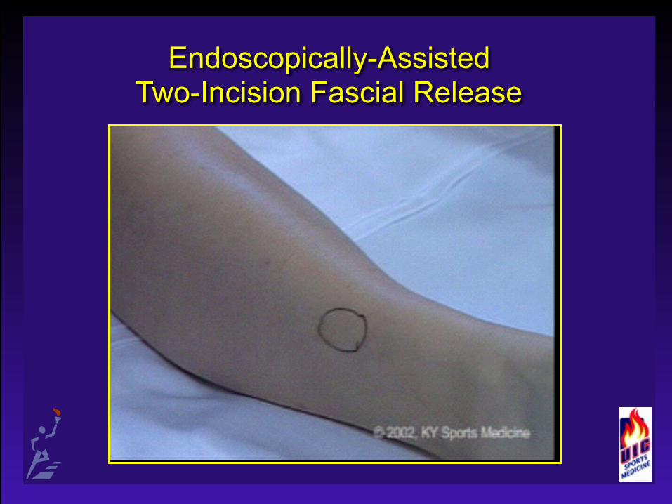
Endoscopically-Assisted Two-Incision Fascial Release
© 2002 Mary Lloyd Ireland, M.D.
© 2002 Mary Lloyd Ireland, M.D.

40% Have a Fascial Herniation
• If present, look for superficial peroneal nerve exiting there
• Begin release at that level • Never close fascia— Rorabeck C, Bourne R, Fowler P, Finlay J, and Nott, L. The role of tissue pressure measurement in diagnosing chronic anterior compartment syndrome. Am J Sports Med 16(2):143-146, 1988.
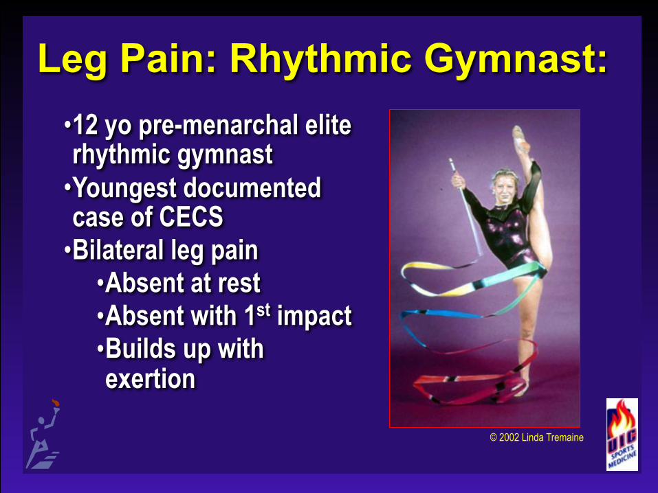
Leg Pain: Rhythmic Gymnast:•12 yo pre-menarchal elite rhythmic gymnast
•Youngest documented case of CECS
•Bilateral leg pain •Absent at rest •Absent with 1st impact •Builds up with exertion
© 2002 Linda Tremaine

Leg Pain: Rhythmic Gymnast•Failed conservative tx
•Massage •Nutrition •Hydration •NSAIDS •Therapy
•Imaging: negative •Radiographs •MRI •Bone scan
© 2002 Linda Tremaine
© 2002 Mark R. Hutchinson, M.D.

Leg Pain: Rhythmic Gymnast
• Compartment measurements
• Resting: All compartments < 15
• Post exertion: • Anterior (R32/L35) • Lateral (R31/L30) • D post (R24/L22) • Sup (R12/L13)
© 2002 Linda Tremaine

Leg Pain: Rhythmic Gymnast• Treatment:
• Let her grow out of it? • Endoscopically assisted fascial release
elected secondary to competitive demands
• Results • Full, pain free competition at 3 months • Team Gold, Individual Bronze at 4-
Continents at 6 months
#26614
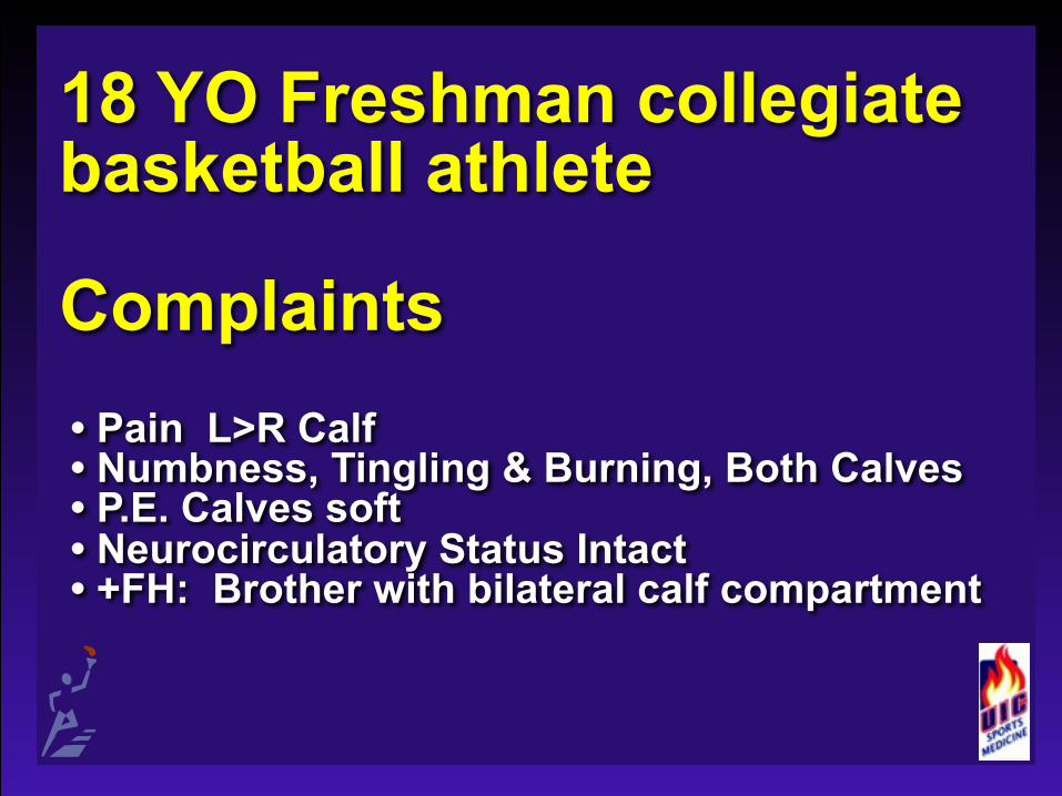
18 YO Freshman collegiate basketball athlete Complaints • Pain L>R Calf • Numbness, Tingling & Burning, Both Calves • P.E. Calves soft • Neurocirculatory Status Intact • +FH: Brother with bilateral calf compartment

Differential Diagnosis
• Exertional compartment syndrome, left leg
• Stress fracture, left tibia

Clinical Course
• Passive stretching and reduction of running activities
• 2 months later, patient returned with continued symptoms; exam unchanged; compartments were soft.
• Patient underwent Stryker testing of bilateral compartments, pre- and post-exercise.

Stryker Compartment Testing
Anterior Posterior Resting Post Exercise Resting Post-Exercise
R 8 62 Not measured
L 20 42 8 16
Nv26614.mpg

© 2002 Mary Lloyd Ireland, M.D.

© 2002 Mary Lloyd Ireland, M.D.

© 2002 Mary Lloyd Ireland, M.D.

Post Op Course
• Crutches, PWB 2 wks. • Walked Normally, 4 wks. • Running, 2 months.
6 Months Post Op
• Bilateral Anterior and Lateral Releases
• No Complaints • Playing Basketball • Compartments Soft, without Bulge
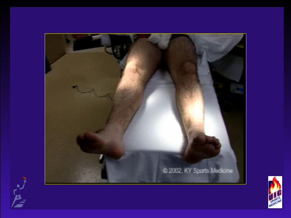
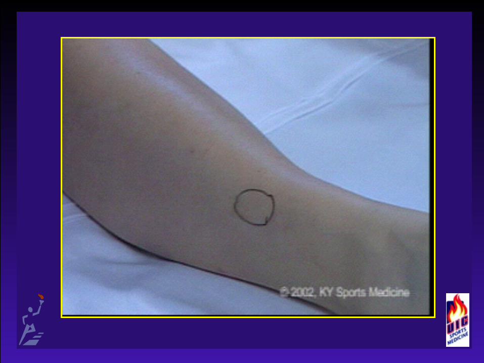
13 YO Female Soccer Athlete
• Bilateral Calf Pain, R>L • Past 6 Months • C/O Knots and Burning Sensation,
Lateral Calf, After 15 Minutes of Running

P.E.
• No Pain or Firmness on Calf Palpation
• Fascial Defect Lateral Distal Third • Neurocirculatory Status Intact • Bilateral Cavus Feet
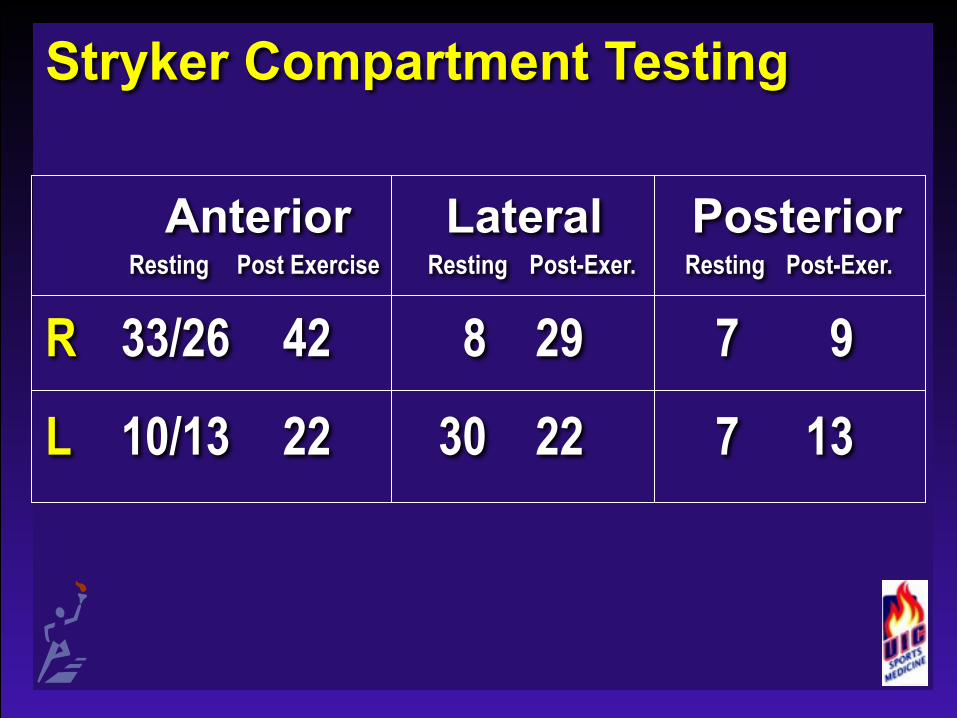
Stryker Compartment Testing
Anterior Lateral Posterior Resting Post Exercise Resting Post-Exer. Resting Post-Exer.
R 33/26 42 8 29 7 9
L 10/13 22 30 22 7 13
#27435
KQ27435Surgery.mpg
#27435
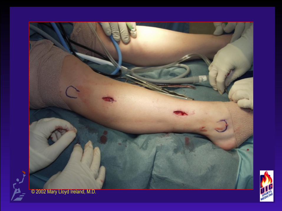
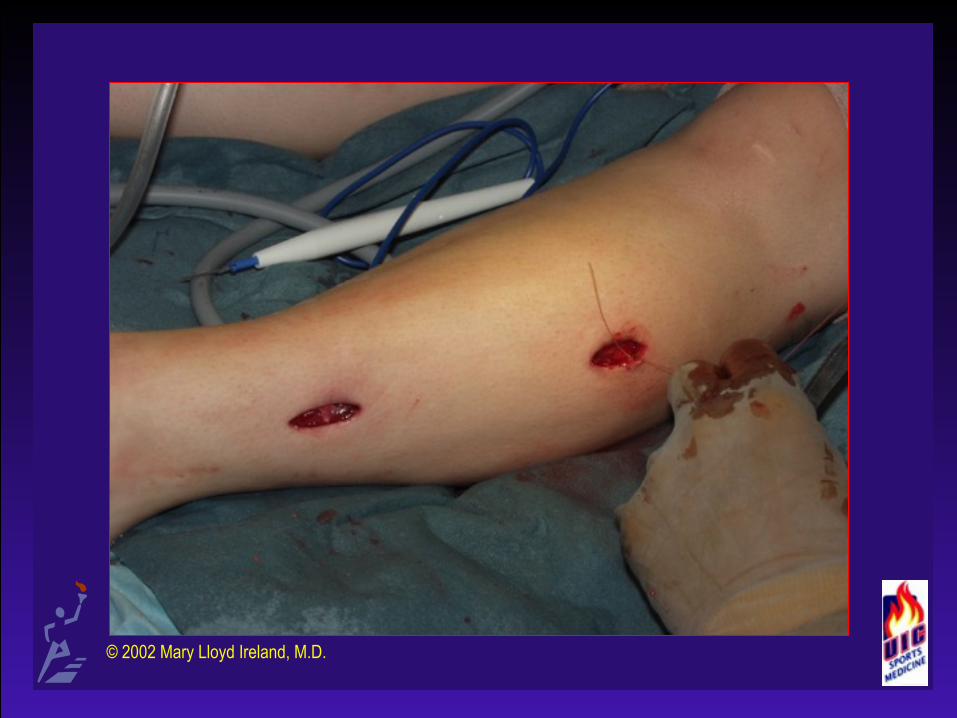
Two-Incision
• Releases Anterior and Lateral Compartment
• Doing Well Post Op • Pain Improved

© 2002 Mary Lloyd Ireland, M.D.
© 2002 Mary Lloyd Ireland, M.D.
© 2002 Mary Lloyd Ireland, M.D.
© 2002 Mary Lloyd Ireland, M.D.
© 2002 Mary Lloyd Ireland, M.D.
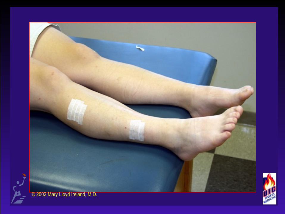
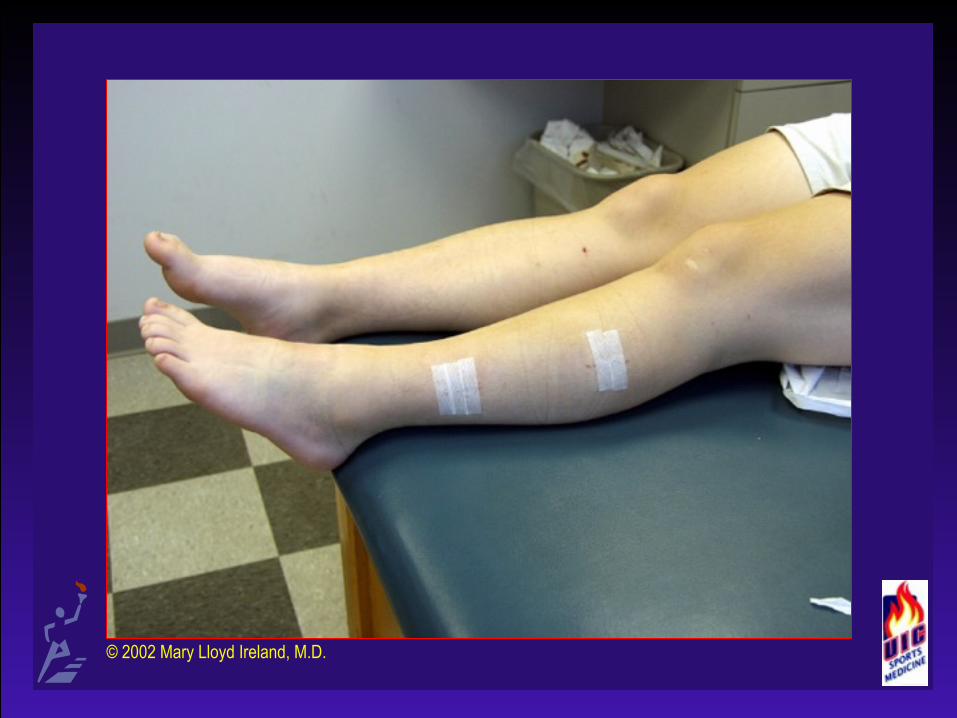
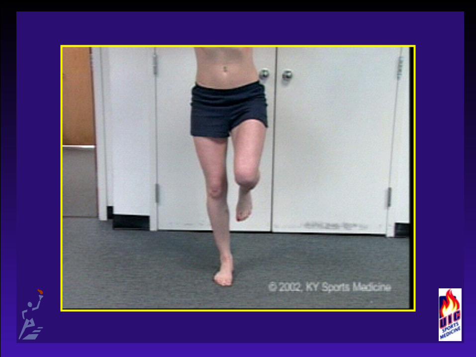
19 YO Freshman Division I Collegiate Cross-Country and Distance Track Athlete
• C/O Pain in Both Calves After Running • Began Cross-Country at Age 10 • No Previous Complaints • Running More Miles, and on Concrete

P.E.
• No Firmness to Compartments or Tibia to Palpation
• Normal Pulses & Neurologic Exam
Previous Workup
• Normal Tib-Fib X-rays • Normal Bone Scan • Lumbar Spine X-rays and MRI Normal • Resting Pressures
• Lateral 12mm • Posterior 10mm
• With Indwelling Needle, Unable to Reproduce Symptoms Due to Pain
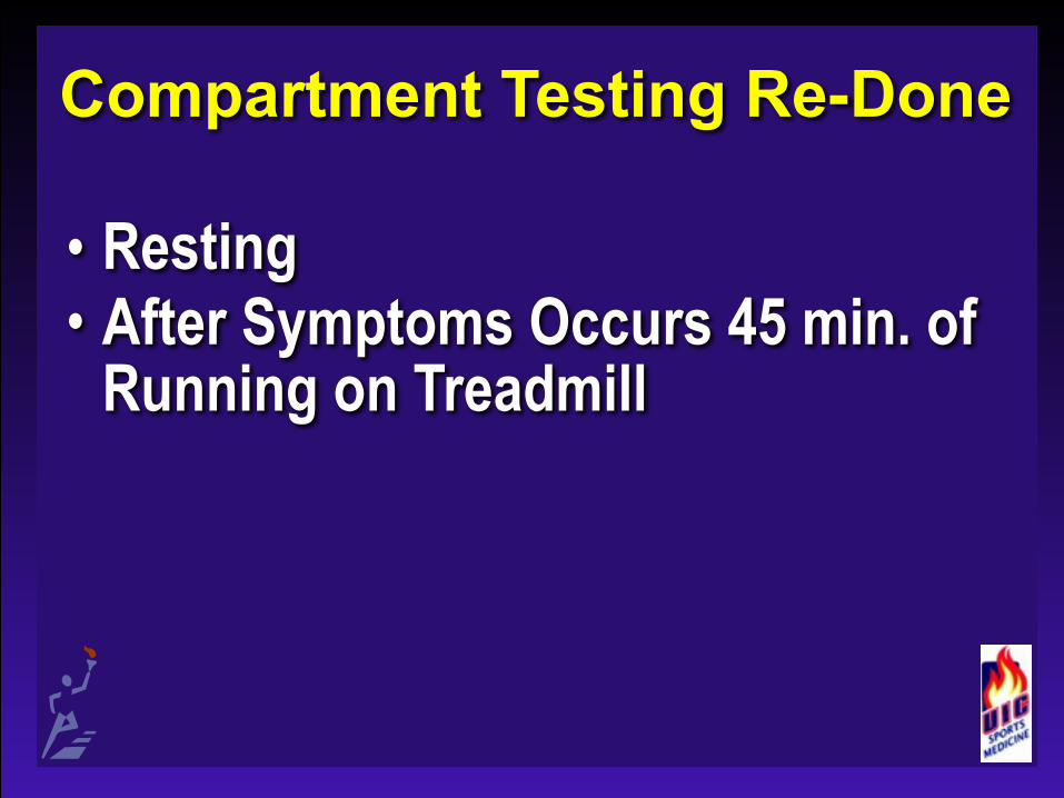
Compartment Testing Re-Done
• Resting • After Symptoms Occurs 45 min. of
Running on Treadmill
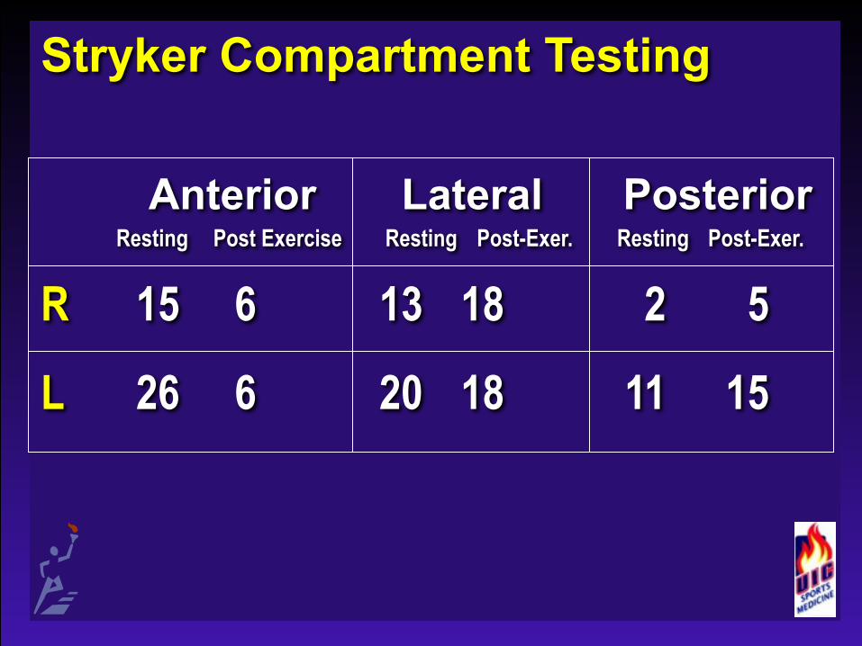
Stryker Compartment Testing
Anterior Lateral Posterior Resting Post Exercise Resting Post-Exer. Resting Post-Exer.
R 15 6 13 18 2 5
L 26 6 20 18 11 15
#27376
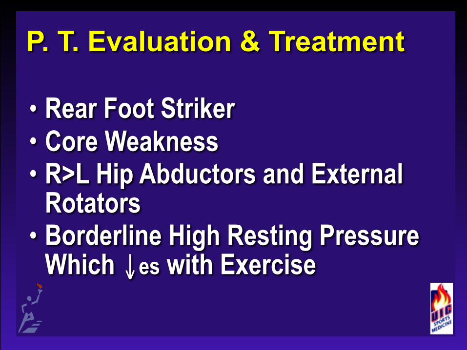
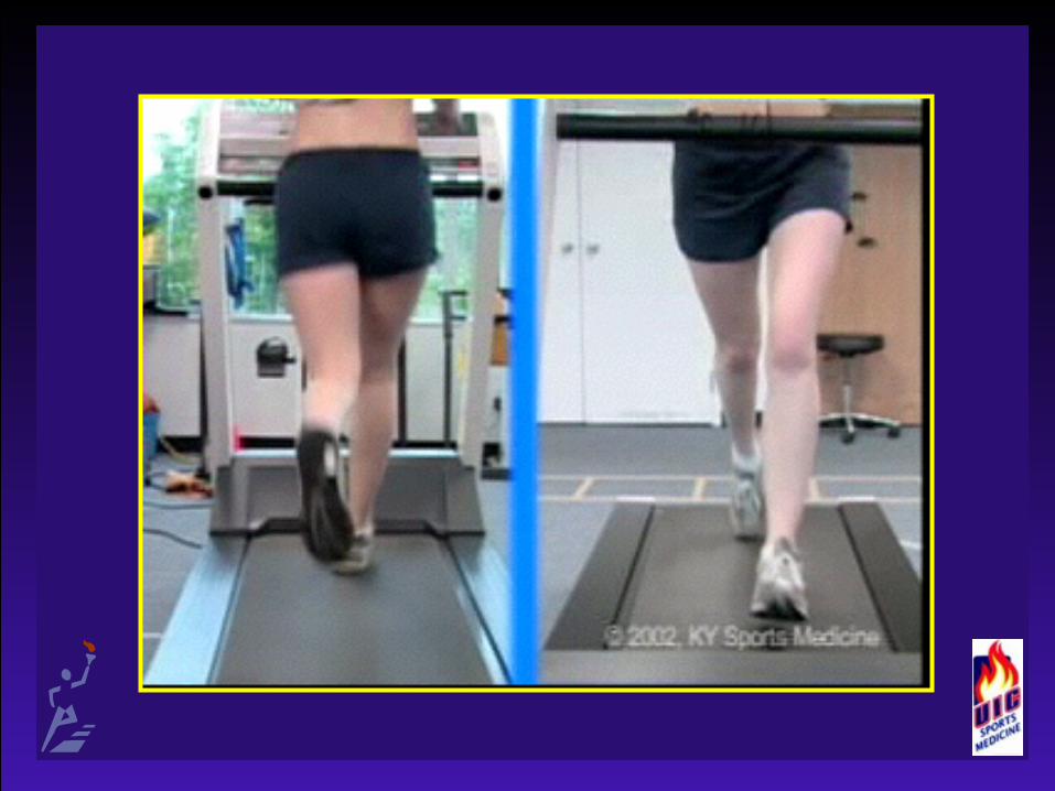
P. T. Evaluation & Treatment
• Rear Foot Striker • Core Weakness • R>L Hip Abductors and External
Rotators • Borderline High Resting Pressure
Which ↓es with Exercise
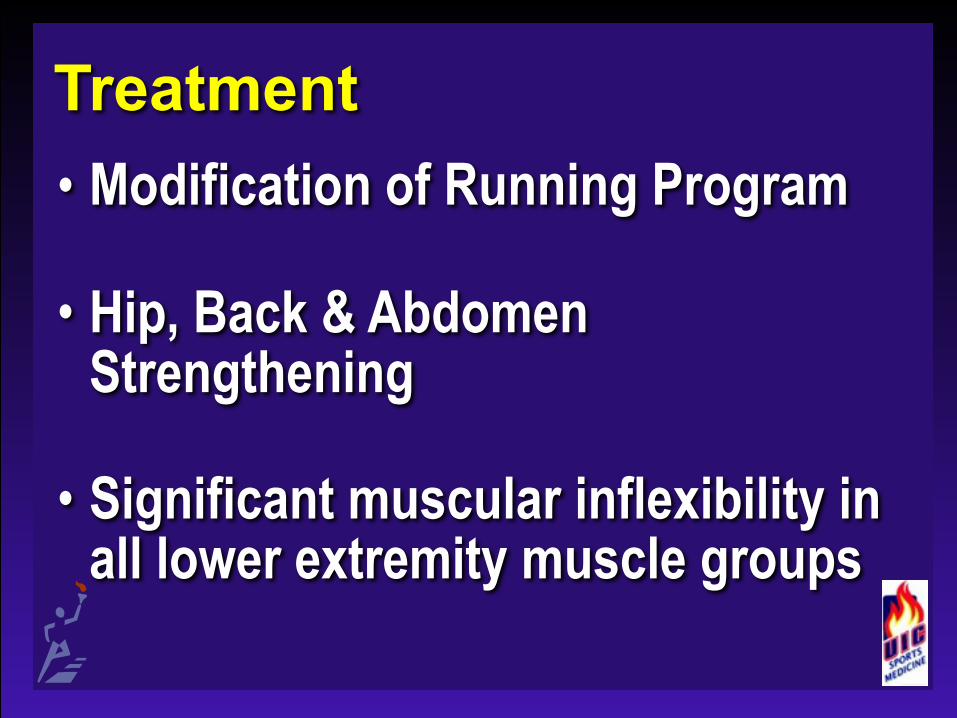
Treatment• Modification of Running Program
• Hip, Back & Abdomen Strengthening
• Significant muscular inflexibility in all lower extremity muscle groups
#27376
Conclusions
• Diagnosis of compartment syndrome must be confirmed by compartmental testing before and after exercise
• Consider broad range of diagnoses for athletes with leg pain.
• Make sure of the diagnosis prior to surgical release

© 2002 Mary Lloyd Ireland, M.D.SuperficialPeroneal n.
Anterior tibial a. and v. and deep peroneal n.
Know the anatomy before performing compartment testing or surgical releases.
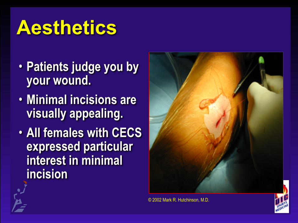
Aesthetics
• Patients judge you by your wound.
• Minimal incisions are visually appealing.
• All females with CECS expressed particular interest in minimal incision
© 2002 Mark R. Hutchinson, M.D.
© 2002 Mary Lloyd Ireland, M.D.
Combination of the arthroscope and long retractors and long scissors optimizes visualization, reduces risk of superficial peroneal nerve injury and bleeding

Clinical Pearls• Always visualize the
superficial peroneal nerve • Confirm dx with
compartment measurements
• Avoid medial release unless indicated
• Diligent intra-operative bleeding control without tourniquet
• Post-operative cryotherapy
© 2002 Mark R. Hutchinson, M.D.

Future Directions• Increasing awareness of problem
in all providers • Does minimal incision surgery allow adequate
fascial release? • What is adequate release? • Non-invasive testing to confirm the diagnosis
(Near infrared spectroscopy)
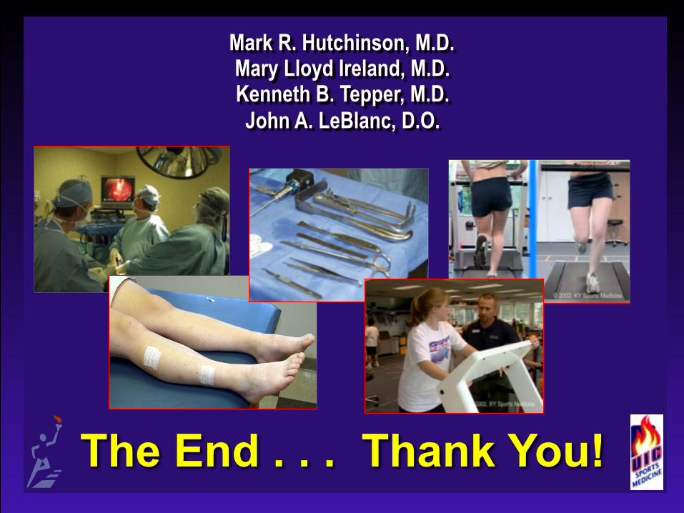
The End . . . Thank You!
Mark R. Hutchinson, M.D. Mary Lloyd Ireland, M.D. Kenneth B. Tepper, M.D.John A. LeBlanc, D.O.