chromosome
TRANSCRIPT
CHROMOSOME
MANJUNATH,G.A
2013-11-176
WHAT EXACTLY IS A CHROMOSOME?
Chromosomes are the rod-shaped, filamentous bodies present in the nucleus, which become visible during cell division.
They are the carriers of the gene or unit of heredity.
Chromosome are not visible in active nucleus due to their high water content, but are clearly seen during cell division.
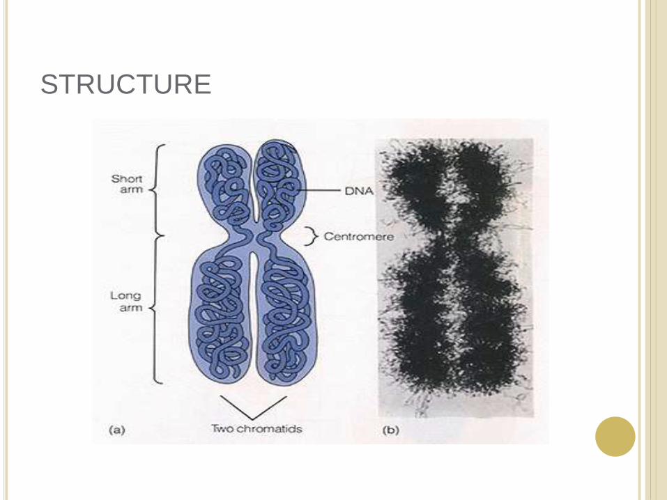
STRUCTURE

Chromosomes were first described by Strausberger in 1875.
The term “Chromosome”, however was first used by Waldeyer in 1888.
They were given the name chromosome (Chromo = colour; Soma = body) due to their marked affinity for basic dyes.
Their number can be counted easily only during mitotic metaphase.

NUMBER OF CHROMOSOMES
Normally, all the individuals of a species have the same number of chromosomes.
Closely related species usually have similar chromosome numbers.
Presence of a whole sets of chromosomes is called euploidy.
It includes haploids, diploids, triploids, tetraploids etc.
Gametes normally contain only one set of chromosome – this number is called Haploid
Somatic cells usually contain two sets of chromosome - 2n : Diploid
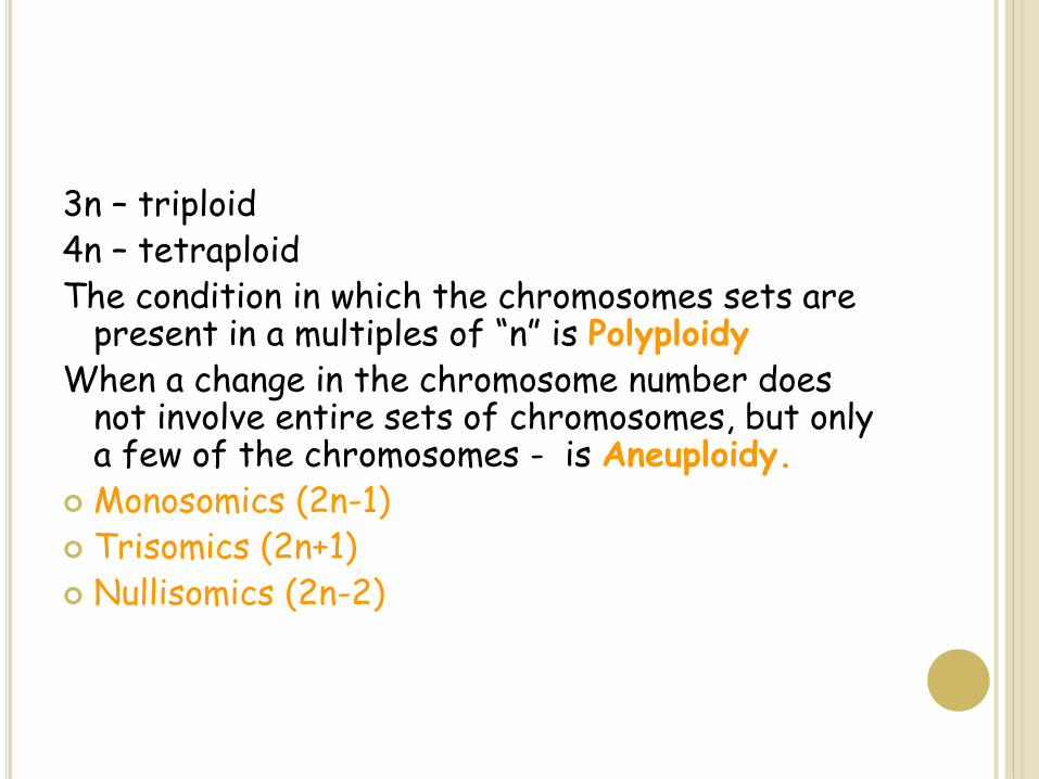
3n – triploid4n – tetraploidThe condition in which the chromosomes sets are
present in a multiples of “n” is PolyploidyWhen a change in the chromosome number does
not involve entire sets of chromosomes, but only a few of the chromosomes - is Aneuploidy.
Monosomics (2n-1) Trisomics (2n+1) Nullisomics (2n-2)
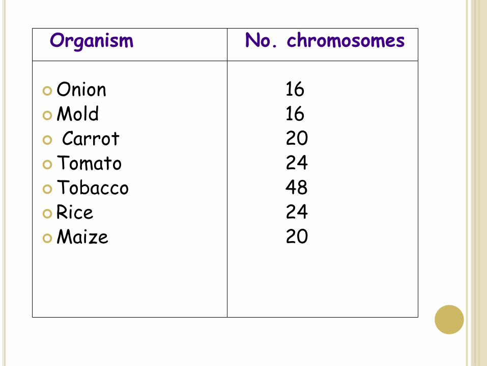
Organism No. chromosomes
Onion 16Mold 16 Carrot 20Tomato 24Tobacco 48Rice 24Maize 20

CHROMOSOME SIZE
In contrast to other cell organelles, the size of chromosomes shows a remarkable variation depending upon the stages of cell division.
Interphase: chromosome are longest & thinnest
Prophase: there is a progressive decrease in their length accompanied with an increase in thickness
Anaphase: chromosomes are smallest.
Metaphase: Chromosomes are the most easily observed and studied during metaphase when they are very thick, quite short and well spread in the cell.
Therefore, chromosomes measurements are generally taken during mitotic metaphase.

KARYOTYPE AND IDIOTYPE
Karyotype : is the general morphology of the somatic chromosome. Generally, karyotypesrepresent by arranging in the descending order of size keeping their centromeres in a straight line.
Idiotype: the karyotype of a species may be represented diagrammatically, showing all the morphological features of the chromosome; such a diagram is known as Idiotype.

CHROMATIN
The complexes between eukaryotic DNA and
proteins are called Chromatin, which typically
contains about twice as much protein as DNA.
The major proteins of chromatin are the histones –
small proteins containing a high proportion of basic
aminoacids (arginine and lysine) that facilitate
binding negatively charged DNA molecule .
There are 5 major types of histones: H1, H2A, H2B,
H3, and H4 – which are very similar among different
sp of eukaryotes.
The histones are extremely abundant proteins in
eukaryotic cells.

CENTROMERE
The region where two sister chromatids of a chromosome appear to be joined or “held together” during mitatic metaphase is called Centromere
When chromosomes are stained they typically show a dark-stained region that is the centromere.
Also termed as Primary constriction
During mitosis, the centromere that is shared by the sister chromatids must divide so that the chromatids can migrate to opposite poles of the cell.
On the other hand, during the first meiotic division the centromere of sister chromatids must remain intact
whereas during meiosis II they must act as they do during mitosis.
Therefore the centromere is an important component of chromosome structure and segregation.

KINETOCHORE
Within the centromere region, most species have several
locations where spindle fibers attach, and these sites
consist of DNA as well as protein.
The actual location where the attachment occurs is called
the kinetochore and is composed of both DNA and
protein.
The DNA sequence within these regions is called CEN
DNA.

TELOMERE
The two ends of a chromosome are known as telomeres.
It required for the replication and stability of the chromosome.
When telomeres are damaged or removed due to chromosome breakage, the damaged chromosome ends can readily fuse or unite with broken ends of other chromosome.

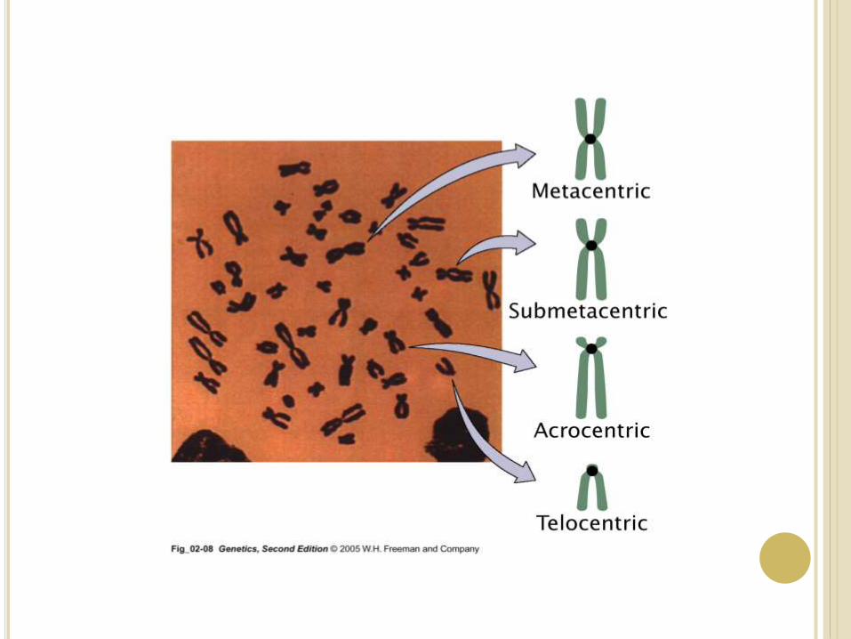
Chromosomes may differ in the position of
the Centromere, the place on the chromosome where
spindle fibers are attached during cell division.
In general, if the centromere is near the middle, the
chromosome is metacentric
If the centromere is toward one end, the chromosome is
acrocentric or submetacentric
If the centromere is very near the end, the chromosome
is telocentric.
EUCHROMATIN AND HETEROCHROMATIN
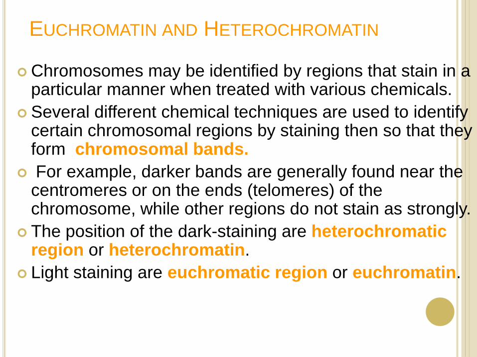
Chromosomes may be identified by regions that stain in a particular manner when treated with various chemicals.
Several different chemical techniques are used to identify certain chromosomal regions by staining then so that they form chromosomal bands.
For example, darker bands are generally found near the centromeres or on the ends (telomeres) of the chromosome, while other regions do not stain as strongly.
The position of the dark-staining are heterochromatic region or heterochromatin.
Light staining are euchromatic region or euchromatin.
Heterochromatin is classified into two groups: (i)
Constitutive and (ii) Facultative.
Constitutive heterochromatin remains permanently in the
heterochromatic stage, i.e., it does not revert to the
euchromatic stage.
In contrast, facultative heterochromatin consists of
euchromatin that takes on the staining and compactness
characteristics of heterochromatin during some phase of
development.


CHROMOSOMAL ABERRATIONS
Sometime due to mutation or spontaneous (without
any known causal factors), variation in
chromosomal number or structure do arise in
nature. - Chromosomal aberrations.
Chromosomal aberration may be grouped into two
broad classes:
1. Structural and 2. Numerical

STRUCTURAL CHROMOSOMAL
ABERRATIONS Chromosome structure variations result from chromosome
breakage.
Broken chromosomes tend to re-join; if there is more than one
break, rejoining occurs at random and not necessarily with the
correct ends.
The result is structural changes in the chromosomes.
Chromosome breakage is caused by X-rays, various
chemicals, and can also occur spontaneously.

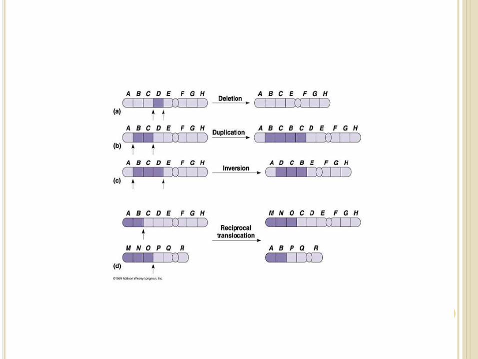
There are four common type of structural aberrations:
1. Deletion or Deficiency
2. Duplication or Repeat
3. Inversion, and
4. Translocation.
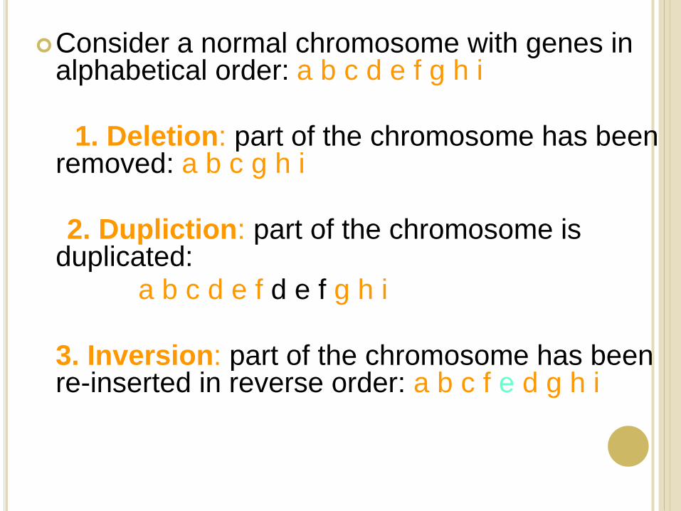
Consider a normal chromosome with genes in alphabetical order: a b c d e f g h i
1. Deletion: part of the chromosome has been removed: a b c g h i
2. Dupliction: part of the chromosome is duplicated:
a b c d e f d e f g h i
3. Inversion: part of the chromosome has been re-inserted in reverse order: a b c f e d g h i
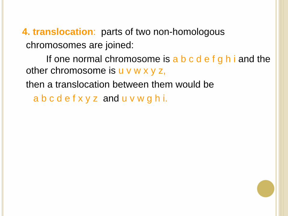
4. translocation: parts of two non-homologous
chromosomes are joined:
If one normal chromosome is a b c d e f g h i and the
other chromosome is u v w x y z,
then a translocation between them would be
a b c d e f x y z and u v w g h i.
Deletion or deficiency
Loss of a chromosome segment is known as deletion or deficiency
It can be terminal deletion or interstitial or intercalarydeletion.
A single break near the end of the chromosome would be expected to result in terminal deficiency.
If two breaks occur, a section may be deleted and an intercalary deficiency created.
Terminal deficiencies might seem less complicated.
But majority of deficiencies detected are intercalary type within the chromosome.
Deletion was the first structural aberration detected by Bridges in 1917 from his genetic studies on X chromosome of Drosophila.

Deletion in Prokaryotes:
Deletions are found in prokaryotes as well, e.g.,
E.coli, T4 phage and Lambda phage.
In E.coli, deletions of up to 1 % of the bacterial
chromosome are known.
In lambda phage, however 20% of the genome may be
missing in some of the deletions.
Deletion in Human:
Chromosome deletions are usually lethal even as
heterozygotes, resulting in zygotic loss, stillbirths, or
infant death.
Sometimes, infants with small chromosome
deficiencies however, survive long enough to permit
the abnormal phenotype they express.
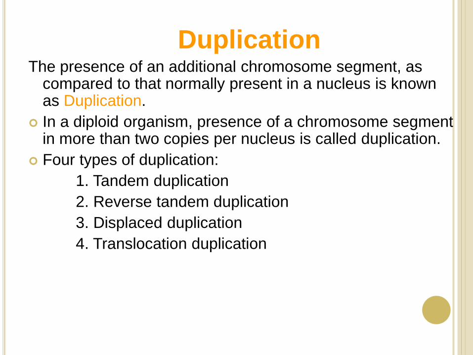
DuplicationThe presence of an additional chromosome segment, as
compared to that normally present in a nucleus is known as Duplication.
In a diploid organism, presence of a chromosome segment in more than two copies per nucleus is called duplication.
Four types of duplication:
1. Tandem duplication
2. Reverse tandem duplication
3. Displaced duplication
4. Translocation duplication

The extra chromosome segment may be located
immediately after the normal segment in precisely the same
orientation forms the tandem
When the gene sequence in the extra segment of a tandem
in the reverse order i.e, inverted , it is known as reverse
tandem duplication
In some cases, the extra segment may be located in the
same chromosome but away from the normal segment –
termed as displaced duplication
The additional chromosome segment is located in a non-
homologous chromosome is translocation duplication.

Inversion
When a segment of chromosome is oriented in the reverse direction, such segment said to be inverted and the phenomenon is termed as inversion.
The existence of inversion was first detected by Strutevant and Plunkett in 1926.
Inversion occur when parts of chromosomes become detached , turn through 1800 and are reinserted in such a way that the genes are in reversed order.
For example, a certain segment may be broken in two places, and the breaks may be in close proximity because of chance loop in the chromosome.
When they rejoin, the wrong ends may become connected.
The part on one side of the loop connects with broken end different from the one with which it was formerly connected.
This leaves the other two broken ends to become attached.
The part within the loop thus becomes turned around or inverted.
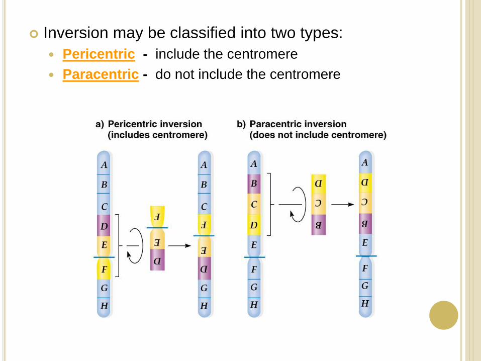
Inversion may be classified into two types:
Pericentric - include the centromere
Paracentric - do not include the centromere
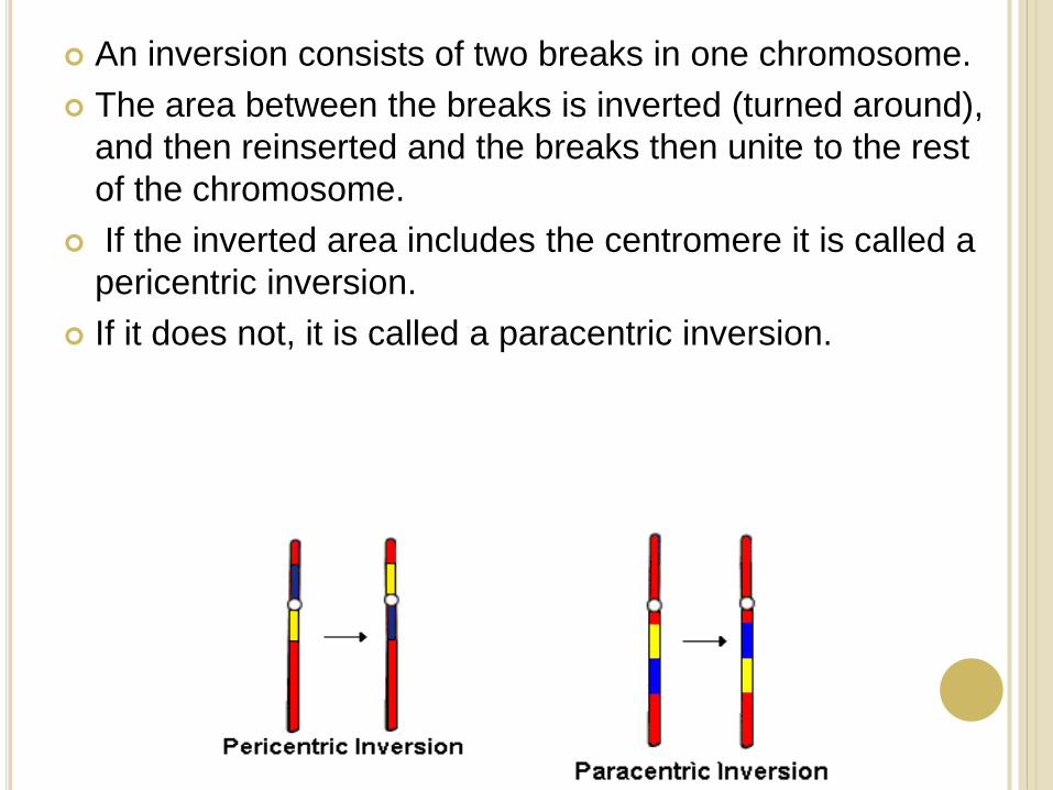
An inversion consists of two breaks in one chromosome.
The area between the breaks is inverted (turned around),
and then reinserted and the breaks then unite to the rest
of the chromosome.
If the inverted area includes the centromere it is called a
pericentric inversion.
If it does not, it is called a paracentric inversion.
Translocation
Integration of a chromosome segment into a
nonhomologous chromosome is known as
translocation.
Three types:
1. simple translocation
2. shift
3. reciprocal translocation.
Simple translocation: In this case, terminal segment of a chromosome is integrated at one end of a non-homologous region. Simple translocations are rather rare.
Shift: In shift, an intercalary segment of a chromosome is integrated within a non-homologous chromosome. Such translocations are known in the populations of Drosophila, Neurospora etc.
Reciprocal translocation: It is produced when two non-homologous chromosomes exchange segments – i.e., segments reciprocally transferred.
Translocation of this type is most common
VARIATION IN CHROMOSOME NUMBER
Euploidy Normal variations of the number of complete sets of chromosomes
Haploid, Diploid, Triploid, Tetraploid, etc…
Aneuploidy Variation in the number of particular chromosomes within a set
Monosomy, trisomy, polysomy
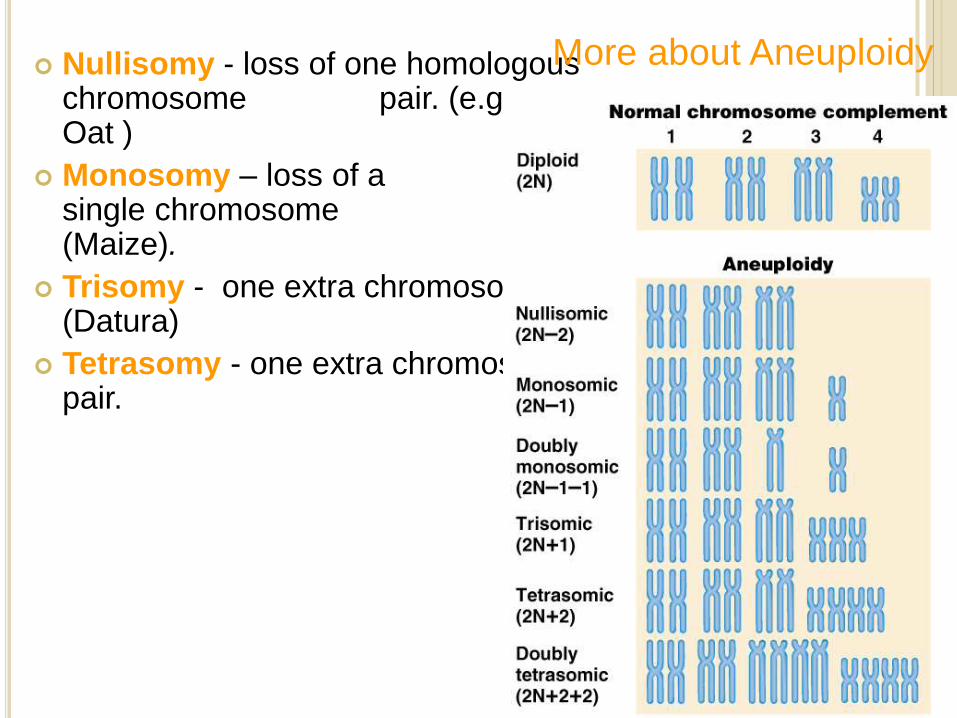
Nullisomy - loss of one homologous chromosome pair. (e.g., Oat )
Monosomy – loss of a single chromosome (Maize).
Trisomy - one extra chromosome. (Datura)
Tetrasomy - one extra chromosome pair.
More about Aneuploidy