geneticdissectionoftheazfregionsofthehumany chromosome
TRANSCRIPT

Hindawi Publishing CorporationJournal of Biomedicine and BiotechnologyVolume 2010, Article ID 936569, 18 pagesdoi:10.1155/2010/936569
Review Article
Genetic Dissection of the AZF Regions of the Human YChromosome: Thriller or Filler for Male (In)fertility?
Paulo Navarro-Costa,1, 2, 3 Carlos E. Plancha,2 and Joao Goncalves1
1 Departamento de Genetica, Instituto Nacional de Saude Dr. Ricardo Jorge, 1649-016 Lisboa, Portugal2 Faculdade de Medicina de Lisboa, Instituto de Histologia e Biologia do Desenvolvimento, 1649-028 Lisboa, Portugal3 Faculdade de Medicina de Lisboa, Instituto de Medicina Molecular, 1649-028 Lisboa, Portugal
Correspondence should be addressed to Paulo Navarro-Costa, [email protected]
Received 17 December 2009; Accepted 23 April 2010
Academic Editor: Brynn Levy
Copyright © 2010 Paulo Navarro-Costa et al. This is an open access article distributed under the Creative Commons AttributionLicense, which permits unrestricted use, distribution, and reproduction in any medium, provided the original work is properlycited.
The azoospermia factor (AZF) regions consist of three genetic domains in the long arm of the human Y chromosome referred toas AZFa, AZFb and AZFc. These are of importance for male fertility since they are home to genes required for spermatogenesis.In this paper a comprehensive analysis of AZF structure and gene content will be undertaken. Particular care will be given tothe molecular mechanisms underlying the spermatogenic impairment phenotypes associated to AZF deletions. Analysis of the 14different AZF genes or gene families argues for the existence of functional asymmetries between the determinants; while some areprominent players in spermatogenesis, others seem to modulate more subtly the program. In this regard, evidence supporting thenotion that DDX3Y, KDM5D, RBMY1A1, DAZ, and CDY represent key AZF spermatogenic determinants will be discussed.
1. Introduction
The notion that functional determinants of spermatogenesismap to the Y chromosome (Y) was established in the 1970s[1]. Ever since the pioneering observation that deletions inthe long arm of the Y chromosome (Yq) could be associatedto defects in sperm production, researchers have tried toprecisely map and identify such factors. In the course ofthis paper, a thorough genetic and functional analysis of theY regions involved in spermatogenesis will be undertaken.These are designated as azoospermia factor (AZF) regionsand they represent an area of significant interest in the fieldof human reproduction. In order to give added insight tothis topic, the present manuscript will start with a briefoverview of the major developments in the mapping of theAZF domains.
2. Historical Perspective on the Mapping of AZF
2.1. The Early Years. Following initial reports tentativelylinking the loss of genetic material in Yq to azoospermia andhypogonadism, Tiepolo and Zuffardi established in 1976 the
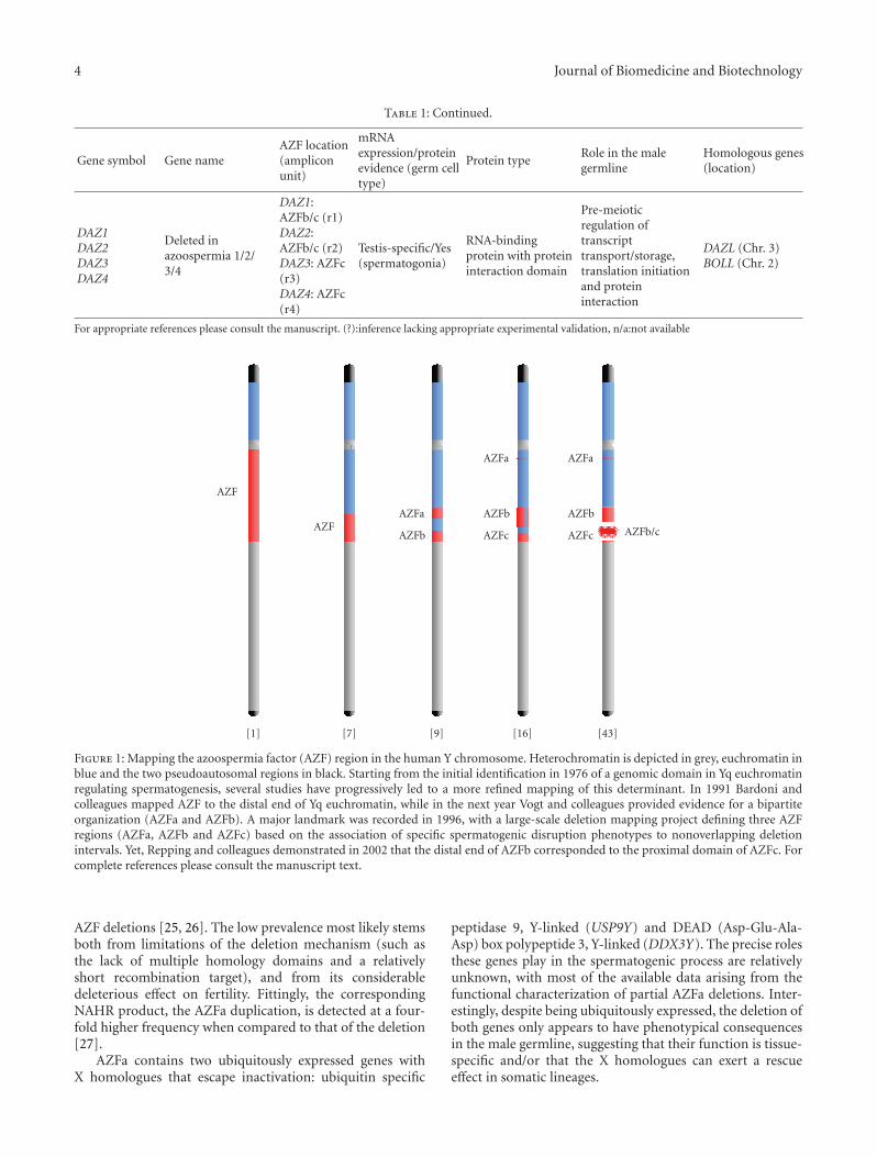
first solid association between Y chromosome deletions andabnormal spermatogenesis [1–4]. The authors screened 1170infertile men for karyotypic abnormalities and observeddeletions removing both fluorescent (heterochromatic) andnonfluorescent (euchromatic) Yq segments in 6 azoospermicmen. This association signalled the advent of a new erain the study of the Y chromosome: the identification andcharacterization of the Yq genetic determinants involved inspermatogenesis (Figure 1). In this regard, significant effortswere invested into mapping the azoospermia factor to aspecific Yq region. These initial studies were based on thedevelopment of linear deletion interval maps using Y-specificDNA probes in samples from infertile men with cytoge-netically visible Y chromosome abnormalities (illustrativeexamples: [5–7]). Although Bardoni and colleagues didmanage to map the human spermatogenesis locus to a moreprecise Yq interval (Yq11.23), the early deletion mappingstrategies were met with meagre success. This stemmed bothfrom the lack of suitable DNA markers, a consequence ofthe highly repetitive organization of Yq; and a dependenceon the relatively rare occurrence of cytogenetically visible Yqabnormalities.

2 Journal of Biomedicine and Biotechnology
2.2. Beyond the Microscope: The Concept of Y Microdeletions.Nevertheless, these efforts were of importance in establishing“rough drafts” of the Yq genomic map, particularly inidentifying markers to be used in subsequent projects.Appropriately, Ma and colleagues in 1992 mapped a markerpanel consisting of 28 DNA probes using a collection ofpatients with Yq structural abnormalities [8]. The realbreakthrough associated to the study was the screening of“chromosomally normal” azoospermic men, leading to theidentification of two deletion patterns not detectable bykaryotype visualization (and therefore dubbed microdele-tions). The implications of this result were paramount.Firstly, it suggested that the AZF region might in facthave a multipartite organization, with the authors refer-ring to them as AZFa and AZFb in a subsequent report(Figure 1) [9]. Secondly, it established the notion thatsmall Yq interstitial deletions not visualized in a stan-dard karyotype analysis might be a causative agent ofspermatogenic failure. Therefore, it became evident thatthe mapping of AZF could benefit from microdeletionscreening programs in infertile men with apparently normalkaryotypes.
Advances in molecular biology techniques, more specif-ically the use of PCR-based analyses of Yq genomic markersheralded a new stage in the quest for the AZF domains [10–12]. Despite the failure of some early studies in confirmingthe existence of two AZF regions, both the notions of multi-ple AZF loci and of the advantages of screening karyotyp-ically normal infertile men (not necessarily azoospermic)gradually became entrenched in the scientific community[13–15]. The corollary of this strategy was the screening of370 idiopathic infertile men (either azoospermic or severeoligozoospermic) with a marker panel consisting of 76Yq sequence-tagged sites (STSs), most of them previouslymapped by Vollrath and colleagues in 1992 [16]. The use of alarge cohort of infertile men was crucial for the identificationof less frequent microdeletion patterns that would otherwisepass undetected. This study revealed the existence of not twobut three AZF regions (AZFa, AZFb, and AZFc) correspond-ing to three deletion intervals, each associated to a specificinfertility phenotype (Figure 1). Therefore, the criterion fordefining the AZF regions was above all functional since it wasbased on particular spermatogenic disruption phenotypesas means to delineate genomic regions. AZFa deletionswere associated to complete absence of germ cells in thetestis tubules (Sertoli cell-only syndrome; SCOS) and AZFbdeletions to maturation arrest at the spermatocyte stage.Contrary to the azoospermia phenotype recorded in AZFaand AZFb deletions, AZFc deletions were shown to becompatible with sperm production (albeit at reduced levels)and could be transmitted to the progeny. More specifically,AZFc deletions were associated to hypospermatogenesis(abnormally decreased sperm production) that stemmedfrom a mixed degree of germ cell atrophy in the testis tubules[15, 17]. Although Vogt and colleagues proposed estimatesof AZF sequence length and a series of gene candidatesresponsible for the AZFb and AZFc deletion phenotypes, theexact length, structure and gene content of the three AZFintervals would only be fully characterized in subsequent
studies. These would show that the functional partitionof AZF into three individual regions was not reflected instructural terms, since the AZFb and AZFc sequences overlap(Figure 1).
2.3. From Microdeletion Screening Programs to Sequencing theY. The concept of AZF microdeletion screening adopted byVogt and colleagues was taken one step further in 1997with the analysis of infertile men irrespectively of theirspermatogenic phenotype [18]. This revealed not only thatYq microdeletions were present in ∼7% of the infertilepopulation, but also, more significantly, that a considerablevariability in sperm counts was associated to such microdele-tions. In fact, some microdeletion types were even detected ininfertile men with normal sperm concentrations. Althoughit was later shown that only some partial AZF deletionsmight be compatible with normozoospermia, this studysignalled the importance of a systematic screening of thesemolecular defects in the infertile population. Accordingly,AZF microdeletions are, alongside karyotype abnormalities,the most common known genetic cause of spermatogenicfailure [19]. Pryor and colleagues also screened fertile men,detecting microdeletions in 2% of the individuals. This ledthe authors to conclude that some microdeletion patternscorrespond to Y variants devoid of any obvious phenotypicalconsequences for male fertility. Thus, an adequate deletionscreening protocol should require a validated selection ofgenetic markers, as well as a precise understanding of theAZF sequence in order to rule out functionally meaninglesspolymorphisms. Such degree of knowledge was dependenton the availability of a reference sequence for the male-specific region of the Y, which only materialized in the early2000s [20].
After this brief overview of the historical landmarks onthe identification of AZF, the following paragraphs containa thorough genetic and functional characterization of thethree intervals. For an abridged analysis of the mapping andfunctional properties of the AZF genes please consult Table 1and Figure 2.
3. The AZFa Region of the Y Chromosome
The AZFa region totals 792 kb and was fully sequencedin 1999 [21]. AZFa maps to proximal Yq (chromosomelocation: ∼12.9–13.7 Mb) and unlike either AZFb or AZFc,is exclusively constituted by single-copy DNA (Figure 2).The region is flanked by two human endogenous retrovirus(HERV) elements, spanning approximately 10 kb each anddisplaying considerable levels of sequence identity. Althoughthe degree of similarity varies along the elements (withthe distal HERV copy having an additional insertion of ∼1.5 kb of transposon material—the L1 insertion), an overallsequence identity of 94% potentiates the occurrence ofHERV-mediated rearrangements [22]. Accordingly, the com-plete AZFa deletion is the result of non-allelic homologousrecombination (NAHR) between the two HERV elements[22–24]. This deletion is always associated with SCOS and isa fairly rare event, representing less than 5% of the reported

Journal of Biomedicine and Biotechnology 3
Table 1: Functional and genomic characterization of the AZF genes.
Gene symbol Gene nameAZF location(ampliconunit)
mRNAexpression/proteinevidence (germ celltype)
Protein typeRole in the malegermline
Homologous genes(location)
USP9YUbiquitin specificpeptidase 9, Y-linked
AZFaUbiquitous/Yes(spermatid)
Ubiquitin-specificprotease
Involved in proteinturnover inspermatogenesis (?)
USP9X (X chr.)
DDX3Y
DEAD(Asp-Glu-Ala-Asp)box polypeptide 3,Y-linked
AZFaUbiquitous/Yes(spermatogoniaonly)
ATP-dependentRNA helicase(deduced bysimilarity)
RNA metabolism inpre-meiotic germ cells(deduced bysimilarity)
DDX3X (X chr.)
CYorf15ACYorf15B
Chromosome Yopen reading frame15A/B
AZFb Ubiquitous/No n/a n/a CXorf15 (X chr.)
RPS4Y2Ribosomal proteinS4, Y-linked 2
AZFb Testis-specific/NoRibosomal proteinsubunit(deduced bysimilarity)
Regulation of mRNAbinding to theribosome (deducedby similarity)
RPS4Y1 (Y chr.)RPS4X (X chr.)
EIF1AYEukaryotictranslation initiationfactor 1A, Y-linked
AZFbUbiquitous/Yes(n/a)
Enhancer ofribosomedissociation andbinding (deduced bysimilarity)
Regulation oftranslationinitiation(deduced bysimilarity)
EIF1AX (X chr.)
KDM5DLysine (K)-specificdemethylase 5D
AZFbUbiquitous/Yes(primaryspermatocytes)
Demethylase of di-and tri-methylatedH3K4
Chromatinremodelling inmeiosis
KDM5C (X chr.)
XKRYXKRY2
XK, Kell bloodgroup complexsubunit-related,Y-linked/2
XKRY : AZFb(yel3)XKRY2:AZFb (yel4)
Testis-specific/NoMultipasstransmembraneprotein (?)
Gamete interaction(?)
—
HSFY1HSFY2
Heat shocktranscription factor,Y-linked 1/2
HSFY1: AZFb(b5)HSFY2: AZFb(b6)
Testis-predominant/Yes(up to thespermatid stage)
Transcription factorGene expressionregulation (?)
HSFX1 (X chr.)HSFX2 (X chr.)
PRYPRY2
PTPN13-like,Y-linked/2
PRY : AZFb(b1)PRY2:AZFb/c (b2)
Testis-specific/Yes(in somepost-meiotic cells)
Signalling molecule(deduced bysimilarity)
Germ cell apoptosis(?)
—
RBMY1A1
RNA binding motifprotein, Y-linked,family 1, memberA1
6 functionalcopies inAZFb, 2 ofthem inampliconunits (t1 andt2)
Testis-specific/Yes(mainly meioticand post-meioticcells)
RNA-bindingprotein with proteininteraction domain
RNA splicing andmetabolism, signaltransduction andmeiotic regulation
RBMX (X chr.)RBMXL1 (Chr. 1)RBMXL2 (Chr. 11)RBMXL9 (Chr.9)
BPY2BPY2BBPY2C
Basic charge,Y-linked, 2 B/C
BPY2:AZFb/c (g1)BPY2B:AZFb/c (g2)BPY2C: AZFc(g3)
Testis-specific/Yes(all spermatogenicstages)
Highly chargedprotein
Regulation of the cytoskeletal network (?)
—
CDY1CDY2
Chromodomainprotein, Y-linked,1/2
CDY1A:AZFb/c (yel1)CDY1B:AZFc (yel2)CDY2A:AZFb (yel3)CDY2B:AZFb (yel4)
Testis-specific/Yes(post-meiotic cells)
Transcriptionalco-repressor withhistoneacetyltransferaseactivity
Gene expressionregulation and andpost-meiotic nuclearremodelling
CDYL (Chr. 6)CDYL2 (Chr. 16)
4 Journal of Biomedicine and Biotechnology
Table 1: Continued.
Gene symbol Gene nameAZF location(ampliconunit)
mRNAexpression/proteinevidence (germ celltype)
Protein typeRole in the malegermline
Homologous genes(location)
DAZ1DAZ2DAZ3DAZ4
Deleted inazoospermia 1/2/3/4
DAZ1:AZFb/c (r1)DAZ2:AZFb/c (r2)DAZ3: AZFc(r3)DAZ4: AZFc(r4)
Testis-specific/Yes(spermatogonia)
RNA-bindingprotein with proteininteraction domain
Pre-meioticregulation oftranscripttransport/storage,translation initiationand proteininteraction
DAZL (Chr. 3)BOLL (Chr. 2)
For appropriate references please consult the manuscript. (?):inference lacking appropriate experimental validation, n/a:not available
[1] [7] [9] [16] [43]
AZF
AZFAZFb
AZFa
AZFc
AZFb
AZFa
AZFc
AZFb
AZFa
AZFb/c
Figure 1: Mapping the azoospermia factor (AZF) region in the human Y chromosome. Heterochromatin is depicted in grey, euchromatin inblue and the two pseudoautosomal regions in black. Starting from the initial identification in 1976 of a genomic domain in Yq euchromatinregulating spermatogenesis, several studies have progressively led to a more refined mapping of this determinant. In 1991 Bardoni andcolleagues mapped AZF to the distal end of Yq euchromatin, while in the next year Vogt and colleagues provided evidence for a bipartiteorganization (AZFa and AZFb). A major landmark was recorded in 1996, with a large-scale deletion mapping project defining three AZFregions (AZFa, AZFb and AZFc) based on the association of specific spermatogenic disruption phenotypes to nonoverlapping deletionintervals. Yet, Repping and colleagues demonstrated in 2002 that the distal end of AZFb corresponded to the proximal domain of AZFc. Forcomplete references please consult the manuscript text.
AZF deletions [25, 26]. The low prevalence most likely stemsboth from limitations of the deletion mechanism (such asthe lack of multiple homology domains and a relativelyshort recombination target), and from its considerabledeleterious effect on fertility. Fittingly, the correspondingNAHR product, the AZFa duplication, is detected at a four-fold higher frequency when compared to that of the deletion[27].
AZFa contains two ubiquitously expressed genes withX homologues that escape inactivation: ubiquitin specific
peptidase 9, Y-linked (USP9Y) and DEAD (Asp-Glu-Ala-Asp) box polypeptide 3, Y-linked (DDX3Y). The precise rolesthese genes play in the spermatogenic process are relativelyunknown, with most of the available data arising from thefunctional characterization of partial AZFa deletions. Inter-estingly, despite being ubiquitously expressed, the deletion ofboth genes only appears to have phenotypical consequencesin the male germline, suggesting that their function is tissue-specific and/or that the X homologues can exert a rescueeffect in somatic lineages.

Journal of Biomedicine and Biotechnology 5
yel3 yel4 b5 b6 b1 t1 t2 b2 g1 r1 r2 Gr1 b3 yel1 g2 r3 r4 g3 yel2 b4 Gr2
1 Mb
100 kb
AZFa deletion = 0.8 Mb
USP9Y DDX3Y
HERVHERV
AZFa
AZFb and AZFc
P3P2
P1
P4
P5
DYZ19u1 u2 u3
XK
RY
CD
Y2A
CD
Y2B
XK
RY
2
HSF
Y1
HSF
Y2
CYo
rf15
AC
Yorf
15B
KD
M5D
EIF
1AY
RP
S4Y
2R
BM
Y1A
1R
BM
Y1A
1R
BM
Y1A
1R
BM
Y1A
1P
RY
RB
MY
1A1
RB
MY
1A1
PR
Y2
BP
Y2
DA
Z1
DA
Z2
CD
Y1A
BP
Y2B
DA
Z3
DA
Z4
BP
Y2C
CD
Y1B
AZFb deletion = 6.23 Mb
AZFb + c deletion = 7.7 Mb
AZFc deletion = 3.5 Mb
Figure 2: Schematics on the genomic architecture and gene content of the reference AZFa, AZFb and AZFc regions of the human Ychromosome. Central ideogram depicts the Y chromosome with the pseudoautosomal regions represented by black boxes at the tips ofthe chromosome (PAR1 and PAR2, respectively), the three heterochromatic domains indicated in grey (the centromeric region, the satelliterepeat array embedded in Yq euchromatin, and the Yq heterochromatic block) and euchromatin in blue. The genomic organization of theAZFa region is depicted in the top half of the figure. This region maps from approximately 12.9 to 13.7 Mb of the chromosome and containstwo single copy genes: USP9Y and DDX3Y (represented in scale by two oriented triangles indicating 5′-3′ polarity). AZFa is flanked by twohuman endogenous retrovirus (HERV) elements that mediate the occurrence of AZFa deletions via non-allelic homologous recombination.The genomic organization of the reference AZFb and AZFc regions, as defined by the occurrence of specific deletion patterns, are depictedin the bottom half of the figure. AZFb maps from approximately 18 to 24.7 Mb of the chromosome and AZFc from ∼23 to ∼26.7 Mb. Bothregions feature multiple stretches of ampliconic sequences, represented by block arrows. The amplicons are divided in six colour-codedsequence families (yellow, blue, turquoise, green, red and grey) with each unit being coded according to a binomial notation indicative offamily type and copy number [20]. The size and orientation of the arrows is representative of amplicon length and polarity, respectively. Theorganization of amplicons in symmetrical arrays of contiguous repeat units (palindromes P1 to P5) is represented by large triangles. AZFbis defined by the P5/proximal P1 deletion (yel3/yel1), and AZFc by the b2/b4 deletion. Single copy domains are depicted in white and theDYZ19 satellite repeat in grey. The spacers between the two red amplicon clusters are identical between them. Transcription unit allocationto these regions is observable below the architecture map. For a more precise mapping of the genetic determinants please consult Figure 3.
3.1. AZFa Gene Content
3.1.1. USP9Y. The USP9Y protein is an ubiquitin-specificprotease and member of the C19 cystein peptidase fam-ily. These enzymes promote the intracellular cleavage of
ubiquitin molecules from ubiquitinated proteins [28, 29].Appropriately, a role for USP9Y in the regulation of proteinturnover during spermatogenesis has been proposed [30,31]. USP9Y shares 91% identity with its X homologue(USP9X) suggesting that both target similar molecules and

6 Journal of Biomedicine and Biotechnology
KDM5DEIF1AYRPS4Y2
CYorf15A
CYorf15B
yel3 yel4 b5 b6 b1 t1 t2 b2 g1 r1 r2 Gr1 b3 yel1 g2 r3 r4 g3 yel2 b4 Gr2
1 Mb
XKRYCDY2HSFY
RBMY1A1PRY
BPY2DAZ
CDY1
(a)
(b)
Figure 3: Mapping and sequence organization of the transcription units located in the reference AZFb and AZFc regions of the human Ychromosome. Top schematics represent the genomic organization of the AZFb and AZFc regions, as defined by Kuroda-Kawaguchi et al.,[65] and Repping et al., [20]. Block arrows represent amplicon units and rectangles single-copy domains. The DYZ19 heterochromatic regionis identified by a discontinuous grey rectangle. a- Single-copy transcription units. Direction of the triangles indicates the 5′-3′ orientation ofthe reading frames. All these sequences map outside of the ampliconic regions. b- Multi-copy sequence families. Number of triangles per lineindicates active copy number inside each sequence family. These range from two (XKRY, CDY1, CDY2, HSFY, and PRY) to six (RBMY1A1).All multi-copy transcription units bar four members of the RBMY1A1 family map to ampliconic domains. Pseudogenes are not depicted.
may overlap functionally [32]. Studies in murine gametoge-nesis have shown that while USP9X expression starts as earlyas in the establishment of the primordial germ cell (PGC)population in both sexes, USP9Y only starts to be expressedin the male germline at the spermatid stage [32, 33]. Thismarkedly distinct expression window hints at a temporalconstrain in the regulation of USP9Y function, a probableconsequence of its molecular targets only being present atlater spermatogenic stages. Yet, available data are inconsistentwith USP9Y being a key player in male gametogenesis.Although USP9Y deletions were initially thought to be exclu-sively associated to azoospermia and the cause of the AZFadeletion phenotype, two more recent reports demonstrateotherwise. Indeed, USP9Y deletions that are compatible withsperm production and with natural conception have alreadybeen identified, the latter corresponding to the completedeletion of the gene [34, 35]. Thus, published data pointsto USP9Y not being essential for male fertility, as observedin other primate lineages where the gene became inactive[36, 37]. Nevertheless, it is still premature to discard theinvolvement of USP9Y in the epistatic regulation of malegametogenesis [38].
3.1.2. DDX3Y. The DDX3Y protein has the hallmarks ofan ATP-dependent RNA helicase belonging to the DEADbox protein family (characterized by the conserved motifAsp-Glu-Ala-Asp). The exact molecular role of DDX3Yis unknown, although the DEAD box proteins have been
implicated in several key processes of RNA metabolismsuch as secondary structure alteration, splicing, spliceo-some assembly and translation initiation [39]. DDX3Y andits X homologue (DDX3X; 91.7% sequence identity) areubiquitously expressed, with expression levels peaking intestis [40, 41]. However, the widespread presence of bothtranscripts in adult tissues does not directly correlate withactual protein expression since DDX3Y, unlike DDX3X,is testis-specific. An additional layer of regulation can beinvoked as both genes encode for testis-specific transcriptscharacterized by an overall shorter length and the presenceof extended untranslated regions (UTRs) [32, 42]. Thisspecificity in transcriptional profiles most likely serves toensure a precise expression window as DDX3Y is detectedpredominantly in the cytoplasm of spermatogonia whereasDDX3X is mainly detected in spermatids [40]. The divergentexpression window of the two genes suggests that DDX3Ymay represent a specialization of DDX3X functions for pre-meiotic developmental stages. Yet, despite differences in celltype expression, the molecular functions of DDX3Y andDDX3X are probably analogous, as evidenced by functionalrescue studies in murine cell lines [41].
Taking into account data from USP9Y deletions as wellas the regulation of DDX3Y function, it is tempting toconsider that the absence of the latter is the main causativeagent for the complete AZFa deletion phenotype. However,this hypothesis still requires the validation warranted by theunambiguous identification of DDX3Y-specific deletions.
Journal of Biomedicine and Biotechnology 7
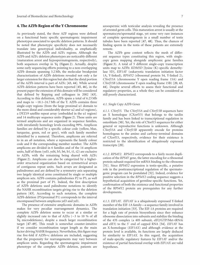
4. The AZFb Region of the Y Chromosome
As previously stated, the three AZF regions were definedon a functional basis: specific spermatogenic impairmentphenotypes associated to specific deletion patterns. It shouldbe noted that phenotypic specificity does not necessarilytranslate into genotypical individuality, as emphaticallyillustrated by the AZFb and AZFc regions. Although theAZFb and AZFc deletion phenotypes are noticeably different(maturation arrest and hypospermatogenesis, respectively),both sequences overlap in Yq (Figure 2). Actually, despitesome early sequencing efforts pointing to a non-overlappingAZFb domain spanning 3.2 Mb, the molecular breakpointcharacterization of AZFb deletions revealed not only a farlarger extension for this region but also that the distal portionof the AZFb interval is part of AZFc [43, 44]. While severalAZFb deletion patterns have been reported [45, 46], in thepresent paper the extension of this domain will be consideredthat defined by Repping and colleagues in 2002 [43].According to this definition, AZFb spans a total of 6.23 Mband maps to ∼18.1–24.7 Mb of the Y. AZFb contains threesingle-copy regions (from the large proximal u1 domain tothe more distal and considerably shorter u2 and u3 regions),a DYZ19 satellite repeat array (embedded in the u1 region)and 14 multicopy sequence units (Figure 2). These units aretermed amplicons and are organized in sequence families,with intrafamily homology levels exceeding 99%. Ampliconfamilies are defined by a specific colour code (yellow, blue,turquoise, green, red or grey), with each family memberidentified by a numeral. Therefore, amplicons are referredto in an ab notation, where a represents the family colourcode and b the corresponding member number. The AZFbamplicons are divided in 6 families and of the 14 ampliconunits, half of them (yel3, yel4, b5, b6, b1, t1, t2) are exclusiveto AZFb, with the remaining being shared with AZFc(Figure 2). Amplicons can also be categorized by a higher-order structural organization based on symmetrical arraysof contiguous repeat units. Such arrays are designated aspalindromes and are defined by a symmetry axis separatingtwo largely identical arms constituted by single or multipleamplicon sets. AZFb contains palindromes P2 to P5, as wellas the proximal part of P1. Indeed, the first descriptionof AZFb deletions used palindrome notations to identifythe NAHR recombination targets giving rise to the deletionpattern [43]. According to such notation, the completeAZFb deletion (P5/proximal P1) corresponds to the intervalencompassed between amplicons yel3 and yel1.
The presence of extensive ampliconic domains in AZFbmakes for very peculiar rearrangement dynamics. Thecomplete AZFb deletion seems to occur at a similar orslightly increased rate to that of AZFa (∼3 to 10 % of allYq microdeletions), despite a much larger recombinationtarget [47–49]. This result is somewhat counter-intuitiveif we consider recombination target length as the mainfactor driving NAHR frequency. Nevertheless, this figure mayrise five-fold if AZFb+c deletions are included, suggestingthat the propensity for rearrangements may vary betweenamplicon units. Regarding the spermatogenic impairmentphenotype of the complete AZFb deletion, patients are
azoospermic with testicular analysis revealing the presenceof arrested germ cells. This maturation arrest is usually at thespermatocyte/spermatid stage, yet some very rare instancesof complete spermatogenesis in a small number of testistubules have been reported [47, 48]. Thus, the chances offinding sperm in the testis of these patients are extremelyremote.
The AZFb gene content reflects the mesh of differ-ent sequence types constituting this region, with single-copy genes mapping alongside ampliconic gene families(Figure 3). A total of 5 different single-copy transcriptionunits map to AZFb: KDM5D [lysine (K)-specific demethy-lase 5D], EIF1AY (eukaryotic translation initiation factor1A, Y-linked), RPS4Y2 (ribosomal protein S4, Y-linked 2),CYorf15A (chromosome Y open reading frame 15A) andCYorf15B (chromosome Y open reading frame 15B) [20, 43,44]. Despite several efforts to assess their functional andregulatory properties, as a whole they can be considered asstill poorly characterized.
4.1. Single Copy AZFb Genes
4.1.1. CYorf15. The CYorf15A and CYorf15B sequences havean X homologue (CXorf15) that belongs to the taxilinfamily and has been linked to transcriptional regulation inosteoblasts [50]. Yet, the role of CYorf15 sequences for eithergeneral or reproductive functions is unknown. AlthoughCYorf15A and CYorf15B apparently encode for proteinshomologous to the amino and carboxy-terminal domainsof CXorf15, respectively, evidence for their existence isrestricted to the identification of ubiquitously expressedtranscripts [20].
4.1.2. RPS4Y2. RPS4Y2 corresponds to a fairly recent dupli-cation of the RPS4Y gene, the latter encoding for a ribosomalprotein subunit required for mRNA binding to the ribosome[51]. Since RPS4Y2 expression is testis-specific, a putativerole in the posttranscriptional regulation of the spermato-genic program can be postulated [52]. Indeed, evidence forpositive selection in the RPS4Y2 coding sequence suggests ahypothetical acquisition of germline-specific functions. Yet,confirmation of both the existence and functional propertiesof the RPS4Y2 protein are prerequisites for any furtherdevelopments.
4.1.3. EIF1AY. EIF1AY is a ubiquitously expressed Y-linkedmember of the EIF-1A family—a sequence family involved intranslation initiation [53]. The EIF-1A proteins are requiredfor a high rate of protein biosynthesis since they enhanceribosome dissociation into subunits and stabilize the bindingof the 43S complex (a 40S subunit, eIF2/GTP/Met-tRNAiand eIF3) to the 5′ end of capped RNA [54]. EIF1AY hasan X-homologue (EIF1AX) and although evidence at theprotein level is available, its functions are largely deducedby similarity to EIF1AX. In this regard, the acquisitionof male-specific regulatory features by EIF1AY and/or theexistence of partial functional overlap with EIF1AX are validhypotheses.

8 Journal of Biomedicine and Biotechnology
4.1.4. KDM5D. KDM5D encodes for a histone H3 lysine4 (H3K4) demethylase that forms a protein complex withthe MSH5 DNA repair factor during spermatogenesis [55,56]. This complex locates to condensed DNA duringthe leptotene/zygotene stage, suggesting an involvement inmale germ cell chromatin remodelling. In accordance, bydemethylating di- and tri-methylated H3K4, KDM5D maybe involved in chromosome condensation during meiosis.Such possibility fits with the instances of maturation arrest atthe spermatocyte stage associated to AZFb deletions. Despitethe apparently male germline-specific functions, this gene isubiquitously expressed and is homologous to KDM5C, an X-borne gene associated to X-linked mental retardation [57–60].
4.2. Multicopy AZFb Genes. Due to the presence of ampli-conic sequences AZFb contains a set of 7 different multi-copy gene families: XKRY (XK, Kell blood group complexsubunit-related, Y-linked), HSFY (heat shock transcriptionfactor, Y-linked), RBMY1A1 (RNA binding motif protein,Y-linked, family 1, member A1), PRY (PTPN13-like, Y-linked) CDY (chromodomain protein, Y-linked), BPY2(basic charge, Y-linked, 2), and DAZ (deleted in azoosper-mia). The members of these gene families make for a total of20 transcription units in the reference AZFb sequence. Sinceseveral of them also map to AZFc, only genes with functionalcopies exclusively located in AZFb will be analysed in thissection.
4.2.1. XKRY. The XKRY gene is expressed specifically intestis and maps to the yellow-coded amplicon family.Although protein evidence is still lacking, sequence analysissuggests that the two active copies of XKRY (mapping to yel3and yel4) encode for a multipass transmembrane transportprotein similar to the XK protein. The latter locates toneuromuscular and hematopoietic cell membranes, with XKmutations causing specific disruption phenotypes [61–63].However, a role for XKRY in spermatogenesis has yet to bevalidated despite tentative links to the fertilization process[64].
4.2.2. HSFY. HSFY encodes for a member of the heatshock factor family of transcriptional activators and displaystestis-predominant expression. This gene maps to the blueamplicons with the two active copies located in b5 andb6. HSFY is subjected to alternative splicing, generating 3different protein-coding transcripts with varying expressionpatterns [66]. HSFY has been identified in spermatogeniccells up to the spermatid stage and in Sertoli cells [67]. Theprotein’s stage-dependent translocation from the cytoplasmto the nucleus is suggestive of a developmentally regulatedfunctional window, consistent with its role as a transcrip-tion factor. Nevertheless, as previously argued, the HSFYpolyclonal antiserum used in the former study might havedetected epitopes from the more widely expressed X homo-logues (HSFX1/2) [32]. In this regard, a subsequent reportby the same team established that the mouse orthologue ofHSFY is predominantly expressed in round spermatids [68].
Regardless of several uncertainties on the exact function andregulation of HSFY, a role in spermatogenesis has alreadybeen proposed based on observations in animal models andin infertile males. More specifically, Vinci and colleaguesdetected a partial AZFb deletion purportedly only affectingthe functional copies of HSFY in an azoospermic man[69]. Yet, it should be noted that the heritability of thedeletion was unknown. More recently, HSFY protein levels inspermatogenic cells were shown to be decreased in sampleswith maturation arrest, associating once again this gene tothe regulation of male gametogenesis [70].
4.2.3. PRY. The PRY gene copies map to the blue amplicons,with the two functional units being restricted to b1 and b2.These are designated as PRY and PRY2, respectively, andencode for a gene product with a low degree of similarityto protein tyrosine phosphatase, nonreceptor type 13. Thelatter corresponds to a signalling molecule involved in theregulation of a myriad of cellular processes, particularlyin programmed cell death (illustrative example: [71]). PRYand PRY2 display testis-specific expression and additionalregulation via alternative splicing [72]. Nevertheless, thealternative transcript seems to correspond to a nonfunctionalisoform since it contains a premature stop codon truncatingthe product at about half. The expression of PRY in germcells is irregular, with the protein being detected only in afew sperm and spermatids [73]. Interestingly, both transcriptand protein levels were shown to be higher in the defectivegerm cell fraction of the ejaculate. Furthermore, PRY levelsare increased in ejaculated sperm obtained from men withabnormal semen parameters, suggesting a link between itsexpression and defective spermatogenesis. Appropriately, arole for PRY in male germ cell apoptosis has been suggestedbased on the observation that approximately 40% of PRY-positive cells show DNA fragmentation. Yet, as acknowledgedby the authors of the paper, results were insufficient to fullyback the claim. Regardless of such considerations, availableevidence points to a postmeiotic expression of PRY inrestricted subsets of developing germ cells.
4.2.4. RBMY1A1. The RBMY1A1 gene family was identifiedin the early 1990s [13]. At the time, the functional propertiesof the then dubbed Y chromosome RNA recognition motifgene (YRRM) made it the first candidate azoospermia factor.Although RBMY1A1 is present in multiple copies along theY chromosome, the six functional units cluster to the AZFbamplicons [74, 75]. This complex arrangement, character-ized by an extensive array of RBMY1A1 pseudogenes andsub-families, had thwarted initial attempts to precisely mapthis determinant [76, 77]. RBMY1A1 is part of the RBM genefamily that also includes an X homologue (RBMX) and a setof autosomal retrogene-derived copies of RBMX (of theseonly RBMXL1, RBMXL2 and RBMXL9 are expressed, andprotein evidence is only available for RBMXL2) (for a review:[78]). Unlike its ubiquitously expressed X homologue,RBMY1A1 is expressed solely in male germ cells, with theprotein displaying a nuclear location [75]. The main featureof the RBM family is the presence of a N-terminal RNA

Journal of Biomedicine and Biotechnology 9
recognition motif (RRM) responsible for the interaction withtarget RNA molecules [74, 79]. In this regard, RBM familymembers display characteristics of canonical RNA-bindingproteins involved in nuclear RNA processing. In fact, thisgene has been linked to the storage and transport of mRNAfrom the nucleus during spermatogenesis [80]. Contrastingwith the other RBM genes, RBMY1A1 also contains a C-terminal protein interaction repeat domain enriched inserine, arginine, glycine, and tyrosine (SRGY) [78]. Thisserves as a probable regulatory region for the modulation ofRBMY1A1 function.
The nuclear localization of RBMY1A1 is pinpointed todomains enriched in pre-mRNA splicing components, asevidenced in prophase I nuclei [81]. In accordance, effortsto identify RBMY1A1-interacting proteins have shown thatpre-mRNA splicing regulators, particularly the SR and theSR-related proteins, are bona fide partners [82, 83]. Theseubiquitously expressed factors also contain RRM domains,therefore their functional modulation via RBMY1A1 inter-action emerges as a distinct possibility [84]. Additionally,RBMY1A1 may modulate cellular processes other thansplicing regulation and mRNA metabolism since it has beenshown to interact with the STAR and T-STAR proteins[85]. These act not only as splicing regulators but also asmembers of signal transduction pathways involved in cellcycle control. In this regard, RBMY1A1 can be involved inseveral aspects of meiotic and premeiotic regulation via theestablishment of multiple protein complexes. Interestingly,the male germ cell-specific expression of RBMY1A1 is alsomimicked by the autosomal RBMXL2 gene. In this case thenuclear localization of the protein during and immediatelyafter meiosis is suggestive of meiotic specialization [86].In accordance, haploinsufficiency of the murine RBMXL2orthologue results in abnormal spermatogenesis in animalmodels [87].
The identification of the RNA targets of RBMY1A1has been partially successful. It is believed that the RRMdomain can bind RNA at both high and low affinity,making the characterization of target molecules complex[78]. Furthermore, the protein has an unique two-stepmechanism for RNA recognition that starts with a sequence-specific interaction with the target molecule before eliciting aconformational modification [88]. This complex mechanismwarrants RBMY1A1 a significant plasticity in terms ofRNA partners. Studies in murine models have identified 12different potential mRNA targets for RBMY1A1, most ofthem expressed in testis starting from the neonatal period[89]. Interestingly, the protein seems to be able to bind to itsown alternative transcript, suggesting a complex regulatorynetwork. The existence of alternative RBMY1A1 transcriptshas also been detected in humans [90].
All the aforementioned properties seem to indicate thatthe disruption of RBMY1A1 plays a significant role in theAZFb deletion phenotype. In reality, both its expressionpattern and putative role in male germ cell developmentsupport the notion that RBMY1A1 deletions perturb themeiotic program. Similarly, the disruption of KDM5D mayalso contribute to the deletion phenotype. In fact, RBMY1A1and KDM5D are located in the germ cell nucleus during
prophase I, suggesting involvement in meiotic orchestration.While this regulation may be directly exerted by KDM5D(via changes in chromatin structure), the role of RBMY1A1might be mediated by effector proteins or by transcriptionalregulation of mRNA targets.
Regardless of the actual contribution of the AZFbgenes for the maturation arrest phenotype, a predominantlystructural effect of the AZFb deletion on meiotic progressioncannot be discarded. The removal of such a large stretchof Yq chromatin (∼6.23 Mb) may result in X-Y pairingimpairment during meiosis and lead to meiotic breakdown.This effect has already been identified in patients withAZFb+c deletions [91]. In such cases, a significant decreasein spermatocyte X-Y bivalent formation was recorded, withonly 29% of the cells having juxtaposed telomere signals. Itcan be argued that the lower rate of sex chromosome pairingarises from DNA conformational changes that underminemeiotic efficiency. However, it is impossible to dissociate theeffect of gene loss from the observed pairing impairment,particularly since we are dealing with genes involved in cellcycle progression. Therefore, and in light of all evidence, thematuration arrest phenotype associated to AZFb deletionsmost probably stems from a combination of genetic disrup-tion with structural defects in the chromosome.
5. The AZFc Region of the Y Chromosome
The AZFc region is one of the most remarkable domains ofthe human genome, displaying a structural and functionalintricacy only paralleled by the major histocompatibilitycomplex in chromosome 6. The sequencing of AZFc rep-resented a monumental effort based on laborious datacompilation and inventive analytical tools [65]. Paradoxi-cally, the effort put into sequencing AZFc revealed that theobtained sequence corresponds to just one of the plethora ofexpected genomic variants in the Y chromosome population.This observation arises from the fact that AZFc is almostexclusively constituted by amplicons. Indeed, the extensivehomology between intra-family ampliconic units is a fertilesubstrate for large-scale AZFc structural rearrangements(deletions, duplications and inversions) as well as moresubtle sequence modifications. Yet, both the moleculardrivers and phenotypical consequences of AZFc variabilityfall outside the scope of the present review, having beenthoroughly discussed elsewhere [92].
5.1. Genomic Assembly of AZFc. Approximately 95% of thereference AZFc sequence is constituted by ampliconic unitsbelonging to five different colour-coded families (blue, green,red, grey and yellow). The remainder corresponds to aduplicated spacer for the red amplicons (present in thetwo red amplicon clusters) and a single copy domain (u3)similar to other Y regions (Figure 2). Structurally, the regioncontains one large (P1) and one smaller (P2) palindrome,as well as the b2-u3-g1 segment. Different models for thegenomic assembly of the reference AZFc structure havebeen proposed. A simple, two-step model states that thepalindromes arose from supragenic tandem duplication

10 Journal of Biomedicine and Biotechnology
followed by inversion [65]. Traces of such events have alreadybeen detected in the P1 palindrome, where Alu elementswere probably involved in a large-scale duplication priorto an IR (inverted repeat)-mediated inversion. Recently, amore complex model for the progressive assembly of theAZFc ampliconic organization was proposed. The modelstates that 3 major waves of amplicon acquisition wererequired for the establishment of AZFc, starting from a basalstructure constituted by the blue, turquoise and the distalpart of the yellow amplicons [93]. In the first wave, thegreen and red amplicons were transposed; in the second themiddle segment of the yellow amplicon was acquired, and inthe third both the proximal yellow amplicon segment andthe grey amplicon were transposed. In parallel with theseacquisition waves, other molecular processes such as dele-tions, duplications and inversions shaped AZFc by operatingon the progressively acquired blocks. A limitation to thismodel is the assumption that the ampliconic families of theancestral AZFc state are identical to those of the referencesequence. This limitation is also evident when calculatingthe minimum-mutation history of AZFc architectures [94].According to the most parsimonious model, the ancestralAZFc architecture was already multicopy, with the majorityof the observed diversity arising from sequence inversions(and to a lesser degree from deletions and duplications) inthe ancestral sequence.
In clinical terms, men with complete AZFc deletionshave variable seminal and testicular phenotypes, with spermproduction levels ranging from azoospermia to severe oligo-zoospermia (but rarely exceeding 1 million sperm/ml) [16,47, 48]. Although in these patients the presence of sperm inthe ejaculate is a frequent event (in ∼50 to 60% of the cases),natural conception is extremely rare due to low sperm counts[16, 95–101]. The variable phenotype associated to thesedeletions suggests an intricate regulation of the AZFc geneticdeterminants, making this region particularly prone to agenetic background effect. Complete AZFc deletions total3.5 Mb (mapping from ∼23 to 26.8 Mb of the chromosome)and are the product of NAHR between the b2 and b4amplicons. They account for approximately 60% of allrecorded AZF deletions, occurring in one out of every ∼4000 males [47, 48, 65]. The ampliconic organization ofAZFc is also responsible for partial deletions that arise fromNAHR between the more internal units. These partial AZFcdeletions are associated to extremely variable spermatogenicdisruption phenotypes (if any), leading to a debate onwhether such rearrangements represent a male infertility riskor not (for selected reading: [92, 102, 103]).
5.2. AZFc Gene Content. As previously stated, 3 protein-coding gene families map to the AZFc interval: BPY2,DAZ and CDY (Figure 3). AZFc is also enriched in othertranscription units, mainly for spliced but apparently non-coding transcripts of the TTTY family (TTTY3 and TTTY4).Additionally, it contains an extensive array of pseudogenes.These correspond to inactive copies of AZFb and AZFc genes(RBMY1A1, PRY, CDY and BPY2), as well as AZFc-exclusivesequence families [GOLGA2LY1 (golgi autoantigen, golgin
subfamily a, 2-like, Y-linked 1) and CSPG4LYP1 (chondroitinsulfate proteoglycan 4-like, Y-linked pseudogene 1)].
5.2.1. BPY2. The BPY2 gene family maps to the green AZFcamplicons (one active copy per amplicon), encoding for atestis-specific highly charged protein tentatively linked tocytoskeletal regulation in spermatogenesis [64, 104]. Thisgene family is further expanded by a set of pseudogenesequences also mapping to the green amplicons [65]. Despitethe existence of a region of homology with chromosome8, no autosomal homologues of BPY2 have been identified[105]. The genomic organization of the gene is quite uniquesince it is constituted by nine exons but only five of whichare translated into amino-acids [106]. The BPY2 proteindisplays a nuclear localization throughout all male germ celldevelopmental stages, persisting even in ejaculated sperm[107]. The exact role played by BPY2 in spermatogenesisis unclear, with most of the available knowledge beinginferred from its protein partners. Using the yeast two-hybrid assay BPY2 has been shown to interact with ubiquitinprotein ligase E3A (UBE3A), a widely-expressed memberof the ubiquitin protein degradation system [108]. Thisinteraction is mediated by the HECT domain of UBE3A.Since UBE3A corresponds to a testis-expressed E3 ubiquitinprotein ligase (responsible for the transfer of the ubiquitingroup to the targeted substrates), BPY2 may modulate itstarget specificity. Additionally, the two-hybrid assays havealso identified microtubule-associated protein 1S (MAP1S)as an interacting protein [104]. MAP1S is a member ofthe microtubule-associated proteins (MAPs) family and isinvolved in microtubule binding, bundling and stabilization,as well as in the crosslinking of microtubules with micro-filaments [109]. Since MAP1S is predominantly expressedin testis, a putative role of BPY2/MAP1S in the control ofthe male germ cell cytoskeletal network has been proposed[104]. The functional properties of the MAP1S complexare regulated by changes to its heavy chain, making thismolecule a suitable target for posttranslational regulation[109]. In this context, BPY2 emerges as a very strongcandidate regulator, possibly through an UBE3A-mediatedubiquitinization event. Protein structure prediction modelsalso suggest the existence of a DNA/RNA binding domain (aHTH-like motif) in BPY2, yet experimental validation is stilllacking [110].
The screening for BPY2 mutations in infertile maleshas thus far been inconclusive, with no identifiable exonmutations in a cohort of 106 SCOS patients [111]. Therefore,despite suggestions that a specific promoter genotype mightbe associated to spermatogenic defects [111], both BPY2function and the phenotypical consequences associated to itsdisruption remain to be elucidated.
5.2.2. CDY. The chromodomain protein family (CDY) con-sists of two Y-encoded genes (CDY1 and CDY2) and twoautosomal copies (CDYL in chromosome 6 and CDYL2 inchromosome 16) [112]. These genes are involved in post-meiotic nuclear remodelling and transcriptional regulation.The Y family members map to the yellow amplicons, with
Journal of Biomedicine and Biotechnology 11
the CDY1 copies in AZFc (amplicons yel1 and yel2) andthe CDY2 copies in AZFb (yel3 and yel4) [44, 65]. A fairlylarge number of pseudogene sequences are also scatteredthroughout AZFb and AZFc. As expected, the Y-linkedcopies have testis-specific expression whereas the autosomalunits display a more general expression pattern (CDYL iseven ubiquitously expressed) [64, 112]. CDY1 displays twoadditional transcript variants (minor and short CDY1) withthe former showing evidence of the excision of a single intron[112, 113]. The expression of these alternative transcriptscorrelates significantly with complete spermatogenesis intesticular samples of azoospermic men [114].
The CDY proteins are characterized by two functionalmotifs: an N-terminal chromatin-binding domain (the chro-modomain) and a C-terminal catalytic domain (responsi-ble for the CoA-dependent acetyltransferase activity). Thechromodomain is a typical signature of proteins involvedin chromatin remodelling and gene expression regulation[115]. Accordingly, in vitro assays have demonstrated thatrecombinant CDY proteins can acetylate histone H4 (and, toa lesser degree, H2A) [116]. Furthermore, it was establishedthat mouse Cdyl (mCdyl) transcript and protein levels peakat the elongating spermatid stage, a time frame coincidingwith histone H4 hyperacetylation [116]. Given the nuclearlocalization of the protein and the post-meiotic expressionwindow, the CDY family is considered a nuclear remodellingfactor promoting histone H4 hyperacetylation [116]. Thelatter, by inducing a more relaxed chromatin configuration,may serve as trigger for the histone-to-protamine transitionand subsequent nuclear condensation.
The function of the CDY proteins is not restricted tohistone acetylation. Studies have associated CDYL and itsparalogues to transcriptional corepressor complexes consist-ing of multiple chromatin modifying proteins [117–119].Accordingly, the primary function of CDYL may be thatof a transcriptional co-repressor, as observed in murinemodels when histone deacetylases (HDACs) bind to itscatalytic domain [117]. The protein acquires its role inchromatin remodelling only when HDACs are degraded(in the elongating spermatid stage) and the CoA-bindingactivity of mCdyl is activated. This fits with data obtainedfrom protein structure analysis indicating that the CDYproteins do not show obvious similarities to canonicalhistone acetyltransferase motifs [120]. Recently, CDY1 hasalso been shown to interact with lysine 9-methylated histones(H3K9me2 and H3K9me3), although the exact functionalrole of this interaction is unknown [121]. The analysis ofsuch binding properties further suggests that CDYL2, notCDYL, is the ancestor of the gene family [122].
The CDY1 and CDY2 proteins are isoforms with anamino-acid identity of 98% and a similar expression window[112]. This contradicts previous views that CDY2 wasrequired at earlier spermatogenic stages [114]. On theother hand, the global identity score between the Y-linkedCDY proteins and CDYL is just 63%. The acceleratedprotein evolution rate of the Y-borne CDY sequences seemsto suggest that these copies have evolved under positiveselection for germline specific functions [123]. Nevertheless,identity levels are slightly higher when comparing just the
functional domains of the Y-derived and autosomal copies.In this regard, functional complementation between CDYgenes may rescue, to some extent, the loss of the AZFb and/orAZFc variants. Fittingly, complete AZFc deletions do notalter H4 hyperacetylation levels in developing spermatidswhen compared to those recorded in nondeleted hyposper-matogenic men [124].
5.2.3. DAZ. The members of the DAZ gene family areRNA binding proteins that play prominent roles in theestablishment and maintenance of the male germ line (forselected reviews: [125, 126]). This gene family consists ofthree different genetic determinants: BOLL (bol, boule-like), DAZL and DAZ [127]. Of these, DAZ maps to AZFc(consequently being organized as a multi-copy gene family)with the remaining two being single-copy autosomal genes.Since the DAZ gene family contains the Y-borne DAZ copies,for the sake of disambiguation the italicized reference (DAZgenes) will refer solely to the Y determinants. The DAZ genesare present in one copy per red AZFc amplicon, for a totalof four in the reference AZFc sequence (DAZ1 to DAZ4)[65, 128]. The particularities of the palindromic organizationof the reference sequence results in the clustering of the DAZcopies to two red amplicon duplets, with the more proximalcluster containing DAZ1 and DAZ2, and the more distalcluster DAZ3 and DAZ4. Nevertheless, variations in genenumber have been recorded among different Y chromosomes[129, 130]. The DAZ genes encode for four protein isoformsvarying in the number of functional domains, with the mostrecent data suggesting that all four are expressed in humantestis [131]. They are expressed in spermatogonia, with theprotein displaying a cytoplasmic localization [131–134].
Historically, DAZ has been the focus of considerableattention both for its link to Yq microdeletion phenotypesand evolutionary origin. In fact, almost 10 years beforethe sequencing of the MSY, DAZ was considered to be theazoospermia factor [17]. This gene also corresponded to thefirst reported instance of an autosome to Y transposition, anobservation that triggered the resurgence of the controversyon the evolutionary fate of the Y chromosome. DAZ was theresult of the transposition of autosomal DAZL to Yq, with thenewly acquired sequence being subjected to several bouts ofintra-and intergenic amplification followed by degenerationof some of the amplified exonic units [128, 135]. Inthis regard, DAZ corresponds to the product of diverseevolutionary forces that have ensured that a RNA-bindingprotein could evolve in a male-specific genomic context.The fact that its reading frame emerged unscathed from allthese intense rearrangements serves as testament of positiveselection, in opposition to previous reports suggesting a lackof selective pressure [128, 136].
The DAZ family proteins are characterized by twofunctional domains: a N-terminal RRM and a C-terminalDAZ repeat. The number of these domains varies betweenthe DAZ genes and may even be polymorphic in the DAZcopies [137]. The DAZ repeat consists of a unit of 24 amino-acids that is involved in protein-protein interactions [126].While the number of DAZ repeat units varies in the DAZ

12 Journal of Biomedicine and Biotechnology
genes (8 to 24), both BOLL, and DAZL contain a singleunit. Several DAZ-interacting proteins have been identified,most of them also displaying RNA-binding properties. Infact, DAZAP1 (DAZ associated protein 1), PUM2 (pumiliohomolog 2), DZIP1 (DAZ interacting protein 1) and DZIP3(DAZ interacting protein 3) are not only able to interactwith DAZ family members but also have RNA bindingactivity (DZIP3 couples this function with that of anubiquitin protein ligase) [138–142]. Additionally, DAZAP2(DAZ interacting protein 2) although devoid of RNA bindingproperties is also a regulator of the spermatogenic program.An interesting property of the DAZ family is that the proteinscan interact with other members in the form of homo- orheterodimers [143]. Therefore, multiple interaction patternsmight modulate the functional status of the protein in astage-dependent manner.
The RNA binding properties of the DAZ family areassociated to the translational activation of developmentallyregulated transcripts. Fittingly, studies in Drosophila haveshown that bol (ortologue of BOLL) mutations diminishedthe protein level of a regulated gene (twine) but not ofits mRNA [144]. These properties can be ascribed to theRRM, an RNP-type motif with a preference for poly(U)and poly(G) UTR sequences [145–148]. The DAZ familyproteins have only one RRM, except the DAZ1 and DAZ4copies that contain 3 and 2, respectively. Several model-based studies have tried to identify target mRNAs, yet the fullrange of these molecules is still open for debate [145–149].This can be illustrated by the meagre overlap of identifiedcandidates between the different studies. It should be notedthat this repertoire of mRNA targets may be more extensivein humans since murine models lack the DAZ genes. Indeed,DAZ orthologues are absent in mammals lower than OldWorld monkeys. Nevertheless, the currently available listof purported mRNA targets shows some very interestingassociations. The majority of the identified transcriptscorrespond to regulators of cell cycle progression and ofgeneral RNA metabolism. Transcripts for genes involved inspermiogenesis also seem to be targeted by murine Dazl,consistent with DAZL expression during cytodifferentiation[125]. Such data, although still awaiting more extensivevalidation, are indicative that the DAZ family plays animportant role in the orchestration of spermatogenesis.Fittingly, overexpression of BOLL, DAZL and DAZ promotesthe formation of haploid cells in human embryonic stem celldifferentiation systems [150].
The molecular mechanism through which the DAZfamily exerts its control over protein expression seems toinvolve the enhancement of translation initiation. A modelhas been proposed based on the binding of the DAZ proteinsto the 3′UTR of target mRNAs followed by the recruitmentof poly(A)-binding proteins to the transcripts [151]. Thesein turn enhance the recruitment of ribosomal subunitsand consequently the onset of translational activation.Accordingly, a report has associated DAZL to the activelytranslating ribosome fraction of testis extracts, although therobustness of this observation has been questioned [125,152]. Moreover, the DAZ family is also involved in thetransport and storage of transcripts [153]. This function
seems to be dependent on the dynein-dynactin complex andleads to the storage of the molecules as transcriptionallyquiescent particles waiting for proper developmental cuesto trigger their activation. This is particularly relevant inlight of the transcriptional shutdown associated to chromatinremodelling in spermiogenesis. On a more general note,the DAZ family corresponds to an active regulator ofthe spermatogenic program, operating at multiple levelsvia mediation of transcript transport/storage, translationinitiation and protein function.
Despite the significant roles in spermatogenesisattributed to the DAZ genes, their complete deletion isnot incompatible either with sperm production (albeitat extremely low levels) or with rare instances of naturalconception [47, 48, 95–97]. This can be explained in partby some functional overlap between DAZ family members.Indeed, DAZ and DAZL share 93% similarity in the RRMregion and 80-90% in the DAZ repeat domain [125]. Inthis context, the loss of the Y-borne DAZ copies may becompensated by DAZL. This functional overlap is illustratedby the fact that a human DAZ transgene can partially rescuethe spermatogenic impairment phenotype of Dazl-null mice[154, 155]. Nevertheless, despite an increase in the germ cellpopulation and meiotic progression up to the pachytenestage, the rescue phenotype is insufficient to ensure maturesperm production. It should be noted that not even a DAZLtransgene was able to extend the rescue effect to post-meioticstages [155], a clear indicator that interspecies differencesplayed a decisive role in determining the degree of rescue.
In summary, AZFc genes play important roles for malefertility. The majority of published studies focus on thefunctional properties of CDY and DAZ. Such reports indicatethat both genes may be the main functional determinants ofthe interval. Yet, the effects of their deletion can presumablybe minimized by some degree of phenotypical rescue ensuredby the autosomal homologues. This might account for theless severe spermatogenic impairment phenotype associatedto AZFc deletions when compared to those removing AZFaor AZFb. Evidence for functionally related copies beingable to partially rescue deletion phenotypes is availablein other genomic contexts. Specifically, the dynamics ofthe SMN1 and SMN2 genes (survival of motor neuron1 and 2, respectively) in the context of spinal muscularatrophy is a prime example [156]. Nevertheless, the stilllargely uncharacterized properties of BPY2 advise somecaution on relegating this gene to a secondary role inspermatogenesis. An additional consideration regards thepossibility of the different copies inside each AZFc genefamily varying slightly in function. In this scenario, eachcopy would contribute differently to the overall function ofthe gene family, with the effects of some determinants beingmore decisive than others. To test this hypothesis, severalauthors have suggested the study of evolutionary branchesof the Y chromosome where partial AZFc deletions havebecome fixed without any apparent consequences for malefertility [37, 157]. Although such authors argue that thecopies remaining in these chromosomes represent the keydeterminants of the AZFc gene families, advances have thusfar been inconclusive. Regardless of such considerations, the

Journal of Biomedicine and Biotechnology 13
variable levels of spermatogenic impairment observed inAZFc deletions are a clear indicator that this region is proneto a more pronounced phenotypical modulation than theother two AZF intervals. In this regard, factors such as geneticbackground and other epigenetic/environmental cues mayplay a crucial role in defining the deletion’s outcome.
Since the complete AZFc deletion removes a still signif-icant Yq stretch, a deleterious effect on cell cycle progres-sion arising from pairing deficiencies may be considered.Fittingly, an association between AZFc deletions and minorimpairment of telomere clustering has been reported [91].AZFc deletions have equally been linked to instances ofprolonged zygotene stage and reduced XY condensation[158]. However, since the level of these disturbances issmall, their effective impact on spermatogenesis is highlyspeculative.
6. Final Considerations and Future Perspectives
Despite the tremendous breakthroughs recorded in the pastfew years, our knowledge of AZF gene function is stillconsiderably limited. The reasons for this can be ascribedto both technical issues and to the inherent complexityof this biological system. In technical terms, the lack ofeasily accessible animal models (AZF sequence architecturesare only present in some primate lineages) and of in vitrospermatogenic cell lines introduce clear restrictions to afaster development of the field. Additionally, the biologicalproperties of the AZF regions further complicate matters, asattested by the tremendous variability associated to the AZFband AZFc sequences, as well as the intricate regulation ofthe corresponding genetic determinants. Clearly, the futureresearch lines to be pursued in this field consist of the fullsequencing of AZF diversity across the Y chromosome popu-lation and of a more in-depth functional characterization ofAZF genes. Both represent considerable challenges that willultimately yield benefits for a significant fraction of infertilecouples. Although it is still too premature to envisage AZFgene therapy approaches, the identification of novel AZFmolecular disturbances and their associated phenotypes isof clear importance for the clinical management of thesepatients.
Based on the state-of-the-art discussed in this paper, theAZF genes display considerable differences in their genomicorganization and molecular role. The available evidence indi-cates that DDX3Y in AZFa, KDM5D and RBMY1A1 in AZFb,and DAZ and CDY in AZFb/c represent key determinants forspermatogenesis. Yet, the characterization of the remainingAZF genes is still quite incipient. Thus, advances in this areaare of paramount importance for a more comprehensiveoutlook on the reproductive fitness of the Y chromosome.Appropriately, the screening of infertile males for specificdeletions or other (epi)genetic alterations in AZF may revealnew clinically-relevant mutations. Coupling such knowledgewith functional data on the affected biological processeswould translate into significant conceptual advances in malereproductive genetics.
Abbreviation List
AZF: Azoospermia factorHDAC: Histone deacetylaseHERV: Human endogenous retrovirusIR: Inverted repeatMAP: Microtubule-associated proteinMSY: Male specific region of the Y chromosomeNAHR: Non-allelic homologous recombinationPGC: Primordial germ cellRRM: RNA recognition motifSCOS: Sertoli cell-only syndromeSTS: Sequence-tagged siteUTR: Untranslated regionX: X chromosomeY: Y chromosomeYq: Long arm of the Y chromosome
Acknowledgments
This work was partially funded by CIGMH (Centro deInvestigacao em Genetica Molecular Humana). P. Navarro-Costa was supported by a Ph.D. fellowship from Fundacaopara a Ciencia e a Tecnologia (no. SFRH/BD/16662/2004).All authors declare that they have no competing interests.
References
[1] L. Tiepolo and O. Zuffardi, “Localization of factors control-ling spermatogenesis in the nonfluorescent portion of thehuman Y chromosome long arm,” Human Genetics, vol. 34,no. 2, pp. 119–124, 1976.
[2] A. C. Chandley and P. Edmond, “Meiotic studies on asubfertile patient with a ring Y chromosome,” Cytogenetics,vol. 10, no. 4, pp. 295–304, 1971.
[3] J. German, J. L. Simpson, and G. A. McLemore Jr., “Abnor-malities of human sex chromosomes. I. A ring Y withoutmosaicism,” Annales de Genetique, vol. 16, no. 4, pp. 225–231,1973.
[4] R. L. Neu, M. J. Barlow Jr., and L. I. Gardner, “A 46,XYq- malewith aspermia,” Fertility and Sterility, vol. 24, no. 10, pp. 811–813, 1973.
[5] N. A. Affara, L. Florentin, N. Morrison et al., “Regionalassignment of Y-linked DNA probes by deletion mappingand their homology with X-chromosome and autosomalsequences,” Nucleic acids research, vol. 14, no. 13, pp. 5353–5373, 1986.
[6] G. Vergnaud, D. C. Page, and M.-C. Simmler, “A deletionmap of the human Y chromosome based on DNA hybridiza-tion,” American Journal of Human Genetics, vol. 38, no. 2, pp.109–124, 1986.
[7] B. Bardoni, O. Zuffardi, S. Guioli et al., “A deletion map ofthe human Yq11 region: implications for the evolution of theY chromosome and tentative mapping of a locus involvedin spermatogenesis,” Genomics, vol. 11, no. 2, pp. 443–451,1991.
[8] K. Ma, A. Sharkey, S. Kirsch et al., “Towards the molecularlocalisation of the AZF locus: mapping of microdeletions inazoospermic men within 14 subintervals of interval 6 of thehuman Y chromosome,” Human Molecular Genetics, vol. 1,no. 1, pp. 29–33, 1992.

14 Journal of Biomedicine and Biotechnology
[9] P. Vogt, A. C. Chandley, T. B. Hargreave, R. Keil, K. Ma,and A. Sharkey, “Microdeletions in interval 6 of the Ychromosome of males with idiopathic sterility point todisruption of AZF, a human spermatogenesis gene,” HumanGenetics, vol. 89, no. 5, pp. 491–496, 1992.
[10] S. Foote, D. Vollrath, A. Hilton, and D. C. Page, “Thehuman Y chromosome: overlapping DNA clones spanningthe euchromatic region,” Science, vol. 258, no. 5079, pp. 60–66, 1992.
[11] D. Vollrath, S. Foote, A. Hilton et al., “The human Ychromosome: a 43-interval map based on naturally occurringdeletions,” Science, vol. 258, no. 5079, pp. 52–59, 1992.
[12] S. Kirsch, R. Keil, A. Edelmann et al., “Molecular analysis ofthe genomic structure of the human Y chromosome in theeuchromatic part of its long arm (Yq11),” Cytogenetics andCell Genetics, vol. 75, no. 2-3, pp. 197–206, 1996.
[13] K. Ma, J. D. Inglis, A. Sharkey et al., “A Y chromosome genefamily with RNA-binding protein homology: candidates forthe azoospermia factor AZF controlling human spermatoge-nesis,” Cell, vol. 75, no. 7, pp. 1287–1295, 1993.
[14] S. Nagafuchi, M. Namiki, Y. Nakahori, N. Kondoh, A.Okuyama, and Y. Nakagome, “A minute deletion of the Ychromosome in men with azoospermia,” Journal of Urology,vol. 150, no. 4, pp. 1155–1157, 1993.
[15] K. Kobayashi, K. Mizuno, A. Hida et al., “PCR analysis of theY chromosome long arm in azoospermic patients: evidencefor a second locus required for spermatogenesis,” HumanMolecular Genetics, vol. 3, no. 11, pp. 1965–1967, 1994.
[16] P. H. Vogt, A. Edelmann, S. Kirsch et al., “Human Ychromosome azoospermia factors (AZF) mapped to differentsubregions in Yq11,” Human Molecular Genetics, vol. 5, pp.933–943, 1996.
[17] R. Reijo, T. -Y. Lee, P. Salo et al., “Diverse spermatogenicdefects in humans caused by Y chromosome deletionsencompassing a novel RNA-binding protein gene,” NatureGenetics, vol. 10, no. 4, pp. 383–393, 1995.
[18] J. L. Pryor, M. Kent-First, A. Muallem et al., “Microdeletionsin the Y chromosome of infertile men,” New England Journalof Medicine, vol. 336, no. 8, pp. 534–539, 1997.
[19] M. Simoni, E. Bakker, and C. Krausz, “EAA/EMQNbest practice guidelines for molecular diagnosis of y-chromosomal microdeletions. State of the art 2004,” Interna-tional Journal of Andrology, vol. 27, no. 4, pp. 240–249, 2004.
[20] H. Skaletsky, T. Kuroda-Kawaguchl, P. J. Minx et al., “Themale-specific region of the human Y chromosome is a mosaicof discrete sequence classes,” Nature, vol. 423, no. 6942, pp.825–837, 2003.
[21] C. Sun, H. Skaletsky, B. Birren et al., “An azoospermic manwith a de novo point mutation in the Y-chromosomal geneUSP9Y,” Nature Genetics, vol. 23, no. 4, pp. 429–432, 1999.
[22] P. Blanco, M. Shlumukova, C. A. Sargent, M. A. Jobling, N.Affara, and M. E. Hurles, “Divergent outcomes of intrachro-mosomal recombination on the human Y chromosome: maleinfertility and recurrent polymorphism,” Journal of MedicalGenetics, vol. 37, no. 10, pp. 752–758, 2000.
[23] C. Kamp, P. Hirschmann, H. Voss, K. Huellen, and P. H.Vogt, “Two long homologous retroviral sequence blocks inproximal Yq11 cause AZFa microdeletions as a result ofintrachromosomal recombination events,” Human MolecularGenetics, vol. 9, no. 17, pp. 2563–2572, 2000.
[24] C. Sun, H. Skaletsky, S. Rozen et al., “Deletion of azoosper-mia factor a (AZFa) region of human Y chromosome causedby recombination between HERV15 proviruses,” HumanMolecular Genetics, vol. 9, no. 15, pp. 2291–2296, 2000.
[25] C. Kamp, K. Huellen, S. Fernandes et al., “High deletionfrequency of the complete AZFa sequence in men withSertoli-cell-only syndrome,” Molecular Human Reproduction,vol. 7, no. 10, pp. 987–994, 2001.
[26] C. Krausz and S. Degl’Innocenti, “Y chromosome and maleinfertility: update, 2006,” Frontiers in Bioscience, vol. 11, no.3, pp. 3049–3061, 2006.
[27] E. Bosch and M. A. Jobling, “Duplications of the AZFa regionof the human Y chromosome are mediated by homologousrecombination between HERVs and are compatible withmale fertility,” Human Molecular Genetics, vol. 12, no. 3, pp.341–347, 2003.
[28] W. M. Baarends, R. van der Laan, and J. A. Grootegoed,“Specific aspects of the ubiquitin system in spermatogenesis,”Journal of Endocrinological Investigation, vol. 23, no. 9, pp.597–604, 2000.
[29] K. H. Lee, G. J. Song, I. S. Kang et al., “Ubiquitin-specificprotease activity of USP9Y, a male infertility gene on the Ychromosome,” Reproduction, Fertility and Development, vol.15, no. 1-2, pp. 129–133, 2003.
[30] G. M. Brown, R. A. Furlong, C. A. Sargent et al., “Charac-terisation of the coding sequence and fine mapping of thehuman DFFRY gene and comparative expression analysis andmapping to the Sxrb interval of the mouse Y chromosome ofthe Dffry gene,” Human Molecular Genetics, vol. 7, no. 1, pp.97–107, 1998.
[31] N. M. Hall, G. M. Brown, R. A. Furlong et al., “Usp9y(ubiquitin-specific protease 9 gene on the Y) is associatedwith a functional promoter and encodes an intact openreading frame homologous to Usp9x that is under selectiveconstraint,” Mammalian Genome, vol. 14, no. 7, pp. 437–447,2003.
[32] P. H. Vogt, C. L. Falcao, R. Hanstein, and J. Zimmer, “TheAZF proteins,” International Journal of Andrology, vol. 31, no.4, pp. 383–394, 2008.
[33] T. Noma, Y. Kanai, M. Kanai-Azuma et al., “Stage- andsex-dependent expressions of Usp9x, an X-linked mouseortholog of Drosophila Fat facets, during gonadal develop-ment and oogenesis in mice,” Mechanisms of Development,vol. 119, no. 1, pp. S91–S95, 2002.
[34] C. Krausz, S. Degl’Innocenti, F. Nuti et al., “Natural transmis-sion of USP9Y gene mutations: a new perspective on the roleof AZFa genes in male fertility,” Human Molecular Genetics,vol. 15, no. 18, pp. 2673–2681, 2006.
[35] A. Luddi, M. Margollicci, L. Gambera et al., “Spermatogene-sis in a man with complete deletion of USP9Y,” New EnglandJournal of Medicine, vol. 360, no. 9, pp. 881–885, 2009.
[36] G. H. Perry, R. Y. Tito, and B. C. Verrelli, “The evolutionaryhistory of human and chimpanzee Y-chromosome gene loss,”Molecular Biology and Evolution, vol. 24, no. 3, pp. 853–859,2007.
[37] C. Tyler-Smith, “An evolutionary perspective on Y-chromosomal variation and male infertility,” InternationalJournal of Andrology, vol. 31, no. 4, pp. 376–382, 2008.
[38] C. Tyler-Smith and C. Krausz, “The Will-o’-the-wisp ofgenetics—hunting for the azoospermia factor gene,” NewEngland Journal of Medicine, vol. 360, no. 9, pp. 925–927,2009.
[39] A. Rosner and B. Rinkevich, “The DDX3 subfamily of theDEAD box helicases: divergent roles as unveiled by studyingdifferent organisms and in vitro assays,” Current MedicinalChemistry, vol. 14, no. 23, pp. 2517–2525, 2007.
[40] H. J. Ditton, J. Zimmer, C. Kamp, E. Rajpert-De Meyts,and P. H. Vogt, “The AZFa gene DBY (DDX3Y) is widely

Journal of Biomedicine and Biotechnology 15
transcribed but the protein is limited to the male germ cellsby translation control,” Human Molecular Genetics, vol. 13,no. 19, pp. 2333–2341, 2004.
[41] T. Sekiguchi, H. Iida, J. Fukumura, and T. Nishimoto,“Human DDX3Y, the Y-encoded isoform of RNA heli-case DDX3, rescues a hamster temperature-sensitive ET24mutant cell line with a DDX3X mutation,” Experimental CellResearch, vol. 300, no. 1, pp. 213–222, 2004.
[42] C. Foresta, A. Ferlin, and E. Moro, “Deletion and expressionanalysis of AZFa genes on the human Y chromosomerevealed a major role for DBY in male infertility,” HumanMolecular Genetics, vol. 9, no. 8, pp. 1161–1169, 2000.
[43] S. Repping, H. Skaletsky, J. Lange et al., “Recombinationbetween palindromes P5 and P1 on the human Y chromo-some causes massive deletions and spermatogenic failure,”American Journal of Human Genetics, vol. 71, no. 4, pp. 906–922, 2002.
[44] A. Ferlin, E. Moro, A. Rossi, B. Dallapiccola, and C. Foresta,“The human Y chromosome’s azoospermia factor b (AZFb)region: sequence, structure, and deletion analysis in infertilemen,” Journal of Medical Genetics, vol. 40, no. 1, pp. 18–24,2003.
[45] Y. Yang, M. Y. Ma, C. Y. Xiao, L. Li, S. W. Li, and S. Z. Zhang,“Massive deletion in AZFb/b+c and azoospermia with Sertolicell only and/or maturation arrest,” International Journal ofAndrology, vol. 31, no. 6, pp. 573–578, 2008.
[46] P. Costa, R. Goncalves, C. Ferras et al., “Identification ofnew breakpoints in AZFb and AZFc,” Molecular HumanReproduction, vol. 14, no. 4, pp. 251–258, 2008.
[47] A. Ferlin, B. Arredi, E. Speltra et al., “Molecular and clinicalcharacterization of Y chromosome microdeletions in infertilemen: a 10-year experience in Italy,” Journal of ClinicalEndocrinology and Metabolism, vol. 92, no. 3, pp. 762–770,2007.
[48] M. Simoni, F. Tuttelmann, J. Gromoll, and E. Nieschlag,“Clinical consequences of microdeletions of the Y chro-mosome: the extended Munster experience,” ReproductiveBioMedicine Online, vol. 16, no. 2, pp. 289–303, 2008.
[49] C. Patrat, T. Bienvenu, L. Janny et al., “Clinical data andparenthood of 63 infertile and Y-microdeleted men,” Fertilityand Sterility, 2008.
[50] V. W. C. Yu, C. Gauthier, and R. St-Arnaud, “Inhibition ofATF4 transcriptional activity by FIAT/γ-taxilin modulatesbone mass accrual,” Annals of the New York Academy ofSciences, vol. 1068, no. 1, pp. 131–142, 2006.
[51] A. W. Bergen, M. Pratt, P. T. Mehlman, and D. Goldman,“Evolution of RPS4Y,” Molecular Biology and Evolution, vol.15, no. 11, pp. 1412–1419, 1998.
[52] O. Andres, T. Kellermann, F. Lopez-Giraldez, J. Rozas,X. Domingo-Roura, and M. Bosch, “RPS4Y gene familyevolution in primates,” BMC Evolutionary Biology, vol. 8, no.1, article 142, 2008.
[53] A. Roll-Mecak, B.-S. Shin, T. E. Dever, and S. K. Burley,“Engaging the ribosome: universal IFs of translation,” Trendsin Biochemical Sciences, vol. 26, no. 12, pp. 705–709, 2001.
[54] S. F. Mitchell and J. R. Lorsch, “Should I stay or should Igo? Eukaryotic translation initiation factors 1 and 1A controlstart codon recognition,” Journal of Biological Chemistry, vol.283, no. 41, pp. 27345–27349, 2008.
[55] M. G. Lee, J. Norman, A. Shilatifard, and R. Shiekhattar,“Physical and functional association of a trimethyl H3K4demethylase and Ring6a/MBLR, a polycomb-like protein,”Cell, vol. 128, no. 5, pp. 877–887, 2007.
[56] C. Akimoto, H. Kitagawa, T. Matsumoto, and S. Kato,“Spermatogenesis-specific association of SMCY and MSH5,”Genes to Cells, vol. 13, no. 6, pp. 623–633, 2008.
[57] C. Santos, L. Rodriguez-Revenga, I. Madrigal, C. Badenas,M. Pineda, and M. Mila, “A novel mutation in JARID1Cgene associated with mental retardation,” European Journalof Human Genetics, vol. 14, no. 5, pp. 583–586, 2006.
[58] S. Iwase, F. Lan, P. Bayliss et al., “The X-linked mentalretardation gene SMCX/JARID1C defines a family of histoneH3 lysine 4 demethylases,” Cell, vol. 128, no. 6, pp. 1077–1088, 2007.
[59] M. Tahiliani, P. Mei, R. Fang et al., “The histone H3K4demethylase SMCX links REST target genes to X-linkedmental retardation,” Nature, vol. 447, no. 7144, pp. 601–605,2007.
[60] F. E. Abidi, L. Holloway, C. A. Moore et al., “Mutations inJARID1C are associated with X-linked mental retardation,short stature and hyperreflexia,” Journal of Medical Genetics,vol. 45, no. 12, pp. 787–793, 2008.
[61] M. Ho, J. Chelly, N. Carter, A. Danek, P. Cracker, and A. P.Monaco, “Isolation of the gene for McLeod syndrome thatencodes a novel membrane transport protein,” Cell, vol. 77,no. 6, pp. 869–880, 1994.
[62] H. H. Jung, A. Danek, and B. M. Frey, “McLeod syndrome: aneurohaematological disorder,” Vox Sanguinis, vol. 93, no. 2,pp. 112–121, 2007.
[63] J. Peng, C. M. Redman, X. Wu et al., “Insights into extensivedeletions around the XK locus associated with McLeodphenotype and characterization of two novel cases,” Gene,vol. 392, no. 1-2, pp. 142–150, 2007.
[64] B. T. Lahn and D. C. Page, “Functional coherence of thehuman Y chromosome,” Science, vol. 278, no. 5338, pp. 675–680, 1997.
[65] T. Kuroda-Kawaguchi, H. Skaletsky, L. G. Brown et al.,“The AZFc region of the Y chromosome features massivepalindromes and uniform recurrent deletions in infertilemen,” Nature Genetics, vol. 29, no. 3, pp. 279–286, 2001.
[66] A. Tessari, E. Salata, A. Ferlin, L. Bartoloni, M. L. Slongo,and C. Foresta, “Characterization of HSFY, a novel AZFbgene on the Y chromosome with a possible role in humanspermatogenesis,” Molecular Human Reproduction, vol. 10,no. 4, pp. 253–258, 2004.
[67] T. Shinka, Y. Sato, G. Chen et al., “Molecular characteri-zation of heat shock-like factor encoded on the human Ychromosome, and implications for male infertility,” Biologyof Reproduction, vol. 71, no. 1, pp. 297–306, 2004.
[68] K. Kinoshita, T. Shinka, Y. Sato et al., “Expression analysis of amouse orthologue of HSFY, a candidate for the azoospermicfactor on the human Y chromosome,” Journal of MedicalInvestigation, vol. 53, no. 1-2, pp. 117–122, 2006.
[69] G. Vinci, F. Raicu, L. Popa, O. Popa, R. Cocos, and K.McElreavey, “A deletion of a novel heat shock gene onthe Y chromosome associated with azoospermia,” MolecularHuman Reproduction, vol. 11, no. 4, pp. 295–298, 2005.
[70] Y. Sato, K. Yoshida, T. Shinka, S. Nozawa, Y. Nakahori,and T. Iwamoto, “Altered expression pattern of heat shocktranscription factor, Y chromosome (HSFY) may be relatedto altered differentiation of spermatogenic cells in testes withdeteriorated spermatogenesis,” Fertility and Sterility, vol. 86,no. 3, pp. 612–618, 2006.
[71] M. Dromard, G. Bompard, M. Glondu-Lassis, C. Puech, D.Chalbos, and G. Freiss, “The putative tumor suppressor genePTPN13/PTPL1 induces apoptosis through insulin receptor

16 Journal of Biomedicine and Biotechnology
substrate-1 dephosphorylation,” Cancer Research, vol. 67, no.14, pp. 6806–6813, 2007.
[72] K. Stouffs, W. Lissens, L. Van Landuyt, H. Tournaye, A.Van Steirteghem, and I. Liebaers, “Characterization of thegenomic organization, localization and expression of fourPRY genes (PRY1, PRY2, PRY3 and PRY4),” MolecularHuman Reproduction, vol. 7, no. 7, pp. 603–610, 2001.
[73] K. Stouffs, W. Lissens, G. Verheyen et al., “Expression patternof the Y-linked PRY gene suggests a function in apoptosisbut not in spermatogenesis,” Molecular Human Reproduction,vol. 10, no. 1, pp. 15–21, 2004.
[74] N.-N. Chai, H. Zhou, J. Hernandez, H. Najmabadi, S. Bhasin,and P. H. Yen, “Structure and organization of the RBMYgenes on the human Y chromosome: transposition andamplification of an ancestral autosomal hnRNPG gene,”Genomics, vol. 49, no. 2, pp. 283–289, 1998.
[75] D. J. Elliott, M. R. Millar, K. Oghene et al., “Expression ofRBM in the nuclei of human germ cells is dependent on acritical region of the Y chromosome long arm,” Proceedingsof the National Academy of Sciences of the United States ofAmerica, vol. 94, no. 8, pp. 3848–3853, 1997.
[76] W. Schempp, A. Binkele, J. Arnemann et al., “Comparativemapping of YRRM- and TSPY-related cosmids in man andhominoid apes,” Chromosome Research, vol. 3, no. 4, pp. 227–234, 1995.
[77] H. Najmabadi, N. Chai, A. Kapali et al., “Genomic structureof a Y-specific ribonucleic acid binding motif-containinggene: a putative candidate for a subset of male infertility,”Journal of Clinical Endocrinology and Metabolism, vol. 81, no.6, pp. 2159–2164, 1996.
[78] D. J. Elliott, “The role of potential splicing factors includingRBMY, RBMX, hnRNPG-T and STAR proteins in spermato-genesis,” International Journal of Andrology, vol. 27, no. 6, pp.328–334, 2004.
[79] Y. Shamoo, N. Abdul-Manan, and K. R. Williams, “MultipleRNA binding domains (RBDs) just don’t add up,” NucleicAcids Research, vol. 23, no. 5, pp. 725–728, 1995.
[80] N. B. Hecht, “Molecular mechanisms of male germ celldifferentiation,” BioEssays, vol. 20, no. 7, pp. 555–561, 1998.
[81] D. J. Elliott, K. Oghene, G. Makarov et al., “Dynamic changesin the subnuclear organisation of pre-mRNA splicing pro-teins and RBM during human germ cell development,”Journal of Cell Science, vol. 111, no. 9, pp. 1255–1265, 1998.
[82] D. J. Elliott, C. F. Bourgeois, A. Klink, J. Stevenin, and H. J.Cooke, “A mammalian germ cell-specific RNA-binding pro-tein interacts with ubiquitously expressed proteins involvedin splice site selection,” Proceedings of the National Academyof Sciences of the United States of America, vol. 97, no. 11, pp.5717–5722, 2000.
[83] J. P. Venables, D. J. Elliott, O. V. Makarova, E. M. Makarov,H. J. Cooke, and I. C. Eperon, “RBMY, a probable humanspermatogenesis factor, and other hnRNP G proteins interactwith Tra2β and affect splicing,” Human Molecular Genetics,vol. 9, no. 5, pp. 685–694, 2000.
[84] D. Longman, I. L. Johnstone, and J. F. Caceres, “Functionalcharacterization of SR and SR-related genes in Caenorhab-ditis elegans,” EMBO Journal, vol. 19, no. 7, pp. 1625–1637,2000.
[85] J. P. Venables, C. Vernet, S. L. Chew et al., “T-STAR/ETOILE:a novel relative of SAM68 that interacts with an RNA-bindingprotein implicated in spermatogenesis,” Human MolecularGenetics, vol. 8, no. 6, pp. 959–969, 1999.
[86] D. J. Elliott, J. P. Venables, C. S. Newton et al., “An evolu-tionarily conserved germ cell-specific hnRNP is encoded by aretrotransposed gene,” Human Molecular Genetics, vol. 9, no.14, pp. 2117–2124, 2000.
[87] I. Ehrmann, C. Dalgliesh, A. Tsaousi et al., “Haploinsuffi-ciency of the germ cell-specific nuclear RNA binding proteinhnRNP G-T prevents functional spermatogenesis in themouse,” Human Molecular Genetics, vol. 17, no. 18, pp. 2803–2818, 2008.
[88] L. Skrisovska, C. F. Bourgeois, R. Stefl et al., “The testis-specific human protein RBMY recognizes RNA through anovel mode of interaction,” EMBO Reports, vol. 8, no. 4, pp.372–379, 2007.
[89] M. Zeng, H. Sun, S. Chen et al., “Identification of targetmessenger RNA substrates for mouse RBMY,” MolecularHuman Reproduction, vol. 14, no. 6, pp. 331–336, 2008.
[90] N. N. Chai, E. C. Salido, and P. H. Yen, “Multiple functionalcopies of the RBM gene family, a spermatogenesis candidateon the human Y chromosome,” Genomics, vol. 45, no. 2, pp.355–361, 1997.
[91] L. Yogev, S. Segal, E. Zeharia et al., “Sex chromosomealignment at meiosis of azoospermic men with azoospermiafactor microdeletion,” Journal of Andrology, vol. 25, no. 1, pp.110–116, 2004.
[92] P. Navarro-Costa, J. Goncalves, and C. E. Plancha, “TheAZFc region of the Y chromosome: at the crossroads betweengenetic diversity and male infertility,” Human ReproductionUpdate. In press.
[93] Y.-H. Yu, Y.-W. Lin, J.-F. Yu, W. Schempp, and P. H. Yen,“Evolution of the DAZ gene and the AZFc region on primateY chromosomes,” BMC Evolutionary Biology, vol. 8, no. 1,artilce 96, 2008.
[94] S. Repping, S. K. M. Van Daalen, L. G. Brown et al., “Highmutation rates have driven extensive structural polymor-phism among human Y chromosomes,” Nature Genetics, vol.38, no. 4, pp. 463–467, 2006.
[95] B. Kuhnert, J. Gromoll, E. Kostova et al., “Case report: nat-ural transmission of an AZFc Y-chromosomal microdeletionfrom father to his sons,” Human Reproduction, vol. 19, no. 4,pp. 886–888, 2004.
[96] X. Y. Xia, Y. X. Cui, L. J. Pan et al., “Analysis of an AZFcdeletion family with natural transmission,” Zhonghua Nan KeXue, vol. 12, no. 8, pp. 720–722, 2006.
[97] A. E. Calogero, M. R. Garofalo, N. Barone et al., “Sponta-neous transmission from a father to his son of a Y chro-mosome microdeletion involving the deleted in azoospermia(DAZ) gene,” Journal of Endocrinological Investigation, vol.25, no. 7, pp. 631–634, 2002.
[98] P. L. Chang, M. V. Sauer, and S. Brown, “Y chromosomemicrodeletion in a father and his four infertile sons,” HumanReproduction, vol. 14, no. 11, pp. 2689–2694, 1999.
[99] V. Gatta, L. Stuppia, G. Calabrese, E. Morizio, P. Guanciali-Franchi, and G. Palka, “A new case of Yq microdeletiontransmitted from a normal father to two infertile sons,”Journal of Medical Genetics, vol. 39, no. 6, p. E27, 2002.
[100] N. Saut, P. Terriou, A. Navarro, N. Levy, and M. J. Mitchell,“The human Y chromosome genes BPY2, CDY1 and DAZare not essential for sustained fertility,” Molecular HumanReproduction, vol. 6, no. 9, pp. 789–793, 2000.
[101] D. S. Cram, K. Ma, S. Bhasin et al., “Y chromosomeanalysis of infertile men and their sons conceived throughintracytoplasmic sperm injection: vertical transmission ofdeletions and rarity of de novo deletions,” Fertility andSterility, vol. 74, no. 5, pp. 909–915, 2000.

Journal of Biomedicine and Biotechnology 17
[102] P. Navarro-Costa, L. Pereira, C. Alves et al., “Characterizingpartial AZFc deletions of the Y chromosome with amplicon-specific sequence markers,” BMC Genomics, vol. 8, article342, 2007.
[103] C. Giachini, I. Laface, E. Guarducci, G. Balercia, G. Forti, andC. Krausz, “Partial AZFc deletions and duplications: clinicalcorrelates in the Italian population,” Human Genetics, vol.124, no. 4, pp. 399–410, 2008.
[104] E. Y. M. Wong, J. Y. M. Tse, K.-M. Yao, V. C. H. Lui, P.-C. Tam,and W. S. B. Yeung, “Identification and characterization ofhuman VCY2-interacting protein: VCY2IP-1, a microtubule-associated protein-like protein,” Biology of Reproduction, vol.70, no. 3, pp. 775–784, 2004.
[105] L. Stuppia, V. Gatta, O. Scarciolla et al., “Identification inchromosome 8q11 of a region of homology with the g1amplicon of the Y chromosome and functional analysis of theBEYLA gene,” Genomics, vol. 85, no. 2, pp. 280–283, 2005.
[106] L. Stuppia, V. Gatta, I. Fogh et al., “Genomic organization,physical mapping, and involvement in Yq microdeletions ofthe VCY2 (BPY 2) gene,” Genomics, vol. 72, no. 2, pp. 153–157, 2001.
[107] J. Y. M. Tse, E. Y. M. Wong, A. N. Y. Cheung, W. S. O,P. C. Tam, and W. S. B. Yeung, “Specific expression ofVCY2 in human male germ cells and its involvement in thepathogenesis of male infertility,” Biology of Reproduction, vol.69, no. 3, pp. 746–751, 2003.
[108] E. Y. M. Wong, J. Y. M. Tse, K.-M. Yao, P.-C. Tam, and W.S. B. Yeung, “VCY2 protein interacts with the HECT domainof ubiquitin-protein ligase E3A,” Biochemical and BiophysicalResearch Communications, vol. 296, no. 5, pp. 1104–1111,2002.
[109] Z. Orban-Nemeth, H. Simader, S. Badurek, A. Trancikova,and F. Propst, “Microtubule-associated protein 1S, a shortand ubiquitously expressed member of the microtubule-associated protein 1 family,” Journal of Biological Chemistry,vol. 280, no. 3, pp. 2257–2265, 2005.
[110] K. Ginalski, L. Rychlewski, D. Baker, and N. V. Grishin,“Protein structure prediction for the male-specific regionof the human Y chromosome,” Proceedings of the NationalAcademy of Sciences of the United States of America, vol. 101,no. 8, pp. 2305–2310, 2004.
[111] J. Choi, E. Koh, H. Suzuki, Y. Maeda, A. Yoshida, and M.Namiki, “Alu sequence variants of the BPY2 gene in provenfertile and infertile men with Sertoli cell-only phenotype,”International Journal of Urology, vol. 14, no. 5, pp. 431–435,2007.
[112] B. T. Lahn and D. C. Page, “Retroposition of autosomalmRNA yielded testis-specific gene family on human Ychromosome,” Nature Genetics, vol. 21, no. 4, pp. 429–433,1999.
[113] S. E. Kleiman, A. Lagziel, L. Yogev, A. Botchan, G. Paz,and H. Yavetz, “Expression of CDY1 may identify completespermatogenesis,” Fertility and Sterility, vol. 75, no. 1, pp.166–173, 2001.
[114] S. E. Kleinman, L. Yogev, R. Hauser et al., “Members ofthe CDY family have different expression patterns: CDY1transcripts have the best correlation with complete sper-matogenesis,” Human Genetics, vol. 113, no. 6, pp. 486–492,2003.
[115] K. Tajul-Arifin, R. Teasdale, T. Ravasi et al., “Identificationand analysis of chromodomain-containing proteins encodedin the mouse transcriptome,” Genome Research, vol. 13, no.6B, pp. 1416–1429, 2003.
[116] B. T. Lahn, Z. L. Tang, J. Zhou et al., “Previously uncharac-terized histone acetyltransferases implicated in mammalianspermatogenesis,” Proceedings of the National Academy ofSciences of the United States of America, vol. 99, no. 13, pp.8707–8712, 2002.
[117] C. Caron, C. Pivot-Pajot, L. A. van Grunsven et al., “Cdyl: anew transcriptional co-repressor,” EMBO Reports, vol. 4, no.9, pp. 877–882, 2003.
[118] Y. Shi, J.-I. Sawada, G. Sui et al., “Coordinated histonemodifications mediated by a CtBP co-repressor complex,”Nature, vol. 422, no. 6933, pp. 735–738, 2003.
[119] P. Mulligan, T. F. Westbrook, M. Ottinger et al., “CDYLbridges REST and histone methyltransferases for gene repres-sion and suppression of cellular transformation,” MolecularCell, vol. 32, no. 5, pp. 718–726, 2008.
[120] H. Wu, J. Min, T. Antoshenko, and A. N. Plotnikov, “Crystalstructures of human CDY proteins reveal a crotonase-likefold,” Proteins: Structure, Function and Bioformatics, vol. 76,no. 4, pp. 1054–1061, 2009.
[121] J. Kim, J. Daniel, A. Espejo et al., “Tudor, MBT and chromodomains gauge the degree of lysine methylation,” EMBOReports, vol. 7, no. 4, pp. 397–403, 2006.
[122] W. Fischle, H. Franz, S. A. Jacobs, C. D. Allis, and S.Khorasanizadeh, “Specificity of the chromodomain Y chro-mosome family of chromodomains for lysine-methylatedARK(S/T) motifs,” Journal of Biological Chemistry, vol. 283,no. 28, pp. 19626–19635, 2008.
[123] S. Dorus, S. L. Gilbert, M. L. Forster, R. J. Barndt, and B. T.Lahn, “The CDY-related gene family: coordinated evolutionin copy number, expression profile and protein sequence,”Human Molecular Genetics, vol. 12, no. 14, pp. 1643–1650,2003.
[124] S. E. Kleiman, B. Bar-Shira Maymon, R. Hauser et al.,“Histone H4 acetylation and AZFc involvement in germcells of specimens of impaired spermatogenesis,” Fertility andSterility, vol. 89, no. 6, pp. 1728–1736, 2008.
[125] P. H. Yen, “Putative biological functions of the DAZ family,”International Journal of Andrology, vol. 27, no. 3, pp. 125–129,2004.
[126] N. Reynolds and H. J. Cooke, “Role of the DAZ genes in malefertility,” Reproductive BioMedicine Online, vol. 10, no. 1, pp.72–80, 2005.
[127] E. Y. Xu, F. L. Moore, and R. A. Reijo-Pera, “A gene familyrequired for human germ cell development evolved from anancient meiotic gene conserved in metazoans,” Proceedingsof the National Academy of Sciences of the United States ofAmerica, vol. 98, no. 13, pp. 7414–7419, 2001.
[128] R. Saxena, J. W. A. De Vries, S. Repping et al., “Four DAZgenes in two clusters found in the AZFc region of the humanY chromosome,” Genomics, vol. 67, no. 3, pp. 256–267, 2000.
[129] P. H. Yen, N. N. Chai, and E. C. Salido, “The humanDAZ genes, a putative male infertility factor on the Ychromosome, are highly polymorphic in the DAZ repeatregions,” Mammalian Genome, vol. 8, no. 10, pp. 756–759,1997.
[130] Y.-W. Lin, D. A. D. Thi, P.-L. Kuo et al., “Polymorphismsassociated with the DAZ genes on the human Y chromo-some,” Genomics, vol. 86, no. 4, pp. 431–438, 2005.
[131] B. Kim, Y. Lee, Y. Kim et al., “Polymorphic expression of DAZproteins in the human testis,” Human Reproduction, vol. 24,no. 6, pp. 1507–1515, 2009.
[132] B. Habermann, H.-F. Mi, A. Edelmann et al., “DAZ (Deletedin AZoospermia) genes encode proteins located in human

18 Journal of Biomedicine and Biotechnology
late spermatids and in sperm tails,” Human Reproduction, vol.13, no. 2, pp. 363–369, 1998.
[133] R. A. Reijo, D. M. Dorfman, R. Slee et al., “DAZ familyproteins exist throughout male germ cell development andtransit from nucleus to cytoplasm at meiosis in humans andmice,” Biology of Reproduction, vol. 63, no. 5, pp. 1490–1496,2000.
[134] W. J. Huang, Y.-W. Lin, K.-N. Hsiao, K. S. Eilber, E. C. Salido,and P. H. Yen, “Restricted expression of the human DAZprotein in premeiotic germ cells,” Human Reproduction, vol.23, no. 6, pp. 1280–1289, 2008.
[135] R. Saxena, L. G. Brown, T. Hawkins et al., “The DAZgene cluster on the human Y chromosome arose from anautosomal gene that was transposed, repeatedly amplifiedand pruned,” Nature Genetics, vol. 14, no. 3, pp. 292–299,1996.
[136] A. I. Agulnik, A. Zharkikh, H. Boettger-Tong, T. Bourgeron,K. McElreavey, and C. E. Bishop, “Evolution of the DAZ genefamily suggests that Y-linked DAZ plays little, or a limited,role in spermatogenesis but underlines a recent Africanorigin for human populations,” Human Molecular Genetics,vol. 7, no. 9, pp. 1371–1377, 1998.
[137] Y. Lin and D. C. Page, “Dazl deficiency leads to embryonicarrest of germ cell development in XY C57BL/6 mice,”Developmental Biology, vol. 288, no. 2, pp. 309–316, 2005.
[138] T. Dai, Y. Vera, E. C. Salido, and P. H. Yen, “Characterizationof the mouse Dazap I gene encoding an RNA-binding proteinthat interacts with infertility factors DAZ and DAZL,” BMCGenomics, vol. 2, article 6, 2001.
[139] S. Tsui, T. Dai, S. Roettger, W. Schempp, E. C. Salido,and P. H. Yen, “Identification of two novel proteins thatinteract with germ-cell-specific RNA-binding proteins DAZand DAZL1,” Genomics, vol. 65, no. 3, pp. 266–273, 2000.
[140] J. Urano, M. S. Fox, and R. A. Reijo-Pera, “Interaction of theconserved meiotic regulators, BOULE (BOL) and PUMILIO-2 (PUM2),” Molecular Reproduction and Development, vol.71, no. 3, pp. 290–298, 2005.
[141] F. L. Moore, J. Jaruzelska, M. S. Fox et al., “Human Pumilio-2 is expressed in embryonic stem cells and germ cells andinteracts with DAZ (Deleted in AZoospermia) and DAZ-likeproteins,” Proceedings of the National Academy of Sciences ofthe United States of America, vol. 100, no. 2, pp. 538–543,2003.
[142] F. L. Moore, J. Jaruzelska, D. M. Dorfman, and R. A. Reijo-Pera, “Identification of a novel gene, DZIP (DAZ-interactingprotein), that encodes a protein that interacts with DAZ(deleted in azoospermia) and is expressed in embryonic stemcells and germ cells,” Genomics, vol. 83, no. 5, pp. 834–843,2004.
[143] M. Ruggiu and H. J. Cooke, “In vivo and in vitro analysisof homodimerisation activity of the mouse Dazl1 protein,”Gene, vol. 252, no. 1-2, pp. 119–126, 2000.
[144] J. Z. Maines and S. A. Wasserman, “Post-transcriptionalregulation of the meiotic Cdc25 protein Twine by the Dazlorthologue Boule,” Nature Cell Biology, vol. 1, no. 3, pp. 171–174, 1999.
[145] J. P. Venables, M. Ruggiu, and H. J. Cooke, “The RNA-binding specificity of the mouse Dazl protein,” Nucleic AcidsResearch, vol. 29, no. 12, pp. 2479–2483, 2001.
[146] S. Maegawa, M. Yamashita, K. Yasuda, and K. Inoue,“Zebrafish DAZ-like protein controls translation via thesequence ‘GUUC’,” Genes to Cells, vol. 7, no. 9, pp. 971–984,2002.
[147] X. Jiao, P. Trifillis, and M. Kiledjian, “Identification oftarget messenger RNA substrates for the murine deleted inazoospermia-like RNA-binding protein,” Biology of Repro-duction, vol. 66, no. 2, pp. 475–485, 2002.
[148] N. Reynolds, B. Collier, K. Maratou et al., “Dazl binds invivo to specific transcripts and can regulate the pre-meiotictranslation of Mvh in germ cells,” Human Molecular Genetics,vol. 14, no. 24, pp. 3899–3909, 2005.
[149] M. Zeng, W. Deng, X. Wang et al., “DAZL binds to thetranscripts of several Tssk genes in germ cells,” Journal ofBiochemistry and Molecular Biology, vol. 41, no. 4, pp. 300–304, 2008.
[150] K. Kee, V. T. Angeles, M. Flores, H. N. Nguyen, and R. A.Reijo-Pera, “Human DAZL, DAZ and BOULE genes mod-ulate primordial germ-cell and haploid gamete formation,”Nature, vol. 462, no. 7270, pp. 222–225, 2009.
[151] B. Collier, B. Gorgoni, C. Loveridge, H. J. Cooke, and N. K.Gray, “The DAZL family proteins are PABP-binding proteinsthat regulate translation in germ cells,” EMBO Journal, vol.24, no. 14, pp. 2656–2666, 2005.
[152] S. Tsui, T. Dai, S. T. Warren, E. C. Salido, and P. H. Yen,“Association of the mouse infertility factor DAZL1 withactively translating polyribosomes,” Biology of Reproduction,vol. 62, no. 6, pp. 1655–1660, 2000.
[153] K. H. Lee, S. Lee, B. Kim et al., “Dazl can bind to dyneinmotor complex and may play a role in transport of specificmRNAs,” EMBO Journal, vol. 25, no. 18, pp. 4263–4270,2006.
[154] R. Slee, B. Grimes, R. M. Speed et al., “A human DAZtransgene confers partial rescue of the mouse Dazl nullphenotype,” Proceedings of the National Academy of Sciencesof the United States of America, vol. 96, no. 14, pp. 8040–8045,1999.
[155] T. Vogel, R. M. Speed, A. Ross, and H. J. Cooke, “Partialrescue of the Dazl knockout mouse by the human DAZLgene,” Molecular Human Reproduction, vol. 8, no. 9, pp. 797–804, 2002.
[156] M. Feldkotter, V. Schwarzer, R. Wirth, T. F. Wienker, andB. Wirth, “Quantitative analyses of SMN1 and SMN2 basedon real-time lightcycler PCR: fast and highly reliable carriertesting and prediction of severity of spinal muscular atrophy,”American Journal of Human Genetics, vol. 70, no. 2, pp. 358–368, 2002.
[157] S. Fernandes, S. Paracchini, L. H. Meyer, G. Floridia, C.Tyler-Smith, and P. H. Vogt, “A large AZFc deletion removesDAZ3/DAZ4 and nearby genes from men in Y haplogroupN,” American Journal of Human Genetics, vol. 74, no. 1, pp.180–187, 2004.
[158] C. Geoffroy-Siraudin, I. Aknin-Seiffer, C. Metzler-Guillemain et al., “Meiotic abnormalities in patientsbearing complete AZFc deletion of Y chromosome,” HumanReproduction, vol. 22, no. 6, pp. 1567–1572, 2007.

Submit your manuscripts athttp://www.hindawi.com
Hindawi Publishing Corporationhttp://www.hindawi.com Volume 2014
Anatomy Research International
PeptidesInternational Journal of
Hindawi Publishing Corporationhttp://www.hindawi.com Volume 2014
Hindawi Publishing Corporation http://www.hindawi.com
International Journal of
Volume 2014
Zoology
Hindawi Publishing Corporationhttp://www.hindawi.com Volume 2014
Molecular Biology International
GenomicsInternational Journal of
Hindawi Publishing Corporationhttp://www.hindawi.com Volume 2014
The Scientific World JournalHindawi Publishing Corporation http://www.hindawi.com Volume 2014
Hindawi Publishing Corporationhttp://www.hindawi.com Volume 2014
BioinformaticsAdvances in
Marine BiologyJournal of
Hindawi Publishing Corporationhttp://www.hindawi.com Volume 2014
Hindawi Publishing Corporationhttp://www.hindawi.com Volume 2014
Signal TransductionJournal of
Hindawi Publishing Corporationhttp://www.hindawi.com Volume 2014
BioMed Research International
Evolutionary BiologyInternational Journal of
Hindawi Publishing Corporationhttp://www.hindawi.com Volume 2014
Hindawi Publishing Corporationhttp://www.hindawi.com Volume 2014
Biochemistry Research International
ArchaeaHindawi Publishing Corporationhttp://www.hindawi.com Volume 2014
Hindawi Publishing Corporationhttp://www.hindawi.com Volume 2014
Genetics Research International
Hindawi Publishing Corporationhttp://www.hindawi.com Volume 2014
Advances in
Virolog y
Hindawi Publishing Corporationhttp://www.hindawi.com
Nucleic AcidsJournal of
Volume 2014
Stem CellsInternational
Hindawi Publishing Corporationhttp://www.hindawi.com Volume 2014
Hindawi Publishing Corporationhttp://www.hindawi.com Volume 2014
Enzyme Research
Hindawi Publishing Corporationhttp://www.hindawi.com Volume 2014
International Journal of
Microbiology