christian g. ramos, sílvia a. sousa, andre´ m. grilo ... · christian g. ramos, sílvia a. sousa,...
TRANSCRIPT

JOURNAL OF BACTERIOLOGY, Apr. 2011, p. 1515–1526 Vol. 193, No. 70021-9193/11/$12.00 doi:10.1128/JB.01375-10Copyright © 2011, American Society for Microbiology. All Rights Reserved.
The Second RNA Chaperone, Hfq2, Is Also Required for Survival underStress and Full Virulence of Burkholderia cenocepacia J2315�
Christian G. Ramos, Sílvia A. Sousa, Andre M. Grilo, Joana R. Feliciano, and Jorge H. Leitao*IBB—Institute for Biotechnology and Bioengineering, Centre for Biological and Chemical Engineering,
Instituto Superior Tecnico, Av. Rovisco Pais, 1049-001 Lisbon, Portugal
Received 18 November 2010/Accepted 19 January 2011
Burkholderia cenocepacia J2315 is a highly virulent and epidemic clinical isolate of the B. cepacia complex(Bcc), a group of bacteria that have emerged as important pathogens to cystic fibrosis patients. This bacterium,together with all Bcc strains and a few other prokaryotes, is unusual for encoding in its genome two distinctand functional Hfq-like proteins. In this work, we show results indicating that the 188-amino-acid Hfq2 proteinis required for the full virulence and stress resistance of B. cenocepacia J2315, despite the presence on itsgenome of the functional 79-amino-acid Hfq protein encoded by the hfq gene. Similar to other Hfq proteins,Hfq2 is able to bind RNA. However, Hfq2 is unique in its ability to apparently form trimers in vitro. Maximaltranscription of hfq was observed in B. cenocepacia J2315 cells in the early exponential phase of growth. Incontrast, hfq2 transcription reached maximal levels in cells in the stationary phase, depending on the CepRquorum-sensing regulator. These results suggest that tight regulation of the expression of these two RNAchaperones is required to maximize the fitness and virulence of this bacterium. In addition, the ability of Hfq2to bind DNA, not observed for Hfq, suggests that Hfq2 might play additional roles besides acting as an RNAchaperone.
The RNA chaperone Hfq was initially identified as a proteinrequired for RNA bacteriophage Q� replication in Escherichiacoli (13). Subsequently, this mediator of small noncoding RNA(sRNA)-mRNA interactions has gained further relevance dueto its role as a master regulator of bacterial metabolism (39).
Much of the knowledge about the roles played by Hfq pro-teins comes from studies with Salmonella (25). Hfq proteinscontrol the translation and decay of several mRNAs (21). Theprotein forms hexamers that bind preferentially to A/U-richsequences of RNAs, mediating the interaction of sRNAs withtheir target mRNAs, thus modifying mRNA translation and/orstability (9, 39).
Several sRNAs that specifically bind to the Hfq protein havebeen shown to control the translation of their target mRNAs inresponse to environmental stresses (9). In addition, accumu-lating evidence indicates that the expression of some virulencegenes depends on the presence of a functional Hfq protein. Ina recent paper, Chao and Vogel reviewed the roles played byHfq in several Gram-negative and Gram-positive pathogens.These roles include the control of toxin production, biofilmformation, quorum-sensing regulators, and type III secretionsystems, among others (7).
The Burkholderia cepacia complex (Bcc) is a group of at least17 closely related species that emerged in the 1980s as life-threatening and difficult-to-eradicate pathogens among pa-tients suffering from cystic fibrosis (CF) (10). More recently,these pathogens have also been recognized as important
threats to hospitalized patients suffering from other diseases(18, 35).
Several prokaryotes harbor genes encoding two distinctHfq-like proteins in their genomes. This is the case for thearchaeal species Methanobacterium thermautotrophicum andArchaeoglobus fulgidus, the bacterial species Bacillus anthracis,Magnetospirillum magnetotacticum, and Novosphingobium aro-maticivorans, and several species of the Burkholderia genus,including all of the members of the Bcc (29).
In a previous work, we performed a functional analysis of theB. cepacia hfq gene, which encodes a 79-amino-acid protein,and identified the hfq2 gene, which encodes an unusually longHfq-like protein composed of 188 amino acid residues (36).
In this report, we describe the functional analysis of thesRNA chaperones Hfq and Hfq2 from the highly virulent clin-ical isolate B. cenocepacia J2315 and compare the transcriptionpatterns of hfq and hfq2 in cells in different growth phases.Results are also presented showing the requirement of bothfunctional RNA chaperones, Hfq and Hfq2, for B. cenocepaciaJ2315 optimal survival of stress and for full virulence.
MATERIALS AND METHODS
Bacterial strains, culture conditions, plasmids, and primers. The bacterialstrains and plasmids used in this study are described in Table 1. The primers andoligonucleotides used are listed in Table 2. B. cenocepacia strains were main-tained on PIA (Pseudomonas isolation agar) plates supplemented with 700 �gml�1 trimethoprim for the B. cenocepacia CJ1 mutant or B. cenocepacia strainstransformed with pMLBAD derivatives. B. cenocepacia H111-R (�cepR) wasmaintained on PIA plates supplemented with 50 �g ml�1 kanamycin. E. colistrains were maintained on LB (Lennox broth) plates supplemented with anti-biotics when appropriate. Liquid cultures were grown at 37°C in LB liquidmedium supplemented with antibiotics when appropriate with orbital agitation(250 rpm). Plasmids pMLBAD, pSAS3, and pCGR9 were used in complemen-tation experiments. Plasmid pCGR9 was obtained by ligating to BamHI/SalI-digested pMLBAD the BamHI/SalI-digested 595-bp amplimer obtained by PCRwith primers UP-HF2-C and Low-HF2-C (Table 2) using B. cenocepacia J2315
* Corresponding author. Mailing address: Institute for Biotechnol-ogy and Bioengineering, Centre for Biological and Chemical Engineer-ing, Instituto Superior Tecnico, Torre Sul, Piso 6. Av. Rovisco Pais,1049-001 Lisbon, Portugal. Phone: 351 218417688. Fax: 351 218419199.E-mail: [email protected].
� Published ahead of print on 28 January 2011.
1515
RETRACTED
on June 18, 2020 by guesthttp://jb.asm
.org/D
ownloaded from
on June 18, 2020 by guest
http://jb.asm.org/
Dow
nloaded from
on June 18, 2020 by guesthttp://jb.asm
.org/D
ownloaded from

chromosomal DNA as the template. Plasmid pJRF1 was obtained by subcloningthe HindIII-XbaI fragment of the hfq gene from pSAS3 and ligating it into theXbaI-HindIII sites of pMLBAD. The resulting plasmid, pJRF1, expresses anantisense RNA, under the control of the PBAD promoter, that is complementaryto the hfq mRNA, thus silencing hfq by induction with arabinose. Plasmids wereintroduced into B. cenocepacia CJ2 by electroporation as previously described(34). Plasmid pSAS2 was used to construct the B. cenocepacia CJ1 mutant.Plasmid pSAS5 (Table 1), obtained by ligating the BamHI/SalI-digested PCRamplimer obtained with primers HF2_EU and HF2_EL (Table 2) to the BamHI/SalI sites of pWH884, was used to express Hfq2 as an N-terminally His-taggedderivative.
DNA manipulation techniques. Total DNA was extracted from cells of expo-nentially growing liquid cultures of B. cenocepacia strains using the DNeasyBlood & Tissue kit (Qiagen). Plasmid isolation and purification, DNA hydrolysiswith restriction enzymes (Clontech-Takara), electrophoresis in agarose gels,DNA ligation, and E. coli transformations were carried out using standardprocedures (30). Amplification of the hfq or hfq2 gene of B. cenocepacia J2315 inreverse transcription (RT)-PCR experiments was achieved with primersHfq2cFWD and Hfq2cREV or Hfq_Cu and Hfq_Cd, respectively (Table 2). B.cenocepacia CJ2 was generated using the lambda red helper plasmid (8). Briefly,plasmid pCGR8 was obtained by TA ligation to pCR2.1 of the 2.0-kb PCRproduct obtained with B. cenocepacia J2315 chromosomal DNA as the template
TABLE 1. Bacterial strains and plasmids used in this work
Bacterial strain or plasmid Genotype or description Reference orsource
StrainsB. cenocepacia J2315 CF patient sputum isolate 11B. cenocepacia J2315(pCGR9) J2315 transformed with pCGR9 This studyB. cenocepacia J2315(pSAS3) J2315 mutant transformed with pSAS3 This studyB. cenocepacia CJ1 (�hfq) J2315 derivative with hfq gene interrupted by tmp gene cassette This studyB. cenocepacia CJ1(pCGR9) CJ1 mutant transformed with pCGR9 This studyB. cenocepacia CJ1(pSAS3) CJ1 mutant transformed with pSAS3 This studyB. cenocepacia CJ2� (hfq2::Cm) J2315 derivative with hfq2 gene interrupted by chloramphenicol
resistance gene cassetteThis study
B. cenocepacia CJ2 (�hfq2) CJ2� derivative with hfq2 gene interrupted by flp recombinase excisionscar without cat gene
This study
B. cenocepacia CJ2(pCGR9) CJ2 mutant transformed with pCGR9 This studyB. cenocepacia CJ2(pSAS3) CJ2 mutant transformed with pSAS3 This studyB. cenocepacia CJ3 (hfqsil �hfq2) CJ2 mutant transformed with pJFR1 This studyB. cenocepacia H111 CF patient sputum isolate 15B. cenocepacia H111-R (�cepR) cepR::Km mutant of H111 15E. coli DH5� F� endA1 glnV44 thi-1 recA1 relA1 gyrA96 deoR nupG �80dlacZ�M15
�(lacZYA-argF)U169 hsdR17(rK� mK
�) �20
E. coli OP50 C. elegans feeding strain 5E. coli Sure endA1 glnV44 thi-1 gyrA96 relA1 lac recB recJ sbcC umuC::Tn5 uvrC e14�
�(mcrCB-hsdSMR-mrr)171 F�proAB� lacIq lacZ�M15 Tn10�Stratagene
E. coli BL21(DE3) F� ompT gal dcm lon hsdSB(rB� mB
�) (DE3 lacI lacUV5-T7 gene 1ind-1 sam-7 nin-5�)
Invitrogen
E. coli MC4100 F� araD139�B/r �(argF-lac)169 � e14� flhD5301 �(fruK-yeiR)725(fruA25) relA1 rpsL150 (Strr) rbsR22 �(fimB-fimE)632(::IS1) deoC1
6
E. coli GS081 E. coli MC4100 �hfq 43
PlasmidspKD46 Lambda red recombinase expression plasmid, contains 2,154 nucleotides
(31088-33241) of phage lambda, including terminator downstream offrp, exo, tL3, and a small open reading frame
8
pKD3 Template plasmid for gene disruption; resistance gene is flanked byFRT sites
8
pCP20 Contains yeast flp recombinase gene, FLP, chloramphenicol andampicillin resistance genes, and a temperature-sensitive replicon
8
pWH884 Apr, used for protein expression, N-terminal His tag 32pMLBAD pBBR1 ori, araC/PBAD, Tpr, mob� 17pCGR4 pET23a� with hfq gene cloned, C-terminal His tag 36pCGR8 pCR 2.1 containing 1.5-kb PCR fragment with hfq-2 gene and 450 bp up-
and downstream, obtained with primers Hfqgc_up and Hfqgc_low,cloned into XbaI/HindIII sites
This study
pCGR8.1 pCGR8 with PCR fragment obtained from pKD3 with primersCM_FWD and CM_REV blunt end ligated into AscI site
This study
pCGR9 pMLBAD with hfq2 gene obtained with primers up_HF2_C andlow_HF2_C cloned into BamHI/SalI sites
This study
pSAS2 pDRIVE with hfq gene interrupted by trimethoprim gene cassette 36pSAS3 pMLBAD with hfq gene 36pSAS5 pWH884 with hfq2 gene obtained with primers HF2_EU and HF2_EL
cloned into BamHI/SalI sitesThis study
pJRF1 pMLBAD with antisense sequence of hfq gene cloned into XbaI/HindIIIsites
This study
pAMG1 pCRII-TOPO with mtvR sRNA cloned downstream of T7 promoter Ramos et al.,unpublished
1516 RAMOS ET AL. J. BACTERIOL.
RETRACTED
on June 18, 2020 by guesthttp://jb.asm
.org/D
ownloaded from
and primers Hfqgc_UP and Hfqgc_LOW (Table 2). Plasmid pCGR8 was di-gested with SmaI, ethanol precipitated, and suspended in elution buffer (10 mMTris, pH 8.0). Plasmid pKD3 (8) was used as a PCR template to obtain thechloramphenicol acetyltransferase (CAT) gene cassette with primers CM FWDand CM REV (Table 2) flanked by the FLP recombinase recognition site aspreviously described (8). The CAT resistance cassette was gel purified, filled inwith the Klenow large fragment of DNA polymerase I (Invitrogen), and bluntend ligated to the digested pCGR8 plasmid, yielding pCGR8.1. All plasmidconstructions were confirmed by nucleotide sequencing.
RT assays were performed using the Superscript one-step RT-PCR kit (Invit-rogen) with appropriate gene-specific primers (Table 2).
Generation of mutants and confirmation of insertional inactivation by PCR.Plasmid pCGR8.1 was digested with EcoRI, and the 1.7-kb fragment correspond-ing to the 900-bp CAT element, the 580-bp hfq2 gene, and the 220-bp hfq2 genesurrounding region was purified after agarose gel electrophoresis and resus-pended in elution buffer (10 mM Tris, pH 8.0). Cells of B. cenocepacia J2315transformed with lambda red helper plasmid pKD46 were grown at 30°C to anoptical density at 600 nm (OD600) of �0.6 in 5-ml SOB (30) liquid cultures with1.5 mg ml�1 ampicillin and 1% (wt/vol) L-arabinose. Cells were made electro-competent and electrotransformed as previously described (22, 36) using 25-�lcell suspensions and 1 to 10 �g of the 1.7-kb digestion product from pCGR8.1.After electroporation, 1 ml of SOC (30) liquid medium was added to the cellsuspension, followed by incubation for 4 h at 37°C. The cell suspension was thenspread onto the surfaces of PIA plates supplemented with 250 �g ml�1 chlor-amphenicol. Cmr transformants were made electrocompetent and transformedwith plasmid pCP20. The resulting cell suspensions were incubated at 30°C for4 h and then spread onto the surface of nonselective PIA and incubated over-night at 42°C for plasmid loss. Colonies were replica plated on PIA plates notsupplemented or supplemented with 250 �g ml�1 chloramphenicol. Coloniesunable to grow in the presence of chloramphenicol were kept for further analysis.
To confirm the insertional inactivation of hfq2 in B. cenocepacia J2315, twocolony PCR experiments were performed. Primers Hfq2cFWD and Hfq2cREVwere used to confirm the occurrence of a double-crossover recombination in thehfq2 gene (with an expected size of 1,480 bp), while primers CM_FWD andCM_REV were used to confirm the loss of the CAT element from the chromo-some of the B. cenocepacia hfq2 insertional mutant. Additionally, the hfq2 mu-tation and the cat excision were confirmed by nucleotide sequencing of the PCRproduct obtained with primers Hfq2cFWD and Hfq2cREV and the chromo-somal DNA from the B. cenocepacia hfq2 insertional mutant as the template. Thedeletion mutant was named CJ2. The B. cenocepacia J2315 hfq insertion mutant
(B. cenocepacia CJ1) was generated using pSAS2 (36) based on previouslydescribed methods (36).
A �hfq2 mutant with the hfq gene silenced (hfqsil) was constructed by cloningthe hfq PCR product digested with XbaI/HindIII into the XbaI/HindIII sites ofpMLBAD, yielding pJRF1, which, upon induction with arabinose, expresses partof the antisense transcript of hfq, thus blocking hfq mRNA translation. pJRF1was electroporated into B. cenocepacia CJ2, and transformants were selected onPIA plates supplemented with 700 �g ml�1 trimethoprim. This conditionalmutant strain (hfqsil �hfq2) was named B. cenocepacia CJ3.
Cloning, overexpression, and purification of the hfq and hfq2 gene products.The Hfq protein was produced as a C-terminally His-tagged derivative expressedin E. coli BL21(DE3) cells harboring pCGR4 (Table 1) based on previouslydescribed methodologies (36). Purified, His-tagged Hfq in hexameric form(Hfq6) was dialyzed with 25 mM Tris-Cl (pH 7.5)–150 mM NaCl–0.5 mM EDTA(17).
The Hfq2 protein was overexpressed as a recombinant protein with six addi-tional histidine residues at its N terminus in E. coli Sure cells harboring pSAS5(33, 40). After purification by affinity chromatography, fractions containing His-tagged Hfq2 were dialyzed overnight against Hfq storage buffer (36) or 20 mMphosphate buffer (pH 8.0) using a Slide-A-Lyzer cassette (Pierce) with a 10-kDacutoff. Purified proteins (as judged by Coomassie staining of a sodium dodecylsulfate-polyacrylamide gel electrophoresis [PAGE] gel) were analyzed by West-ern blotting based on the methods of Sousa et al. (36) using the Penta-His HRPConjugate antibody (Qiagen) and TMB (Sigma) as the substrate.
RNA extraction and purification. Total RNA was obtained from cells of B.cenocepacia strains J2315, CJ1, CJ2, H111, and H111-R harvested by centrifu-gation at appropriate time points using the Ribopure RNA isolation kit (Am-bion). RNA concentration was estimated in an ND 1000 spectrophotometer(Nanodrop). RNA quality was assessed by visual inspection after electrophoresisin agarose/formaldehyde denaturing gels.
Electrophoretic mobility shift assays (EMSAs). The 136-nucleotide small non-coding RNA mtvR (C. G. Ramos, A. M. Grilo, and J. H. Leitao, unpublisheddata) was used in EMSAs. For this purpose, mtvR was transcribed in vitro fromthe pAMG1 plasmid using the MegaShortScript kit (Ambion), purified from a 7M urea–8% polyacrylamide gel, and 5� end labeled with fluorescein isothiocya-nate (FITC)-12-UTP (Amersham) using T4 polynucleotide kinase (NEB). Sam-ples were prepared in a total volume of 25 �l of RNA binding buffer (36)containing 0, 5, 25, or 50 pmol of purified, His-tagged Hfq2 or 0, 0.1, 1, 5, 25, or50 mM Hfq and 10 pmol of FITC-labeled mtvR sRNA. Nonlabeled yeast tRNA(Ambion) was added in excess to each sample to minimize nonspecific binding.
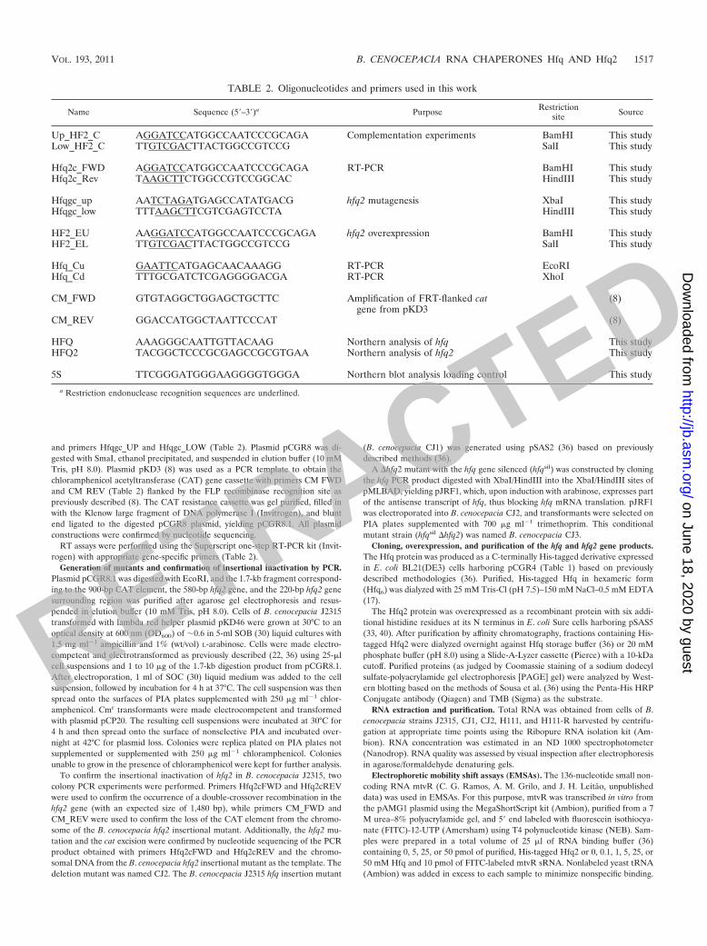
TABLE 2. Oligonucleotides and primers used in this work
Name Sequence (5�–3�)a Purpose Restrictionsite Source
Up_HF2_C AGGATCCATGGCCAATCCCGCAGA Complementation experiments BamHI This studyLow_HF2_C TTGTCGACTTACTGGCCGTCCG SalI This study
Hfq2c_FWD AGGATCCATGGCCAATCCCGCAGA RT-PCR BamHI This studyHfq2c_Rev TAAGCTTCTGGCCGTCCGGCAC HindIII This study
Hfqgc_up AATCTAGATGAGCCATATGACG hfq2 mutagenesis XbaI This studyHfqgc_low TTTAAGCTTCGTCGAGTCCTA HindIII This study
HF2_EU AAGGATCCATGGCCAATCCCGCAGA hfq2 overexpression BamHI This studyHF2_EL TTGTCGACTTACTGGCCGTCCG SalI This study
Hfq_Cu GAATTCATGAGCAACAAAGG RT-PCR EcoRIHfq_Cd TTTGCGATCTCGAGGGGACGA RT-PCR XhoI
CM_FWD GTGTAGGCTGGAGCTGCTTC Amplification of FRT-flanked catgene from pKD3
(8)
CM_REV GGACCATGGCTAATTCCCAT (8)
HFQ AAAGGGCAATTGTTACAAG Northern analysis of hfq This studyHFQ2 TACGGCTCCCGCGAGCCGCGTGAA Northern analysis of hfq2 This study
5S TTCGGGATGGGAAGGGGTGGGA Northern blot analysis loading control This study
a Restriction endonuclease recognition sequences are underlined.
VOL. 193, 2011 B. CENOCEPACIA RNA CHAPERONES Hfq AND Hfq2 1517
RETRACTED
on June 18, 2020 by guesthttp://jb.asm
.org/D
ownloaded from

After incubation for 15 min at 30°C, the mixtures were loaded onto a 10%polyacrylamide gel in 1 Tris-borate-EDTA (TBE) buffer containing 1% (vol/vol) glycerol. Electrophoresis was performed at 4°C and 150 V for 2 h. RNAswere electroblotted at 200 mA for 1 h onto a BrightStar-Plus (Ambion) mem-brane soaked in 0.5 TBE and UV cross-linked. RNAs were detected by chemi-luminescence using alkaline phosphatase-conjugated anti-FITC antibody (Am-ersham) in combination with the CDP-Star alkaline phosphatase substrate(Novagen).
Discontinuous native protein gel electrophoresis and circular dichroism (CD)experiments. Discontinuous native protein gel electrophoresis of purified, His-tagged Hfq2 was performed based on previously described methods (37). Briefly,aliquots of 20-�l protein samples (6 �g) were added to 5 �l of loading buffer (100mM Tris-Cl [pH 8.0], 40% glycerol, 0.5% Brilliant Blue G) and incubated for 10min at room temperature. To confirm the molecular mass of the Hfq2 oligomer,20-�g samples of the proteins aldolase (158 kDa), bovine serum albumin (66 and132 kDa), and ovalbumin (44 kDa) were used as molecular mass standards. Thesamples were visualized after 8% (wt/vol) discontinuous native PAGE (30) at 4°Cand 100 V for 2 h.
CD spectroscopy experiments were performed using 36 �g of purified, His-tagged Hfq2 in 20 mM phosphate buffer (pH 8.0) and processed as previouslydescribed (3). The far UV CD spectrum was recorded at 25°C in a cell with a1-mm path length using a PiStar-180 spectrometer (Applied Photophysics) at ascan speed of 60 nm/min, a time constant of 1 s, and a bandwidth of 2 nm. Theneural network analysis method was used to estimate the Hfq2 protein secondarystructure from the far UV CD spectrum data (1). Results are the means of atleast 10 wavelength scans.
Northern analysis. The levels of hfq or hfq2 mRNA were assessed by Northernblot analysis using 2 �g of total RNA purified from cells harvested from culturesin different growth phases. Samples containing 2 �g of total RNA were loadedinto an 8 M urea–6% polyacrylamide gel and separated by electrophoresis in 1 TBE at a constant current of 35 mA. After electrophoresis, total RNA waselectrotransferred to a Hybond-N� membrane (Amersham) at 20 V for 16 h at4°C using 0.5 TBE.
Oligonucleotide probes (Table 2) were FITC labeled using the Fluorescein-High Prime labeling kit (Roche) and purified using the Quick-Spin column forlabeled DNA purification (Roche). Prehybridization and hybridization were per-formed as previously described (14, 16). The 5S RNA was used as a loadingcontrol in all Northern blot experiments.
Hybridization signals were detected using a Typhoon 8600 variable-modeimager scanner (Amersham Biosciences Europe) after selecting the fluoresceinemission laser, blue filter, 500-V excitation, and 200-�m resolution. RelativemRNA abundance analysis was estimated with the UN-SCAN-IT gel softwaresuite (Silk Scientific, Inc.) using the band intensities of the 5S RNA as a refer-ence. Affinity constants (KD) were calculated by assuming a single binding siteand using the equation KD � [protein-bound RNA]/[total RNA].
DNA binding experiments. The ability of Hfq2 to bind DNA was assessedusing gel shift assays as previously described (28). For this purpose, 100 fmol ofthe FITC-labeled 124-bp DNA fragment (corresponding to part of the araCpromoter and coding region) was obtained by restriction of plasmid pMLBADwith EcoRV and ClaI, followed by agarose gel electrophoresis, band excision,and purification with the NucleoSpin Extract II kit (Macherey-Nagel). The pu-rified DNA fragments were incubated for 30 min at 25°C with His-tagged Hfq orHfq2 (0.2, 1, and 2 �M) in a total volume of 20 �l of DNA binding buffer (28)supplemented with 1 mM ATP and 100 nmol of yeast tRNA (Ambion). Controlexperiments were performed using the DNA probe and 500 pM Pbr (28). Allmixtures contained 10 nmol of the unlabeled 322-bp fragment obtained bypUC19 restriction with PvuII as competing nonlabeled DNA. After incubation,the reaction products were subjected to native 10% PAGE at 4°C and 150 V for3 h. Retardation corresponding to DNA-protein complexes was detected using aTyphoon 8600 variable-mode imager scanner with the fluorescein emission laser,blue filter, 650-V excitation, and 200-�m resolution (Amersham BiosciencesEurope). The affinity constant (KD) was calculated by assuming a single bindingsite and using the equation KD � [protein-bound DNA]/[total DNA].
Stress susceptibility experiments. The susceptibility of strains of B. cenocepa-cia (J2315, J2315 with plasmid pSAS3 or pCGR9, the CJ1 mutant, CJ1 withplasmid pSAS3 or pGCR9, the CJ2 mutant, CJ2 with plasmid pGCR9 or pSAS3,and the CJ3 mutant) and E. coli (MC4100, GS081, and GS081 with plasmidpSAS3 or pCGR9) to the stresses imposed by growth in solid LB mediumcontaining 1% (wt/vol) L-arabinose and supplemented with 3% (wt/vol) NaCl,150 �M methyl viologen (pH 5.0; [obtained with 100 mM phosphate-bufferedLB]), or growth at 42°C were performed as previously described (28). Results arethe means of at least five independent experiments.
Nematode infection experiments. Nematode killing assays and nematode di-gestive tract bacterial colonization experiments were performed based on previ-ously described methods (36) using Caenorhabditis elegans mutant strain DH26.Reported values are the means of triplicate results of at least three independentexperiments using a nematode population of 1,250 � 25 worms. Nematodes werephotographed at 24, 48, and 72 h postinfection (p.i.) to visually evaluate thecondition of their health (magnification shown is 40).
Nucleotide and amino acid sequence analysis and structure predictions. Nu-cleotide and amino acid sequences were analyzed using bioinformatic toolsavailable at the National Center for Biotechnology Information, the ExPaSyProsite, or the European Bioinformatics Institute. The Virtual Footprint suitewas used for promoter analysis (24) using Pseudomonas aeruginosa promotersequences.
Statistical analysis. All of the data presented were subjected to a one-wayanalysis of variance of mean values. Error bars represent the standard deviationof the mean.
RESULTS
Two distinct hfq-like genes are found in B. cenocepacia J2315chromosome I. In a previous work, we performed a functionalanalysis of the 79-amino-acid protein Hfq (BCAL1879 in thegenome sequence of B. cenocepacia J2315, Fig. 1A) from theclinical isolate B. cepacia IST408 and described the identifica-tion of a second 188-amino-acid putative Hfq-like protein in B.cenocepacia J2315, Hfq2 (36), which was the focus of this work.The gene encoding Hfq2 (BCAL1538) is also located in chro-mosome I, about 370 kb from the gene for Hfq (Fig. 1B), andis flanked by genes encoding predicted membrane proteins,exported proteins, regulatory proteins, and an acyl coenzymeA (CoA) synthetase (Fig. 1B).
Inspection of the complete genomes of other members ofthe Bcc revealed that all encode proteins highly similar to Hfqand Hfq2. Moreover, these Hfq-like protein-encoding genesare located in similar loci (data not shown).
Construction of hfq and hfq2 B. cenocepacia J2315 mutants.In order to gain insights into the biological functions of thissecond Hfq-like protein, two mutant strains were constructed,CJ2� and CJ2. CJ2� was derived from B. cenocepacia J2315 bythe insertion of a cat gene flanked by FRT sites. CJ2 wasobtained by excision of the cat gene from CJ2�, introducingthree stop codons after nucleotide position 211, thus inactivat-ing the hfq2 gene. The B. cenocepacia CJ1 mutant was alsoconstructed from B. cenocepacia J2315 by insertional inactiva-tion of the hfq gene with a trimethoprim resistance cassette(Fig. 1A and C) using plasmid pSAS2 and based on previouslydescribed methodologies (36). Despite several attempts to con-struct a �hfq �hfq2 double mutant, we were not successful.Therefore, a conditional mutant with the hfq gene silenced(hfqsil) was obtained by introducing pJRF1 into �hfq2 mutantstrain CJ2, yielding hfqsil �hfq2 mutant strain CJ3. Upon in-duction with arabinose, an hfq antisense RNA is expressedfrom plasmid pJRF1, thus silencing hfq in the �hfq2 back-ground. In order to confirm that the hfq and hfq2 genes werenot expressed in the CJ1 and CJ2� mutants, respectively, totalRNA was extracted from cells of wild-type strain B. cenocepa-cia J2315 and of the mutants CJ1 and CJ2� (before excision ofthe cat gene) and used in RT experiments. No amplificationbands corresponding to the hfq or hfq2 mRNA could be de-tected for the B. cenocepacia CJ1 and CJ2� mutants, respec-tively, thus confirming the inactivation of the hfq and hfq2genes (Fig. 2A). The CJ2 mutant was used thereafter to avoideffects due to the use of chloramphenicol.
1518 RAMOS ET AL. J. BACTERIOL.
RETRACTED
on June 18, 2020 by guesthttp://jb.asm
.org/D
ownloaded from

The hfq and hfq2 genes are differentially expressed. In orderto gain further clues to the regulation of both the hfq and thehfq2 genes, we investigated their mRNA levels in B. cenocepa-cia strain J2315. The levels of mRNA corresponding to hfq2increased about 2-fold, reaching maximal values in cells in thestationary phase of growth (Fig. 2B). In contrast, the levels ofmRNA corresponding to hfq were maximal in cells in the earlyexponential phase of growth (Fig. 2B), decreasing almost 2.5-fold in cells in the stationary phase (Fig. 2C).
The mRNA levels of hfq2, but not hfq, are quorum sensingdependent. A bioinformatic search for putative promoter se-quences upstream of the hfq2 gene revealed a putative luxbox-like consensus sequence located 50 bp upstream of thehfq2 gene ATG codon (Fig. 1B). In B. cenocepacia, CepR is aLuxR family transcription regulator which binds to lux boxsequences (42). Therefore, we used total RNA extracted fromthe well-studied B. cenocepacia isolate H111 and its derivativecepR mutant in Northern blot experiments to assess the mRNAlevels of hfq2 in a cepR-defective mutant. As shown in Fig. 2C,the band corresponding to the hfq2 mRNA is significantlyweaker in the cepR mutant than in the B. cenocepacia H111wild-type strain and is only detected at 24 h of growth.
The Hfq2 C terminus contains a repeated motif. Comparedto the 79-amino-acid protein Hfq, which is 100% identical tothe B. cepacia IST408 Hfq protein previously characterized byour research group (36), the B. cenocepacia J2315 Hfq2 proteinis 109 amino acid residues longer (with a total of 188 residues)(Fig. 3A). Alignment of the B. cenocepacia J2315 Hfq andHfq2 proteins and with other large Hfq proteins revealed that
the two proteins share a significant level of conservation be-tween residues 10 and 65, with 60% identity and 75% similarity(Fig. 3B). The typical conserved core motifs of Hfq chaperoneswere also highly conserved in Hfq2, namely, the Sm1 and Sm2motifs, structurally conserved loops 3 and 5, and RNA bindingresidues 10Gln, 62Ile, and 63Ser (Fig. 3A). The 8His, 9Pro,10Gln, 11Asn, 62Ile, 63Ser, 64Thr, 65Ile, and 66Gln residues inHfq2, which are predicted to be involved in substrate binding,were conserved only in Hfq proteins from Burkholderia strains(Fig. 3A).
Interestingly, the C-terminal extension of Hfq2 and the Hfqhomologues from B. cenocepacia MCO-3, B. vietnamensis G4,and B. multivorans ATCC 17616 were all composed of a glycine-rich region (70G to 173G), a putative DNA binding domain (89Gto 129E), and the repetitive motif RE(P/S)RRXX(E/G)(G/S),which occurs five times in Hfq2, as highlighted in Fig. 3A.
Hfq2 forms trimers in vitro. Secondary-structure predictionsindicate that 6% of the amino acid residues of Hfq2 fold intoan �-helix and 31% fold into a �-sheet. In loose agreementwith these predictions, calculations performed based on CDspectroscopy results indicate that B. cenocepacia Hfq2 iscomposed of 5% �-helix and 47% �-sheet (Fig. 4A). Thisdiscrepancy probably derives from the fact that the compu-tational structure prediction of B. cenocepacia J2315 Hfq2was based on the 69-amino-acid crystal structure of the E.coli Hfq protein.
The ability of purified, His-tagged Hfq2 (Fig. 4B) to formmultimeric structures was investigated by discontinuous nativePAGE. A major band with approximately three times the mo-
FIG. 1. Genetic organization of the hfq and hfq2 genes in B. cenocepacia J2315. (A) Genetic organization of the hfq gene locus showing thetrimethoprim (Tmp) cassette insertion. (B) Genetic organization of the hfq2 gene locus. Open reading frames BCAL1535 (membrane protein withunknown function), BCAL1536 (�54-dependent transcriptional regulator), BCAL1537 (putatively exported lipoprotein), BCAL1538 (Hfq2 chap-erone), BCAL1539 (putative exported protein), BCAL1540 (transmembrane lipoprotein), BCAL1541 (acyl-CoA synthetase), BCAL1542 (TetRfamily regulatory protein), and BCAL1543 (major facilitator superfamily protein) are represented in scale. The positions of the three nonsense stopsignals introduced after nucleotide 211 are indicated by the filled flag (hfq2::Frt). Primers used in RT-PCR experiments are indicated by the lettersF (Hfq2c_Fwd) and R (Hfq2c_Rev). The predicted CepR-dependent promoter is also indicated. (C) PCR amplification of the hfq and hfq2 genesfrom B. cenocepacia wild-type (WT) strain J2315 and mutant strains CJ1 (�hfq) and CJ2 (�hfq2).
VOL. 193, 2011 B. CENOCEPACIA RNA CHAPERONES Hfq AND Hfq2 1519
RETRACTED
on June 18, 2020 by guesthttp://jb.asm
.org/D
ownloaded from

lecular mass of monomeric Hfq2 suggests that the proteinmight exist as a trimer in its active/native form in vitro. Thisresult contrasts with the findings for other Hfq proteins and theHfq protein of B. cenocepacia J2315, which forms hexamers invitro (36). A faint band, highlighted in Fig. 4B with an asterisk,with an apparent molecular mass compatible with a tetramericform of Hfq2 was also observed. Nevertheless, the trimericform is predominant.
B. cenocepacia J2315 Hfq and Hfq2 bind to the sRNA mtvR.Since both Hfq and Hfq2 possess all of the motifs required forRNA binding, we tested their ability to bind the mtvR sRNA byusing EMSAs. Results shown in Fig. 5 clearly show that bothproteins are able to bind this sRNA. Interestingly, Hfq seems tobind mtvR more efficiently (60-fold lower KD) than Hfq2 does.
Hfq2, but not Hfq, binds DNA. Since a DNA binding domainspanning amino acid residues 87 to 128 was bioinformaticallypredicted for Hfq2, we performed DNA binding assays. Forthis purpose, we used either purified, His-tagged Hfq or Hfq2and the 124-bp EcoRV/ClaI DNA fragment of the araC pro-moter of pMLBAD (17). The purified, His-tagged Pbr regula-
tory protein, which is involved in the regulation of B. cenoce-pacia K56-2 phenazine biosynthesis and cellular processesrelated to stress resistance and virulence (28), was used as apositive control. Results presented in Fig. 6 clearly show thatHfq2 is able to bind DNA, consistent with the predicted oc-currence of a DNA binding domain. An apparent bindingconstant of 92 nM for the Pbr regulator to the DNA fragmentwas estimated, while a value of 44.2 �M was estimated for thebinding of Hfq2 to this particular DNA fragment. Similar ex-periments performed using Hfq instead of Hfq2 did not revealthe binding of Hfq to this DNA fragment (data not shown).The biological significance of the ability of Hfq2 to bind DNAis unknown, although we speculate that the protein might actas a transcription regulator.
Hfq2 is required for resistance to stress. We compared theabilities of wild-type B. cenocepacia J2315 and its derivativemutants CJ1 and CJ2 (carrying nonfunctional hfq and hfq2genes, respectively) to survive stress conditions mimickingthose faced by the bacterium when colonizing/infecting the CFlung, namely, growth at 42°C, oxidative stress conditions im-
FIG. 2. Expression analysis of the hfq and hfq2 genes. (A) RT of hfq and hfq2 RNA samples obtained from cells of B. cenocepacia wild-type(WT) strain J2315 and the CJ1 (�hfq) and CJ2� (�hfq2) mutants. Control experiments were carried out using chromosomal DNA. (B) Northernblot analysis of mRNA corresponding to hfq and hfq2 in B. cenocepacia J2315. Relative abundance was quantified for both hfq (black bars) andhfq2 (white bars) in B. cenocepacia J2315 using the RNA levels of the 5S rRNA as a control. Error bars represent the standard deviations of themeans. (C) Detection of hfq and hfq2 mRNA levels in B. cenocepacia wild-type strain H111 and a cepR mutant strain by Northern blotting.Quantification of hfq mRNA in the B. cenocepacia wild-type H111 (black bars) and �cepR mutant (white bars) strains and of hfq2 transcripts inthe B. cenocepacia wild-type H111 (gray bars) and �cepR mutant (dashed bars) strains was performed using the 5S rRNA mRNA levels as acontrol. All experiments were repeated at least three times.
1520 RAMOS ET AL. J. BACTERIOL.
RETRACTED
on June 18, 2020 by guesthttp://jb.asm
.org/D
ownloaded from

posed by methyl viologen, acidic pH, and osmotic stress in-duced by the addition of 3% NaCl to the culture medium. TheCJ3 (hfqsil �hfq2) strain was also included in this analysis. CJ3carries a nonfunctional hfq2 gene, and upon induction withL-arabinose, it expresses an antisense RNA targeting hfq. Incomparison with wild-type strain J2315 overexpressing the hfq
or hfq2 gene, the results clearly indicate that mutation of thehfq or the hfq2 gene rendered the respective strain CJ1 or CJ2more susceptible to the tested stress. This indicates that bothgenes are required for optimal survival of various stresses (Fig.7). The increased susceptibility of these mutants was at leastpartially rescued by the ectopic expression of hfq or hfq2. This
FIG. 3. Hfq2 is highly conserved within the Bcc. (A) Alignment of the B. cenocepacia J2315 Hfq and Hfq2 proteins with other Hfq-like proteinsshowing the predicted secondary structure of Hfq2 above the amino acid residues. The Sm1 and Sm2 motifs are indicated. RNA binding domainsare represented by blue boxes. Residues also predicted to be involved in RNA binding are highlighted in yellow. The DNA binding domain is withinthe gray box. Boxed sequences denote the identified repeats. Identical amino acid residues are marked with an asterisk, and conserved andsemiconserved substitutions are marked with double and single dots, respectively. Abbreviations: Bcen, B. cenocepacia; Bviet, B. vietnam-ensis; Bmul, B. multivorans; Nmening, Neisseria meningitidis; Mcatar, Moraxella catarrhalis. (B) Superimposition of the monomeric predictedthree-dimensional structures of Hfq and Hfq2 from B. cenocepacia J2315. The structural alignment is highlighted in yellow, with a root meansquare deviation of 0.910 Å.
FIG. 4. Hfq2 forms trimeric structures in vitro. (A) Far UV CD spectrum of Hfq2 from B. cenocepacia J2315. The regions of the spectrumcorresponding to the �-helix and �-sheet are indicated. (B) Discontinuous native PAGE showing a predominant Hfq2 band at �63 kDa compatiblewith a trimeric form of the protein. The monomeric form is also visible. Possible higher-order complexes (marked by an asterisk) can be seen. BSA,bovine serum albumin.
VOL. 193, 2011 B. CENOCEPACIA RNA CHAPERONES Hfq AND Hfq2 1521
RETRACTED
on June 18, 2020 by guesthttp://jb.asm
.org/D
ownloaded from

partial complementation has been observed before (28) and isderived from insufficient amounts of L-arabinose to fully in-duce the PBAD promoter. Remarkably, the CJ3 strain exhibitedthe highest susceptibility to the stresses studied, reinforcing theobservation that both the hfq and hfq2 genes are required foroptimal survival of B. cenocepacia J2315 under stress condi-tions. Overexpression of hfq rendered B. cenocepacia wild-typestrain J2315 more resistant to methyl viologen and NaCl, whileoverexpression of hfq2 rendered the wild-type strain more re-sistant to growth at 42°C and pH 5.0. Both the hfq and hfq2genes of B. cenocepacia J2315 were used in complementationexperiments with E. coli hfq mutant strain GS081. Resultsshown in Fig. 8 indicate that both genes are functional in E.coli, being able to partially rescue the observed growth defectsof the E. coli hfq mutant when it is exposed to oxidative stressimposed by methyl viologen. It is worth noting that hfq2 wasable to fully complement the ability of the E. coli hfq mutant togrow at 42°C or pH 5.0. Remarkably, when hfq2 was providedin trans to the E. coli hfq mutant, this strain became lesssusceptible to the osmotic stress imposed by 3% NaCl, com-pared with E. coli wild-type strain MC4100.
hfq and hfq2 are both required for full virulence of B. ceno-cepacia J2315. The role played by hfq and hfq2 in B. cenoce-pacia J2315 virulence using C. elegans as an infection modelwas investigated based on slow-killing assays (36). The abilitiesof wild-type B. cenocepacia J2315, hfq mutant strain CJ1, hfq2mutant CJ2 strain, and the CJ3 antisense knockdown strain, aswell as the wild-type strain overexpressing hfq or hfq2 in trans,to kill C. elegans larvae at the L4 stage were compared.
Results shown in Fig. 9A clearly indicate that overexpressionof hfq rendered the wild-type strain more virulent. This wasalso observed, although to a lesser extent, until the second dayof infection by the wild-type strain overexpressing hfq2. Com-pared to the wild-type strain, the CJ3 strain was significantlyattenuated, and this effect was particularly evident at 3 or 4days p.i. Figure 9 also clearly indicates that mutations in hfq(panel B) or hfq2 (panel C) significantly reduced the virulenceof B. cenocepacia J2315. Although general overexpression of
hfq or hfq2 by the CJ1 or CJ2 mutant led to numbers ofsurviving worms comparable to or below those of worms sur-viving infection by the wild-type strain, a reduction in survivalwas observed when worms were challenged with the CJ2 mu-tant overexpressing hfq.
The total number of CFU surviving within the nematode’sdigestive tract was consistent with the relative virulence of thewild-type strain, the CJ1 and CJ2 mutants, and the comple-mented CJ1 and CJ2 mutants (Fig. 9D). Visual inspection ofworms infected at 24, 48, or 72 h p.i. with B. cenocepacia J2315,the CJ2 mutant, or the complemented CJ2 mutant showed thatworms infected with the CJ2 mutant appeared healthier, inagreement with the requirement of a functional hfq2 gene forthe full virulence of B. cenocepacia J2315 (panel E).
DISCUSSION
Bcc members are among a small number of prokaryotes thatencode two distinct copies (highly conserved among Bcc mem-bers [27]) of Hfq-like proteins in their genomes, and to the bestof our knowledge, this is the first study of a bacterium harbor-ing two distinct and functional Hfq-like proteins.
The similarity found between Hfq2 and the Sm1 motif of theSm and Lsm proteins from eukaryotes and archaea suggeststhat these bacterial proteins derive from an ancestral Sm pro-tein (29). In the case of archaea, it was proposed that theancestral Sm-like protein was subjected to several duplicationevents, producing the present related Sm-like proteins in yeast,humans, and other eukaryotes (29). Since no significant ho-mology of the C-terminal extension of Hfq2 could be foundwithin other bacterial genome sequence databases besides Bcc,and considering the limited number of bacteria with two dis-tinct Hfq-like genes, we hypothesize that both hfq and hfq2have arisen in the genomes of Bcc through the duplication ofan ancestor gene, followed by evolutionary events that led totwo distinct and functional Hfq-like proteins in the current Bccgenomes.
A recent analysis of the genome sequence of B. cenocepaciaJ2315 suggested that this bacterium has recently adapted to the
FIG. 6. B. cenocepacia J2315 Hfq2 is able to bind DNA. The abilityof Hfq2 to bind DNA was evaluated by band shift assays. The Pbrtranscription regulator (28) was used as a positive control. All bindingkinetic parameters were calculated by assuming a fixed-term single-sitebinding isotherm.
FIG. 5. B. cenocepacia J2315 Hfq2 is able to bind sRNAs.(A) EMSA experiments using 0, 0.2, 1, and 2 �M purified, His-tagged[Hfq2]3 and 400 nM FITC-12-UTP 5�-end-labeled mtvR sRNA.(B) With increasing [Hfq]6, additional Hfq multimers bind to the mtvRsRNA. [Hfq2]3, trimeric form of Hfq2; [Hfq]6, hexameric form of Hfq;R-H, complexes formed by [Hfq2]3 and the mtvR sRNA; R.H, com-plexes formed by [Hfq]6 and the mtvR sRNA (R).
1522 RAMOS ET AL. J. BACTERIOL.
RETRACTED
on June 18, 2020 by guesthttp://jb.asm
.org/D
ownloaded from

ecological niche that is the human host, evidenced by a genomewell equipped with functions associated with virulence in theCF lung, where the organism must face adverse conditions,including oxidative stress conditions associated with survivalwithin macrophages (12). Our results show that both Hfq-likeproteins are required for and contribute to the bacterium’ssurvival of stresses related to the colonization and infection of
the CF host, particularly evidenced by the phenotypes of stresssusceptibility observed in the CJ3 strain, which harbors a non-functional hfq2 gene and expresses an antisense RNA targetinghfq. This strain was constructed because we were unable togenerate a stable �hfq �hfq2 double mutant.
Interestingly, results also show that overexpression of hfq inthe wild-type strain led to increased resistance to pH 5.0 and
FIG. 7. Both Hfq and Hfq2 are required for stress resistance. The susceptibility of the indicated B. cenocepacia strains to various stresses wastested by spot inoculating serially diluted bacterial suspensions with an initial OD640 of 1.0. wt, wild type.
FIG. 8. B. cenocepacia J2315 Hfq and Hfq2 are able to complement an E. coli hfq mutation. The ability of the B. cenocepacia hfq or hfq2 geneto complement the hfq mutation in E. coli GS081 was evaluated by subjecting the indicated E. coli strains to the stresses indicated. phfq, E. coliGS081 transformed with pSAS3; phfq2, E. coli GS081 transformed with pCGR9; wt, wild type.
VOL. 193, 2011 B. CENOCEPACIA RNA CHAPERONES Hfq AND Hfq2 1523
RETRACTED
on June 18, 2020 by guesthttp://jb.asm
.org/D
ownloaded from

3% NaCl, while the wild-type strain overexpressing hfq2 exhib-ited increased resistance to thermal stress and to 150 �Mmethyl viologen. These results suggest that, besides a commonrole, as evidenced by the ability of both Hfq and Hfq2 tocomplement an E. coli hfq mutant used in this study, eachprotein might play additional roles related to specific stresses.This specificity possibly derives from the different C termini ofHfq and Hfq2.
Large Hfq proteins with an extended C terminus have beendescribed in some bacterial species of the beta- and gamma-proteobacteria. Glycine-rich domains between the N and Ctermini (31) have been described for the few large Hfq proteinscharacterized so far. B. cenocepacia Hfq2 also contains a gly-cine-rich domain and a repetitive pattern that occurs five timesin its C terminus (27), although no biological function has yetbeen assigned. A repetitive but distinct pattern was describedfor Acinetobacter baylyi Hfq which was also found in DnaJ-likechaperones (41), but no functions were attributed to thesefeatures of the protein. The biological function of the C ter-minus of the 210-amino-acid Hfq protein from Moraxella ca-tarrhalis is also unknown (2).
In the case of E. coli, Veccerek et al. (40) have shown thattruncated variants of Hfq lacking the C-terminal extension,effectively bind sRNAs but are defective in the establishmentof sRNA-mRNA base pairing, as well as in Hfq autoregulation,suggesting that the C terminus constitutes an unrecognized
RNA interaction surface with specificity for mRNAs (40). Al-though E. coli Hfq and B. cenocepacia Hfq2 are quite distinct,we cannot exclude the possibility that the B. cenocepacia J2315Hfq2 C terminus plays roles similar to those of the E. coli HfqC terminus. A putative DNA binding domain was also identi-fied in the C-terminal extension of Hfq2. Although we presentresults clearly showing that Hfq2 is able to bind DNA, both thespecificity of the interaction and its biological significance re-main unknown. Curiously, a recent report on the ability of E.coli Hfq to bind DNA highlighted the importance of the Cterminus in DNA displacement, especially for residues beyondposition 75 (38). This amino acid stretch is absent from the B.cenocepacia J2315 Hfq protein.
Results of structure prediction and CD spectroscopy exper-iments indicate that the Hfq2 glycine-rich C terminus is pre-dominantly unstructured. We anticipate that this disorderedregion contributes to the difficulty in assembling the proteininto a stable multimeric form greater than three monomers. Itis worth noting that this contrasts with the hexameric struc-tures of all of the bacterial small Hfq proteins studied so far (4,19, 26). Moreover, a recent report by Moskaleva et al. (23)highlights the important role played by histidine residue 57 inthe establishment of a stable hexameric structure. Curiously, inB. cenocepacia Hfq2, 57H is replaced by 57R, which, togetherwith steric constraints, may account for its inability to formhexamers.
FIG. 9. Hfq and Hfq2 are both required for full B. cenocepacia J2315 virulence in the nematode C. elegans. (A) Abilities of B. cenocepaciawild-type strain J2315 (wt, black bars), J2315 harboring pSAS3 (wt � phfq, dashed bars) or pCGR9 (wt � phfq2, squared bars), and CJ3 (hfqsil
�hfq2, dotted bars) to kill the nematode C. elegans DH26. (B) Abilities of B. cenocepacia hfq mutant CJ1 (�hfq, gray bars) and CJ1 harboring pSA3(�hfq � phfq, dashed gray bars) or pCGR9 (�hfq � phfq2, squared gray bars) to kill the nematode C. elegans DH26. (C) Abilities of the B.cenocepacia hfq2 mutant CJ2 (�hfq2, white bars) and CJ2 harboring pSAS3 (�hfq2 � phfq, dashed bars) or pCGR9 (�hfq � phfq2, squared bars)to kill the nematode C. elegans DH26. (D) Abilities of B. cenocepacia J2315 (wt, black bars), CJ1 (�hfq, gray bars), CJ2 (�hfq2, white bars), CJ1harboring pSA3 (�hfq � phfq, dashed gray bars), and CJ2 harboring pCGR9 (�hfq � phfq2, squared bars) to colonize the nematode’s digestivetract, expressed as the number of CFU/worm. (E) The health status of worms after infection for 24, 48, or 72 h with the indicated strains wasassessed by visual inspection. Worms in panel E were randomly chosen and photographed.
1524 RAMOS ET AL. J. BACTERIOL.
RETRACTED
on June 18, 2020 by guesthttp://jb.asm
.org/D
ownloaded from

Given the distinct pattern of regulation of expression ob-served for the two hfq-like genes of B. cenocepacia J2315, withthe RNA levels of hfq maximal in cells in the early exponentialphase of growth and the levels of hfq2 RNA being maximal inthe stationary phase of growth, it is expected that Hfq andHfq2 levels must be strictly regulated in the bacterial cell, withthis strictly balanced level of the two proteins being requiredfor optimal survival of stress and efficient virulence. Interest-ingly, our data point out that quorum sensing triggers theexpression of hfq2, allowing its maximal expression when hfqmRNA levels are minimal. The differential regulation of hfqand hfq2 might fulfill specific cellular requirements for each ofthe two proteins in cultures in different growth phases. Ourdata also suggest that, besides playing the same roles in cellphysiology, Hfq and Hfq2 might have distinct functions. This iswell illustrated by the increased resistance to high temperatureand low pH conferred by the overexpression of Hfq in wild-type strain J2315 and by the increased resistance of the wild-type strain to osmotic and oxidative stress when overexpressingHfq2. Both proteins also seem to play somehow distinct rolesin virulence. In fact, while Hfq2 overexpression has a morepronounced effect on the rate of C. elegans killing, overexpres-sion of Hfq led to higher numbers of bacterial CFU colonizingthe nematode digestive tract. Nevertheless, both proteins arerequired for virulence, since the simultaneous inactivation ofthe two Hfq-like proteins results in reduced virulence com-pared with that of the respective single mutants. Taking thesedata together and considering the high level of conservation ofthese proteins in Bcc bacteria, results from this work suggestthat although there is a partial redundancy between the func-tions of these two RNA chaperones, they probably have addi-tional functions in the biology of Bcc bacteria.
Ongoing work envisages the understanding of the specificroles played by each of the two Hfq-like proteins in the ex-pression of genes associated with the stress resistance andvirulence of B. cenocepacia J2315.
ACKNOWLEDGMENTS
We thank the Caenorhabditis Genetics Center (University of Min-nesota) for the kind gift of C. elegans DH26, Leo Eberl for B. ceno-cepacia H111 and H111-cepR, and the Coli Genetic Stock Center(Yale University) for plasmids pKD46, pKD13, and pCP20. We thankA. Azevedo and G. Gomes for help with CD experiments.
This work was supported by Fundacao Ciencia e Tecnologia, Por-tugal (contracts PTDC/BIA-MIC/65210/2006 and PTDC/EBB-BIO/098352/2008); a postdoctoral grant to S.A.S.; and a Fundacao CalousteGulbenkian doctoral grant to C.G.R.
REFERENCES
1. Andrade, M. A., P. Chacon, J. J. Merelo, and F. Moran. 1993. Evaluation ofsecondary structure of proteins from UV circular dichroism using an unsu-pervised learning neural network. Protein Eng. 6:383–390.
2. Attia, A. S., et al. 2008. Moraxella catarrhalis expresses an unusual Hfqprotein. Infect. Immun. 76:2520–2530.
3. Brahms, S., and J. Brahms. 1980. Determination of protein secondary struc-ture in solution by vacuum ultraviolet circular dichroism. J. Mol. Biol. 138:149–178.
4. Brennan, R. G., and T. M. Link. 2007. Hfq structure, function and ligandbinding. Curr. Opin. Microbiol. 10:125–133.
5. Brenner, S. 1974. The genetics of Caenorhabditis elegans. Genetics 77:71–94.6. Casadaban, M. J. 1976. Transposition and fusion of the lac genes to selected
promoters in Escherichia coli using bacteriophage lambda and Mu. J. Mol.Biol. 104:541–555.
7. Chao, Y., and J. Vogel. 2010. The role of Hfq in bacterial pathogens. Curr.Opin. Microbiol. 13:24–33.
8. Datsenko, K. A., and B. L. Wanner. 2000. One-step inactivation of chromo-
somal genes in Escherichia coli K-12 using PCR products. Proc. Natl. Acad.Sci. U. S. A. 97:6640–6645.
9. Gottesman, S. 2004. The small RNA regulators in Escherichia coli: roles andmechanisms. Annu. Rev. Microbiol. 58:303–328.
10. Govan, J. R., and V. Deretic. 1996. Microbial pathogenesis in cystic fibrosis:mucoid Pseudomonas aeruginosa and Burkholderia cepacia. Microbiol. Rev.60:539–574.
11. Govan, J. R. W., et al. 1993. Evidence for transmission of Pseudomonascepacia by social contact in cystic fibrosis. Lancet 342:15–19.
12. Holden, M. T., et al. 2009. The genome of Burkholderia cenocepaciaJ2315, an epidemic pathogen of cystic fibrosis patients. J. Bacteriol.191:261–277.
13. Kajitani, M., A. Kato, A. Wada, Y. Inokuchi, and A. Ishihama. 1994. Reg-ulation of the Escherichia coli hfq gene encoding the host factor for phageQ�. J. Bacteriol. 176:531–534.
14. Kawano, M., A. A. Reynolds, J. Miranda-Rios, and G. Storz. 2005. Detectionof 5�- and 3�-UTR-derived small RNAs and cis-encoded antisense RNAs inEscherichia coli. Nucleic Acids Res. 33:1040–1050.
15. Kothe, M., et al. 2003. Killing of Caenorhabditis elegans by Burkholderiacepacia is controlled by the cep quorum-sensing system. Cell. Microbiol.5:343–351.
16. Lagos-Quintana, M., R. Rauhut, W. Lendeckel, and T. Tuschl. 2001. Iden-tification of novel genes coding for small expressed RNAs. Science 294:853–858.
17. Lefebre, M. D., and M. A. Valvano. 2002. Construction and evaluation ofplasmid vectors optimized for constitutive and regulated gene expression inBurkholderia cepacia complex isolates. Appl. Environ. Microbiol. 68:5956–5964.
18. Leitao, J. H., et al. 2010. Pathogenicity, virulence factors, and strategies tofight against Burkholderia cepacia complex pathogens and related species.Appl. Microbiol. Biotechnol. 87:31–40.
19. Link, T. M., P. Valentin-Hansen, and R. G. Brennan. 2009. Structure ofEscherichia coli Hfq bound to polyriboadenylate RNA. Proc. Natl. Acad. Sci.U. S. A. 106:19292–19297.
20. Meselson, M., and R. Yuan. 1968. DNA restriction enzyme from E. coli.Nature 217:1110–1114.
21. Møller, T., et al. 2002. Hfq: a bacterial Sm-like protein that mediates RNA-RNA interaction. Mol. Cell 9:23–30.
22. Moreira, L. M., et al. 2003. Identification and physical organization of thegene cluster involved in the biosynthesis of Burkholderia cepacia complexexopolysaccharide. Biochem. Biophys. Res. Commun. 312:323–333.
23. Moskaleva, O., et al. 2010. The structures of mutant forms of Hfq fromPseudomonas aeruginosa reveal the importance of the conserved His57 forthe protein hexamer organization. Acta Crystallogr. Sect. F Struct. Biol.Cryst. Commun. 66(Pt. 7):760–764.
24. Munch, R., et al. 2005. Virtual Footprint and PRODORIC: an integrativeframework for regulon prediction in prokaryotes. Bioinformatics 21:4187–4189.
25. Papenfort, K., and J. Vogel. 2010. Regulatory RNA in bacterial pathogens.Cell Host Microbe 8:116–127.
26. Rajkowitsch, L., and R. Schroeder. 2007. Dissecting RNA chaperone activ-ity. RNA 13:2053–2060.
27. Ramos, C. G., A. M. Grilo, and J. H. Leitao. 2010. Small non-coding RNAsin prokaryotes: roles in pathogenesis and potential therapeutic targets. Lam-bert Academic Publishing, Saarbrucken, Germany.
28. Ramos, C. G., S. A. Sousa, A. M. Grilo, L. Eberl, and J. H. Leitao. 2010. TheBurkholderia cenocepacia K56-2 pleiotropic regulator Pbr, is required forstress resistance and virulence. Microb. Pathog. 48:168–177.
29. Salgado-Garrido, J., E. Bragado-Nilsson, S. Kandels-Lewis, and B.Seraphin. 1999. Sm and Sm-like proteins assemble in two related complexesof deep evolutionary origin. EMBO J. 18:3451–3462.
30. Sambrook, J., and D. W. Russell. 2001. Molecular cloning: a laboratorymanual, 3rd ed. Cold Spring Harbor Laboratory Press, Cold Spring Har-bor, NY.
31. Schilling, D., and U. Gerischer. 2009. The Acinetobacter baylyi hfq geneencodes a large protein with an unusual C terminus. J. Bacteriol. 191:5553–5562.
32. Schirmer, F., S. Ehrt, and W. Hillen. 1997. Expression, inducer spectrum,domain structure, and function of MopR, the regulator of phenol degra-dation in Acinetobacter calcoaceticus NCIB8250. J. Bacteriol. 179:1329–1336.
33. Sousa, S. A., L. M. Moreira, and J. H. Leitao. 2008. Functional analysis ofthe Burkholderia cenocepacia J2315 BceAJ protein with phosphomannoseisomerase and GDP-D-mannose pyrophosphorylase activities. Appl. Micro-biol. Biotechnol. 80:1015–1022.
34. Sousa, S. A., et al. 2007. The Burkholderia cepacia bceA gene encodes aprotein with phosphomannose isomerase and GDP-D-mannose pyrophos-phorylase activities. Biochem. Biophys. Res. Commun. 353:200–206.
35. Sousa, S. A., C. G. Ramos, and J. H. Leitao. 2011. Burkholderia cepaciacomplex: emerging multihost pathogens equipped with a wide range ofvirulence factors and determinants. Int. J. Microbiol. 2011:607575.
36. Sousa, S. A., C. G. Ramos, L. M. Moreira, and J. H. Leitao. 2010. The hfq
VOL. 193, 2011 B. CENOCEPACIA RNA CHAPERONES Hfq AND Hfq2 1525
RETRACTED
on June 18, 2020 by guesthttp://jb.asm
.org/D
ownloaded from

gene is required for stress resistance and full virulence of Burkholderiacepacia to the nematode Caenorhabditis elegans. Microbiology 156:896–908.
37. Sutherland, B. W., J. Toews, and J. Kast. 2008. Utility of formaldehydecross-linking and mass spectrometry in the study of protein-protein interac-tions. J. Mass Spectrom. 43:699–715.
38. Updegrove, T. B., J. J. Correia, R. Galletto, W. Bujalowski, and R. M.Wartell. 2010. E. coli DNA associated with isolated Hfq interacts with Hfq’sdistal surface and C-terminal domain. Biochim. Biophys. Acta 1799:588–596.
39. Valentin-Hansen, P., M. Eriksen, and C. Udesen. 2004. The bacterial Sm-like protein Hfq: a key player in RNA transactions. Mol. Microbiol. 51:1525–1533.
40. Veccerek, B., L. Rajkowitsch, E. Sonnleitner, R. Schroeder, and U. Blasi.2008. The C-terminal domain of Escherichia coli Hfq is required for regu-lation. Nucleic Acids Res. 36:133–143.
41. Walsh, P., D. Bursa, Y. C. Law, D. Cyr, and T. Lithgow. 2004. The J-proteinfamily: modulating protein assembly, disassembly and translocation. EMBORep. 5:567–571.
42. Williams, P., and M. Camara. 2009. Quorum sensing and environmentaladaptation in Pseudomonas aeruginosa: a tale of regulatory networks andmultifunctional signal molecules. Curr. Opin. Microbiol. 12:182–191.
43. Zhang, A., K. M. Wassarman, J. Ortega, A. C. Steven, and G. Storz. 2002.The Sm-like Hfq protein increases OxyS RNA interaction with targetmRNAs. Mol. Cell 9:11–22.
1526 RAMOS ET AL. J. BACTERIOL.
RETRACTED
on June 18, 2020 by guesthttp://jb.asm
.org/D
ownloaded from

Retraction for Ramos et al., The Second RNA Chaperone, Hfq2, IsAlso Required for Survival under Stress and Full Virulence ofBurkholderia cenocepacia J2315
Christian G. Ramos, Sílvia A. Sousa, André M. Grilo, Joana R. Feliciano, and Jorge H. Leitão
IBB—Institute for Biotechnology and Bioengineering, Centre for Biological and Chemical Engineering, Instituto Superior Técnico, Av. Rovisco Pais, 1049-001 Lisbon,Portugal
Volume 193, no. 7, p.1515–1526, 2011. Problems related to images published in this paper have been brought to our attention. Figure8 contains duplicated images as well as images previously published in articles in Microbiology and Microbial Pathogenesis, i.e., thefollowing:
S. A. Sousa, C. G. Ramos, L. M. Moreira, and J. H. Leitão, Microbiology 156:896 –908, 2010. http://dx.doi.org/10.1099/mic.0.035139-0.
C. G. Ramos, S. A. Sousa, A. M. Grilo, L. Eberl, and J. H. Leitão, Microb. Pathog. 48:168 –177, 2010. http://dx.doi.org/10.1016/j.micpath.2010.02.006.
Therefore, we retract the paper. We deeply regret this situation and apologize for any inconvenience to the editors and readers of Journalof Bacteriology, Microbial Pathogenesis, and Microbiology.
Copyright © 2014, American Society for Microbiology. All Rights Reserved.
doi:10.1128/JB.02242-14
RETRACTION
3980 jb.asm.org Journal of Bacteriology p. 3980 November 2014 Volume 196 Number 22