chemistry ofdownload.e-bookshelf.de/download/0002/7167/66/l-g-0002716766... · instrumental...
TRANSCRIPT


CHEMISTRY OFMETALLOPROTEINS

WILEY SERIES IN PROTEIN AND PEPTIDE SCIENCE
VLADIMIR N. UVERSKY, Series Editor
Metalloproteomics • Eugene A. Permyakov
Instrumental Analysis of Intrinsically Disordered Proteins: Assessing Structure andConformation • Vladimir Uversky and Sonia Longhi
Protein Misfolding Diseases: Current and Emerging Principles and Therapies •
Marina Ramirez-Alvarado, Jeffery W. Kelly, and Christopher M. Dobson
Calcium Binding Proteins • Eugene A. Permyakov and Robert H. Kretsinger
Protein Chaperones and Protection from Neurodegenerative Diseases• Stephan Witt
Transmembrane Dynamics of Lipids • Philippe Devaux and Andreas Herrmann
Flexible Viruses: Structural Disorder in Viral Proteins • Vladimir Uversky and SoniaLonghi
Protein Families: Relating Protein Sequence, Structure, and Function • Christine A.Orengo and Alex Bateman
Chemistry of Metalloproteins: Problems and Solutions in Bioinorganic Chemistry •Joseph J. Stephanos and Anthony W. Addison

CHEMISTRY OFMETALLOPROTEINS
Problems and Solutions inBioinorganic Chemistry
JOSEPH J. STEPHANOS
ANTHONY W. ADDISON

Copyright 2014 by John Wiley & Sons, Inc. All rights reserved
Published by John Wiley & Sons, Inc., Hoboken, New JerseyPublished simultaneously in Canada
No part of this publication may be reproduced, stored in a retrieval system, or transmitted in any formor by any means, electronic, mechanical, photocopying, recording, scanning, or otherwise, except aspermitted under Section 107 or 108 of the 1976 United States Copyright Act, without either the priorwritten permission of the Publisher, or authorization through payment of the appropriate per-copy fee tothe Copyright Clearance Center, Inc., 222 Rosewood Drive, Danvers, MA 01923, (978) 750-8400,fax (978) 750-4470, or on the web at www.copyright.com. Requests to the Publisher for permissionshould be addressed to the Permissions Department, John Wiley & Sons, Inc., 111 River Street, Hoboken,NJ 07030, (201) 748-6011, fax (201) 748-6008, or online at http://www.wiley.com/go/permission.
Limit of Liability/Disclaimer of Warranty: While the publisher and author have used their best effortsin preparing this book, they make no representations or warranties with respect to the accuracy orcompleteness of the contents of this book and specifically disclaim any implied warranties ofmerchantability or fitness for a particular purpose. No warranty may be created or extended by salesrepresentatives or written sales materials. The advice and strategies contained herein may not be suitablefor your situation. You should consult with a professional where appropriate. Neither the publisher norauthor shall be liable for any loss of profit or any other commercial damages, including but not limited tospecial, incidental, consequential, or other damages.
For general information on our other products and services or for technical support, please contact ourCustomer Care Department within the United States at (800) 762-2974, outside the United Statesat (317) 572-3993 or fax (317) 572-4002.
Wiley also publishes its books in a variety of electronic formats. Some content that appears in print maynot be available in electronic formats. For more information about Wiley products, visit our web siteat www.wiley.com.
Library of Congress Cataloging-in-Publication Data:
Stephanos, Joseph J., author.Chemistry of metalloproteins: problems and solutions in bioinorganic chemistry/by Joseph J.
Stephanos, Anthony W. Addison.p.; cm. – (Wiley series in protein and peptide science)
Includes bibliographical references and index.ISBN 978-1-118-47044-2 (paperback)
I. Addison, A. W., author. II. Title. III. Series: Wiley series in protein and peptide science.[DNLM: 1. Metalloproteins–chemistry–Examination Questions. QU 18.2]QP5515720.6076–dc23
2013041995
Printed in the United States of America
10 9 8 7 6 5 4 3 2 1
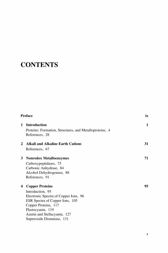
CONTENTS
Preface ix
1 Introduction 1Proteins: Formation, Structures, and Metalloproteins, 4References, 28
2 Alkali and Alkaline Earth Cations 31References, 67
3 Nonredox Metalloenzymes 71Carboxypeptidases, 75Carbonic Anhydrase, 84Alcohol Dehydrogenase, 88References, 91
4 Copper Proteins 95Introduction, 95Electronic Spectra of Copper Ions, 96ESR Spectra of Copper Ions, 105Copper Proteins, 117Plastocyanin, 119Azurin and Stellacyanin, 127Superoxide Dismutase, 131
v

Hemocyanin, 135Ascorbic Oxidase, 139References, 142
5 Iron Proteins 147Introduction, 147Electronic Spectra of Iron Ions, 148Mössbauer Spectroscopy of Iron Ions, 155ESR Spectra of Iron (III), 161Iron Bioavailability, 166Siderophores, 171Iron Storage and Transfer Proteins, 184Ferritin, 184Transferrin, 187Dioxygenase Iron Proteins, 195Iron–Sulfur Proteins, 207Rubredoxin, 207Ferredoxins, 2122Fe–2S Ferredoxins, 2124Fe–4S Ferredoxins, 221Aconitase, 226Hydroxylases, 229Hydrogenases, 232Nitrogenases, 240Binuclear Iron Proteins, 251Hemerythrin, 253Ribotide Reductase, Purple Acid Phosphate, and
Methane Monooxygenase, 260Hemoproteins: Classification and Behavior of Heme in
Absence of Globins, 267Myoglobin and Hemoglobin, 275Myoglobin, 275Hemoglobin, 280Cytochrome C, 298Electron Transfer in Porphyrins and Metalloporphyrins, 301Catalases, 311Peroxidases, 315Cytochrome P-450, 322Electronic Spectra of Hemoproteins, 327ESR Spectra of Hemoproteins, 362References, 378
vi CONTENTS

6 Vitamin B12 393References, 405
7 Chlorophyll 407References, 421
Index 423
CONTENTS vii


PREFACE
This book is an attempt to reveal the chemical concepts that rule the biological actionof metalloproteins. The emphasis is on building up an understanding of basic ideasand familiarization with basic techniques. Enough background information is pro-vided to introduce the field from both chemical and biological areas. It is hoped thatthe book may be of interest to workers in biological sciences, and so, primarily for thispurpose, a brief survey of relevant properties of transition metals is presented.
The book is intended for undergraduates and postgraduates taking courses incoordination chemistry and students in biology and medicine. It should also be a valueto research workers who would like an introduction to this area of inorganicchemistry. It is very suitable for self-study; the range covered is so extensive thatthe book can serve as a student’s companion throughout his or her university career.At the same time, teachers can turn to it for ideas and inspirations.
The book is divided into seven chapters and covers a full range of topics inbioinorganic chemistry. It is well-illustrated and each chapter contains suggestions forfurther reading, providing access to important review articles and papers of relevance.A reference list is also included, so that the interested reader can readily consult theliterature cited in the text.
It is hoped that the present book will provide the basis for a more advanced study inthis field.
JOSEPH J. STEPHANOS
ANTHONY W. ADDISON
ix

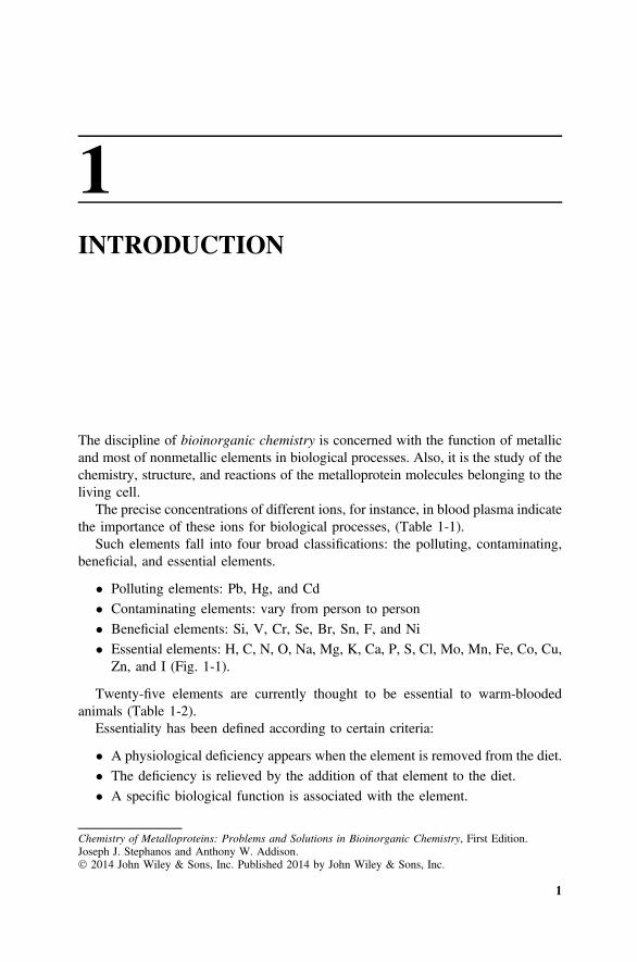
1INTRODUCTION
The discipline of bioinorganic chemistry is concerned with the function of metallicand most of nonmetallic elements in biological processes. Also, it is the study of thechemistry, structure, and reactions of the metalloprotein molecules belonging to theliving cell.
The precise concentrations of different ions, for instance, in blood plasma indicatethe importance of these ions for biological processes, (Table 1-1).
Such elements fall into four broad classifications: the polluting, contaminating,beneficial, and essential elements.
• Polluting elements: Pb, Hg, and Cd
• Contaminating elements: vary from person to person
• Beneficial elements: Si, V, Cr, Se, Br, Sn, F, and Ni
• Essential elements: H, C, N, O, Na, Mg, K, Ca, P, S, Cl, Mo, Mn, Fe, Co, Cu,Zn, and I (Fig. 1-1).
Twenty-five elements are currently thought to be essential to warm-bloodedanimals (Table 1-2).
Essentiality has been defined according to certain criteria:
• A physiological deficiency appears when the element is removed from the diet.
• The deficiency is relieved by the addition of that element to the diet.
• A specific biological function is associated with the element.
1
Chemistry of Metalloproteins: Problems and Solutions in Bioinorganic Chemistry, First Edition.Joseph J. Stephanos and Anthony W. Addison. 2014 John Wiley & Sons, Inc. Published 2014 by John Wiley & Sons, Inc.

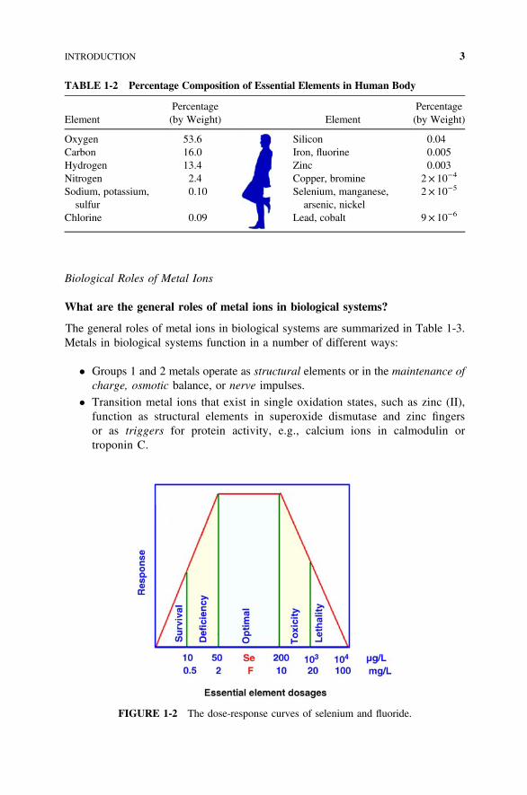
Every essential element follows a dose–response curve, shown in Fig. 1-2. Atlowest dosages the organism does not survive, whereas in deficiency regions theorganism exists with less than optimal function.
The ten ions classified as trace metal are Fe, Cu, Mn, Zn, Co, Mo, Cr, Sn, V, andNi, and the four classified as bulk metals are Na, K, Mg, and Ca. The nonmetallicelements are H, B, C, N, O, F, Si, P, S, Cl, Se, and I.
FIGURE 1-1 Distribution of elements essential for life (Cotton and Wilkinson, 1980).
TABLE 1-1 Ion Concentration inExtracellular Blood Plasma
Ion mM Ion mM
Na+ 138 SO42� 1
Cl� 100 Fe 0.02K+ 4 Zn2+ 0.02Ca2+ 3 Cu2+ 0.015Mg2+ 1 Co2+ 0.002HPO4
2� 1 Ni2+ 0
2 INTRODUCTION
Biological Roles of Metal Ions
What are the general roles of metal ions in biological systems?
The general roles of metal ions in biological systems are summarized in Table 1-3.Metals in biological systems function in a number of different ways:
• Groups 1 and 2 metals operate as structural elements or in the maintenance ofcharge, osmotic balance, or nerve impulses.
• Transition metal ions that exist in single oxidation states, such as zinc (II),function as structural elements in superoxide dismutase and zinc fingersor as triggers for protein activity, e.g., calcium ions in calmodulin ortroponin C.
TABLE 1-2 Percentage Composition of Essential Elements in Human Body
ElementPercentage
(by Weight) ElementPercentage
(by Weight)
Oxygen 53.6 Silicon 0.04Carbon 16.0 Iron, fluorine 0.005Hydrogen 13.4 Zinc 0.003Nitrogen 2.4 Copper, bromine 2× 10�4
Sodium, potassium,sulfur
0.10 Selenium, manganese,arsenic, nickel
2× 10�5
Chlorine 0.09 Lead, cobalt 9× 10�6
FIGURE 1-2 The dose-response curves of selenium and fluoride.
INTRODUCTION 3

• Transition metals that exist in multiple oxidation states serve as electroncarriers, e.g., iron ions in cytochromes or in iron–sulfur clusters of the enzymenitrogenase or copper ions in azurin and plastocyanin.
• As facilitators of oxygen transport, e.g., iron ions in hemoglobin or copper ionsin hemocyanin.
• As sites at which enzyme catalysis occurs, e.g., copper ions in superoxidedismutase or iron and molybdenum ions in nitrogenase.
• Metal ions may serve multiple functions, depending on their location within thebiological system, so that the classifications in Table 1-3 are somewhat arbitraryand/or overlapping.
PROTEINS: FORMATION, STRUCTURES, AND METALLOPROTEINS
This section is designed to introduce the chemistry of proteins. The text broadlyincludes where and how the proteins are formed, along with the structure andformation of metalloproteins.
Following the introduction of organelles and their functions within the cell, thediscussion will be concerned with the general structure of deoxyribonucleic acid(DNA) and how the nucleus maintains its control of cell growth, division, andformation of [messenger, transfer, and ribosomal ribonucleic acid (mRNA, tRNA,
TABLE 1-3 Role of Metal Ions and Examples
Metal Functions and Examples
Na+, K+ Charge transfer, osmotic balance, nerve impulsesMg2+ Structure in hydrolases, isomerases, phosphate transfer, and
trigger reactionsCa2+ Structure, charge carrier, phosphate transfer, trigger reactionsZn2+ (tetrahedral) Structure in zinc finger, gene regulation, anhydnase,
dehydrogenaseZn2+ (square pyramidal) Structure in hydrolases, peptidasesMn2+ (octahedral) Structure in oxidases, photosynthesisMn3+ (tetragonal) Structure in oxidase, photosynthesisFe2+ Electron transfer, nitrogen fixation in nitrogenase, dioxygen
transport in hemoglobin and myoglobinFe3+ Electron transfer in oxidasesCu+, Cu2+ Electron transfer in type I blue copper proteins, oxidases and
hydroxylases in type II blue copper proteins, hydroxylasesin type III blue copper proteins, dioxygen transport inhemocyanin
Co2+ (tetrahedral) Alkyl group transfer, oxidasesCo+, Co2+, Co3+ (octahedral) Alkyl group transfer in B12
Ni2+ (square planar) Hydrogenase, hydrolasesMo4+, Mo5+, Mo6+ Nitrogen fixation in nitrogenose, oxo transfer in oxidases
4 INTRODUCTION

rRNA)]. This is followed by how mRNA and tRNA master the formation ofproteins within a cell. Then, primary, secondary, tertiary, and quaternary structuresof the formed proteins and the factors that control each of these structures arediscussed.
Specific points about the ligation of various metal ions to different amino acidswithin the proteins are made, and the binding stabilities of various metal ions towarddifferent amino acids are arranged.
The general formulas, side chains, and corresponding names of the commonnatural α-amino acids, the formation of the peptide chain from the amino acids, andthe physiological roles of proteins are described.
The chemistry of the prosthetic and cofactors is explored. Enough basic bio-chemistry is presented to enable the student to understand the discussions that follow.
Organelles and Their Functions
Identify the organelles and their functions within the cell.
• Cells are the building blocks of all living things.
• There are similarities in the appearance, chemical constituents, and activities ofall cells (Fig. 1-3).
• Different structures within the cell are called organelles.
• Each organelle has an important, specific function in the cell.
• The mitochondria are responsible for conversion of food into usable energy(metabolism):
� They contain enzymes for cell metabolism.
� More than 50% of the energy produced by mitochondrial oxidation ofcarbohydrates is recaptured as adenosine diphosphate (ADP) and convertedinto adenosine triphosphate (ATP).
FIGURE 1-3 (a) Animal cell and (b) plant cell.
PROTEINS: FORMATION, STRUCTURES, AND METALLOPROTEINS 5

� The derived energy is trapped in ATP molecules (Scheme 1-1).
� ATP can diffuse rapidly throughout the cell, delivering energy to sites whereit is required for cellular processes.
• In green plants, chloroplasts contain chlorophyll molecules and otherpigments.
� Chlorophyll and other pigments in chloroplasts absorb light energy fromthe sun and use it to produce ATP, glucose, and oxygen.
• Ribosomes are round particles (mRNA) that are sent by the nucleus to activateprotein synthesis.
� The mRNA causes a specific protein molecule to be synthesized from thepool of amino acids present in the cell cytoplasm.
• The nucleus, or command station, contains information for the development andoperation of the cell.
� This information is stored chemically in long molecular strands called DNA.A combination of DNA and protein forms fine strands of chromatin. When acell is about to divide, the chromatin strands coil up and become denselypacked, forming chromosomes.
� The number of chromosomes varies with the species: Humans have 23, thefruit fly has 4, corn has 10, and the mosquito has 3.
Structure of DNA
What is the general structure of deoxyribonucleic acids, DNA?
• Polymerization of nucleoside phosphates produces the nucleic acids, DNA andRNA.
SCHEME 1-1 Derived energy is trapped in adenosine triphosphate molecules (ATP).
6 INTRODUCTION

• DNA is a giant molecule with molecular weight of order 1 billion or more.
• The information is chemically stored by nitrogen-base molecules that arebonded to the sugar residues of the sugar–phosphate chain.
• There are four nitrogen bases:
(a) Two purines, which are bicyclic molecules:
(b) Two pyrimidines, which are monocyclic:
• The order in which they appear on the chain makes up the molecular message(Fig. 1-4).
• The DNA molecule is also capable of duplicating itself and dividing.
PROTEINS: FORMATION, STRUCTURES, AND METALLOPROTEINS 7

• Under a microscope we can see the duplicated chromosomes divide equally asthe cell divides.
• The DNA double strand forms when the bases on the two adjacent single strandsform hydrogen bonds:
FIGURE 1-4 Order of N bases on chain.
8 INTRODUCTION

• Adenine and thymine form a hydrogen bonded pair, or complementary basepair.
• Cytosine and guanine also form a complementary base pair (Fig. 1-5).
• These complementary base pairs are conformed by the base ratios: G/C= 1 andA/T= 1 (Table 1-4).
FIGURE 1-5 DNA double strand.
TABLE 1-4 Nitrogen-Base Content of DNA from Different Organisms
Species TissueSource
CalfThymus
CrabAll tissue
Algea (Euglcna)Chloroplast
Virus (Coliphaga ×174)Replicative Form
A 29.0 47.3 38.2 26.3T 28.5 47.3 38.1 26.4A/T 1.01 1.00 1.00 1.00G 21.2 2.7 12.3 22.3C 21.2 2.7 11.3 22.3G/C 1.00 1.00 1.09 1.00
Note: Data in mole percent.
PROTEINS: FORMATION, STRUCTURES, AND METALLOPROTEINS 9

Cell Growth and Division
How does the nucleus maintain its control of cell growth and division?
• During ordinary cell division called mitosis, two new cells result from a singleparent.
• Each daughter has the same number of chromosomes as the parent.
• If DNAis themolecular stuffof thechromosome, itmustbeable to reproduce itself.
• The DNA double helix rewinds and separates into two single strands (Fig. 1-6).
• As the unwinding occurs, the single strands act as templates for synthesis of newcomplementary strands.
• When the parent DNA double helix has completed its unwinding, two newDNA double-stranded molecules are formed.
• The process by which new DNA is formed is called replication.
Protein Synthesis
How can proteins be synthesized in cells?
• The order of the N bases on the DNA molecule determines the order of aminoacids in the protein molecule.
• While DNA is in the nucleus, the proteins are synthesized on ribosomes outsidethe nucleus as follows:
FIGURE 1-6 DNA double helix rewinds and separates into two single strands.
10 INTRODUCTION

� As the DNA double helix unwinds, the N base segment becomes exposed.
� The DNA molecule serves as template for the synthesis of mRNA molecule.
� The synthesis of mRNA is analogous to the replication synthesis of DNA(Fig. 1-7).
� mRNA has structure similar to DNA but contains:
• Ribose instead of deoxyribose
• N-base uracil instead of thymine:
• After mRNA is synthesized, it is transported out of the nucleus and becomesattached to the ribosomes, where the protein syntheses begin (Fig. 1-8).
• At the ribosomes, the order of the bases on the mRNA determines the aminoacid sequence in the protein molecule.
• The amino acid sequence is determined by a triplet code on the mRNA molecule.
• A group of three N bases represents a code for signifying a single amino acid(Scheme 1-2).
• The amino acids are brought to the mRNA at the ribosomes by much smallerRNA molecules called tRNA.
• Each tRNA has a triplet of bases, which is complementary to an amino acid codeon mRNA.
• The tRNA molecules bring the amino acids to the ribosomes as they move alongthe mRNA strand, and the amino acids are knit into the growing protein chain.
FIGURE 1-7 Synthesis of mRNA.
PROTEINS: FORMATION, STRUCTURES, AND METALLOPROTEINS 11

• After the tRNA has discharged its amino acid passenger, it moves out into thecytoplasm, finds another amino acid, and returns to the ribosome surface.
Common Natural α-Amino Acids
Give the general formula, side chain, and corresponding name of the commonnatural α -amino acids.
• There are 20 common natural amino acids.
SCHEME 1-2 Genetic codes.
FIGURE 1-8 Protein synthesis.
12 INTRODUCTION

• The general formula for an α-amino acids is
• They are summarized in Table 1-5.
TABLE 1-5 L-α-amino Acids
NameSide Chain
(R at pH= 7) pKa Nature
Glycine, Gly, G —H 2.35, 9.78 Structural, spacerAlanine, Ala, A —CH3 2.35, 9.87 HydrophobicValine, Val, V 2.29, 9.74 Hydrophobic
Leucine, Leu, L 2.33, 9.74 Hydrophobic
Isoluecine, Ile, I 2.32, 9.76 Hydrophobic
Phenylalanine, Phe, F 2.16, 9.18 Hydrophobic
Proline, Pro, P 1.95, 10.64 Hydrophobic,structural
Tryptophan, Trp, W 2.43, 9.44 Hydrophobic
Serine, Ser, S 2.19, 9.21 Ambivalent
Threonine, Thr, T 2.09, 9.11 Hydrophobic
(continued )
PROTEINS: FORMATION, STRUCTURES, AND METALLOPROTEINS 13
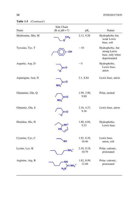
Table 1-5 (Continued )
NameSide Chain
(R at pH= 7) pKa Nature
Methionine, Met, M 2.13, 9.28 Hydrophobic butweak Lewisbase, soft
Tyrosine, Tyr, T ∼10 Hydrophobic, butstrong Lewisbase, only whendeprotonated
Aspartic, Asp, D ∼5 Hydrophobic,Lewis base,anion
Asparagine, Asn, N 2.1, 8.84 Lewis base, anion
Glutamine, Gln, Q 1.99, 3.90,9.90
Polar, neutral
Glutamic, Glu, E 2.16, 4.27,9.36
Lewis base, anion
Histidine, His, H 1.80, 6.04,9.33
Hydrophobic,Lewis base
Cysteine, Cys, C 1.92, 8.35,10.46
Lewis base,anion, soft
Lysine, Lys, K 2.16, 9.18,10.79
Polar, cationic,protonated
Arginine, Arg, R 1.82, 8.99,12.48
Polar, cationic,protonated
14 INTRODUCTION

Peptide Chain Formation
How can the peptide chain be formed from the amino acids?
• Linear polymerization by condensation to yield amide peptide linkage (Scheme1-3).
• All proteins are polypeptides.
Protein physiological functions
What are the physiological roles of proteins?
• The physiological roles of proteins are:
� Structural: finger nails, hair, and skin
� Transport: oxygen, electrons, and iron
� Catalysis: enzymes responsible for all synthesis of proteins, DNA, andorganics
SCHEME 1-3 Polypeptide formation.
PROTEINS: FORMATION, STRUCTURES, AND METALLOPROTEINS 15

Structural Features of Proteins
Define: primary, secondary, tertiary, and quaternary structures. And what arethe factors that control each of these structures?
• The properties and functions of a particular protein depend on the sequence ofthe amino acids in the protein, or the primary structure.
� The primary structure determines higher levels of structures.
� These structural details are crucial to the biological role of a protein.
• The secondary structure arises from the relative disposition of atoms in thepolypeptide “backbone”:
The groups of four gray-shaded atoms are coplanar. Free rotation occursabout the bond connecting the carbon with the carbonyl and the nitrogen.Therefore, the extended polypeptide chain is a semirigid structure with two-thirds of the atoms of the backbone held in a fixed plane.
� Examples of secondary structures:
(a) random coil
(b) α–helix (Fig. 1-9)
(c) β–pleated (Fig. 1-10), associated as (i) parallel and (ii) antiparallel
(d) reverse turns (Fig. 1-11)
(e) omega loops (Fig. 1-12)
Both reverse turns and omega loops appear at the outer surface of themolecules.
• A Tertiary structure refers to the folding of the already secondary structuredamino acids to form a three-dimensional (3D) structure. The overall 3Darchitecture of the polypeptide backbone:
� Fibrous proteins: coils (Fig. 1-13).
� Globular proteins: compact, ellipsoidal, spherical, until denatured. Thefolded tertiary, globular, structure of myoglobins is imposed over thehelical secondary structure. Structures from X-ray diffraction are shownin Fig. 1-14.
� Synthetic polypeptides have random or simply repetitive structures.
16 INTRODUCTION

FIGURE 1-9 α-Helix structure of protein.
FIGURE 1-10 β-Pleated structure.
FIGURE 1-11 Reverse turn.
PROTEINS: FORMATION, STRUCTURES, AND METALLOPROTEINS 17

FIGURE 1-14 Tertiary structure of oxymyoglobin at 1.6 Å resolution, PDB 1MBO (Phillips,1980).
FIGURE 1-12 Omega loop.
FIGURE 1-13 Fibrous oligomers, PDB 1G6U (Ogihara et al., 2001).
18 INTRODUCTION