chemiluminescent reactions catalyzed by nanoparticles of gold, silver, and gold/silver alloys
TRANSCRIPT

Eastern Michigan UniversityDigitalCommons@EMU
Master's Theses and Doctoral Dissertations Master's Theses, and Doctoral Dissertations, andGraduate Capstone Projects
3-2012
Chemiluminescent reactions catalyzed bynanoparticles of gold, silver, and gold/silver alloysSaqib Ul Abideen
Follow this and additional works at: http://commons.emich.edu/theses
Part of the Chemistry Commons
This Open Access Thesis is brought to you for free and open access by the Master's Theses, and Doctoral Dissertations, and Graduate Capstone Projectsat DigitalCommons@EMU. It has been accepted for inclusion in Master's Theses and Doctoral Dissertations by an authorized administrator ofDigitalCommons@EMU. For more information, please contact [email protected].
Recommended CitationAbideen, Saqib Ul, "Chemiluminescent reactions catalyzed by nanoparticles of gold, silver, and gold/silver alloys" (2012). Master'sTheses and Doctoral Dissertations. 405.http://commons.emich.edu/theses/405
Chemiluminescent Reactions Catalyzed by Nanoparticles of Gold, Silver, and
Gold/Silver Alloys
by
Saqib Ul Abideen
Thesis
Submitted to the Department of Chemistry
Eastern Michigan University
In partial fulfillment of the requirements
For the degree of
MASTER OF SCIENCE
in
Chemistry
Thesis committee:
Timothy Brewer, PhD, Chair
Donald Snyder, PhD
Heather Holmes, PhD
March 2012
Ypsilanti, Michigan
.

ii
ACKNOWLEDGEMENTS
Thank you, first and foremost, to my research advisor and mentor, Dr. Timothy Brewer,
who made this project possible and who has always encouraged and supported my own unique
interests. He has always challenged me to do my very best work to think critically and
compassionately, and his guidance and belief in me has allowed me to grow both professionally
and personally. He acknowledges and shares in my success, no matter how small; he is someone
I have always been proud to work with and have looked up to since the start of this project.
Thank you to Dr. Ross Nord, Chemistry Departmental Head, for providing us with a
great learning place and for endorsing my thesis. Thank you to Dr. Krish Rengan, who always
guided me in all the problems no matter how small or big they were.
Thank you to my other committee members, Dr. Donald Snyder and Dr. Heather Holmes,
who have offered invaluable guidance and assistance with this project. Their feedback,
thoughtful comments, and unique expertise along the way have not only made my thesis better
but have also contributed substantially to my growth as a researcher.
Thank you to my parents, who support me in their own unique way and who inspire me
to be and do better. At last, but definitely not least, thank you to my wife, Anila, and daughters,
Irwa and Khadija; their patience, understanding, love, and support is unending and has been
present since our beginning, and without them I could not do this or any work.

iii
ABSTRACT
Chemiluminescence (CL) reactions are catalyzed by metals nanoparticles, which display unique
catalytic properties due to an increased surface area. The present study describes the catalytic
effects of nanoparticles (NP) of silver, gold, and alloys of Au/Ag nanoparticles on the
chemiluminescent reaction taking place between luminol and potassium ferricyanide. It was
found that silver nanoparticles and alloy nanoparticles enhance the CL process when their sizes
remained in the range of 30 nm to 50 nm. The data show that the intensity and rate of
chemiluminescence were influenced by the mole fraction of gold and silver in the alloy. Data to
this chemiluminescence reaction are modeled by a double exponential curve, which indicates
that two competing processes are occurring.

iv
TABLE OF CONTENTS
Acknowledgement .......................................................................................................................... ii
Abstract .......................................................................................................................................... iii
List of Schemes ................................................................................................................................v
List of Figures ................................................................................................................................ vi
List of Tables ............................................................................................................................... viii
Introduction ......................................................................................................................................1
Project Goal ..................................................................................................................................16
Experimental ..................................................................................................................................17
Results and Discussion ..................................................................................................................24
Conclusion and Future Studies ......................................................................................................45
References ......................................................................................................................................46
v
LIST OF SCHEMES
1. Chemiluminescence reaction of luminol ..................................................................................10
2. Mechanism of enhancement by nanoparticles on luminol-ferricyanide reaction .....................37

vi
LIST OF FIGURES
1. Schematic representation of different sizes of nanoparticles ......................................................2
2. Density of states in metal (A) and semiconductors (B) nanocrystals showing the band edge
separation .........................................................................................................................................3
3. The Lycurgus cup .......................................................................................................................4
4. Interaction of electromagnetic radiation with metal nanospheres and nanorods ........................5
5. Sizes of nanoparticles show distinct light scattering properties .................................................5
6. Silver nanoparticles of different sizes .........................................................................................6
7. SPR in silver nanoparticles .........................................................................................................7
8. Stability of AgNP induced by repulsive forces from borohydride ions......................................8
9. Characterization of silver nanoparticles......................................................................................9
10. Schematic of flow injection chemiluminescence system........................................................11
11. Absorption spectra of AgNP with diameters ranging from 10-100 nm ..................................27
12. Absorption spectra of AgNP using borohydride method ........................................................28
13. Size effect on the absorption spectra of silver nanoparticles ..................................................29
14. Formation of gold nanoparticles .............................................................................................30
15. Absorption spectra of gold nanoparticles ...............................................................................31
16. Absorption spectra of gold nanoparticles with different diameters ........................................32
17. Absorption spectra of Au/Ag nanoparticles alloy with varying mole fractions .....................34

vii
18. Nanoparticles of gold, silver and Au/Ag alloy .......................................................................35
19. Effect of gold mole fraction on absorption spectra .................................................................36
20. Blank chemiluminescence reaction between luminol and potassium ferricyanide ................38
21. Effect of AgNP on chemiluminescence of luminol and pottasium ferricyanide ....................39
22. Effects of AuNP on chemiluminescence ................................................................................40
23. Effects of Au/Ag alloy NP on Chemiluminescence of luminol reacting with potassium
ferricyanide ....................................................................................................................................41
24. Two processes going on in the presence of AgNP .................................................................43
25. Two processes going on in the presence of Au/Ag alloy NP .................................................43
26. Chemiluminescence data fit ....................................................................................................44
viii
LIST OF TABLES
1. Synthesis of silver nanoparticles using citrate method .............................................................19
2. Synthesis of gold nanoparticles ................................................................................................20
3. Synthesis of Au/Ag alloy nanoparticles ....................................................................................21
4. Various conditions of chemiluminescence reactions ................................................................22
5. Effect of change of concentration ratio of AgNP stability .......................................................25
6. Nanoparticles of Au/Ag alloy formation ..................................................................................33
7. Duration of light affected by different sizes of silver nanoparticles .........................................38
8. Nanoparticles and their respective peak intensities in chemiluminescence ..............................42
1
Introduction
The purpose of this research project is to synthesize nanoparticles of gold, silver, and
Au/Ag alloys with a wide range of size and shape and determine how these properties affect the
catalysis of chemiluminescence reactions. Before proceeding with experimental details, a brief
introduction to nanoparticles, chemiluminescence reactions, and the role of nanoparticles as
catalysts is presented.
Nanoparticles
A bulk material has constant physical properties regardless of its size, but at the nano
scale level these properties change. Bulk materials larger than one micrometer have a smaller
percentage of atoms at the surface than to the total number of atoms of the material. For the
smaller particles the percentage of surface atoms increases, leading to changes in physical and
chemical behavior of the materials. A nanoparticle has a size range between 10 nm to 100 nm.
Figure 1 shows the relationship between the particle size and number of atoms. Unexpected
physical and chemical behavior of matter occurs at the nanometer scale, paving the way for a
number of scientific exploitations, making nanoparticles a great area of scientific research.
The conversion of particles to nanoparticles results in unique properties which are
governed by two major factors. First with the decrease in the size of the particles, the number of
atoms at the surface in comparison to the number of atoms in the center of the crystal increases
dramatically.1

2
Figure 1. Schematic representation of different sizes of nanoparticles 1
Second, when the particle size decreases, the electron hole pairs are much closer and
columbic interactions between them cannot be neglected. This results in an increase in the
spacing of the electronic levels and band gaps. The large band gap means that more energy is
needed to excite the electrons from the valence band to the conduction band. This phenomenon
is observed for metal nanoparticles where a color change is caused by the decrease in the size of
particles.1, 2

3
Figure 2. Density of states in metal (A) and semiconductor (B) nanocrystals showing the
band edge separation2
History of Nanoparticles
Nanoparticles have a very old history dating back more than 25 centuries. Silver and
copper nanoparticles were used by artisans for glittering effects. In the fourth or fifth century
B.C., the first syntheses of metallic gold nanoparticles were reported in China and Egypt. 3 Since
their discovery, nanoparticles have mainly been exploited for their medicinal and aesthetic
properties. Gold colloidal solution was named as Aurum Potabile, which was a suspension of
gold nanoparticles in volatile oils and was used as elixir of youth and a cure for heart diseases,
epilepsy, and tumors.4 One of the most interesting examples of the use of nanoparticles by the
early artists was the famous Lycurgus cup, which could be seen in the British museum in

4
London, crafted by the Romans in the 4th century. 5 Due to the presence of gold and silver alloy
nanoparticles, it reflected green light while transmitting red light.
Figure 3. The Lycurgus cup 5
Optical properties of nanoparticles
The color of metal nanoparticles depends upon their size and shape. When incident light
interacts with metal nanoparticles, it oscillates the conduction electrons on the surface.6 This
effect is known as surface plasmon resonance. The electric field of the incident radiation induces
the formation of dipoles in the nanoparticles. A restoring force in the nanoparticles tries to
compensate, resulting in a unique resonance wavelength responsible for the distinctive color of
the nanoparticles. In Figure 4A, electromagnetic radiation interacts with a spherical metal
nanoparticle to induce a dipole. The dipole oscillates in phase with the incoming light. In Figure
4B, electromagnetic radiation is interacting with metal nanorods. The interaction produces
transverse and longitudinal oscillations of the electrons in the rods, resulting in an absorption
peak for each oscillation.

5
Figure 4. Interaction of electromagnetic radiation with metal nanospheres and nanorods6
Figure 5 shows spherical silver and gold nanoparticles of different sizes. 7 For
nanoparticles of different sizes, interaction of the electron waves with the incoming light results
in unique colors.
Figure 5. Sizes of nanoparticles show distinct light scattering properties 7

6
Siver Nanoparticles
Silver nanoparticles exhibit the distinctive features of high electrical conductivity, low
sintering temperature, enhanced catalytic properties, better chemical reactivity, and protection
against bacteria, and their colors vary with changes in size and shape.
Figure 6. Silver nanoparticles of different sizes 8
The unique features of silver nanoparticles are caused by the properties8of surface
plasmon resonance, chemical reactivity, and stability. Figure 6 shows the images of silver
nanoparticles using a scanning electron microscope with various magnifications to help
characterize the size and shape of these materials.8
(a) Surface Plasmon Resonance
When light of a specific wavelength falls on nanoparticles, the conduction electrons on
metal surface undergo a collective oscillation known as surface plasmon resonance (SPR), which
is responsible for the unique optical properties of silver nanoparticles. A change in the color
results when the particle size changes or the refractive index is altered. The oscillations
produced by the coupling of free electrons with incident light are depicted in Figure 7.8
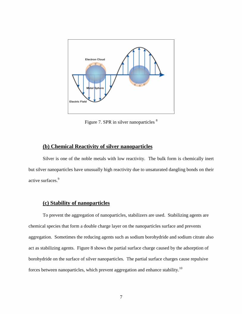
7
Figure 7. SPR in silver nanoparticles 8
(b) Chemical Reactivity of silver nanoparticles
Silver is one of the noble metals with low reactivity. The bulk form is chemically inert
but silver nanoparticles have unusually high reactivity due to unsaturated dangling bonds on their
active surfaces.9
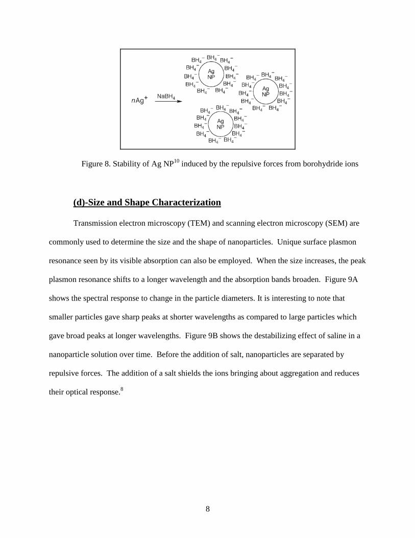
(c) Stability of nanoparticles
To prevent the aggregation of nanoparticles, stabilizers are used. Stabilizing agents are
chemical species that form a double charge layer on the nanoparticles surface and prevents
aggregation. Sometimes the reducing agents such as sodium borohydride and sodium citrate also
act as stabilizing agents. Figure 8 shows the partial surface charge caused by the adsorption of
borohydride on the surface of silver nanoparticles. The partial surface charges cause repulsive
forces between nanoparticles, which prevent aggregation and enhance stability.10
8
Figure 8. Stability of Ag NP10 induced by the repulsive forces from borohydride ions
(d)-Size and Shape Characterization
Transmission electron microscopy (TEM) and scanning electron microscopy (SEM) are
commonly used to determine the size and the shape of nanoparticles. Unique surface plasmon
resonance seen by its visible absorption can also be employed. When the size increases, the peak
plasmon resonance shifts to a longer wavelength and the absorption bands broaden. Figure 9A
shows the spectral response to change in the particle diameters. It is interesting to note that
smaller particles gave sharp peaks at shorter wavelengths as compared to large particles which
gave broad peaks at longer wavelengths. Figure 9B shows the destabilizing effect of saline in a
nanoparticle solution over time. Before the addition of salt, nanoparticles are separated by
repulsive forces. The addition of a salt shields the ions bringing about aggregation and reduces
their optical response.8

9
Figure 9. Characterization of silver nanoparticles 8
Chemiluminescence Reactions
Chemiluminescence reactions emit light in the visible or infrared region. For these
emissions, an excitation source is not needed. 11 This differs from fluorescence, in which
molecules first absorb incident radiation and then emit radiation of a longer wavelength without
any chemical reaction occurring.
Chemiluminescence reactions are categorized into the following three types:
1. Reactions that involve highly oxidized peroxide species. The peroxides convert into an
excited intermediate when reacted, returning to their ground states after release of energy
in the form of light.
2. Bioluminescent reactions are defined as chemiluminescent reactions that take place in
living organisms.
3. Electrochemiluminescent reactions are characterized by the use of an electrical current to
initiate a light emitting reaction.

10
Chemiluminescent reactions involving oxidized peroxide species are widely used in the
pharmaceutical industry, clinical science, analytical chemistry, and forensics. The emitted light
is easily measured while the time course and amplitude of light emission is dependent upon
factors such as concentrations and pH. Detection reagents used for these reactions (i.e., luminol)
can be manufactured in bulk and are, thus, not expensive. Additionally, chemiluminescence
involves no background signal because interference signals are absent.
Luminol is one of the most easily available and studied chemiluminescence reagents.
Luminol is also used because of its availability and inexpensive cost. Luminol reacts with
common oxidizing agents like H2O2 and K3Fe(CN)6.
Scheme 1 represents the mechanism of the luminol chemiluminescent reaction. In the
first step, a base removes the protons from nitrogen leaving a negative charge on the compound.
Then the negative charge moves to the carbonyl oxygen creating an enolate. In the next step,
cyclic peroxide is formed from the addition of oxygen. The loss of nitrogen, an excellent leaving
group, results in an excited state form of 3-aminophthalate, which returns to the ground state
through the emission of a photon.12,13
Scheme 1: Chemiluminescence reaction of Luminol12, 13

11
Chemiluminescence Measurement
Typically, luminometers are used for the detection and measurement of light emitted in
chemiluminescent reactions. A luminometer consists of a light-tight sample holder and a
photomultiplier tube that is capable of detecting extremely low levels of light. When the light
signals are strong, luminometers equipped with photodiodes are used. Figure 10 shows a
schematic of a flow injection chemiluminescence detection system14, 16 in which luminol, water,
and hydrogen peroxide are added through channels A, B, and C. The flow rate of 1.5 ml/min is
maintained with a peristaltic pump. Catalyst or nanoparticle colloid is added through D. The
chemiluminescence reaction takes place at F, and light signal is detected by photo multiplier
tubes.
Figure 10. Schematic of flow injection chemiluminescence system 14, 16
12
Chemiluminescence reactions catalyzed by nanoparticles
Use of nanoparticles to catalyze the chemiluminescence reactions is of interest to the
scientific community. Some interesting findings of using nanoparticles to catalyze
chemiluminescence reactions are described below.
In a study of the chemiluminescence reaction between hydrogen peroxide and luminol,
silver nanoparticles of sizes less than 7 nm were shown to increase the intensity and duration of
the light produced, though the effect varied with the experimental conditions used. The optimal
chemiluminescence reaction conditions were found when the concentration of luminol was 0.30
mM, hydrogen peroxide was 0.15 M, and silver nanoparticles were 44.0 nM with a pH of 12.0.
Under these optimal conditions, silver nanoparticles of 20 nm size gave the highest light
intensity lasting for the longest duration of time. This effect was proposed to be due to the
increase in surface area and surface electron density in the catalytic reaction involving
nanoparticles. It was proposed that silver nanoparticles catalyze the reaction by breaking
hydrogen peroxide into two radicals that swiftly oxidized the luminol, which resulted in the
production of light. This theory was supported by bonding the silver nanoparticles with bovine
serum albumin (BSA) and thioglycolic acid. This reduced the catalytic activity by not splitting
the hydrogen peroxide, thus resulting in no CL reaction.15
In another study, silver island films formed by the reduction of silver nitrate on glass
slides enhanced the chemiluminescent reaction between hydrogen peroxide and phenyl oxalate
ester. The results were elucidated by performing control experiments in which half of a glass
slide was coated with silver island films, and the remaining half was not coated. It was observed
that the portion covered with silver island film intensified the light signal four to five times more
than the bare glass portion. This article proposed that the silver nanoparticles in the island film

13
acted as surface plasmons, which were excited by the chemically-induced electronically-excited
luminophore. 16
Catalytic properties of silver, gold, and platinum nanoparticle colloids were also
investigated on the chemiluminescent reaction between hydrogen peroxide and luminol. Under
optimal reaction conditions, the catalytic activity of silver nanoparticles was better than that of
the nanoparticles of gold and platinum.17
A study of silver nanoparticles as a heterogeneous catalyst in the luminol-hydrogen
peroxide chemiluminescence system was performed in the presence of KI. Iodine radical
adsorbs readily on the surface of nanoparticles poisoning the catalyst and reducing the
chemiluminescence reaction that occurs. When the chemiluminescence reaction between
luminol and hydrogen peroxide on silver nanoparticles was performed in the presence of amino
acids such as cysteine, histidine, methionine, tyrosine, and tryptophan, no chemiluminescence
reaction took place. The authors proposed that amino acids adsorbed on the surface of silver
nanoparticles inhibited the chemiluminescence reaction.17
Other researchers determined that gold nanoparticles were better catalysts than
nanoparticles of silver and platinum due to their chemical stability and high resistance to surface
oxidation. Gold nanoparticles of 15 nm were used to catalyze the reduction of dissolved oxygen
by a mixture of luminol and hydrazine. The reduction of oxygen by hydrazine produced
hydrogen peroxide, which was then oxidized by luminol to produce the 3-aminophthalate anion
and an intense visible light signal. When the same experiment was performed in the absence of
gold nanoparticles, no light emission was observed. These results led to the conclusion that gold
nanoparticles had a catalytic effect on the reduction of hydrazine into hydrogen peroxide. A
linear relationship was observed between the intensity of chemiluminescence and the

14
concentration of hydrazine. An analytical method was developed for the determination of
hydrazine concentration using gold nanoparticles with a wide linear dynamic range, a low
detection limit of 30 nM, and good precision.18
A previous study concluded that the effect of gold nanoparticles on chemiluminescence
of luminol and ferricyanide was size-dependent. Gold nanoparticles less than 5 nm quenched the
chemiluminescence reaction between luminol and ferricyanid because the nanoparticles smaller
than 5 nm have a very high surface energy, which leads to high redox activity. The experimental
results indicated that the particles were partially oxidized by potassium ferricyanide, thus
inhibiting the reaction. A change in oxidation state of gold nanoparticles was validated by x-ray
photoelectron spectroscopic (XPS) studies. On the other hand, when the size of gold
nanoparticles was more than 10 nm, enhancement of the chemiluminescence of luminol and
ferricyanide was seen. In this case X-ray photoelectron spectroscopy (XPS) showed no change
in the oxidation state of gold nanoparticles before and after the reaction. The enhancement of
chemiluminescence was due to the catalytic activity of gold nanoparticles involved in the
electron transfer process.19
In another study, gold nanoparticles of 6 to 99 nm were used to catalyze the
chemiluminescent reaction between luminol and hydrogen peroxide. Small nanoparticles had a
low charge density and their surface was highly activated. Therefore, the intensity of
chemiluminescence signal was weak as compared to large size. Gold nanoparticles with a size of
38nm produced intense light signals. When the size of gold nanoparticles started to increase from
38 nm, the light signal started to decrease because the active surface area of nanoparticles started
to decrease. The role of gold nanoparticles as catalysts was confirmed by x-ray photoelectron
spectroscopy (XPS) and transmission electron microscopy (TEM) investigations, which revealed

15
that gold nanoparticles remained chemically and physically unaffected after the completion of
the reaction. Gold nanoparticles catalyzed the reaction by breaking down hydrogen peroxide to
produce superoxide. Superoxide converted luminol into activated ion on the surface of gold
nanoparticles. The activated ion of luminol emitted light when it returned to its ground state.
Organic compounds containing hydroxyl, amino, and mercapto groups reacted readily with
oxygen-containing intermediate radicals reducing the chemiluminescence intensity. A
quantitative method was developed for the determination of the concentration of organic
compounds containing hydroxyl, amino, and mercapto groups with a low detection limit of 10
nM, and the linear range of all compounds reached 3 orders of their magnitude.20
Another study found that Au/Ag alloy nanoparticles followed the same principles of
catalysis as gold and silver individual nanoparticles but found them superior to other
nanoparticles as their properties can be engineered by altering the mole fractions of gold and
silver. The chemiluminescence of the luminol-hydrogen peroxide system was strongly enhanced
by the addition of nanoparticles composed of Au/Ag alloy in the molar ratio of 5:4.
Nanoparticles composed of the alloy of gold and silver catalyzed the dissociation of hydrogen
peroxide, forming the hydroxyl radical and super oxide anion. These intermediates reacted with
luminol to produce the phthalate anion in its excited state, which emitted visible light upon return
to its ground state.21

16
Project Goal
Work done previously on the role of gold and silver nanoparticles as catalysts in
chemiluminescence reactions raised questions of which shape and size of nanoparticles was the
best catalyst under various conditions. This will be the main part of this project. This project
will also investigate the stability and synthesis of alloys of gold and silver nanoparticles and the
conditions for optimal catalysis of the luminol chemiluminescence reaction. The research
project will entail the synthesis of nanoparticles of gold, silver, and their alloys with a specific
size and then study the chemiluminescence reactions to discover their catalytic behavior in the
kinetics of the reactions.

17
Experimental
The experimental project to investigate the catalytic properties of nanoparticles on
chemiluminescence reactions was designed with three steps. The first step was the synthesis of
silver, gold, and silver/gold alloy nanoparticles through previously published methods. The
second step was to optimize the reaction conditions for chemiluminescent reactions between
luminol, hydrogen peroxide, and potassium ferricyanide. The last portion of the project was to
study the influence of nanoparticles on the chemiluminescence reaction.
Chemicals:
Silver nitrate (AgNO3), sodium borohydride (NaBH4) ascorbic acid, sodium hydroxide,
and trisodium citrate dihydrate (C6H5Na3O7.2H2O) were purchased from Sigma Aldrich, USA.
Hydrogen peroxide (H2O2), luminol, auric acid (H4AuCl4), and potassium ferricyanide
(K4FeCN6) were purchased from Fischer Scientific, USA. All chemicals used were of analytical
grade. Water was filtered to be ultra-pure with a conductivity less than 18 microsiemens.
Synthesis of nanoparticles
Silver nanoparticles, gold nanoparticles, and gold/silver alloy nanoparticles were
synthesized by well-established procedures using reducing agents such as sodium borohydride or
sodium citrate dihydrate to reduce either silver nitrate or auric acid.
1-Synthesis of Silver Nanoparticles:
Silver nanoparticles were synthesized by the use of sodium borohydride and sodium
citrate dihydrate as reducing agents.

18
1.1- Sodium Borohydride Method
A solution of 2.0 mM sodium borohydride was prepared by dissolving 0.00790 g of
sodium borohydride in 100 mL of ultrapure water. The solution was kept on ice to avoid the
decomposition of sodium borohydride. In all experiments, freshly-prepared and ice-cold sodium
borohydride was used. A solution of 1.00 mM AgNO3 was prepared by dissolving 0.0195 g of
AgNO3 in 100 mL of ultra-pure water. These solutions were then mixed together in different
portions to synthesize various size nanoparticles.
A batch of silver nanoparticles was synthesized by adding 5.0 mL of 1.0 mM AgNO3
drop-wise to 30.0 mL of icy, freshly-prepared 2.0 mM sodium borohydride. The addition took a
total time of around two minutes, and the resulting solution was constantly stirred. After five
minutes, a light yellow colored solution of silver nanoparticles was obtained.
A second batch of silver nanoparticles was prepared by adding 10.0 mL of 1.0 mM
AgNO3 to 30.0 mL of 2.0 mM freshly-prepared ice-cold sodium borohydride. After five minutes
with constant stirring, a darker yellow solution of silver nanoparticles was obtained.
1.2 Sodium Citrate Method
A 0.3 mM sodium citrate dihydrate solution was prepared by dissolving 0.00823 g of
sodium citrate dihydrate in 100 mL of ultra-pure water. A solution of 0.3 mM silver nitrate was
prepared by diluting the 1.0 mM stock solution of silver nitrate. Table 1 shows the amounts of
solutions used to synthesize various samples of silver nanoparticles. Each sample was heated to
a near boil for ten minutes.

19
Table 1: Synthesis of silver nanoparticles using citrate method
Sample Amount of Sodium Citrate Amount of AgNO3
1.2a 2.00 ml 8.00 ml
1.2b 4.00 ml 6.00 ml
1.2c 5.00 ml 5.00 ml
1.2d 6.00 ml 4.00 ml
1.2e 8.00 ml 2.00 ml
1.3-Synthesis of Large Silver Nanoparticles
Silver nanoparticles synthesized in previous methods were used as seed solutions for the
synthesis of larger silver nanoparticles. Volumes from 2.0 mL to 8.0 mL were reacted with 10
mL of 0.3 mM ascorbic acid, 10 mL of 0.1 mM NaOH and then treated with the drop-wise
addition of 10 mL of 0.30 mM AgNO3 with vigorous stirring. The reactions were done in a
constant boiling water bath.
2-Synthesis of Gold Nanoparticles:
A 0.1 M HAuCl4.4H2O was prepared by dissolving 1.0296 grams of the compound in
25.0 mL of ultra-pure water. This solution was then diluted to 1.0 mM before being used in the
synthesis procedure. A 40.0 mM sodium citrate dihydrate solution was prepared by dissolving
1.1764 g in 100.0 mL of ultra-pure water. From this, solutions of 20.0 mM and 10.0 mM sodium
citrate dihydrate were prepared by dilution. A 40.0 mM solution of sodium borohydride was
prepared by dissolving 0.151 g in 100.0 ml of ultra-pure water. From the 40.0 mM solutions of
NaBH4, solutions with concentration of 20.0 mM, and 10.0 mM were prepared. All samples

20
used 6.0 mL of 1.0 mM HAuCl4 and various amounts of reductants in the mixtures with various
heating time as shown in Table 2.
Table 2: Synthesis of gold nanoparticles
Sample 3.0 mL Heating time
2.1 40.0mM sodium citrate 5 min.
2.2 20.0mM sodium citrate 15 min.
2.3 10.0mM sodium citrate 30 min.
2.4 40.0mM NaBH4 3 min.
2.5 20.0mM NaBH4 3 min
2.6 10.0mM NaBH4 5 min.
3-Synthesis of Silver-Gold Alloy Nanoparticles:
Solutions of 0.10 mM AgNO3 and HAuCl4 were used, while the concentrations of sodium
borohydride and sodium citrate were 0.010 M. To 95 mL of ultra-pure water was added 1.0 mL
of sodium citrate and varying amounts of AgNO3 and HAuCl4 totaling 1.0 µL. To this solution,
1.0 mL of sodium borohydride was added, and then the volume was made up to the mark in a
100 ml volumetric flask and followed by vigorously stirring. The mole fractions of the gold and
silver were changed by adding different amounts of AgNO3 and HAuCl4 as shown in Table 3.

21
Table 3: Synthesis of Au/ Ag alloy nanoparticles
Sample Amount of 0.10 mM of
AgNO3
Amount of 0.10 mM of
HAuCl4
1 1.00 µL 0.00
2 0.75 µL 0.25 µL
3 0.50 µL 0.50 µL
4 0.25 µL 0.75 µL
5 0.00 1.00 µL
II-Optimum Chemiluminescence Reactions:
Luminol oxidizes in a reaction with hydrogen peroxide and potassium ferricyanide to
produce the phathalate ion in the excited state. The best chemiluminescent reaction conditions
depend upon the concentration of both oxidizing agents and varying reaction conditions such as
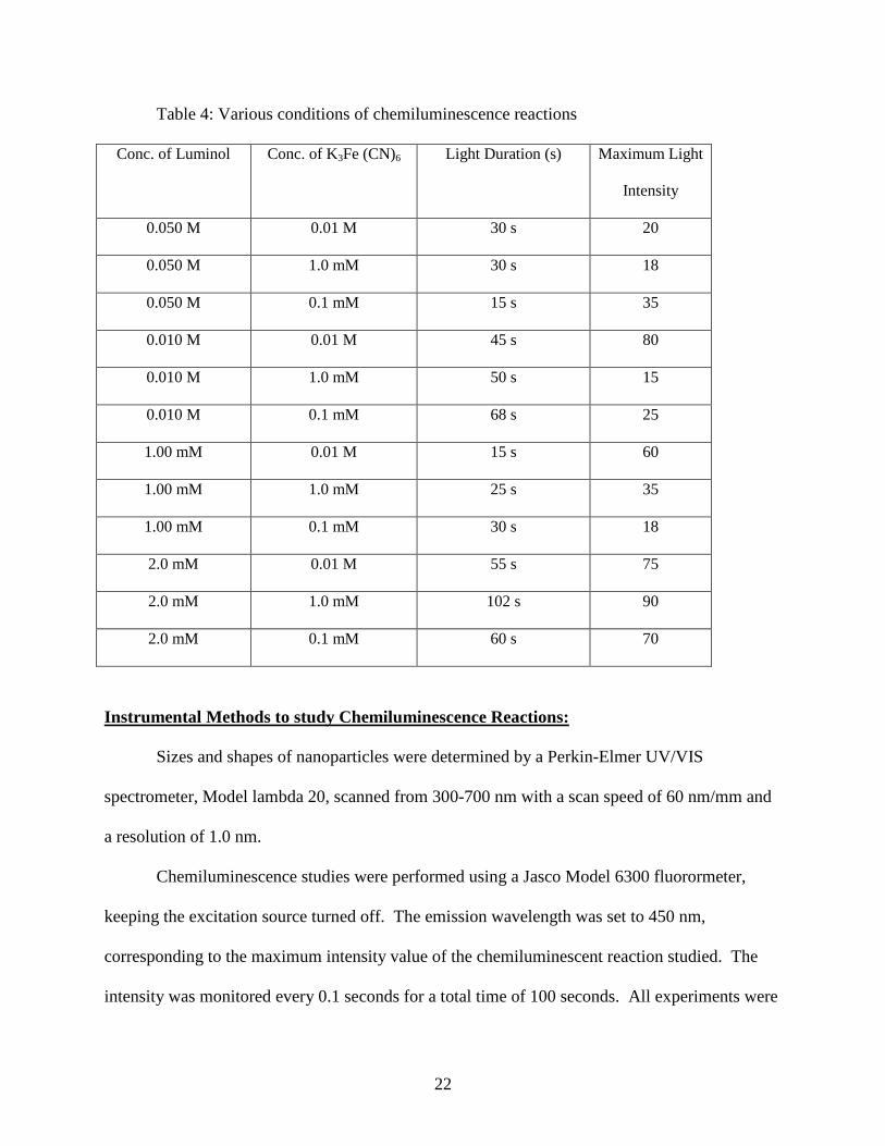
pH and temperature and can be characterized by the maximum light emission. Table 4 shows the
various reaction conditions used in this study. Concentrations of luminol were varied from 0.01
M to 2.00 mM along with varying the concentration of K3Fe (CN)6 from 0.01 M to 0.10 mM
and the pH from 9 to 12.
22
Table 4: Various conditions of chemiluminescence reactions
Conc. of Luminol Conc. of K3Fe (CN)6 Light Duration (s) Maximum Light
Intensity
0.050 M 0.01 M 30 s 20
0.050 M 1.0 mM 30 s 18
0.050 M 0.1 mM 15 s 35
0.010 M 0.01 M 45 s 80
0.010 M 1.0 mM 50 s 15
0.010 M 0.1 mM 68 s 25
1.00 mM 0.01 M 15 s 60
1.00 mM 1.0 mM 25 s 35
1.00 mM 0.1 mM 30 s 18
2.0 mM 0.01 M 55 s 75
2.0 mM 1.0 mM 102 s 90
2.0 mM 0.1 mM 60 s 70
Instrumental Methods to study Chemiluminescence Reactions:
Sizes and shapes of nanoparticles were determined by a Perkin-Elmer UV/VIS
spectrometer, Model lambda 20, scanned from 300-700 nm with a scan speed of 60 nm/mm and
a resolution of 1.0 nm.
Chemiluminescence studies were performed using a Jasco Model 6300 fluorormeter,
keeping the excitation source turned off. The emission wavelength was set to 450 nm,
corresponding to the maximum intensity value of the chemiluminescent reaction studied. The
intensity was monitored every 0.1 seconds for a total time of 100 seconds. All experiments were

23
performed in a dark room to minimize interference from stray light. In the blank experiments,
1.0 ml of ultra-pure water was added to the cuvette, followed by the simultaneous addition of 1.0
mL each of luminol and oxidizing agents using micro pipettes. In the nanoparticle experiments,
1.0 ml of the nanoparticle solution was added to the cuvette followed by the simultaneous
addition of 1.0 mL each of luminol and oxidizing agents using micro-pipettes.

24
Results and Discussion
Stability and Formation of Silver Nanoparticles
The reaction involved in the formation of nanoparticles was a simple reduction reaction,
in which silver nanoparticles were obtained as a result of reduction of AgNO3 by appropriate
reducing agents such as sodium borohydride, shown by equation (1)
AgNO3 + NaBH4 → Ag + 1/2 H2 + NaNO3 + 1/2 B2H6 (1)
The stability of the silver nanoparticles can be controlled either by using stabilizing
agents like polyvinyl alcohol (PVA) or by adjusting the concentration of reducing agent. The
ions formed from the reducing agents surround the particles and prevent their aggregation by
creating repulsive forces among them.
When sodium borohydride was used as the reducing agent, two approaches were used. In
the first approach, the concentration of AgNO3 was changed, while the concentration of sodium
borohydride was kept constant. Table 5 shows the concentration of reducing agent and AgNO3.
When the concentration ratio of sodium borohydride to AgNO3 was 2.00, a light-yellow silver
nanoparticle solution was obtained. The product was stable for more than six months and silver
nanoparticles did not aggregate. For a concentration ratio of 2.22 or 1.80, the color of
nanoparticle solution turned grayish after five minutes, which indicated that the nanoparticle
solution decomposed or aggregated. When the concentration ratio was 2.10 or 1.90, the color of
the colloidal solution was deep yellow or violet and the nanoparticles aggregated after 30
minutes.

25
The distinctive colors of the silver nanoparticle solutions were due to the plasmon
absorbance in which the electromagnetic radiation was absorbed by conduction electrons on the
surfaces of nanoparticles.
Table 5: Effect of change of concentration ratio on AgNP stability
No Conc. of
NaBH4
Conc. of
AgNO3
Conc.
Ratio(NaBH4/AgNO3)
Color Duration of AgNP stability
1 2.00 mM 0.90 mM 2.22 Grayish Approximately 5 min.
2 2.00 mM 0.95 mM 2.10 Deep yellow Approximately 30 min.
3 2.00 mM 1.00 mM 2.00 Yellow Very Stable
4 2.00 mM 1.05 mM 1.90 Violet Approximately 25 min.
5 2.00 mM 1.10 mM 1.80 Grayish Approximately 5 min.
In the second approach for the synthesis of silver nanoparticles, the concentration of
sodium borohydride was kept constant, but the volume of AgNO3 used was changed. The
change of the volume of AgNO3 had no effect on the stability of the nanoparticles, and they
remained stable for more than six months. The color of silver nanoparticle solution was deep
yellow in those samples where the volume of AgNO3 used was doubled, as compared to those
samples where a smaller volume of AgNO3 was used, where the color was faint yellow.
In the second method, sodium citrate was used as the reducing agent. It was expected
that a light yellow silver nanoparticle solution would be obtained after the addition of sodium

26
citrate to the boiling solution of AgNO3 but no color change was observed. This indicated that
no reaction took place. Various volumes of citrate and silver solutions were used, but none of
them worked under the conditions in Table 1. The plausible explanation for the results was that
the reaction conditions may have had the wrong ratio of concentrations of sodium citrate and
silver nitrate or the solutions were contaminated slightly. Future experiments in which different
concentrations of sodium citrate and silver nitrate are used would prove or disprove this
hypothesis.
These results showed that the limiting factor in the stability of silver nanoparticles was
the concentration and choice of the reducing agent. A change in the type of reducing agent such
as sodium citrate, which is a weak reducing agent, resulted in no reaction. In those reactions in
which the concentration ratio of NaBH4 to AgNO3 was 2, the nanoparticles were stable, but
ratios different from 2.0 resulted in the decomposition of the colloidal solutions.
Stability of the nanoparticles was due to the fact that when silver nanoparticles were
formed, the borohydride ions surrounded the nanoparticles and prevented them from
aggregating. The ions stopped their decomposition, resulting in a clear solution without the
formation of a black residual at the bottom.
Determination of Nanoparticle Size
The size of nanoparticle was determined with UV/Vis spectroscopy. The absorption
spectra of the nanoparticles of silver, gold, and gold/silver alloys were compared with the
literature values to determine the size of nanoparticles. Figure 11 shows the published
absorption spectra of silver nanoparticles with size ranging from 10 nm to 100 nm.22 Smaller

27
silver nanoparticles absorbs wave lengths near 400 nm, whereas larger silver nanoparticles have
peaks that broaden and shift towards larger wavelengths.
Figure 11. Absorption spectra of AgNP with diameters ranging from 10-100 nm22
Figure 12 shows the absorption spectra of silver nanoparticles formed by the reduction of
borohydride in which the volume of AgNO3 solution was changed. In both the cases the reaction
resulted in the formation of small-sized stable nanoparticles with an absorption peak at 396 nm.
When the absorption spectra from Figure 11 and 12 are compared, the sizes of silver
nanoparticles formed by the borohydride method were found to be 10 nm.

28
Figure 12. Absorption spectra of AgNP using sodium borohydride method.
Charcterization of Large Silver Nanoparticles
It is well known that the absorption peaks shift towards the longer wavelengths as the
sizes increase. The shift of the absorption peaks towards longer wavelengths is attributed to the
strong scattering and absorption of visible light due to surface plasmon resonance which arises
from the collective oscillation of the conduction electrons due to their interaction with the
incident light. The seed solution shows an absorption peak at 396 nm. When the amount of seed
solution was less than 2.0 mL, the absorption peak had a negligible shift as compared to the
absorption peak of the seed solution. This showed that there was no change in the size. But
when seed solution was 2.0-3.0 mL, there was a slight absorption peak shift from 396 nm to 400
nm, as shown in Figure 13.

29
Figure 13. Size effect on the absorption spectra of silver nanoparticles
Those samples in which the amount of seed solution was 4.0-8.0 mL displayed a larger
absorption peak shift from 396 nm to 415 nm, 428 nm, and 435 nm as the amount of seed
solution increased. According to literature values and comparison with Figure 11, it was shown
that when the absorption peak was at 400 nm, the size range of nanoparticles would be 10-15
nm. The absorption peak at 415 nm corresponds to a size from 20-30 nm, while an absorption
peak at 425 nm corresponds to the size range from 30-40 nm. An absorption peak at 438 nm
indicates a size range corresponding to 40-60 nm nanoparticles. It was found that various sizes
of nanoparticles were dependent on the amount of seed used in the synthesis.
Preparation of Gold Nanoparticles
Reduction of auric acid with sodium citrate produces gold nanoparticles. The reaction
follows a simple reduction mechanism route, in which the aurate H4AuCl4 solution reduces to

30
metallic gold atoms that aggregate to produce spherical nanoparticles. The concentration of the
reducing agent plays a vital role in the stability of the nanoparticles. Figure 14 shows a
schematic of the reduction reaction taking place between auric acid and sodium citrate producing
gold nanoparticles.
Figure 14. Formation of gold nanoparticles
The stability and size of gold nanoparticles was determined by the concentration of the
citrate solution involved in the synthesis. Three different concentrations of sodium citrate
solution (40.0 mM, 20.0 mM, and 10.0 mM) were used to reduce the aurate solution. When the
concentration of the citrate solution was 40.0 mM, the size of the gold nanoparticles was small,
as indicated by the UV-Vis. absorption peak at 525 nm as shown in Figure 15. These
nanoparticles were very stable because the citrate ions that were in excess surrounded the
nanoparticles and prevented them from aggregation. When 20 mM citrate solution was used, the
size of gold nanoparticles was medium and they were relatively unstable as determined by their
decomposition taking about three hours. When a dilute citrate solution of 10 mM was used for
reduction, nanoparticles aggregated almost instantly and resulted in the decomposition of
colloidal solution shown by a black residual mass.
31
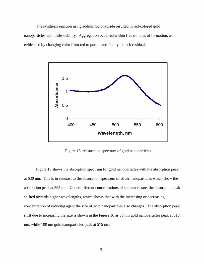
The synthesis reaction using sodium borohydride resulted in red-colored gold
nanoparticles with little stability. Aggregation occurred within five minutes of formation, as
evidenced by changing color from red to purple and finally a black residual.
0
0.5
1
1.5
400 450 500 550 600
Wavelength, nm
Ab
sorb
ance
Figure 15. Absorption spectrum of gold nanoparticles
Figure 15 shows the absorption spectrum for gold nanoparticles with the absorption peak
at 530 nm. This is in contrast to the absorption spectrum of silver nanoparticles which show the
absorption peak at 395 nm. Under different concentrations of sodium citrate, the absorption peak
shifted towards higher wavelengths, which shows that with the increasing or decreasing
concentration of reducing agent the size of gold nanoparticles also changes. The absorption peak
shift due to increasing the size is shown in the Figure 16 as 30 nm gold nanoparticles peak at 510
nm, while 100 nm gold nanoparticles peak at 575 nm.

32
Figure 16. Absorption spectra of gold nanoparticles with different diameters 23
Synthesis and Study of Au-Ag Alloy
Au/Ag nanoparticle alloys were synthesized by the reduction of varying mole fractions of
HAuCl4 and AgNO3 by sodium borohydride in the presence of sodium citrate. This method
posed two challenges. The first challenge was the formation of AgCl, and the second was the
formation of core shell Au/Ag. The precipitate of AgCl was formed due to the presence of high
concentration of chloride ions produced from the reduction of HAuCl4, which combined with the
Ag ions and precipitated as AgCl. To avoid this problem, the solutions were prepared in a way
such that the concentrations were less than the Ksp of AgCl(s). The second problem of a mix of
Au/Ag core shell formation was solved by adopting an appropriate synthetic procedure and
analyzing carefully the resulting absorption spectra. In the case of core shells, two peaks would

33
have appeared, each representing gold and silver, but in our approach we obtained only one peak
which proved that the method synthesized nanoparticle alloys of gold and silver.
Table 6: Nanoparticle Au/Ag alloy formation
Sample No Metal Mole Fraction Color Absorbance
Au Ag
1 0.0 1 Bright Yellow 390 nm
2 0.2 0.8 Deep Yellow 410 nm
3 0.4 0.6 Bluish Yellow 450 nm
4 0.5 0.5 Blue 465 nm
5 0.8 0.2 Purple 490 nm
6 1.0 0.0 Bright Red 520 nm
Table 6 shows the results for the alloy formation. When 1.0 µL of 0.01 M AgNO3 was
added to 95.0 mL of water followed by the addition of sodium citrate and sodium borohydride, a
yellow color immediately appeared. The yellow color indicated the formation of silver
nanoparticles. When 0.2 µL of HAuCl4 and 0.8 µL of AgNO3 was added, followed by the
addition of capping agent and reducing agent, a deep yellow color appeared, indicating the
formation of alloy. Alloy formation was further validated by the UV/Vis spectroscopy, in which
a single characteristic peak appeared. Three more alloy samples were prepared by changing the
mole fractions of gold and silver but keeping their total volume exactly equal to 1.0 µL, resulting
in single absorption peaks between 390 nm (pure silver) and 515 nm (pure gold).

34
Figure 17. Absorption spectra of Au/Ag nanoparticle alloy with varying mole fractions
Figure 17 shows the UV/Vis spectra of these synthesized alloys, which suggests that
when 100% of AgNO3 or 100 % HAuCl4 was used, the product formed was pure silver or gold
nanoparticles, but when varying mole fractions of gold solution and silver salt solution were
used, the product obtained was the alloy with only one characteristic peak between that of pure
gold and pure silver nanoparticles. When the mole fraction of silver was decreased gradually,
the absorption peaks shifted towards the higher wavelengths.

35
Figure 18. Nanoparticles of gold, silver and Au/Ag alloy
A strong relationship was observed between the mole fractions of gold and silver and the
color of nanoparticle alloy samples. Figure 18 shows a change in the color of nanoparticles with
different mole fractions of gold. When the mole fraction of silver was dominant, they were
yellow or deep yellow in color, while those samples with gold dominant produced colors of red
to light pink.
Figure 19 shows a plot of the gold mole fraction against the maximum absorbance. The
absorbance peak wavelength increases in a linear fashion with an increase in the gold mole
fraction. In some experiments, very dilute solutions of nanoparticles of gold and silver were
mixed together and their absorption spectra were taken. The spectra showed two absorption
peaks, indicating a solution of gold and silver nanoparticles with no alloy formation.

36
Figure 19. Effect of gold mole fraction on absorption spectra
Chemiluminescence Reactions
The chemiluminescence reaction of luminol, when performed in the presence of
hydrogen peroxide as oxidizing agent and copper (II) sulfate as catalyst, produced a large
luminescence signal. It was expected that when the catalyst, Cu ions, would be replaced by
nanoparticles, the intensity of light signals would not be affected. When the reaction was
performed in the presence of nanoparticles of gold, silver, or their alloy, no light emission could
be seen. Many different concentrations of luminol, hydrogen peroxide, and nanoparticle solution
were tried, but luminescence signal was never seen in our trials.
In another approach, we changed the oxidizing agent of potassium ferricyanide in the
absence of any catalyst. Optimum chemiluminescence reaction conditions were accomplished
when the concentration of oxidizing agent, K3Fe (CN) 6, was 1.00 mM, luminol was 2.0 mM, and
the pH was 10. Under these reaction conditions the emitted light intensity was ~100 and its
R² = 0.9826
0
0.2
0.4
0.6
0.8
1
1.2
375 395 415 435 455 475 495 515 535
Mo
le fra
cti
on
of
Au
Absorbance peak wavelength, nm

37
duration was 80 s. When nanoparticles of silver, gold, and Au/Ag alloy were used under these
conditions, a more intense signal of a shorter duration was observed, indicating the catalytic
activity.
Mechanism
Previous studies have shown that the size and oxidation state of nanoparticles remained
unchanged when chemiluminescence reactions were performed in their presence. This indicates
that nanoparticles act as catalysts with the reduction of luminol radicals taking place on their
surface during an exchange interaction between the unpaired electron of luminol radical and the
conduction band electrons of the nanoparticles, as shown in Scheme 2.
Scheme 2: Mechanism of enhancement by nanoparticles on the luminol-ferricyanide
reaction

38
Chemiluminescence Reactions Catalyzed by Silver Nanoparticles
Silver nanoparticles were divided into three groups based on their size and ability to
catalyze the chemiluminescence reaction. Figure 20 shows the blank chemiluminescence
reaction of luminol oxidized by K3Fe (CN) 6 with a maximum light intensity of 55 with duration
of 60 s.
0102030405060
0 20 40 60 80 100
Time (s)
Inte
nsi
ty
Figure 20. Blank chemiluminescence reaction between luminol and potassium
ferricyanide
When the same reaction was performed in the presence of silver nanoparticles, the peak
intensities increased and reaction was much faster, as shown in Figure 21. When the reaction
was performed in the presence of small-sized silver nanoparticles of about 20 nm, the peak
intensity was 340 while the duration of the reaction was 8 seconds.

39
Figure 21. Effects of AgNP on the chemiluminescence of luminol and potassium ferricyanide
It should be noted that the concentration of silver nanoparticles was kept constant in all
the experimental studies. For all the experiments, the medium-sized silver nanoparticles (~50
nm) were found to be the best catalyst in terms of intensity as shown in Table 7. Therefore, it
appeared that surface area of nanoparticles played a role in the intensity of emitted light in the
chemiluminescence reactions.
Table 7: Duration of light affected by different sizes of silver nanoparticles
Nanoparticle size Maximum intensity Duration (S)
20 nm 340 8
50 nm 325 15
70 nm 250 25
Chemiluminescence Reactions Catalyzed by Gold Nanoparticles
Green 20 nm AgNPRed 50 nm AgNPBlue 70 nm AgNP
-50
0
50
100
150
200
250
300
350
400
0 5 10 15 20 25 30
Time (s)
Inte
nsi
ty

40
The concentration of reducing agent was varied during the synthesis of gold nanoparticles
in an attempt to produce a wide size range. Corresponding absorption spectra showed little
change in the wavelength of maximum absorption, indicating a narrow range of particle size.
When gold nanoparticles were used as catalyst in chemiluminescence reactions, the effect was
very small, as shown in Figure 22. In the presence of gold nanoparticles there was slight change
in the intensity of the emission, but the duration for blank and catalyzed reactions remained
about the same.
Figure 22. Effects of AuNP on chemiluminescenc
Chemiluminescence Reactions Catalyzed by Ag-Au Alloy Nanoparticles
From our studies it was shown that silver nanoparticles in general were better catalysts
than gold nanoparticles. These results were further confirmed by investigating the catalytic
effects of Au/Ag nanoparticle alloys. Figure 23 shows spectra for nanoparticles with varied
Blue:BlankRed: In the presence of AuNP
-20
0
20
40
60
80
100
120
140
160
180
0 20 40 60 80 100 120 140
Time (s0
Inte
nsity

41
ratios of Ag and Au. When the gold was 0.20, the catalyzed chemiluminescence had an intensity
of 300 with duration of 30 seconds. However, when the mole fraction of gold was 0.80, the
intensity of the light signal was only 140 with approximately the same duration. This indicated
that alloy nanoparticles with a greater mole fraction of silver are better catalysts for the
chemiluminescence reactions.
Figure 23. Effects of Au/Ag alloy NP on Chemiluminescence of luminol reacting with
potassium ferricyanide
Kinetics studies of CLReactions Catalyzed by Ag-Au Alloy Nanoparticles
The kinetics studies were performed by using Origin software 2.0. Table 8 shows peak
intensities and durations for chemiluminescence reactions of luminol and potassium ferricyanide
catalyzed by silver nanoparticles and alloys of Au/Ag nanoparticles.
Blank: red Green: Au/Ag 80/:20 Blue: Au/Ag 20:80
-50
0
50
100
150
200
250
300
350
0 10 20 30 40 50 60
Time (s)
Inte
nsity

42
Table 8: Nanoparticles and their respective peak intensities in chemiluminescence
Type Peak intensity Duration (s)
Blank 2.0 m M Luminol+ 1.0mM Fe 88 100
Ag:Au 100:0 500 25
Ag:Au 80:20 300 50
Ag:Au 60:40 280 50
Ag:Au 50:50 180 60
Ag:Au 40:60 110 80
Ag:Au 20:80 130 80
Ag:Au 0:100 85 100
Blank 2.0 m M Luminol+ 1.0mM Fe 45 100
Conc. AgNP 325 20
0.3 ml Conc. AgNP+0.7ml H2O 160 60
0.5 ml Conc. AgNP+0.5ml H2O 250 30
0.7 ml Conc. AgNP+0.3ml H2O 330 20
The decay times for the double exponential curves for the alloy nanoparticles are shown
in Figures 24 and 25. The curves show dependence on the concentration of nanoparticles as well
as on the environment in which the chemiluminescence decay is taking place. Figure 24 shows
the chemiluminescence reaction taking place in the presence of silver nanoparticles. It shows
that two processes go side by side. Process A is slow and is environment dependant, while
process B is catalyzed by the silver nanoparticles, and its decay is faster than process A.

43
Figure 24. Two processes going on in the presence of AgNP
In Figure 25, process B shows the chemiluminescence reaction in the presence of Au/Ag
alloys. When the concentration of silver was higher, the decay was very fast.
Figure 25. Two processes going on in the presence of Au/Ag alloy NP

44
Figure 26 shows the chmiluminescence decay fit to a single exponential decay as shown
in purple and to a double exponential decay as shown in red.
Figure 26. The chemiluminescence data fit
The two mechanisms of the decay are still undetermined and need further
experimentation and analysis to develop a feasible theory for the process.

45
5-Conclusion and Future studies
Silver, gold, and Au/Ag alloy nanoparticles were synthesized by citrate and borohydride
methods and were verified by UV/Vis spectroscopy. Their catalytic properties were studied
using the chemiluminescent reaction of luminol and potassium ferricyanide. Catalysis by silver
nanoparticles was stronger than catalysis by gold nanoparticles. Au/Ag alloy nanoparticles
showed a linear correlation between wavelength of maximum absorption and mole fraction of
alloy. Catalysis by alloy nanoparticles was more effective for those alloys that had greater mole
fraction of silver than gold.
Future work should focus on a variety of methods for alloy synthesis and examine
quantitative decay rates of chemiluminescence reactions with their dependence on the mole
fractions of gold and silver.

46
References
1- Nanoparticles: from theory to application, edited by Gunter Schmid, Weinheim :
Wiley-VCH, 2004.
2- Alivisatos, A.P.; Perspectives on the physical chemistry of semiconductor
nanocrystals. J. Phys. Chem. 1996, 100, 13226-13239
3- Daniel, M.C.; Astruc, D.; Gold Nanoparticles: Assembly, Supramolecular
Chemistry,Quantum-Size-Related Properties, and Applications toward Biology,
Catalysis,and Nanotechnology. Chem. Rev. 2004, 104, 293-346
4- Fricker, S.P.; Medical uses of Gold compounds:Past, Present and Future. Gold
Bulletin, 1996, 29(2), 53-60
5- Freestone, I.; Meeks, N.; Sax, M.; Higgitt, C.; The Lycurgus Cup - A Roman
Nanotechnology, Gold Bulletin, 2007, 4 4
6- Liz-Marzan, L.M.; Nanomatals: formation and color. Materialstoday, 2004, 26-31
7- Rosi, N.L; Mirkin, C.A.; Nanostructures in Biodiagnostics. Chem. Review, 2005,
105(4), 1547-1562
8- http://www.sigmaaldrich.com/materials-science/nanomaterials/silver-
nanoparticles.html (last visited on 10/11/2011)
9- Li, L.; Zhu, Y.; High chemical reactivity of silver nanoparticles toward hydrochloric
acid. J. of Colloid Interface Sci. 2006, 303, 415-418
10- Solomon, S.D.; Bahadory, M.; Jeyarajasingam, A.V.; Rutkowsky, S.A. synthesis and
study of silver nanoparticles. J. of Chem. Edu. 2007, 84(2), 322-325

47
11- Mulfinger, L.; Solomon,S. D.; Bahadory, M.; Jeyarajasingam, A. V.; Rutkowsky, S.
A.; Boritz, C. ; Synthesis of Silver Nanoparticles, J. Chem. Educ., 2007, 84 (2), p
322.
12- White,E. H.; Zafiriou, O.; Kagi, H.H.; Hill, J H. M.; Chemiluminescene of Luminol:
The Chemical Reaction, J. Am. Chem. Soc., 1964, 86 (5), pp 940–941.
13- White E.H.; Bursey M.M.; Chemiluminescence of luminol and related hydrazides: the
light emission step. J. Am. Chem. Soc., 1964, 86 (5), 941-942
14- Economou A.; Papargyris D.; Stratis J.; Automated flow-injection instrument for
chemiluminescence detection using a low-cost photodiode detector. J. Chem. Edu.
2004, 81(3), 406-410
15- Chen, H.; Gao, F.; He, R.; Cui, D. Chemiluminescence of luminol catalyzed by silver
nanoparticles. J. of Colloid Interface Sci. 2007, 315, 158-163.
16- Chowdhury, M. H.; Aslan, K.; Malyn, S. N.; Lakowicz, J. R.; Geddes, C. D. Metal-
enhanced chemiluminescence. J. of Fluoresc. 2006, 16, 295-299.
17- Guo, J.; Cui, H.; Zhou, W.; Wang, W. Ag nanoparticle-catalyzed chemiluminescence
reaction between luminol and hydrogen peroxide J. of Photochem. Photobiol.,A 2008,
193, 89-96.
18- Safavi, A.; Absalan, G.; Bamdad, F. Effect of gold nanoparticles as novel
nanocatalyst on luminol-hydrazine chemiluminescence system and its analytical
application. Analytica Chimica Acta. 2008, 610, 243-248.
19- Duan, C.; Cui, H.; Zhang, Z.; Liu, B.; Guo, J.; Wang, W. Size dependent inhibition
and enhancement by gold nanoparticles of luminol-ferricyanide chemiluminescence.
J. Phys. Chem. 2007, 111, 4561-4566.

48
20- Zhang, Z.; Cui, H.; Lai, C.; Liu, L. Gold nanoparticles-catalyzed luminol
chemiluminescence and its analytical applications. Anal. Chem. 2005, 77, 3324-3329.
21- Li, S.; Tao, S.; Wang, F.; Hong, J.; Wei, X. Chemiluminescence reactions of luminol
system catalyzed by nanoparticles of a gold/silver alloy Microchim Acta 2010, 169,
73-78.
22- http://nanocomposix.com/kb/silver/optical-properties
23- Leiterer, J.; Deliben, F.; Emmerling, F.; Thünemann A.F.; Panne, U.; structure
analysis using acoustically levitated droplets, analytical and bioanalytical chemistry,
2011, 391, 1221-1228,