characterization profile of plant … 336.pdfcharacterization profile of plant mediated biogenic...
TRANSCRIPT

Review Article
Pritam Singh Shekhawat, IJPRBS, 2013; Volume 2(3
Available Online At www.ijprbs.com
CHARACTERIZATION PROFILE OF PLANT MEDIATED BIOGENIC SILVER
NANOPARTICLES: AN OVERVIEW
PRITAM SINGH SHEKHAWAT
1. Center for Conversing Technologies,
2. Department of Zoology, University of Rajasthan, Jaipur
Accepted Date:
16/01/2013
Publish Date:
27/06/2013
Keywords
Biosynthesis,
AgNPs,
UV-Vis,
SEM,
TEM,
XRD,
Nano-biotechnology,
Not available (NA).
Corresponding Author
Mr. Pritam Singh Shekhawat
IJPRBS-QR CODE
Article
, IJPRBS, 2013; Volume 2(3): 1-24
Available Online At www.ijprbs.com
CHARACTERIZATION PROFILE OF PLANT MEDIATED BIOGENIC SILVER
NANOPARTICLES: AN OVERVIEW
PRITAM SINGH SHEKHAWAT1*, KAPIL SINGH
ASHISH RANJAN SINGH2, ISHWAR SINGH
2
Center for Conversing Technologies, University of Rajasthan, Jaipur-302055, India
of Zoology, University of Rajasthan, Jaipur -302055, India
Abstract
Nano-biotechnology is at leading edge of research development,
making an impact in all spheres of human life. The size of
nanoparticles is comparable to that of most of biological molecules
(e.g., proteins, DNA) and structures (e.g., viruses and bacteria)
therefore; nanoparticles can be developed for diagnostic devices,
contrast agents, analytical tools, physical therapy applications, and
drug delivery vehicles. This review illustrates possibilities of
development of reliable experimental protocols for the bio synthesis
of nanomaterial’s using different plant extracts by performing
analytical comparisons of characterization techniques like Ultra
Violet visible spectroscopy, Scanning Electron Microscope,
Transmission Electron Microscope and X
methods of synthesis have paved way for the “greener synthesis” of
nanoparticles and these have proven to be better methods due to
slower kinetics, being environmentally friendly, less expensive. Also
they offer better manipulation and control over stabilization of
nanoparticles.
ISSN: 2277-8713
IJPRBS
CHARACTERIZATION PROFILE OF PLANT MEDIATED BIOGENIC SILVER
*, KAPIL SINGH1,
302055, India.
302055, India.
biotechnology is at leading edge of research development,
making an impact in all spheres of human life. The size of
able to that of most of biological molecules
(e.g., proteins, DNA) and structures (e.g., viruses and bacteria)
therefore; nanoparticles can be developed for diagnostic devices,
contrast agents, analytical tools, physical therapy applications, and
very vehicles. This review illustrates possibilities of
development of reliable experimental protocols for the bio synthesis
of nanomaterial’s using different plant extracts by performing
analytical comparisons of characterization techniques like Ultra
let visible spectroscopy, Scanning Electron Microscope,
Transmission Electron Microscope and X-Ray Diffraction. Biological
methods of synthesis have paved way for the “greener synthesis” of
nanoparticles and these have proven to be better methods due to
ower kinetics, being environmentally friendly, less expensive. Also
they offer better manipulation and control over stabilization of
PAPER-QR CODE

Review Article ISSN: 2277-8713
Pritam Singh Shekhawat, IJPRBS, 2013; Volume 2(3): 1-24 IJPRBS
Available Online At www.ijprbs.com
INTRODUCTION:
The term "nanotechnology" was first
defined by Norio Taniguchi, Tokyo Science
University in 1974 [1] as follows
“Nanotechnology mainly consists of the
processing, separation, consolidation, and
deformation of materials by one atom or
one molecule”. In nature also living cells are
the good examples of machines in which
basic function of all kind of metabolism
operate at the Nano dimension and
perform like generation of energy,
extraction of targeted materials, etc. at very
high efficiency. Most of subcellular
components are also of nano dimension for
example ribosome, enzymes, lysosome, the
Golgi apparatus, the interior structure of
the mitochondrion, the photosynthetic
reaction center [2].
In recent year nanotechnology becomes
great importance to human being because
of wide range of application where classical
means even can’t think. The development
of new resistant strains of bacteria to
current antibiotics [3], has become a
serious problem in public health because of
smaller size nanoparticle may provide an
alternative to conventicle bactericides. Due
to size dependence properties of
nanoparticles it has a potential application
in Nano-biotechnology. It is an emerging
field which made its contribution to most of
spheres of human life such as application of
nano-scale drug system or Nano-medicine
for diagnosis and cure of diseases and
disease causing means. The potential
benefits of Nano-materials in biomedical
and industrial applications for human
health and environment are now accepted
in the literature [4, 5].
Metal nanoparticles have received
considerable attention in recent years
because of their unique properties and
potential applications in catalysis [6],
plasmonics [7], optoelectronics [8],
biological sensor [9, 10] and pharmaceutical
applications [11]. Nano-particles show
entirely different properties comparative to
bulk material because of their nanometer
size they have a higher surface to volume
ratio. The specific surface area of
nanoparticles is directly proportional to
their biological effectiveness due to the
increase in surface energy [12].
Silver with atomic no. 47 and symbol ‘Ag’ is
a white and brilliant metal. Pure silver is

Review Article ISSN: 2277-8713
Pritam Singh Shekhawat, IJPRBS, 2013; Volume 2(3): 1-24 IJPRBS
Available Online At www.ijprbs.com
ductile, malleable and has high electrical
and thermal conductivity as well as the low
contact resistance [13]. Silver has known to
be a metal that came into use even before
Neolithic revolution. Even the Greeks used
it for cooking and to keep water safe [14,
15]. Silver is a health additive in traditional
Chinese and Indian Ayurvedic medicine
from long time period. The first recorded
medicinal use of silver was reported during
8th century [16]. Silver has long recognized
as inhibitory effect on microbes present in
medical and industrial process [17] and
have antimicrobial properties with low
toxicity [18].
Silver compounds (silver sulfadiazine
cream) have also been used in the medical
field to treat burns and a variety of
infections [19]. Amenitop (A silica gel
microspheres containing silver-thiosulfate
complex) is mixed into plastics for lasting
antibacterial protection [20]. The urinary
tract related bacterial pathogens are found
to be susceptible to oligo-dynamic silver
[21, 22]. Silver nanoparticle have been
studied as a mean for antibiotic delivery
[23], to synthesize composites which can be
used as disinfecting filters [24] and coating
materials [25].
Synthesis and characterization of
nanoparticles is benchmark in research
because size and shape of nanoparticles
controls its physical and chemical
properties [26, 27]. The current review
throws light on different characterization
techniques like visual observation of color
change, UV-Vis spectroscopy, Scanning
Electron Microscope (SEM), Transmission
Electron Microscope (TEM) and X-Ray
Diffraction (XRD) to analysis the
biosynthesized silver nanoparticles.
Analytical study of these techniques for
different plants extract helps to sort the
better condition for synthesis of good
quality silver nanoparticles. In nature also
nano-dimension molecules are synthesized
by organism for example DNA, RNA,
protein, many of signaling molecules and
much more. Interestingly metallic
nanoparticles synthesis by microorganism
also observed. Some well-known examples
of microorganisms synthesizing inorganic
materials include magneto tactic bacteria
synthesizing magnetite nanoparticles [28-
30], diatoms synthesizing siliceous materials
[31-33], and S-layer bacteria producing
gypsum and calcium carbonate layers [34].

Review Article ISSN: 2277-8713
Pritam Singh Shekhawat, IJPRBS, 2013; Volume 2(3): 1-24 IJPRBS
Available Online At www.ijprbs.com
Silver nanoparticles be synthesized through
array of methods like reduction in solutions
[35], chemical and photochemical reactions
in reverse micelles [36], thermal de-
composition of silver compounds [37],
radiation assisted [38], electrochemical
[39], sono-chemical [40], microwave
assisted process [41] and recently via green
chemistry route i.e. biological synthesis
[42]. In chemical synthesis method many of
the reactants, substrate and by products
may be toxic and Hazardous w.r.t to
biological applications [43]. Chemical
methods of nanoparticle synthesis may
leads to nano-pollution (Nano-pollution is a
generic name for all waste generated by
nano-devices or during the nano-materials
manufacturing process). Because of size in
nanometer easily penetrate animal and
plant cells causing unknown effects [44].
Most human-made nanoparticles do not
appear in nature, so living organisms may
not have appropriate means to deal with
nano-waste, so there is a growing need to
develop alternative to deal with this. Now a
day’s green synthesis means using
biological molecules to reduce silver at the
nano-scale level is used by researchers to
overcome problems of chemical methods.
Bio synthesis proves better methods
because of slower kinetics [45], eco-
friendliness, no use of toxic substrates, and
compatibility for pharmaceutical and other
biomedical applications. Green synthesis is
also cost effective, can be easily scaled up
for large scale synthesis and no need to use
high pressure, energy, temperature as in
case of chemical and physical methods [46].
Biological materials like plant leaf extract
[47], bacteria [48], fungi [49] and enzymes
[50] are used for the green synthesis of
silver nanoparticles. It has been reported
that the rate of reduction of metal ions
using plants has been found to be much
faster as compared to micro-organisms and
stable formation of metal nanoparticles [51]
and also there is no need of maintaining cell
culture.
Different researchers Group synthesized
silver nanoparticles using various plant
extracts like, Carica papaya [52], Allium
cepa [53], Azadirachtaindica [54], Capsicum
annum [55], Cassia auriculata [18],
Citrulluscolocynthis [56], Elaeagnuslatifolia
[46], Eucalyptus hybrid [57], Euphorbia hirta
[58], Lactuca sativa [59], Neriumindicum
[58], Ocimum sanctum [60],
Padinatetrastromatica [61],

Review Article ISSN: 2277-8713
Pritam Singh Shekhawat, IJPRBS, 2013; Volume 2(3): 1-24 IJPRBS
Available Online At www.ijprbs.com
Partheniumhysterophorous [62],
Phyllostachys[63], Pomegranate seeds, [51],
Ricinuscommunis [64], Rosmarinusofficinalis
[65], Saururuschinensis [66],
Solanumtorvum [67], Spinaciaoleracea [ 59],
Syzygiumcumini[68], Zingiberofficinale [69].
SILVER NANOPARTICLE’S SYNTHESIS
Table 1 and 2 contains list of different
plants whose extracts has being used to
synthesis silver nanoparticle via green route
synthesis. The biosynthesis of nanoparticles
as an emerging field of the intersection
between nanotechnology and
biotechnology, received increasing
attention due to a growing need to develop
environmentally friendly technologies in
material synthesis [70].
Chemical Reduction v/s Green Synthesis
Before emergence of green synthesis
Chemical reduction is the most frequently
used method for the synthesis of silver
nanoparticles [Silver nanoparticles] as
colloidal dispersions in water or organic
solvents [71. 72]. The reduction of silver
ions (Ag+0 in aqueous solution generally
yields colloidal silver with particle diameters
of several nanometers [72], but the scene
was not easy as mentioned. In the chemical
method reduction of various chemical
complexes with Ag (+) ions lead to the
formation of silver atoms Ag (0), which is
followed by agglomeration into oligomer
clusters. These clusters eventually lead to
the formation of colloidal Ag particles
[73].So there is a need of a strong reducing
agent to produce small Ag particles.
In Different studies the enlargement of
particles in the secondary step from about
20–45 nm to 120–170 nm was reported [74-
76] probably due to the aggregation of two
or more nanoparticles together. Therefore
Chemical reduction method need a
stabilizer to prevent unwanted
agglomeration of the colloids, Also the
initial solution was not reproducible and
specialized equipment was needed [77].
The biosynthesis of silver nanoparticles thus
proved a better method than the chemical
methods due to slower kinetics [45], cost
effective, and also there is no special
experimental requirement. Slower kinetics
helps better manipulation of nanoparticle
synthesis and their stabilization.
Followed Protocol For Nanoparticle
Synthesis
Review Article ISSN: 2277-8713
Pritam Singh Shekhawat, IJPRBS, 2013; Volume 2(3): 1-24 IJPRBS
Available Online At www.ijprbs.com
Most of the researchers used following
pattern for bio-synthesizing silver
nanoparticles. Extract of plant parts (mostly
leaves) were obtained by washing of leaves
by tap and distill water and then allows
leaves to dry. Dried power was either
directly mixed to the 1mM (Aqueous) silver
nitrate (AgNO3) solution or extracts of plant
part by using Sox let apparatus and then
filters by using whattman filter paper.
Amount of extract and silver nitrate
solution can be varied but concentration of
AgNO3 is kept 1mM in almost all references
and also the quantity of silver nitrate
solution is 70-90 % v/v to that of plant
extract. Mixing of extract and AgNO3
solution was done at room temperature
approx. synthesis of nanoparticles was
observed visually by changes in color of
solution. After some hours of incubation
the solution was centrifuged and
nanoparticles were separated out. Details
of synthesis protocol used by researchers
can be seen at the references.
CHARACTERIZATION
The effects of Silver nanoparticles on size
dependent toxicity with various
concentrations already explained earlier
[78] suggest that a size controlled synthesis
is verymuch necessary when it comes to
cellular interactions and analytical study of
characterization techniques helps to sort
out better synthesizing conditions for good
quality of Silver nanoparticles.
Characterization of synthesized silver
nanoparticles also provides information
about correctness and efficiency of
methods used. Synthesized nanoparticles
can be characterize by visual observation of
color change, UV-Vis spectroscopy,
Scanning Electron Microscope (SEM),
Transmission Electron Microscope (TEM), X-
Ray Diffraction (XRD), Fourier Transform
irradiation microscopy (FTIR), Energy-
dispersive X-ray (EDX), Atomic Force
Microscope(AFM), etc.In this current review
we were focusing light on analytical study
of results of visual observation, UV-Vis
spectroscopy, SEM, TEM, and XRD for the
synthesized silver nanoparticles.
Visual Observation
On mixing the plant extract with AgNO3
solution, the color of the solution changes.
This color change is may be due to
excitation of surface Plasmon vibrations in
the silver metal nanoparticles [79].

Review Article ISSN: 2277-8713
Pritam Singh Shekhawat, IJPRBS, 2013; Volume 2(3): 1-24 IJPRBS
Available Online At www.ijprbs.com
Conflictions are there in color variation but
uniquely final color of solution was found to
be brown it may be dark or yellowish or
reddish in combination, so we can say that
appearance of brown color in solution
provide preliminary support for synthesis of
silver nanoparticles. Details of color change
are mentioned in Table 1.
UV-Vis Spectroscopy
Reduction of Ag+ ions during mixing of the
extract of plant part and aqueous silver
nitrate solution was easily followed by UV-
Vis spectroscopy using a standard
spectrophotometer with a resolution of
±1nm between 200-800nm. It is generally
recognized that UV–Vis spectroscopy could
be used to examine size and shape
controlled nanoparticles in aqueous
suspensions [80]. High Dilution of final
solution after mixing is must to reduce the
possible error due to high density of
solution. UV-VIS absorption
spectrophotometer is used to investigate
the LSPR phenomenon. Silver nanoparticles
exhibits interesting optical properties
directly related to Localized Surface
Plasmon Resonance (LSPR) [66].
The frequency and width of the surface
Plasmon absorption depends on the size
and shape of the metal nanoparticles as
well as on the dielectric constant of the
metal itself and the surrounding medium
[81-82]. The surface Plasmon resonance
plays a major role in the determination of
optical absorption spectra of metal
nanoparticles, which shifts to a longer
wavelength with increase in particle size
[83]. According to Mie's theory, only a
single SPR band is expected in the
absorption spectra of spherical
nanoparticles, whereas anisotropic particles
could give rise to two or more SPR bands
depending on the shape of the particles.
The number of SPR peaks increases asthe
symmetry of the nanoparticle decreases
[84].
In Absorption spectra of the reaction media
following outcome are possible:-
1. Single and strong peak between 410-
465 nm: Solution only contains the
silver nanoparticle that is isotropic in
shape and uniform in size. This band is
called the surface plasmon resonance
[64].

Review Article ISSN: 2277-8713
Pritam Singh Shekhawat, IJPRBS, 2013; Volume 2(3): 1-24 IJPRBS
Available Online At www.ijprbs.com
2. Single but widen peak between 410-465
nm: Solution containing the silver
nanoparticles arranged in poly
dispersed manner [61], but of similar
size.
3. More than one peaks between 410-465
nm: Solution containing the silver
nanoparticles ofdifferent size.
4. Sometime peaks near 210nm, 280nm,
other than mentioned range observed,
this indicates the presence of impurities
which may be protein, amino acid or
other biomolecules of plant extract or
may be due to unknown by products
and compounds [64].
SEM and TEM Characterization
UV-Vis spectroscopy provides only
preliminary information, it does not stats
about Nanoparticle’s morphology and
Distribution, so structural analysis was done
by using either Scanning Electron
Microscope (SEM) or Transmission Electron
Microscope (TEM). Thin films of the sample
were prepared on a carbon coated copper
grid by just dropping a very small amount of
the sample on the grid, extra solution was
removed using a blotting paper and then
the film on the SEM/TEM grid were allowed
to dry by putting it under a mercury lamp
for 5-10 minutes [66]. Operating conditions
may subject to vary according to the model
of machine used for analytical study.
As listed in Table 1, Silver nanoparticles
exhibit a broad size distribution mainly of
spherical shape, although other shapes
were also found. It is known that the shape
of metal nanoparticles considerably change
their optical and electronic properties [85].
SEM and TEM analysis explore spherical
shaped silver nanoparticles of size ranged
between 03-140 nm. due to the aggregation
of two or more nanoparticles together
larger particles size was observed which in
turn Because of the presence of excess
amounts of reducing moieties and the
interactions between stabilizing molecules
bound to the surface of particles and
secondary reduction process on the surface
of the preformed nuclei [86]. Smaller the
size of nanoparticle larger its surface area
to volume ratio thus with decrease in size
interaction of nanoparticles with pathogen
increases and become a potent
antimicrobial agent. There is a possibility
that a smaller nanoparticle shows uniform
distribution, without aggregation i.e. mono-
dispersed. Distribution of nanoparticles was

Review Article ISSN: 2277-8713
Pritam Singh Shekhawat, IJPRBS, 2013; Volume 2(3): 1-24 IJPRBS
Available Online At www.ijprbs.com
either mono-dispersed or poly-dispersed
depending upon the types of nanoparticles
formed. In the references where XRD also
done addition to SEM/TEM size of Silver
nanoparticles obtained by both means was
comparative. It was noticeable that the
edges of the particles were lighter than the
centers, suggesting that some bioorganic
compounds such as proteins capped the
silver NPs [87] contributing to reduction of
Ag+ ions to Ag. However, it is not yet clear
which protein or compound is responsible
for bio reduction of silver.
X-Ray Diffraction
Characterization technique like UV-Vis,
SEM, TEM are compatible with the
impurities present in Solution containing
Silver nanoparticles and plant extract
(obtained by help of soxhlet apparatus),
because they only stats about qualitative
detection of silver nanoparticles not
quantitative. Further demonstration and
quantitative analysis of biosynthesized
Silver nanoparticles was done by X-Ray
Diffraction (XRD). X-ray powder diffraction
is a rapid analytical technique primarily
used for phase identification of a crystalline
material and can provide information on
unit cell dimensions [65].
For quantitative study of sample should be
in pure form, so biosynthesized silver
nanoparticles were subjected to centrifuged
at 8000-12000 rotation per minute (rpm)
for 15- 30 minutes in most of the
references. Obtained silver nanoparticles
were subjected to the re-dispersion into
small amount of deionized water and allow
freezing. After freeze drying of the purified
silver particles, the structure and
composition were analyzed by XRD [46],
using X-ray diffract photometer operated at
a voltage of 40kV and a current of 30mA
with Cu Kα radiation in a θ- 2 θ
configuration and range of 20°- 80° [64].
The crystallite domain size was calculated
from the width of the XRD peaks, assuming
that they are free from non-uniform strains,
using the Debye-Scherer’s formula:
D= 0.94 λ / β Cos θ
where D is the average crystallite domain
size perpendicular to the reflecting planes, λ
is the X-ray wavelength, β is the full width
at half maximum (FWHM), and θ is the
diffraction angle [61]. To eliminate
additional instrumental broadening the
Review Article ISSN: 2277-8713
Pritam Singh Shekhawat, IJPRBS, 2013; Volume 2(3): 1-24 IJPRBS
Available Online At www.ijprbs.com
FWHM was corrected, using the FWHM
from a large grained Si sample:
β corrected = (FWHM2 sample- FWHM
2si)
1/2
This modified formula is valid only when the
crystallite size is smaller than 100 nm [88].
As listed in Table 2 XRD results shows 3
characteristics peaks between 20°-
80°respectively to the different plane like
[111], [200], [220], [112], [311], etc.
Comparative observation of peaks results
shows that most of the time 1st
peak was
observed approximately at 38° (range of
28.09°- 38.18°) in the plane [111], other
peaks like [220] and [101] were also
observed. Similarly 2nd
peak was observed
approximately at 44° (range of 32.46°-
64.52°) in the plane [200], other peaks were
also observed like [111] and [122] and 3rd
peak was observed approximately at 64°
(range of 44°- 67.52°) in the plane [220],
other peaks were also observed like [112],
[311] and [400].
Analysis of peak indicated that the structure
of biosynthesized silver nanoparticles is
face-centered cubic (fcc) and have
crystalline geometry in all the mentioned
references. Also in most of the case peak
was of sharp nature, this sharpening of the
peaks shows that the biosynthesized
particles are of the nano-scale dimension
[57]. Sometimes peaks other than above
mentioned also obtained, probably due to
human error or may be due to presence of
impurities in minute quantity or may be the
all silver ions not reduced but forms
agglomerates.
CONCLUSION AND FUTURE PROSPECTS
This review illustrates possibilities of
development of reliable experimental
protocols for the synthesis of nano-
materials by performing analytical
comparison of characterization techniques
like visual observation, UV-Vis
spectroscopy, Scanning Electron
Microscope (SEM), Transmission Electron
Microscope (TEM) and X-Ray Diffraction
(XRD). Appearance of brown color in
solution of silver nitrate and plant extract
provide preliminary support for synthesis of
silver nanoparticles. This appearance of
color directly related to Localized Surface
Plasmon Resonance [LSPR] [66]. Based on
LSPR the UV-visible optical absorption
properties are analyzed and found the
absorbance peak between 410-465 nm. In
some cases shift of SPR wavelengths was

Review Article ISSN: 2277-8713
Pritam Singh Shekhawat, IJPRBS, 2013; Volume 2(3): 1-24 IJPRBS
Available Online At www.ijprbs.com
also observed indicating formation of
smaller silver nanoparticles but UV-Vis tells
a little about dispersion and morphology of
nanoparticles, so characterization by SEM
or TEM is required.
SEM or TEM images reveled that in almost
all references silver nanoparticles were of
spherical in shape and they are found either
in mono-dispersed or poly-dispersed
arrangement. Biosynthesized nanoparticles
size ranged between 03-140 nm depending
upon the capping efficiency of unknown
compounds of plant extract. For further
structural characterization XRD results were
analyzed and interestingly they revealed
that silver nanoparticles shows 3
characteristics peaks between 20°- 80°. In
most of cases 1st
, 2nd
and 3rd
peaks were
observed at 38, 44 °and 64 ° respectively.
Studying plane [111], [200], [220] confirmed
the crystalline nature of silver nanoparticles
with face centered geometry and this
crystallographic surface structure with their
large surface to volume ratio may increase
chemical activity of nanoparticles.
Analytical study of the characterization
techniques helps to sort the better
condition for synthesis of good quality silver
nanoparticles. The presented method in
the table’s references can be economic,
environment friendly and effective
alternative for the large scale synthesis of
silver nanoparticles in nanotechnology
processing industries. However the
elucidation of exact mechanism of
nanoparticles production using living
organisms needs much more
experimentations. Reduction of silver
nanoparticle accomplished mainly because
of phytochemicals like polyphenols,
ascorbic acid capsaicinoids which might
have played the important role in
fabrication of Ag NPs [55]. The flavonoid
and terpenoid constituents which present
in Eucalyptus hybridaleaf extract are the
surface active molecules stabilizing the
nanoparticles [57]. Further experiments are
needed in order to determine the atoms in
the functional groups that are involved in
the binding and stability of Silver
nanoparticles. Issues such as
monodispersity and shape selectivity for
obtaining phase pure monodisperse
nanoparticles are yet to be addressed and
focused on.
A very promising application of
nanoparticles is its use in targeted drug

Review Article ISSN: 2277-8713
Pritam Singh Shekhawat, IJPRBS, 2013; Volume 2(3): 1-24 IJPRBS
Available Online At www.ijprbs.com
delivery or also in “multi-targeting”, which
is essential in the case of several diseases
[89]. Although it requires an understanding
of the mechanisms of nanoparticles
entering and leaving the cells [90]. Silver
nanoparticle can be effectively applied in
biomedical application by the use of
biologically synthesized nanoparticles,
comparative to chemical synthesis protocol
which may produce toxic nano-waste,
requires highly specified protocols and even
the synthesized nanoparticle were not
stable also. The biosynthesized silver
nanoparticles can be used as potential free
radical scavengers and also against the
various damages caused by free radicals
[68]. The potential benefits of nano-
materials in biomedical applications for
human health is because particle size scale
is similar to that of biological molecules
(e.g., proteins, DNA) and structures e.g.,
viruses and bacteria). Nanoparticles can be
developed for diagnostic devices, contrast
agents, analytical tools, physical therapy
applications, and drug delivery vehicles.
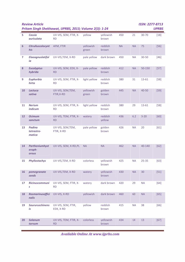
Table 1: Plants listed for Bio-synthesis of silver nanoparticles and their characterization
results.
S.
N.
Plantae extract
used
Charterization
Techniques
Color of Solution λmax
(nm)
Size of AgNPs
(nm)
Refe-
rnce
Initial Final UV-VIS X-
RD
SEM/TE
M
1 Allium
cepa
UV-VIS,TEM, DLS light yellow dark brown 413 NA 31-48 [53]
2 Azadirachtaindi
ca
UV-VIS, TEM, FTIR, X-
RD
NA yellowish
brown
450 NA 5-35 [54]
3 Capsicum
annum
UV-VIS,TEM, X-
RD,SAED
yellowish
green
reddish
brown
441 NA 02-06 [55]
4 Carica
papaya
UV-VIS, SEM, X-RD,
FTIR
watery yellowish
brown
450 15 25-50 [52]
Review Article ISSN: 2277-8713
Pritam Singh Shekhawat, IJPRBS, 2013; Volume 2(3): 1-24 IJPRBS
Available Online At www.ijprbs.com
5 Cassia
auriculata
UV-VIS, SEM, FTIR, X-
RD
yellow yellowish
brown
450 21 30-70 [18]
6 Citrulluscolocynt
his
AFM, FTIR yellowish
green
reddish
brown
NA NA 75 [56]
7 Elaeagnuslatifol
ia
UV-VIS,TEM, X-RD pale yellow dark brown 450 NA 30-50 [46]
8 Eucalyptus
hybrida
UV-VIS, SEM, EDX, X-
RD
pale yellow reddish
brown
412 NA 50-150 [57]
9 Euphorbia
hirta
UV-VIS, SEM, FTIR, X-
RD
light yellow reddish
brown
380 31 13-61 [58]
10 Lactuca
sativa
UV-VIS, SEM,TEM,
FTIR,X-RD
yellowish
green
golden
brown
445 NA 40-50 [59]
11 Nerium
indicum
UV-VIS, SEM, FTIR, X-
RD
light yellow reddish
brown
380 29 13-61 [58]
12 Ocimum
sanctum
UV-VIS, TEM, FTIR, X-
RD
watery reddish
yellow
436 6.2 3-20 [60]
13 Padina
tetrastro-
matica
UV-VIS, SEM,TEM,
FTIR, X-RD
pale yellow golden
brown
426 NA 20 [61]
14 Partheniumhyst
eroph-
orous
UV-VIS, SEM, X-RD,PL NA NA 462 NA 40-140 [62]
15 Phyllostachys
UV-VIS,TEM, X-RD colorless yellowish
brown
425 NA 25-35 [63]
16 pomegranate
seeds
UV-VIS,TEM, X-RD watery yellowish
brown
430 NA 30 [51]
17 Ricinuscommuni
s
UV-VIS, SEM, FTIR, X-
RD
watery dark brown 420 29 NA [64]
18 Rosmarinusoffici
nalis
UV-VIS, X-RD yellowish dark brown 460 60 NA [65]
19 Saururuschinens
is
UV-VIS, SEM, FTIR,
EDX, X-RD
yellow reddish
brown
415 NA 38 [66]
20 Solanum
torvum
UV-VIS, TEM, FTIR, X-
RD
colorless yellowish
brown
434 14 13 [67]

Review Article ISSN: 2277-8713
Pritam Singh Shekhawat, IJPRBS, 2013; Volume 2(3): 1-24 IJPRBS
Available Online At www.ijprbs.com
21 Spinaciaolerace
a
UV-VIS, SEM,TEM,
FTIR, X-RD
yellowish
green
golden
brown
440 NA 40-70 [59]
22 Syzygium
cumini
UV-VIS, SEM, FTIR, X-
RD,EDX
light brown yellowish
brown
450 3.5 93 [68]
23 Zingiberofficinal
e
UV-VIS, TEM, FTIR, X-
RD
colorless yellowish
brown
430 NA 10-66 [69]
Table 2: Plants listed for Bio-synthesis of silver nanoparticles and their XRD, SEM or TEM
results.
S.
N.
Plantae extract
used
Silver nanoparticles Braggs reflections at 2θ in XRD for: Refer
nce
Morpho-
logy
Distributi
on
1st
Peak
Resp.
Plane
2nd
Peak
Resp.
Plane
3rd
Peak
Resp.
Plane
1 Allium
cepa
spherical polydisper
sed
NA NA NA NA NA NA [53]
2 Azadir-achta
indica
spherical polydisper
sed
38 111 45 200 64 220 [54]
3 Capsicum
annum
spherical monodisp
ersed
38 111 44 200 64 220 [55]
4 Carica
papaya
cubic polydisper
sed
38.11 111 64.52 110 67.52 112 [52]
5 Cassia
auriculata
spherical polydisper
sed
38.02 111 NA NA NA NA [18]
6 Citrulluscolocynt
his
spherical NA NA NA NA NA NA NA [56]
7 Elaeagnus
latifolia
spherical NA 38.06 111 44.64 200 64.58 220 [46]
8 Eucalyptus
hybrida
cubic monodisp
ersed
30.8 111 38 NA 44 NA [57]
9 Euphorbia
hirta
spherical monodisp
ersed
38.11 111 44.46 200 65.24 220 [58]
10 Lactuca
sativa
spherical polydisper
sed
38.18 111 44.37 200 64.48 220 [59]

Review Article ISSN: 2277-8713
Pritam Singh Shekhawat, IJPRBS, 2013; Volume 2(3): 1-24 IJPRBS
Available Online At www.ijprbs.com
11 Nerium
indicum
spherical monodisp
ersed
38.11 111 44.46 200 64.48 220 [58]
12 Ocimum
sanctum
spherical polydisper
sed
37.6 111 44.7 200 76.3 311 [60]
13 Padinatetrastr-
omatica
spherical monodisp
ersed
28.09 220 32.46 122 42.18 400 [61]
14 Partheniumhyste
roph-orous
spherical polydisper
sed
38.1 111 44.6 200 64.8 220 [62]
15 Phyllost-achys
spherical polydisper
sed
38 111 44 200 64 220 [63]
16 Pomegr-anate
seeds
spherical NA NA NA NA NA NA NA [51]
17 Ricinus
communis
NA monodisp
ersed
38.11 111 44.27 200 64.42 220 [64]
18 Rosma-
rinusofficinalis
NA NA 32.37 111 38.4 111 44.85 200 [65]
19 Saururus
chinensis
spherical polydisper
sed
32.22 NA 32.96 NA 38.08 111 [66]
20 Solanum
torvum
spherical monodisp
ersed
38 111 44 200 64 220 [67]
21 Spinacia
oleracea
spherical polydisper
sed
38.18 111 44.37 200 46.18 200 [59]
22 Syzygium
Cumini
spherical monodisp
ersed
32.4 101 38.2 111 44.4 200 [68]
23 Zingiber
officinale
spherical polydisper
sed
38 111 45 200 64 220 [69]
Review Article ISSN: 2277-8713
Pritam Singh Shekhawat, IJPRBS, 2013; Volume 2(3): 1-24 IJPRBS
Available Online At www.ijprbs.com
REFERENCE
1. Taniguchi N. Proc. Intl. Conf. Prod. Eng.
Tokyo, Part II, Japan Society of Precision
Engineering 1974, 18-23.
2. Goodsell D S. Bio-nanotechnology:
Lessons from nature. Hoboken, New York:
Wiley-Liss 2004, 224-237.
3. Kyriacou S V, Brownlow W J &Xu N.
Using nanoparticle optics assay for direct
observation of the function of antimicrobial
agents in single live bacterial cells.
Biochemistry, 43 (2004) 140.
4. David M B, Martin P & William A S.
Research strategies for safety evaluation of
nanomaterials, Part III: Nanoscale
technologies for assessing risk and
improving public health.ToxicolSci, 88
(2005) 298-306.
5. Lanone S &Boczkowski J. Biomedical
applications and potential health risks of
nanomaterials: molecular
mechanisms.CurrMol Med, 6 (2006) 651-
663.
6. Kamat P V. Photophysical,
photochemical and photocatalytic aspects
of metal nanoparticles.J PhysChem B, 106
(2002) 7729-7744.
7. Maier S A, Brongersma M L, Kik P G,
Meltzer S, Requicha A A G, et al,
Plasmonics- A Route to Nanoscale Optical
Devices.AdvancedMaterials, 19 (2001)
1501-1505.
8. Gracias D H, Tien J, Breen T, Hsu C &
Whitesides G M. Forming electrical
networks in three dimensions by self
assembly.Science, 289 (2002) 1170-1172.
9. Mirkin C A, Letsinger R L, Mucic R C
&Storhof J J A. DNA-based method for
rationally assembling nanoparticles into
macroscopic materials. Nature, 382 (1996)
607-609.
10. Han M, Gao X, Su J Z &Nie S. Quantum-
dot tagged micro beads for multiplexed
optical coding of biomolecules. Nature
Biotechnology, 19 (2001) 631-635.
11. Chan W C W &Nie S. Quantum dot bio
conjugates for ultrasensitive non isotopic
detection. Science, 281 (1998) 2016-2018.

Review Article ISSN: 2277-8713
Pritam Singh Shekhawat, IJPRBS, 2013; Volume 2(3): 1-24 IJPRBS
Available Online At www.ijprbs.com
12. Willems, Van den Wildenberg, W
&Espana sl. Roadmap report on
nanoparticles Barcelona, Spain, 2005.
13. Nordberg G, Gerhardsson L, Seiler H G,
Sigel H & Sigel A (Eds.). Handbook on
Toxicity of Inorganic Compounds. Marcel
Dekker, New York 1988, 619.
14. Bhatt J S A. Heralding a new future-
Nano biotechnology. CurrSci, 85 (2003) 147-
154.
15. James E M & Browning N D. Practical
aspects of atomic resolution imaging and
analysis in STEM. Ultramicroscopy, 78
(1999) 125-139.
16. Moyer CA. A treatment of burns: Trans
Stud Coll Physicians, Philadelphia, 33 (1965)
53-63.
17. Reda M, Sheshtwy E I, Abdullah M
&Nayera A. In situ production of silver
nanoparticles on cotton fabric and its
antimicrobial evaluation.Cellulose, 18
(2011) 75-82.
18. Udayasoorian U, Kumar V
&Jayabalahrishnan R M. Extracellular
synthesis of silver nanoparticles using leaf
extract of Casciaauriculate. Digest J
Nanomat and Biostr, 6 (2011) 279-283.
19. Feng Q L, Wu J, Chen G Q, Cui F Z, Kim T
M et al. A mechanistic study of the
antibacterial effect of silver ions on
Escherichia coli and Staphylococcus aureus.
J Biomed Mater Res, 52 (2000) 662.
20. Gupta A. Silver S. Silver as a biocide: will
resistance become a problem? Nat
Biotechnol 1998;16:888.
21. Niemeyer C M. Self-assembled
nanostructures based on DNA: towards the
development of Nano biotechnology.
CurrOpinChemBiol, 4 (2000) 609.
22. Peterson B R, Ayusman S, Varun S &
Megan M M. Silver bromide
nanoparticle/polymer composites: dual
action tunable antimicrobial materials.
AmerChemSoc, 128 (2006) 808.
23. Li P, Li J, Wu C, Wu Q & Li J. Synergistic
antibacterial effects of β-lactam antibiotic
combined with silver nanoparticles.
Nanotechnology, 16 (2005)1912.
24. Jain P &Pradeep T. Potential of silver
nanoparticle-coated polyurethane foam as

Review Article ISSN: 2277-8713
Pritam Singh Shekhawat, IJPRBS, 2013; Volume 2(3): 1-24 IJPRBS
Available Online At www.ijprbs.com
an antibacterial water filter.
BiotechnolBioeng, 90 (2005) 59.
25. Li Y, Leung P, Yao L, Song Q W & Newton
E. Antimicrobial effect of surgical masks
coated with nanoparticles Hosp. J Infec, 62
(2006) 58.
26. Steven, Emory R, Haskins W E &Niel S.
Direct observation of size-dependent
optical enhancement in single metal
nanoparticles. J Am ChemSoc, 120 (1998)
8009.
27. Alivisatos A P. Perspectives on the
physical chemistry of semiconductor
nanocrystals of Physical Chemistry. J
PhysChem, 100 (1996) 13226-13239.
28. 28. Lovley D R, Stolz J F, Nord G L &
Phillips E J P. Anaerobic production of
magnetite by a dissimilatory iron-reducing
microorganism Microbial reduction of
uranium. Nature, 330 (1987) 252.
29. Philse A P & Maas D. Magnetic colloids
from magnetotactic bacteria: chain
formation and colloidal stability. Langmuir,
18 (2002) 9977.
30. Dickson D P E, Nanostructured
magnetism in living systems.J MagnMagn
Mater, 203 (1999) 46.
31. Mann S. Molecular tectonics in
biomineralization and biomimetic materials
chemistry. Nature, 365 (1993) 499.
32. Oliver S, Kuperman A, Coombs N, Lough
A &Ozin G A. Lamellar alumino phosphates
with surface patterns that mimic diatom
and radiolarian microskeletons. Nature, 378
(1995) 47.
33. Kröger N, Deutzmann R &Sumper M.
Polycationic peptides from diatom biosilica
that direct silica nanosphere formation.
Science, 286 (1999) 1129.
34. Sleytr U B, Messner P, Pum D & Sara M.
Crystalline bacterial cell surface layers (S
layers): from supra molecular cell structure
to biomimetic and nanotechnology. Angew
Chem. Int. Ed, 38 (1999) 1034.
35. Goia D V &Matijevic E N. Preparation of
monodispersed metal particles. J Chem, 22
(1998) 1203.
36. Taleb C, Petit M&Pileni P. Optical
Properties of Self- Assembled 2D and 3D

Review Article ISSN: 2277-8713
Pritam Singh Shekhawat, IJPRBS, 2013; Volume 2(3): 1-24 IJPRBS
Available Online At www.ijprbs.com
Superlattices of Silver Nanoparticles. Chem
Mater, 9 (1997)950.
37. Esumi K, Tano T, Torigoe K & Meguro K.
Preparation and characterization of
bimetallic palladium-copper colloids by
thermal decomposition of their acetate
compounds in organic solvents. Chem
Mater, 2 (1990) 564.
38. Henglein. Reduction of Ag(CN)2 on silver
and platinum colloidal
nanoparticles.Langmuir, 17 (2001) 23.
39. Rodriguez S L, Blanco M C & Lopez M A.
Electrochemical synthesis of silver
nanoparticles. J PhysChemB, 104 (2000)
9683.
40. Zhu J J, Liu S W, Palchik O, Koltypin
Y&Gedanken A. Shape-controlled synthesis
of silver nanoparticles by pulse
sonoelectrochemical methods.Langmuir, 16
(2000) 6396.
41. Pastoriza-Santos & Liz-Marzan.
Synthesis of silver nanoprisms in
DMF.Langmuir, 18 (2002) 2888.
42. Begum N A, Mondal S, Basu S, Laskar R
A &Mandal D, Biogenic synthesis of Au and
Ag nanoparticles using aqueous solutions of
black tea leaf extracts.Colloids and Surfaces
B: Biointerfaces, 71( 2009) 113.
43. Ankamwar. B., Chaudhary, M. &Sastry,
M. Gold nanotriangles biologically
synthesized using tamarind leaf extract and
potential application in vapor sensing. Synth
React Inorg Metal-Org NanometalChem 10
(2005) 1665.
44. Prasad S, Singh M, Singh S &Gambhir S.
Nanotechnology in medicine and
antibacterial effect of silver
nanoparticles.Digest J Nanomat and Biostr,
3 (2008) 115-122.
45. Vaidyanathan R, Kalishwaralal K,
Gopalram S &Gurunathan S. Retracted:
Nanosilver—The burgeoning therapeutic
molecule and its green synthesis.
Biotechnology Advances, 27 (2009) 924-
937.
46. Phanjom P, Sultana A, Sharma H,
Ramchiary J, Goswami K et al. Plant-
mediated synthesis of silver nanoparticles
using Elaeagnuslatifolialeaf extract. Digest J
Nanomat and Biostr, 7 (2012) 1117-1123.
47. Parashar V, Parashar R, Sharma B
&Pandey A C. Parthenium leaf extract
mediated synthesis of silver nanoparticles: a

Review Article ISSN: 2277-8713
Pritam Singh Shekhawat, IJPRBS, 2013; Volume 2(3): 1-24 IJPRBS
Available Online At www.ijprbs.com
novel approach towards weed utilization.
Digest J NanomatBiostr, 4 (2009) 45-50.
48. Saifuddin N, Wong C W &Yasumira A N.
Rapid biosynthesis of silver nanoparticles
using culture supernatant of bacteria with
microwave irradiation. E-J Chem, 6 (2009)
61-70.
49. Bhainsa K C & D’Souza S F. Extracellular
biosynthesis of silver nanoparticles using
the fungus Aspergillusfumigatus. Colloids
and Surfaces B: Biointerfaces, 47 (2006)
160-164.
50. Willner B, Basnar B &Willner B.
Nanoparticle–enzyme hybrid systems for
Nano biotechnology. FEBS J, 274 (2007)
302-309.
51. Chauhan S, Upadhyay M K, Rishi N &
Rishi S. Phytofabrication of silver
nanoparticles using pomegranate fruit
seeds.Int J NanomatBiostr, 1 (2011) 17-21.
52. Kothari S L, Jain D, Daima H K
&Kachhwaha S. Synthesis of plant-mediated
silver nanoparticles using papaya fruit
extract and evaluation of their anti
microbial activities. Digest J Nanomat and
Biostr, 4 (2009) 557-563.
53. Tripathi R M, Singh R P &Saxena A.
Biological synthesis of silver nanoparticles
by using onion (I) extract and their
antibacterial activity. Digest J Nanomat and
Biostr, 5 (2010) 427-432.
54. Shankar S S,Rai A, Ahmad A &Sastry M.
Rapid synthesis of Au, Ag, and bimetallic Au
core–Ag shell nanoparticles using Neem
(Azadirachtaindica) leaf broth.Journal of
Colloid and Interface Science, 275 (2004)
496-502.
55. Prasad K &Jha A K. Green fruit of chili
(Capsicum annum l.) synthesizes nano
silver. Digest J Nanomat and Biostr, 6 (2011)
1717-1723.
56. Satyavani K, Ramanathan T
&Gurudeeban S. Green synthesis of silver
nanoparticles by using stem derived callus
extract of bitter apple Citrulluscolocynthis.
Digest J Nanomat and Biostr, 6 (2011) 1019-
1024.
57. Dubey M, Bhadauria S &Kushwah. B
SGreen synthesis of nanosilver particles
from extract of Eucalyptus hybrida(safeda)
leaf. Digest J Nanomat and Biostr, 4 (2009)
537-543.

Review Article ISSN: 2277-8713
Pritam Singh Shekhawat, IJPRBS, 2013; Volume 2(3): 1-24 IJPRBS
Available Online At www.ijprbs.com
58. Paul J A, Priya M M&Selvi B K, Green
synthesis of silver nanoparticles from the
leaf extracts of Euphorbia hirta and
Neriumindicum, Digest J. Nanomat and
Biostr, 6 (2011) 869-877.
59. Kanchana A, Agarwal I, Sunkar S, Nellore
J &Namasivayam K. Biogenic Silver
Nanoparticles from SpinaciaOleraceaand
Lactuca Sativa and their potential
antimicrobial activity. Digest J Nanomat and
Biostr, 6 (2011) 1741-1750.
60. Raju B D P, Mallikarjuna K, Narasimha G,
Dillip G R, Praveen B et al. Green synthesis
of silver nanoparticles using Ocimumleaf
extract and their characterization. Digest J
Nanomat and Biostr, 6 (2011) 181-186.
61. Shivaraj R, Venckatesha R,
Jegadeeswaran P. Green synthesis of silver
nanoparticles from extract of
Padinatetrastromaticaleaf. Digest J
Nanomat and Biostr, 7 (2012) 991-998.
62. Kumbhakar P, Sarkar R &Mitra A K.
Green synthesis of silver nanoparticles and
its optical properties.Digest J Nanomat and
Biostr, 5 (2010) 491-496.
63. Kumar S V, Jegan A, Ramasubbu A,
Balamurugan M &Saravanan S.
Environmental benign synthesis and
characterization of silver nanoparticles
using phyllostachyssp leaves extract. Digest
J Nanomat and Biostr, 6 (2011) 325-330.
64. Srivastava J N, Singh A, Mittal S,
Srivastava R &Dassa S. Biosynthesis of silver
nanoparticles using Ricinuscommunis leaf
extract and its antibacterial activity.Digest J
Nanomat and Biostr, 7 (2012) 1157-1163.
65. Sulaiman G M, Mohammad A A W,
Wahed H E A & Ismail M M. Biosynthesis,
antimicrobial and cytotoxic effects of silver
nanoparticles using
Rosmarinusofficinalisextract.Digest J
Nanomat and Biostr, 8 ( 2013) 273 - 280.
66. Lee K D, Prasad T N V K V, Nagajyoti P C
&Sreekanth T V M. Bio-fabrication of silver
nanoparticles using leaf extract of
Saururuschinenis. Digest J. Nanomat and
Biostr, 6 (2011) 121-133.
67. Govindaraju K, Tamilselvan S, Kiruthiga
V &Singaravelu G. Biogenic silver
nanoparticles by Solanumtorvum and their
promising antimicrobial activity. J
Biopesticides 3-Special Issue (2010) 394-
399.

Review Article ISSN: 2277-8713
Pritam Singh Shekhawat, IJPRBS, 2013; Volume 2(3): 1-24 IJPRBS
Available Online At www.ijprbs.com
68. Narendhirakannan R T & Banerjee J.
Biosynthesis of silver nanoparticles from
Syzygiumcumini(l.) seed extract and
evaluation of their in vitro antioxidant
activities. Digest J Nanomat and Biostr, 6
(2011) 961-968.
69. Singh H, Singh C, Sharma V, Naik P K
&Khandelwal V. A green biogenic approach
for synthesis of gold and silver
nanoparticles using Zingiberofficinale.
Digest J. Nanomat and Biostr,6 (2011) 535-
542.
70. Huang J, Li Q, Sun D, Lu Y, Su Y et al.
Biosynthesis of silver and gold nanoparticles
by novel sundried
Cinnamomumcamphoraleaf,
Nanotechnology, 18 (2007) 105104.
71. Tao A, Sinsermsuksaku P & Yang P.
Polyhedral silver nanocrystals with distinct
scattering signatures. AngewChemInt Ed, 45
(2006) 4561-4597.
72. Wiley B, Sun Y, Mayers B & Xia Y. Shape-
controlled synthesis of metal
nanostructures: the case of silver.Chem A
Eur J, 11 (2005) 454-463.
73. Kapoor S, Lawless D, Kennepohl P,
Meisel D &Serpone N. Reduction and
aggregation of silver ions in aqueous gelatin
solutions. Langmuir, 10 (1994) 3018.
74. Schneider S, Halbig P, Grau H & Nickel
U. Reproducible preparation of silver sols
with uniform particle size for application in
surface-enhanced Raman
spectroscopy.PhotochemPhotobiol, 60
(1994) 605.
75. Schirtcliffe N, Nickel U & Schneider S.
Reproducible preparation of silver sols with
small particle size using borohydride
reduction: for use as nuclei for preparation
of larger particles. J Colloid Interface Sci,
211 (1999) 122-129.
76. Rivas L, Sanchez-Cortes S, Garcia-Ramos
J V &Morcillo G. Growth of silver colloidal
particles obtained by citrate reduction to
increase the Raman enhancement factor.
Langmuir, 17 (2001) 574.
77. Nickel U, Castell A Z, Poppl K &
Schneider S. A Silver colloid produced by
reduction with hydrazine as support for
highly sensitive surface-enhanced Raman
spectroscopy. Langmuir, 16 (2000) 9087.
78. Carlson C, Hussain S M, Schrand A M,
Braydich-Stolle L K, Hess K L et al. Unique
cellular interaction of silver nanoparticles:

Review Article ISSN: 2277-8713
Pritam Singh Shekhawat, IJPRBS, 2013; Volume 2(3): 1-24 IJPRBS
Available Online At www.ijprbs.com
Size-dependent generation of reactive
oxygen species. J PhysChem B, 112 (2008)
13608-13619.
79. Mulvaney P. Surface plasmon
spectroscopy of nanosized metal particles.
Langmuir, 12 (1996) 788.
80. Sondi I &Sondi B S. Silver nanoparticles
as antimicrobial agent: a case study on E.
coli as a model for Gram-negative bacteria
J. Colloid Interface Sci., 275 (2004) 177.
81. Mukherjee P, Senapati S, Mandal D,
Ahmad A, Khan M I et al, Intracellular
synthesis of gold nanoparticles by a novel
alkalotolerantactinomycete, Rhodococcus
species. Chem. Bio. Chem, 3 (2002) 461.
82. Gonzalo J, Serna R, Sol J, Babonneau D
&Afonso C N. Morphological and interaction
effects on the surface plasmon resonance
of metal nanoparticles. J PhysCondens
Matter, 15 (2003) 3001.
83. David E, Elumalai E K, Prasad T N V K V
&Nagajyothi P C. A bird’s eye view on
Biogenic Silver nanoparticles and their
applications. Der ChemicaSinica, 2 (2011)
88-97.
84. Sosa I O, Noguez C & Barrera R G.
Optical properties of metal nanoparticles
with arbitrary shapes. J PhysChem B, 107
(2003) 6269.
85. Xu H &Käll M. Morphology effects on
the optical properties of silver
nanoparticles. J NanosciNanotechnol, 4
(2002)254.
86. Song J Y & Kim B S. Rapid biological
synthesis of silver nanoparticles using plant
leaf extracts. Bioprocess BiosystEng, 32
(2008) 79.
87. Li Y, Yates J A & Chen J J. Identification
and characterization of sea squirt
telomerase reverse transcriptase. Gene,
400 (2007) 16.
88. Cullity B D. Elements of X-ray
Diffraction, Edison-Wesley Publishing
Company Inc, USA 1978.
89. Woodleand M C & Lu PY. Nanoparticles
deliver RNAi therapy.Nanotoday, 8 (2005)
34-41.
90. Devika C B, Arezou, Ghazani A & Warren
C W C. Determining the size and shape
dependence of gold nanoparticle uptake

Review Article ISSN: 2277-8713
Pritam Singh Shekhawat, IJPRBS, 2013; Volume 2(3): 1-24 IJPRBS
Available Online At www.ijprbs.com
into mammalian cells. Nano Lett, 6 (2006)
662-668.