characterization of thsd7a- and pla2r1- specific
TRANSCRIPT

Characterization of THSD7A- and PLA2R1-specific antibodies and their role in the
pathogenesis of membranous nephropathy
Kumulative Dissertation zur Erlangung des Doktorgrades (Dr. rer. nat.)
an der Fakultät für Mathematik, Informatik und Naturwissenschaften
Fachbereich Biologie der Universität Hamburg
vorgelegt von Larissa Seifert (geb. 21.12.1988)
Hamburg, 2020
1. Gutachter: Priv.-Doz. Dr. Gunther Zahner 2. Gutachter: Professor Wolfgang Streit Datum der Disputation: 26.06.2020 Prüfungskommission: Professor Wolfgang Streit Priv.-Doz. Gunther Zahner Professor Friedrich Koch-Nolte Priv.-Doz. Hartwig Lüthen Vorsitz: Priv.-Doz. Hartwig Lüthen
Table of contents
1. Introduction .............................................................................................................. 2
1.1 Clinical and histological features of membranous nephropathy ........................... 2
1.2 Rat Animal Models of MN ................................................................................... 4
1.3 Identification of antigens in patients with MN ...................................................... 5
1.4 Clinical role of autoantibody measurement ......................................................... 8
1.5 Structure of PLA2R1 and identification of autoantibody binding domains ...........10
1.6 Pathogenicity of autoantibodies .........................................................................11
2. Materials, Methods and Results ............................................................................14
3. Discussion ..............................................................................................................15
3.1 Structure of THSD7A and the role of autoantibody binding-sites in THSD7A-
associated MN .............................................................................................................15
3.2 Heterologous models of THSD7A- and PLA2R1-associated MN .......................22
4. Summary .................................................................................................................30
5. Zusammenfassung .................................................................................................31
6. Literature .................................................................................................................33
7. Individual contribution ...........................................................................................39
8. Acknowledgements ................................................................................................40
9. Eidesstattliche Erklärung .......................................................................................41
10. Appendix ..............................................................................................................42
Introduction
2
1. Introduction
1.1 Clinical and histological features of membranous nephropathy
The kidney is the most important detoxification organ of the body; it filters the blood and
excretes metabolic end products and toxins into the urine. Additionally, it participates in the
regulation of water and electrolyte balance, acid-base balance and blood pressure. The
nephron is the structural and functional unit of the kidney consisting of the renal corpuscle
connected with a renal tubule, the latter responsible for both resorption and secretion of
substances. The renal corpuscle is composed of a capillary tuft called glomerulus encased
by the Bowman's capsule. In healthy kidneys the glomeruli are responsible for the filtration
of the primary urine. The filtration barrier of the glomerulus consists of a unique fenestrated
endothelium around the glomerular capillary and a glomerular basement membrane (GBM),
both negatively charged. As the third component of the barrier the glomerular epithelial
cells, also called podocytes, build highly differentiated foot processes with a connecting “slit
diaphragm” that localize on top of the GBM. The filtration barrier is permeable for water and
smaller molecules but almost not for negatively charged and/or large molecules. Therefore,
plasma proteins pass through the filter only to a very small extent. A large amount of all
filtrated small molecules and water are reabsorbed during the following passage through
the tubular system of the nephron, resulting in the concentrated, excreted urine.
In case of damages of the filtration barrier, high amounts of protein escape into the urine
leading to a condition called proteinuria. The stage of a nephrotic syndrome has been
reached if severe proteinuria is accompanied by peripheral edema, hypoalbuminemia and
hyperlipidemia [1, 2]. In approximately 30% of cases, membranous nephropathy (MN) is
responsible for those symptoms and thus is the most common cause of nephrotic syndrome
in adult Caucasians [3]. In immunofluorescence microscopy, MN is characterized by a gran-
ular pattern of IgG together with components of the complement system along the GBM.
Electron microscopy reveals subepithelial electron-dense deposits on the outer aspect of
the GBM, close to the podocytes. Additionally, the podocyte foot processes are extensively
effaced and the GBM is expanded. It has long been assumed that the formation of subepi-
thelial deposits in combination with complement activation leads to the disturbance of the
filtration barrier and the development of nephrotic syndrome, without notable proliferation
or infiltration of inflammatory cells in the glomerulus [4, 5].
In 80% of cases MN is primary, occurring in the absence of an established cause. Never-
theless there are several conditions which are capable to induce a so called secondary MN,
such as lupus erythematosus, cancer, some infections such as hepatitis B or C, or the use
of certain drugs such as penicillamine or gold [6-9]. Even though the histopathological pat-

Introduction
3
tern is very similar in these two types of MN there are some features that distinguish be-
tween them. Electron-dense deposits in primary MN are typically subepithelial and in-
tramembranous, while mesangial deposits are uncommon [10]. In contrast, in secondary
forms of MN those deposits are often located subendothelial and mesangial, suggesting a
circulating immune complex [6, 11, 12]. The immune deposits in primary MN mostly contain
IgG4, whereas in secondary cases IgG1, IgG2 and IgG3 are the dominant subtypes [13,
14].
Figure 1: A schematic presentation of the filtration barrier under normal and disease conditions. 1: podocyte, 2: glomerular basement membrane, 3: endothelium B Electron microscopic image under normal and disease conditions. C PAS staining of a healthy and a damaged glomerulus. D Immunohistochemical staining for IgG in
a biospsy of a patient positive for MN. All images were kindly provided by Prof. Dr. Thorsten Wiech.

Introduction
4
The clinical outcome of MN is very diverse. While about 40% of patients accomplish spon-
taneous remission, another 30% end up with end stage renal disease, with a poor response
to immunosuppressive treatment [15, 16]. Those patients are in need of dialysis or trans-
plantation. Notably, even a successful transplantation is no guarantee for a favorable out-
come. In approximately 40% of patients who received a kidney graft, disease will re-emerge
and 45% of those patients will lose their graft due to this recurrence [17].
Because of the natural heterogeneity of this disease, it is of great importance to find predic-
tors of the individual disease outcome. Such predictors would allow to select patients for
immunosuppressive treatment who will have a benefit regarding kidney function and, con-
versely, to reduce adverse events from immunosuppression in individuals who will likely not
benefit from such treatment. Thus, further progress in patient care depends on a deeper
pathophysiological understanding of the disease processes and the identification of molec-
ular signatures that help to predict the disease outcome.
1.2 Rat Animal Models of MN
The Heymann Nephritis (HN) in rats is the first model of experimental membranous
nephropathy that was established. Two different forms were classically used to study the
pathogenesis of MN, the so called active and passive HN. The active model relies on active
immunization of rats with an extract of rat tubular proteins, which induces generation of
autoantibodies [18]. In the passive model, heterologous antibodies from animals that were
immunized with rat brush border protein extracts are transferred to rats [19, 20].
In both models, animals develop granular glomerular deposits and proteinuria. The follow-
ing key characteristics of MN could be deduced from the models of active and passive Hey-
mann nephritis:
Binding of circulating (auto)antibodies to a resident antigen leads to in situ formation
of subepithelial immune complexes [21, 22].
The subepithelial localization of the detected IgG indicates that the target antigen is
expressed on glomerular podocytes, which represent the outer layer of the glomer-
ular filtration barrier [21, 22].
Antibody binding and deposition along the glomerular filtration barrier activates the
complement system, which contributes to the glomerular damage in MN [20, 23, 24].
Yet, the causal link between complement activation and disease induction is not conclu-
sively clarified. On the one hand rats which are de-complemented with cobra venom factor
Introduction
5
present no proteinuria in passive HN, despite the demonstrable formation of immune de-
posits [20]. On the other hand nephrotic syndrome is inducible in C6-deficient rats with ac-
tive and passive HN [25, 26].
The relevant antigen in HN was later identified as a member of the LDL-receptor family,
with a molecular weight of about 600 kDa. Due to its size this podocyte membrane protein
is called Megalin (LRP2; low density lipoprotein receptor-related protein 2). Megalin is ex-
pressed on rat tubular cells and podocytes [27, 28]. In rats immunized with a small N-termi-
nal fragment of Megalin an epitope spreading from the immunization fragment to more distal
domains takes place over the time course of the experiment. This spreading is necessary
for full onset of disease with remarkable proteinuria [29].
However, all these results can only be transferred to the human MN to a limited extent since
human podocytes do not express Megalin. It is neither detectable in immune deposits, nor
circulating antibodies are found in sera of patients with MN. Hence, Megalin is not involved
in human MN.
1.3 Identification of antigens in patients with MN
The first identified human antigen in MN was the neutral endopeptidase (NEP). Antibodies
against this protein cause nephrotic syndrome with possible renal failure by crossing the
placenta and binding to podocytes of neonates [30]. The mothers of these children are ge-
netically NEP deficient and were subjected to allo-immunization due to miscarriages, or
during the course of the pregnancy [31, 32]. Although these cases of neonatal MN are very
rare they are the first proof that a human podocyte protein can serve as an antigen targeted
by circulating antibodies.
In 2009 the M-type phospholipase A2 receptor 1 (PLA2R1) was discovered as an antigen
in MN. Beck et al. [33] tested patient sera in Western blot analyses on protein extracts of
glomeruli that were isolated from healthy human kidneys. Approximately 70% of all sera
tested in this study contained antibodies that bound to a 185 kDa protein. Subsequent mass
spectrometry revealed that this protein was PLA2R1. Indeed all previous analyzed sera also
recognized recombinant PLA2R1 in Western blot analyses. The reactivity of sera is only
given under non-reducing conditions, suggesting a conformational epitope depending on
disulfide bonds.
PLA2R1 is detectable in healthy human podocytes and the staining is remarkably enhanced
under disease conditions. Subepithelial deposits in diseased podocytes contain PLA2R1
often co-localized with IgG4 (the dominant IgG subtype in PLA2R1- associated MN), indi-
cating an in situ formation of the deposits similar to the ones in HN. Moreover, antibodies
Introduction
6
eluted from frozen human biopsy samples detect recombinant PLA2R1 in Western blot
analyses, proving that the antibodies deposited in patient glomeruli are PLA2R1-specific.
PLA2R1 belongs to the mannose receptor family. All members of this family are transmem-
brane proteins consisting of an N-terminal cysteine rich (CysR) domain, a single fibronectin
type II (FnII) domain, followed by 8 to 10 C-type lectin-like domains (CTLDs). Their cyto-
plasmic tail contains motifs that lead to constitutive endocytotic recycling [34, 35]. The exact
function of PLA2R1 is not clear until now. However, it is known that PLA2R1 serves as a
receptor for secretory PLA2 (sPLA2), which is a strong pro-inflammatory enzyme. Thereby,
PLA2R1 might act in two directions, as a clearance receptor or as a positive regulator of
sPLA2. This interaction can lead to different effects, for example protein kinase activation,
producing lipid activators or activating DNA damage pathways [36, 37]. Furthermore
PLA2R1 was proposed to alter the migratory responses to collagen type I and IV. Recom-
binant PLA2R1 binds to collagen I and thereby interrupts the interaction between collagen
and integrin β1 [38, 39]. Interestingly, anti-PLA2R1 autoantibodies from patients with MN
interfere with the adhesion of podocytes to collagen type IV, suggesting that the disturbance
of podocyte interaction with collagen type IV in the glomerular basement membrane may
be involved in MN pathogenesis [40].
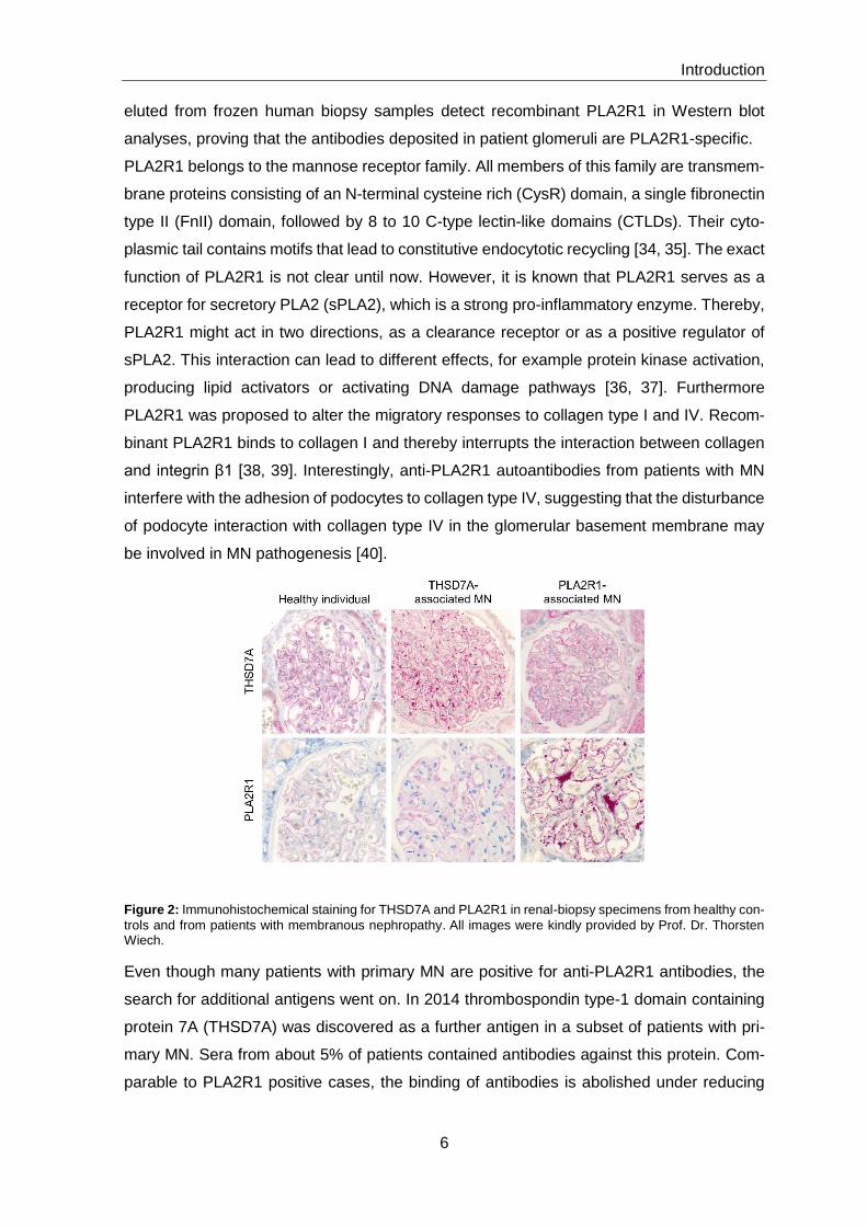
Figure 2: Immunohistochemical staining for THSD7A and PLA2R1 in renal-biopsy specimens from healthy con-
trols and from patients with membranous nephropathy. All images were kindly provided by Prof. Dr. Thorsten Wiech.
Even though many patients with primary MN are positive for anti-PLA2R1 antibodies, the
search for additional antigens went on. In 2014 thrombospondin type-1 domain containing
protein 7A (THSD7A) was discovered as a further antigen in a subset of patients with pri-
mary MN. Sera from about 5% of patients contained antibodies against this protein. Com-
parable to PLA2R1 positive cases, the binding of antibodies is abolished under reducing

Introduction
7
conditions in Western blot analyses and the dominant IgG subtype is IgG4 [41]. In line with
the results for PLA2R1, IgG eluted from biopsies of THSD7A-positive patients detects re-
combinant THSD7A in Western Blot analyses. THSD7A is detectable in human podocytes
with an enhanced staining under disease conditions. Worthy of note, 20% of THSD7A pos-
itive patients also presented with malignancies. THSD7A was found in their tumors, meta-
static lymph nodes and also in subepithelial immune deposits, indicating a potential causal
link between cancer and MN [42, 43].
THSD7A is a transmembrane glycoprotein with a large, heavily glycosylated, extracellular
region, consisting of 11 thrombospondin type 1 domains (TSP-1 domains) [41]. It is promi-
nently expressed on podocytes of humans and rodents, where it is located at the basal
aspects of the foot processes [44]. During angiogenesis THSD7A mediates cell migration
and tube formation in association with focal adhesion proteins in endothelial cells [45, 46].
In human cultured podocytes THSD7A expression enhances adhesion and reduces the
cells’ ability to migrate [47]. In summary, THSD7A likely plays a role in podocyte adhesion
through the thrombospondin type 1 domains.
In very rare cases patients present with autoantibodies against both antigens THSD7A and
PLA2R1. They show no different clinical features compared to single-positive patients and
until now it is not known whether this double positivity is just a coincidence or caused by
intermolecular spreading [48-50].
Despite the identification of THSD7A and PLA2R1, there are still cases of “double-negative”
patients. Their target antigens remain elusive. In search of those antigens, new combina-
tions of methods were implemented in experimental setups. For instance, laser microdis-
section and subsequent tandem mass spectrometry analysis of biopsies provided data for
two new antigens, exostosin 1 and exostosin 2 (EXT1/EXT2), in immune deposits of
PLA2R1-negative patients. The proteins co-localize with the granular IgG along the GBM in
immunohistochemistry but no circulating antibodies against EXT1/EXT2 are detectable in
sera of EXT1/EXT2 positive patients. Importantly, most patients positive for EXT1/EXT2
show clinical and biopsy features of associated autoimmune diseases. 20 out of 38 patients
with EXT1/2-associated MN had co-existing systemic lupus erythematosus, indicating that
EXT1/2 are markers of secondary (autoimmune) MN. However, the lack of autoantibodies
challenges the concept that these are true antigens. Rather, EXT1/2 may be proteins that
are upregulated by podocytes that have been injured as a consequence of another under-
lying glomerular disorder or circulating proteins that are trapped in the injured glomerulus
[51].
Introduction
8
In general, exostosins are endoplasmic reticulum-resident glycosyltransferases adding gly-
cosaminoglycan to the core protein to synthesize heparin sulfate proteoglycans, an essen-
tial component of the GBM. Specifically, EXT1 and EXT2 are co-polymerases which act in
the elongation of the heparin sulfate chain. EXT1 and EXT2 are expressed in podocytes
and in various other mammalian tissues. The most likely explanation why both proteins are
found together in all stainings is their capacity to form heterodimers with enhanced stability
and activity [52-54].
The most recently identified antigen in MN is the neural epidermal growth factor-like 1 pro-
tein (NELL-1). As previously for EXT1/EXT2, also in this study microdissection, combined
with tandem mass spectrometry, was used for the identification. In glomeruli dissected from
PLA2R1-negative patients, the protein NELL-1 is detectable in high counts. Immunohisto-
chemistry reveals a bright capillary wall staining for NELL-1 in co-localization with the sub-
epithelial deposits. Sera of a few NELL-1 positive patients contain NELL-1 specific antibod-
ies, dominantly of the IgG1 subtype these antibodies recognize their antigen exclusively
under non-reducing conditions. None of the positive patients present features of secondary
MN, such as malignancies, autoimmune disease or infections, indicating a primary type of
MN [55]. NELL-1 consists of several conserved motifs including the NH2-terminal throm-
bospondin1-like molecule relevant for heparin binding, a coiled coil domain, four von Wil-
lebrand-type domains, and six EGF-like repeats, which serve as protein kinase C binding
domains [56, 57]. Additionally, the C-terminus mediates osteoplastic cell adhesion [58, 59].
NELL-1 is secreted mainly in tubules of the kidney. Conversely, its expression in the glo-
meruli is very low, suggesting that NELL-1 may be deposited in the GBM as an extracellular
component [58, 60].
The studies that provide the basis for the thesis presented here focus mainly on the two
most established antigens in MN, PLA2R1 and THSD7A.
1.4 Clinical role of autoantibody measurement
The discovery of PLA2R1 as a target antigen was a milestone for the understanding, diag-
nosis and treatment of MN. Although anti-PLA2R1 antibodies are found in some cases of
secondary MN, there is no evidence for PLA2R1 positivity in other nephropathies, autoim-
mune diseases or in healthy individuals [61, 62]. With 100% specificity and about 78% sen-
sitivity, these antibodies are a powerful biomarker for MN [63]. Remarkably, in some cases
PLA2R1 can still be detected in deposits by immunohistochemistry in the absence of circu-
lating antibodies. Conversely there are other patients with circulating antibodies but no

Introduction
9
PLA2R1-deposits [64, 65]. These findings suggest a combined serological and biopsy-
based approach for the diagnosis of MN is beneficial.
Because of the promising role for anti-PLA2R1 antibodies as a biomarker in PLA2R1-asso-
ciated MN, an immunofluorescence assay (IF) and an enzyme-linked immunosorbent assay
(ELISA) were developed to quantify circulating antibodies. In particular the ELISA can easily
be used outside of expert laboratories, which paves the way for a direct application in patient
diagnosis [66, 67]. The introduction of the PLA2R1 IF assay and ELISA lead to several
insights into the progression of PLA2R1-associated MN. Anti-PLA2R1 antibody titers
strongly correlate with disease activity. Antibody titers are high when patients present a
nephrotic level of proteinuria. In contrast, during remission antibodies against PLA2R1 de-
cline or disappear before proteinuria fully resolves. This is most likely caused by the fact
that the deposit remodeling and restauration of the filtration barrier takes some time [33,
68]. Furthermore, it takes patients with high titers at the time of diagnosis substantially
longer to go into remission, defined as a decrease in proteinuria to subnephrotic levels, i.e.
less than 3.5 g/g albumin-to-creatinine, in comparison to patients with lower titers [69]. High
anti-PLA2R1 antibody titers are associated with more rapid loss of renal function [70]. Sev-
eral studies examined the course of antibody titers against PLA2R1 under immunosuppres-
sive treatment. One possible drug for such a treatment is rituximab, a monoclonal antibody
against the B cell marker CD20. By binding to CD20, rituximab is capable of depleting B
cells and, as a consequence thereof, antibodies. Under treatment with rituximab the rate of
remission for PLA2R1 related and unrelated cases is similar, indicating that the mere pres-
ence of anti-PLA2R1 antibodies does not predict the outcome of the patient treatment [71].
However, in patients with PLA2R1-associated MN the rate of remission is inversely corre-
lated with antibody titer: Partial or complete depletion of antibodies against PLA2R1 pre-
cedes remission, while an increase or re-emergence of antibodies predicts a possible renal
relapse [71-73].
Taken together, anti-PLA2R1 antibody levels are associated with disease activity, remis-
sion, and outcome, and measurement of anti-PLA2R1 antibodies is useful for diagnosis,
individual risk assessment, and treatment monitoring in patients with MN.
For THSD7A antibody measurement in clinical practice only an IF-assay is available, but
most recently an ELISA was invented and used for the analysis of patient sera [50]. Notably,
it is more difficult to generate valid, statistically relevant data for THSD7A-associated MN,
due to the small number of patients in this group. Nonetheless, also anti-THSD7A antibody
titers strongly correlate with the level of proteinuria. Patients going into complete remission
also become negative for antibodies against THSD7A. On the contrary, ongoing proteinuria
correlates with persisting anti-THSD7A antibodies [43, 50]. Thus, the antibodies against

Introduction
10
THSD7A, just as the ones against PLA2R1, are a powerful biomarker for monitoring disease
activity during follow up and treatment of THSD7A-associated MN.
1.5 Structure of PLA2R1 and identification of autoantibody binding do-
mains
PLA2R1 is a multidomain protein with 10 consecutive extracellular domains: an N-terminal
cysteine rich (CysR) domain followed by a single fibronectin type II (FnII) domain and 8 C-
type lectin-like domains (CTLDs) [33, 34]. Cryo-electron microscopy revealed that the do-
mains of PLA2R1 can present at least in two different conformations.
Under acidic conditions it folds in a dense conformation, consisting of two ring-like struc-
tures. The smaller ring on top is formed by the CysR domain, the FnII domain and the first
two CTLDs. The larger ring underneath contains CTLD1-6, whereas CTLD6 interacts with
FnII to close the ring. Additionally there is a possible low affinity interaction between CysR
and CTLD-4. On the contrary, transferring the protein to a physiological or basic environ-
ment leads to various extended conformations. It is presumed that in these extended con-
formations the epitopes may be more accessible for the binding of the PLA2R1-autoanti-
bodies [74].
Figure 3: Schematic presentation of PLA2R with an N-terminal cysteine-rich (CysR) region, a fibronectin-like
type II domain (FnII), a tandem repeat of eight C-type lectin-like domains (CTLDs), a transmembrane domain, and a short intracellular C-terminal domain. (b) A ball-and-stick model of the domains gained from cryo-electron microscopy. Blue arrow, red arrow: pH-dependent interaction, black arrow: pH-independent dynamic interac-tions. Model adapted from [74].
Several studies investigated the autoantibody binding sites in PLA2R1 by means of trun-
cated versions of the protein. Kao et al. [75] identified a fragment consisting of CysR-FnII-
CTLD1 as the epitope containing region. Later Fresquet et al. [76] localized one epitope to
a 31 amino acid sequence within CysR. Even though this study proved that the immu-
nodominant epitope of PLA2R1 lies within the most N-terminal region of the protein the

Introduction
11
antibody reaction is not restricted to this area; CTLD1 and CTLD7 are also epitope contain-
ing regions [77].
In a retrospective study [77], patients whose sera only recognized CysR were younger, had
lower proteinuria and exhibited a higher frequency of spontaneous remission during follow
up. In contrast, those patients with recognition for all three epitope containing domains pre-
sented with a much worse disease outcome. It was suggested that this additional recogni-
tion is caused by epitope spreading from the immunodominant CysR towards the other
epitope containing domains, during the time course of the disease [77]. Seitz-Polski et al.
[78] showed that the epitope spreading associates with a decreased remission rate during
follow up, independent of age, sex or baseline antibody level and strongly correlates with
the anti-PLA2R1 antibody titer.
The topic of epitope spreading for PLA2R1-associated MN, has been controversially dis-
cussed over the last years. Most recently, Reinhard et al. [79] conducted a study in 150
consecutive patients with PLA2R1-associated MN. In line with previous studies, they found
that all tested sera recognized CysR. However, all sera also recognized at least one more
epitope in the more C-terminal region of the molecule, i.e. CTLD7 or a newly identified
epitope in CTLD8.
Moreover, the binding of CTLD7 or 8 did not depend on the presence of antibodies against
CTLD1. The authors pointed out that the detection of antibodies strongly relies on their
concentration in patient serum, experimental setup and detection method chosen. 31 pa-
tients with antibodies against N- and C-terminal domains of PLA2R1 went into remission in
this study, challenging the concept that a spreading of the immune response to the C-ter-
minal region itself is a predictor of an unfavorable clinical outcome. [79]. In fact, this data
indicates no relevant use of “epitope spreading” as marker for prognosis or treatment out-
come in PLA2R1-associated MN. If there is an epitope spreading it must have taken place
before the diagnosis of the disease. Of course, a multi-specific immune response from the
beginning on is also possible.
Despite the great progress in case of PLA2R1, the questions discussed above remained
widely unanswered for THSD7A. Consequently, the first part of the present work aimed at
taking a closer look at the structure of THSD7A, identify the antibody binding sites and
correlate the results with available clinical data of all tested patients.
1.6 Pathogenicity of autoantibodies
While the formal proof that podocyte-directed autoantibodies were causative for the devel-
opment of MN was still missing, there was indirect evidence from clinical data that this is
Introduction
12
indeed the case: (i) high autoantibody titers strongly correlate with ongoing disease activity
and a poor clinical outcome [33, 68], (ii) an immunological remission (i.e. the disappearance
of detectable serum autoantibodies) precedes the clinical remission and (iii) an increase of
autoantibody titers frequently associates with a relapse of the disease [71-73]. Additionally,
the disease can recur in the transplanted kidney, which associates with persistent or relaps-
ing autoantibody titers [44, 66, 80].
But the question remained: Are the circulating antibodies really the pathogenic factor that
drives disease in primary MN?
PLA2R1 is not expressed in rodent glomeruli, a fact that has made it difficult to finally prove
this theory. However this is not the case for THSD7A. The protein is strongly expressed on
rodent podocytes, offering the opportunity to perform transfer experiments with antibodies
derived from patient sera [81, 82]. Indeed antibodies present in hole patient sera as well as
affinity-purified THSD7A-specific human IgG (huIgG) bound to THSD7A in mouse glomer-
uli, resembling the histomorphological pattern of MN, and lead to the development of pro-
teinuria. Given the purified antibodies, mice only presented with transient proteinuria and
without a strong autologous phase of the disease. In contrast, mice receiving the whole
serum produced large amounts of anti-huIgG antibodies at later time points of the experi-
ment, causing a persisting proteinuria for the whole observation period of 70 days. In line
with this, C3 deposits were found in mice injected with whole serum, but were absent in
mice receiving purified antibodies [44]. These findings indicate that the autologous mouse
antibodies bound to huIgG are necessary for a sufficient complement activation and mainte-
nance of the disease. Yet, for the initiation of podocyte injury and proteinuria, complement
activation doesn’t seem to be mandatory.
THSD7A-associated MN is a rare disease, which rules out the usage of human serum for
larger experimental setups. To address this issue, a heterologous model of THSD7A-asso-
ciated MN was invented [83]. This model depends on heterologous antibodies, derived from
rabbits immunized with human and mouse THSD7A. Transfer of purified rabbit-IgG (rbIgG)
to BALB/c mice leads to pronounced granular subepithelial IgG deposits, electron-dense
deposits and foot process effacement. Interestingly, proteinuria was stronger in this exper-
iment compared with the administration of huIgG. A possible explanation could be a higher
dose of antibodies and a higher polyclonality of the rabbit antibodies, effectively binding to
their antigen in mice. Despite the massive proteinuria and a pronounced autologous phase,
with mouse anti-rbIgG antibodies one week after starting the experiment, practically no C3
could be detected.
The lacking glomerular expression of PLA2R1 in mice necessitates the generation of a
transgenic mouse line in order to enable similar proof-of-principle experiments. A previous
Introduction
13
attempt to induce MN in mice expressing human PLA2R1 fused to a GPI anchor on their
podocytes was not successful, likely due to insufficient antigen presentation or membrane
incorporation of the human protein [74, 84, 85].
Thus, in the second study presented here, a new approach to establish a reliable and re-
producible mouse model of PLA2R1-associated MN was pursued.

Materials, Methods and Results
14
2. Materials, Methods and Results
The following two publications contain the materials and methods that were used as well as
the results of the experiments performed as basis of this cumulative dissertation. The com-
plete original publications are included into the appendix.
“The Most N-Terminal Region of THSD7A Is the Predominant Target for Autoimmun-
ity in THSD7A-Associated Membranous Nephropathy.”
Seifert L, Hoxha E, Eichhoff AM, Zahner G, Dehde S, Reinhard L, Koch-Nolte F, Stahl RAK,
Tomas NM
J Am Soc Nephrol. 2018 May. 29(5)
“A novel mouse model of phospholipase A2 receptor 1-associated membranous
nephropathy mimics podocyte injury in patients.”
Meyer-Schwesinger C, Tomas NM, Dehde S, Seifert L, Hermans-Borgmeyer I, Wiech T,
Koch-Nolte F, Huber TB, Zahner G
Kidney Int. 2019 Nov.

Discussion
15
3. Discussion
3.1 Structure of THSD7A and the role of autoantibody binding-sites in
THSD7A-associated MN
In the first study presented in this thesis (The most N-terminal region of THSD7A is the
predominant target for autoimmunity in THSD7A-associated membranous nephropathy), a
deeper look at the predicted structure of THSD7A was taken and epitope containing do-
mains were identified. Structure based alignments of THSD7A with data of TSP-1 domains
from the protein data bank (pdb) revealed that 21 TSP-1 (d1_d21) domains are part of the
THSD7A structure. These domains show a high homology either to the TSP-1 domains of
thrombospondin 1 (THBS1, pdb code 3r6b) or complement component 6 (C6, pdb code
3t5o, containing two TSP-1 domains) and F-spondin (pdb 1szl, containing one TSP-1 do-
main) [86-88]. Both types of TSP-1 domains form three antiparallel peptide strands, their
structure only differs in the position of their third disulfide bridge. While it connects the sec-
ond and the third strand in THBS1 (C3-C4), it is located between C4 on the second and C0
on the first strand in C6/F-spondin.
Figure 4: A Schematic presentation of THSD7A.The extracellular part consists of 21 thrombospondin type 1 (TSP-1) domains and one coiled coil domain. B Predicted three-dimensional structure and schematic view of
THBS1- and complement component 6 (C6)–like domains with three strand-connecting disulfide bonds each (yellow/orange).
Interestingly, the first two N-terminal domains of THSD7A are both THBS1 like domains,
followed by two C6-like domains separated by a highly basic coiled coil domain. The do-
mains d5 to d21 then show an alternating pattern of THBS1- and C6-like domains. THBS1
is a multi-domain matrix glycoprotein that is involved in cellular responses to growth factors,
cytokines and injury. It regulates cell proliferation, migration and apoptosis in a variety of
physiological and pathological settings [89]. In particular, the TSP-1 domains of thrombos-
pondin 1 interact in a complex network with proteins of the extracellular matrix [90], cell
receptors [91-93] and proteases [94] resulting in activation of downstream signaling path-
Discussion
16
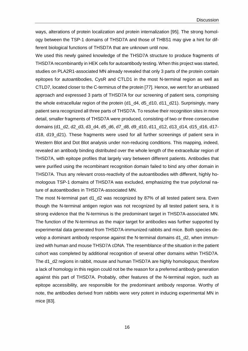
ways, alterations of protein localization and protein internalization [95]. The strong homol-
ogy between the TSP-1 domains of THSD7A and those of THBS1 may give a hint for dif-
ferent biological functions of THSD7A that are unknown until now.
We used this newly gained knowledge of the THSD7A structure to produce fragments of
THSD7A recombinantly in HEK cells for autoantibody testing. When this project was started,
studies on PLA2R1-associated MN already revealed that only 3 parts of the protein contain
epitopes for autoantibodies, CysR and CTLD1 in the most N-terminal region as well as
CTLD7, located closer to the C-terminus of the protein [77]. Hence, we went for an unbiased
approach and expressed 3 parts of THSD7A for our screening of patient sera, comprising
the whole extracellular region of the protein (d1_d4, d5_d10, d11_d21). Surprisingly, many
patient sera recognized all three parts of THSD7A. To resolve their recognition sites in more
detail, smaller fragments of THSD7A were produced, consisting of two or three consecutive
domains (d1_d2, d2_d3, d3_d4, d5_d6, d7_d8, d9_d10, d11_d12, d13_d14, d15_d16, d17-
d18, d19_d21). These fragments were used for all further screenings of patient sera in
Western Blot and Dot Blot analysis under non-reducing conditions. This mapping, indeed,
revealed an antibody binding distributed over the whole length of the extracellular region of
THSD7A, with epitope profiles that largely vary between different patients. Antibodies that
were purified using the recombinant recognition domain failed to bind any other domain in
THSD7A. Thus any relevant cross-reactivity of the autoantibodies with different, highly ho-
mologous TSP-1 domains of THSD7A was excluded, emphasizing the true polyclonal na-
ture of autoantibodies in THSD7A-associated MN.
The most N-terminal part d1_d2 was recognized by 87% of all tested patient sera. Even
though the N-terminal antigen region was not recognized by all tested patient sera, it is
strong evidence that the N-terminus is the predominant target in THSD7A-associated MN.
The function of the N-terminus as the major target for antibodies was further supported by
experimental data generated from THSD7A-immunized rabbits and mice. Both species de-
velop a dominant antibody response against the N-terminal domains d1_d2, when immun-
ized with human and mouse THSD7A cDNA. The resemblance of the situation in the patient
cohort was completed by additional recognition of several other domains within THSD7A.
The d1_d2 regions in rabbit, mouse and human THSD7A are highly homologous; therefore
a lack of homology in this region could not be the reason for a preferred antibody generation
against this part of THSD7A. Probably, other features of the N-terminal region, such as
epitope accessibility, are responsible for the predominant antibody response. Worthy of
note, the antibodies derived from rabbits were very potent in inducing experimental MN in
mice [83].

Discussion
17
In 2019 Stoddard et al. [96] presented an in silico 3-D structure of THSD7A. In line with our
results, they defined the extracellular domains of the protein as a mixture of THBS1-like and
C6/F-spondin like domains. While they also ended up with 21 domains, the numbering
shifted. In contrast to our work, they labelled the coiled coil domain as an additional THBS1-
like domain with a polybasic region, resulting in the alternating pattern of THBS1- and C6-
like domains, already from domain 2 on. They assumed that the polybasic region mentioned
above serves as a glycosaminoglycan binding site for heparan sulfate or similar proteogly-
cans. Heparan sulfate is a component of the glomerular basement membrane. THSD7A
may serve as an adhesion protein, anchoring the basal aspects of podocyte foot processes
to the GBM. Disruption of this anchor by autoantibodies binding in the C-terminal region of
the antigen represents a potential mechanism of glomerular injury in MN. Whereas we iden-
tified a THBS1-like domain at the C-terminal end of the extracellular region of THSD7A, the
study points out that this domain most probably does not fold into a proper structure, due to
a low number of residues in the sequence corresponding to the C-strand of the predicted
domain. As a consequence, they defined the extracellular region of THSD7A as the part
from domain 1 to domain 21, the latter being the equivalent to domain 20 in our terminology.
Strikingly, the predicted favorable epitopes in the extracellular region of THSD7A investi-
gated by Stoddard et al. correspond very well with our experimental data. In their analysis,
18 domains of THSD7A are predicted to contain epitopes and 3 domains are not. Compared
with our results, this holds true for all fragments we tested except for one. None of the
patient sera that we tested recognized the fragment d3-cc-d4, corresponding to domains 3,
4 and 5 in their study. Additionally, Stoddard et al. suggest that hydrophobic and polar un-
charged residues are most likely to be involved in epitope sites. In their study this was the
case for all predicted epitope containing domains, with eleven having mostly hydrophobic
residues, five consisting of mostly polar uncharged residues and two with a mixture of both
kinds of residues. Taken together our data and the study by Stoddard et al. strongly support
and strengthen each other.
Next, epitope recognition patterns were correlated with anti-THSD7A antibody levels and
clinical characteristics of the investigated patient cohort. Patients whose sera recognized
more than two fragments of THSD7A had higher antibody titer measured in IF than patients
with recognition of only one or two fragments. Furthermore, they presented with higher pro-
teinuria (not reaching statistical significance) and tended to go into remission less often
during follow-up. After analyzing the sera of 31 untreated patients diagnosed with THSD7A-
associated MN, experiments with follow-up sera from 16 of them were performed and re-
sults were correlated with available clinical data. During follow-up, the epitope profile re-
Discussion
18
mained unchanged in five patients. They had stable anti-THSD7A titers and suffered ne-
phrotic-range proteinuria during the whole time of observation. Seven patients lost recogni-
tion of one or more constructs, which was accompanied by a decrease of antibody level
and remission of proteinuria. Taken together, several conclusions can be drawn from our
data. First, the vast majority of patient sera contain antibodies against the most N-terminal
region of the antigen. This strongly suggests that this area is of enhanced immunogenicity.
One could speculate that the disease starts with a break of tolerance in this region. Second,
most patients recognize multiple additional domains along the antigen. This demonstrates
that THSD7A-associated MN is a polyclonal disease with poly-reactive autoantibodies.
Third, a high overall anti-THSD7A antibody titer correlates with the number of recognized
domains and a decrease in the overall titer is associated with a reduction in the number of
recognized domains. This situation strongly suggests that the epitope recognition profiles
depend on the anti-THSD7A antibody titer. However, from these clinical data alone it is not
possible to deduce whether the epitope profile or the number of recognized epitopes have
a direct impact on disease severity. It is possible that the more epitopes are bound the
stronger are the immunological effector mechanisms, such as complement activation, that
take place at the podocyte foot processes. Further experimental studies are warranted to
dissect the pathogenic role of the targeted domains in THSD7A-associated MN.
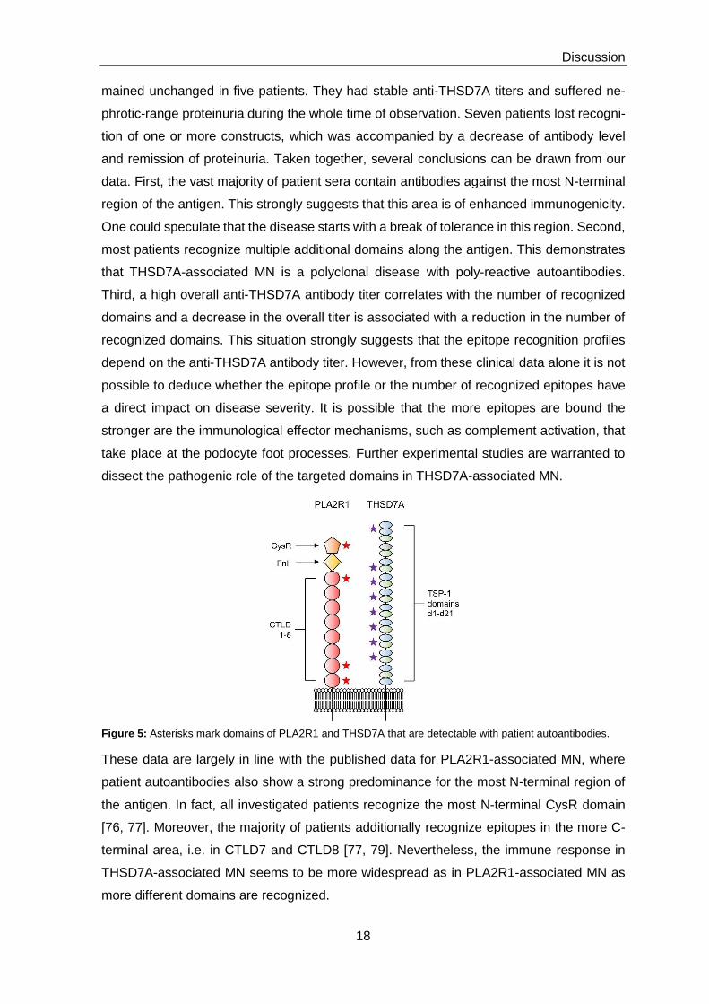
Figure 5: Asterisks mark domains of PLA2R1 and THSD7A that are detectable with patient autoantibodies.
These data are largely in line with the published data for PLA2R1-associated MN, where
patient autoantibodies also show a strong predominance for the most N-terminal region of
the antigen. In fact, all investigated patients recognize the most N-terminal CysR domain
[76, 77]. Moreover, the majority of patients additionally recognize epitopes in the more C-
terminal area, i.e. in CTLD7 and CTLD8 [77, 79]. Nevertheless, the immune response in
THSD7A-associated MN seems to be more widespread as in PLA2R1-associated MN as
more different domains are recognized.

Discussion
19
Similar to our data also in PLA2R1-associated MN the total antibody level against PLA2R1
correlates with the number of recognized domains and a lower total PLA2R1 antibody level
usually leads to a higher remission rate and a better outcome of the disease [69].
Although four patients had a change in their epitope recognition pattern during follow up,
we could not find clear evidence for any epitope spreading over time.
Inter-and intra-molecular epitope spreading was observed in several antibody-mediated au-
toimmune diseases, such as bullous pemphigoid, multiple sclerosis and encephalomyelitis
[97-100]. The phenomenon is defined by an expansion of epitope specificity from an initial
immune response against a dominant epitope within a self or foreign protein, towards sub-
dominant and/or cryptic epitopes on that protein (intramolecular spreading) or other proteins
(intermolecular spreading). Because MN is considered to be a very slowly developing dis-
ease and patients are usually not under clinical observation at the origin of disease, it is
very difficult to verify epitope spreading in human disease. To address this problem specific
animal models that mimic the human disease are very useful [101].
Indeed, for the experimental MN model of Heymann Nephritis an epitope spreading from
the immunization construct, a small N-terminal fragment of Megalin, to more distal domains
could be shown [29].
Epitope spreading was reported in 2014 for PLA2R1-associated MN, and it was suggested
to be useful for prediction of disease development regarding spontaneous remission and/or
response to treatment [77]. Besides Western Blot analyses, an ELISA with PLA2R1 frag-
ments was used to investigate domain specific antibody titers. Two groups were defined in
these studies, (i) patients whose sera only recognize the CysR domain and (ii) patients in
whom the recognition had spread towards CTLD1 and CTLD7. Members of the second
group were older, showed active disease and a poor renal prognosis. During follow up,
antibodies against CTLD1 and CTLD7 disappeared with disease remission and reappeared
with disease relapse [77, 78]. Additionally, a reversal of epitope spreading could be induced
by treatment with rituximab.
The role of epitope spreading in PLA2R1-associated MN was severely weakened by the
most recent study of Reinhard et al. [79] which not only identified a new recognition site of
antibodies in CTDL8, but could not find any evidence for epitope spreading in their cohort
during the whole time course of observation. All tested patients recognized more than one
fragment of PLA2R1 at baseline. Some patients achieved spontaneous remission, regard-
less of existing antibodies against N-and C-terminal domains in their sera. They [79] also
mentioned that the method applied, the dilution of sera, the composition of sera and the
accuracy in performance are besides further aspects of great importance for the detection
of epitope specific antibodies in PLA2R1-associated MN. For example, if they used higher
Discussion
20
dilutions of sera, recognition of domains in the C-terminal area disappeared, while new
recognitions appeared if lower dilutions were used. Taken together this refutes the occur-
rence of an epitope spreading during the time of follow up. It is more likely that the antibody
repertoire already exists from the beginning of the disease. Consequently, only the autoan-
tibody titer decides whether recognition is seen at the domain level or not.
In fact, we could not rule out either that epitope profiles would have changed if we had
applied additional methods, like an ELISA, or changed the serum dilutions. A dilution of
1:100 was used for all experiments, raising the possibility that antibody concentration
against specific fragments of THSD7A were below the detection limit for some patient sera.
Therefore it has to be carefully considered whether profiles of recognized protein fragments
are a valid tool to predict disease outcome at the moment.
Nonetheless, if larger cohorts of patients with THSD7A-associated MN are available in the
future it would be a good approach to establish a similar ELISA as in PLA2R1-associated
MN because the identification of the precise epitopes in THSD7A-associated MN is still of
high interest for several reasons. Some domains of THSD7A may interact with other mole-
cules, so that antibody binding to these domains specifically interferes with the natural func-
tion of the protein, leading to a structural and functional alteration of slit diaphragm perme-
ability. It is already known, that anti-THSD7A antibodies induce cytoskeletal rearrange-
ments in primary cultured murine glomerular epithelial cells [44].
Furthermore, the autoimmunity in THSD7A- associated MN is possibly caused by a molec-
ular mimicry between microbial antigens and host proteins [102, 103]. Knowing the precise
epitopes would allow deeper analyses to address this theory. The term molecular mimicry
describes the pathogenic consequence of cross-reactivity between common B or T cell re-
active epitopes of microorganisms or environmental agents and the host. It can occur in
several different forms including complete identity at the protein level, homology at the pro-
tein level, structural similarity and similarity at the level of amino acid sequences [104]. For
instance, PLA2R1 and THSD7A share a signature motif in their N-terminal domains, which
is suggested to be an epitope involved in the initial B cell triggering event in MN. Parts of
this motif are also present in sequences of proteins from Clostridium, Saccharomyces cere-
visiae and Pseudomonas HrcC Type, raising the opportunity that exposure to microbes may
play a role in the development of MN [105].
Importantly, the precise epitopes could serve as a basis for innovative and individualized
treatments, specific for THSD7A-associated MN (see below). To design and apply those
future therapies properly and expediently, the remaining question should be answered: is it
an individual epitope profile, the number of recognized epitopes, the antibody titer alone or
a combination of these factors that drives disease?

Discussion
21
One way to answer this question is to generate multiple domain-specific antibodies and
investigate the conditions that are required for disease development in mice. For this pur-
pose we already started to generate monoclonal antibodies using THSD7A-knockout mice
(Thsd7a-/- mice). The mice are immunized with the mouse orthologues of the TSP-1 do-
mains that were most frequently recognized by patient autoantibodies, afterwards spleens
and lymph nodes are taken and fused with a multiple myeloma cell line. Cell clones that
produce THSD7A-specific antibodies are then selected for further cell culture and produc-
tion of larger amounts of antibodies. IgG subtypes can be determined and antibodies can
even be sequenced and potentially be modified to different IgG subtypes. Once a proper
antibody library is generated, antibodies will be transferred to mice. Thereby, modification
of the variables „antibody amount“(i.e. a certain anti-THSD7A titer), „epitope profile“(i.e. a
certain domain recognition profile) and „antibody polyreactivity“(i.e. targeting different num-
bers of domains) can be freely modified in order to further comprehend what drives disease
on a molecular level. One example would be the transfer of antibodies against two domains
versus transfer of antibodies against four or five domains, while the injected amount is
adapted to identical antibody titers circulating in the serum of mice. This experimental setup
will allow to understand whether antibodies targeting multiple domains along the antigen
induce more severe disease (e.g. via more efficient complement activation) in comparison
to a more restricted antibody distribution. Preliminary experiments have already shown that
polyclonal IgG against THSD7A derived from Thsd7a-/- mice has indeed the capacity to in-
duce experimental MN in wild type BALB/c mice.
However, such monoclonal antibodies have a variety of potential applications regarding the
investigation of disease pathogenesis. (1) Our collaboration partner has established a po-
docyte cell line that stably expresses THSD7A. These antibodies can now be applied on
this podocyte cell line and transcriptomic and proteomic analyses over time may reveal
different signaling programs, depending on which or how many epitopes are targeted. (2)
In preliminary studies, we have identified the in vitro THSD7A interactome, i.e. the direct or
indirect binding partners of THSD7A. The monoclonal antibodies can be used to analyze
whether such protein-protein interactions are disrupted due to binding of certain domains
by IgG. Those interactions may be an integral part of the yet unknown function of THSD7A
for the podocyte. (3) The precise structure of the THSD7A antigen has not been defined yet
by experimental methods such as cryo-electron microscopy or x-ray crystallography. It is
unclear how the antigen structure is involved in THSD7A function under physiological con-
ditions or whether an alteration in antigen structure plays a role in MN pathogenesis. To this
end, the monoclonal antibodies can be co-crystallized with the antigen in direct comparison
to the crystal structure of the unbound antigen.

Discussion
22
It is essential to emphasize that this approach is based on an immunological reaction in
mice, resulting in antibodies against mouse THSD7A. Even though mouse and human
THSD7A share over 90% of amino acid sequence homology, the application of such inno-
vative therapeutic approaches in patients with MN will rely on further characterization of the
antigen/antibody interaction in human disease.
In summary, in the first study presented in this thesis, the structure of THSD7A was ana-
lyzed for the first time using in silico structure-based alignments of THSD7A with TSP-1
domains. Additionally, the epitope-containing domains were identified that are involved in
the pathogenesis of MN, and patient recognition patterns were correlated with clinical data.
In vivo immunization experiments revealed that the most N-terminal part of the antigen con-
tains particular immunogenicity, as an antibody response against this region was not only
found in patient sera but also in sera of mice and rabbits after immunization with THSD7A.
The presented data build the basis for a variety of studies that will shed further light on the
role of the targeted epitopes in this antibody-mediated autoimmune disease and, not last,
may help to develop specific, pathogenesis-based treatments in the near future.
3.2 Heterologous models of THSD7A- and PLA2R1-associated MN
In the second study presented in this thesis, the direct pathogenicity of anti-PLA2R1 anti-
bodies was demonstrated for the first time and a reproducible animal model of PLA2R1-
associated MN was established by means of transgenic overexpression of the PLA2R1
antigen in podocytes of BALB/c mice. It is known for some time that autoantibodies against
THSD7A are the pathogenic factor in THSD7A-associated MN. Purified antibodies, derived
from sera of patients with THSD7A-associated MN, are capable of inducing proteinuria and
the typical histopathological features of MN in wild type BALB/c mice [44]. In order to trans-
fer this insight into a reproducible and valid experimental model of MN, a heterologous ap-
proach, using rabbit antibodies against THSD7A was pursued [83].
These experiments were feasible because THSD7A is expressed on mouse podocytes with
over 90% homology with the human protein.
The situation is more complicated for PLA2R1-associated MN as the protein is not ex-
pressed on mouse podocytes. To enable experiments in mice, a transgenic approach had
to be applied. The first attempts, to express the human PLA2R1 on mouse podocytes were
not successful. Most probably due to the complex structure of PLA2R1 with high internal
flexibility of the human PLA2R1 ectodomains [74], resulting in insufficient antigen expres-
sion or possible membrane incorporation of the human protein in mice.

Discussion
23
Therefore we decided to design a mouse line that expresses mouse PLA2R1 (mPLA2R1-
positive mice) on its podocytes, comparable to the natural expression of THSD7A on podo-
cytes of wild type BALB/c mice.
First the expression pattern and the possible consequences of the transgenic protein ex-
pression were examined. Under basal conditions the mice express the transgenic
mPLA2R1 protein strongly in their cytoplasm and cell membrane. In the podocyte mem-
brane, mPLA2R1 is specifically localized to podocyte foot processes, since PLA2R1 stain-
ing partially merged with the slit diaphragm protein nephrin. The number of podocytes iden-
tified was not changed in mPLA2R1 expressing mice in comparison to non-PLA2R1 ex-
pressing littermates. Furthermore, in light microscopy and electron microscopy, no altera-
tion in the glomerular morphology, especially for the GBM, the podocyte foot processes or
the glomerular endothelium could be detected. We also looked for signs of ER-stress and
a possible overload of cellular degradation/repair systems, caused by the transgenic ex-
pression of the large, strongly glycosylated protein in podocytes. All performed experiments
demonstrated podocyte tolerance towards transgenic mPLA2R1 expression with normal
expression of all investigated stress markers in both mPLA2R1-negative and -positive mice.
Importantly, the albumin/creatinine ratio in PLA2R1-positive mice showed no anomalies
when compared to PLA2R1-negative mice, leading to the conclusion that a transgenic
mouse line that is suitable for further applications was successfully generated.
In an attempt to reproduce the proof of principle experiment that was already successful for
THSD7A, human antibodies against PLA2R1, derived from patients with PLA2R1-associ-
ated MN, were transferred to the transgenic mice. While the application of the antibody lead
to a slightly positive staining of huIgG in the glomerulus, no proteinuria or additional features
of MN could be observed. Most probably the homology of 72% between human and mouse
PLA2R1 is not enough for a sufficient binding of patient antibodies to the mouse orthologue
in the transgenic mice. This assumption was also supported by the finding that the detection
of mPLA2R1 by human autoantibodies was very weak in Western blot analyses.
Further approaches for a successful expression of the human protein in mice are in pro-
gress to finally prove the pathogenicity of human anti-PLA2R1 antibodies in PLA2R1-asso-
ciated MN.
In order to investigate whether anti-PLA2R1 antibodies in general are capable of inducing
proteinuria and the classical histopathology of MN, antibodies against mPLA2R1 were pro-
duced in rabbits immunized with mPLAR1 cDNA, similar to the approach that was under-
taken for the heterologous model of THSD7A -associated MN [83]. In accordance with pa-
tient sera from PLA2R1- associated MN, the rabbit antibodies strongly recognized the N-

Discussion
24
terminal region of mouse PLA2R1 (CysR-CTLD1). While the antibody binding was also de-
tectable in fragments compromising CTLD2-6 and CTLD2-8, in contrast to patient data, no
recognition was found for the CTLD7-8 region. This strengthens the hypothesis that, like it
the case for THSD7A, also the N-terminal region of PLA2R1 is of particularly enhanced
immunogenicity.
In line with the characteristics of patient anti-PLA2R1 antibodies, the rabbit antibodies
showed no binding under reducing conditions in Western Blot analyses, indicating binding
to conformation dependent epitopes.
The purified total rabbit antibodies were injected into PLA2R1-positive and -negative mice
and the animals were observed for seven days. PLA2R1-positive mice developed severe
proteinuria persisting over the whole time course of the experiment, while PLA2R1-negative
mice or those who received control rbIgG (i.e. rbIgG without recognition of mPLA2R1) re-
mained unaffected. Additionally, the mice presented the typical histomorphological signs of
human MN. Granular and subepithelial deposition of rbIgG, as well as an enhanced mem-
brane staining for mPLA2R1 could be detected. Electron microscopy revealed electron-
dense deposits in a strictly subepithelial location within the GBM and areas of foot process
broadening in mPLA2R1-positive mice. The slit diaphragm is an early target of antibody
immune complex formation in experimental THSD7A-associated MN [47]. This might be
also the case for the mPLA2R1-anti-mPLA2R1 complex formation in our experiment, sug-
gested by electron-dense deposits in close proximity to the podocyte slit diaphragms and a
co-localization of mPLA2R1 and the deposited rbIgG with the slit-diaphragm protein nephrin
found in high-resolution confocal imaging.
For the first time we demonstrate that anti-PLA2R1 antibodies directly cause MN in animals.
Moreover, we generated the first animal model of PLA2R1-associated MN which is reliable
and reproducible at the same time. The proof for the pathogenicity of anti-PLA2R1 antibod-
ies is of particular importance as it builds a pathomechanistic rationale for future therapies
aiming for the elimination of the pathogenic factor (i.e. the antibodies) or of the source of
the pathogenic factor (i.e. the antibody-producing cells).
The experimental model is highly comparable with the one of THSD7A-associated MN, with
one major difference. While glomerular deposition of C3 was barely detectable in the model
of THSD7A- associated MN, here C3 was found in partial co-localization with the bound
rbIgG at day seven, suggesting complement activation in the area of immune complex dep-
osition. However, whether the absence of C3 in the model of THSD7A-associated MN holds
true, is currently under investigation using highly sensitive methods.
Until today it remains a major question how antibodies lead to the glomerular damage in
MN, in particular the foot process effacement of podocytes and the consecutive loss of

Discussion
25
plasma proteins into the urine. Several injury pathways are conceivable and have been
discussed extensively in the field of MN. With both PLA2R1- and THSD7A-related models
now successfully running; we have the opportunity to systematically investigate different
postulated mechanisms:
(1) Antibodies bind to the target antigen, leading to the activation of the complement cas-
cade which ends with the formation of the final product C5b9, also known as the membrane
attack complex. C5B9 gets inserted into the podocyte membranes, resulting in a "sublytic“-
damage of the cells. The knowledge about this role of the complement system for the de-
velopment of glomerular damage in MN has been derived from several investigations in the
Heymann nephritis model [20, 23, 24]. However, other studies have strongly challenged this
pathophysiological concept [25, 26]. It has to be considered that the classic protocols for
both active and passive HN involve antibodies against a multitude of tubular and glomerular
proteins. In active HN these antibodies develop in rats due to direct immunization with a
cocktail of rat tubular proteins. In passive HN the antibodies were derived from immunization
of sheep with the identical protein cocktail and subsequently transferred to rats. This stands
in strong contrast to the pathophysiology of MN, where the immune system predominantly
targets one membrane-expressed podocyte antigen (i.e. PLA2R1 or THSD7A).
The heterologous mouse models of PLA2R1- and THSD7A-associated MN now allow in-
vestigating the role of the complement system in a more specific way. To this regard, a
mouse line genetically lacking C3 was generated. C3 is the center complement component
without it all activation pathways of the complement system stop at the level of C3 cleavage,
preventing the membrane attack complex to be formed. We already crossed the mouse line
to a BALB/c background and currently crossing it with the PLA2R1-positive mice to set up
proof of principle experiments involving anti-THSD7A and anti-PLA2R1 antibodies. In addi-
tion, we have established the use of small interfering RNA (siRNA) depleting specific com-
plement components, to answer the question whether a complement-targeted therapy is
promising in MN. siRNAs are small RNA molecules that are 20 to 25 base pairs in length.
They are important for the regulation of gene expression and can temporarily switch off
certain genes, in our case genes for the expression of complement components [106]. We
will inject this siRNA before the induction or during the course of the experimental MN mod-
els. Thus, we can examine whether the complement factors are sufficiently suppressed in
mice, and whether the outcome of the disease is ameliorated in the models.
(2) Important structural functions of the antigen are disrupted due to the binding of the au-
toantibodies. Such a mechanism has already been demonstrated for the antibody-mediated
autoimmune skin disease pemphigus vulgaris. This disease is characterized by separation
of skin layers and consequently blistering of the skin caused by antibodies against

Discussion
26
desmoglein-3, a protein of the cadherin family that is a component of the skin desmosome
[107]. Both PLA2R1 and THSD7A are large multidomain membrane proteins expressed on
podocyte foot processes, likely interacting with other surrounding molecules. For example,
PLA2R1 was shown to interact with collagen type IV (the collagen subtype that is the main
constituent of the GBM) [38, 39], and the TSP-1 domains of THSD7A may interact with
heparan sulfate (an essential component of the GBM) [96]. Thus, it is possible that PLA2R1
and THSD7A contribute to the integrity of the glomerular filtration barrier by anchoring po-
docyte foot processes to the outer aspect of the GBM. Anti-PLA2R1 [40] and anti-THSD7A
antibodies might directly interfere with this structural function. In the heterologous mouse
models of PLA2R1 and THSD7A-associated MN, it will be possible to investigate this hy-
pothesis directly, e.g. by structural and ultrastructural spacial resolution of the glomerular
filtration barrier in high-resolution immunofluorescence imaging and ultrastructural electron
microscopy analyses.
(3) If autoantibodies bind to the target antigen, intracellular signaling is altered. This is the
case, for example, in Grave’s disease, a disorder of the thyroid gland which is characterized
by clinically relevant hyperthyroidism. The Autoantibodies target the thyrotropin receptor (or
TSH receptor) activate it and induce a G-protein signal cascade with intracellular formation
of cAMP. This leads to activation of all functional aspects of the thyroid cells, including thy-
roid hormone release [108]. Until today the physiological functions of PLA2R1 and THSD7A
are largely unknown and it is unclear whether PLA2R1 and THSD7A serve as receptors for
specific ligands and transmit signals from the outside to the inside of cells, e.g. podocytes.
PLA2R1 may be a regulator of inflammation by binding sPLA2s [36, 37], even though the
in vivo relevance of this mechanism is not well understood.
With regard to THSD7A it was recently shown that the expression of the protein is accen-
tuated at filopodia on podocytes. THSD7A overexpression in cell culture podocytes is as-
sociated with increased cell size and cell adhesion (e.g. to collagen IV) and a decreased
cell migratory potential [47]. This suggests that THSD7A may be involved in outside-in sig-
naling. Thus, it is possible that anti-PLA2R1 and anti-THSD7A antibodies interfere with (to
date largely unknown) signaling pathways and that this process contributes to the cellular
damage in MN. The heterologous models of PLA2R1- and THSD7A-associated MN can be
applied to elucidate such potential pathomechanisms. After the disease is induced by trans-
fer of rbIgG, whole glomeruli or even podocytes, endothelial cells and mesangial cells can
be isolated using FACS-sorting [109]. The isolated samples can be investigated for altera-
tions in signaling pathways, e.g. by performing transcriptomic and proteomic analyses. This
may reveal PLA2R1- and THSD7A-dependent signaling pathways that are activated by the
respective antibodies. The identified pathways then can be validated in patients with MN

Discussion
27
(e.g. by immunostaining for the identified targets) and further investigated regarding their
involvement in MN pathogenesis, e.g. by generating mice that are genetically deficient for
the identified target. Finally, such pathways may serve as druggable targets, which can be
evaluated in the models of PLA2R1- and THSD7A-associated MN.
Besides investigations regarding PLA2R1- and THSD7A-related pathomechanisms in MN,
the models can be applied to evaluate innovative, antigen-specific treatments in vivo:
(1) Epitope blocking therapy using non-pathogenic antibodies/nanobodies. For such an ap-
proach, it would be feasible to use specifically engineered antagonists to block several
epitopes, for example nanobodies. The nanobodies would bind to the antigen and thereby
prevent the binding of the pathogenic antibody. Nanobodies are small, recombinantly pro-
duced antigen binding VHH fragments, derived from cameloid heavy chain IgG antibodies
[110]. It has already been shown that nanobodies are capable of blocking epitopes on mol-
ecules and interrupt the biological function of the targeted protein [111, 112]. For such a
blocking strategy, the already generated monoclonal antibodies (see 3.1) could be applied
in case that they share recognition of identical (or at least overlapping) epitopes with the
patient autoantibodies. The antigen recognition site of the monoclonal antibodies can be
analyzed by antibody sequencing and then be fused to an immunologically inactive IgG or
a nanobody VHH backbone. Such blocking antibodies could be applied in the models of
PLA2R1- and THSD7A-associated MN, e.g. initially as a pretreatment strategy to evaluate
whether the disease will be attenuated as a consequence of epitope blocking.
(2) Antibody extraction using epitope-specific immunoadsorption. This method has been
proven to be a possible tool in the treatment of pemphigus vulgaris (see above). When
patient sera were adsorbed on sepharose, loaded with the parts of desmoglein-3 that bind
the patient autoantibodies, all pathogenically active antibodies were eliminated [113]. In a
neonatal mouse model of pemphigus vulgaris such depleted sera failed to induce the dis-
ease at all, giving a strong hint for the usefulness of this strategy as future therapy [114]. In
the heterologous models of PLA2R1- and THSD7A-associated MN comparable proof-of-
concept studies could be performed.
(3) Antibody extraction using endogenous degradation systems. For this approach, frag-
ments of THSD7A and PLA2R1, containing the epitopes of pathogenic antibodies, are fused
to the constant region of the mouse IgG heavy chain (Fc-region) and produced recombi-
nantly. Genetic modifications in the Fc region (the region of the antibody that binds to Fc
receptors) can enhance binding to the FcγIIb receptor. This receptor is highly expressed on
liver sinusoidal epithelial cells, which play an important role in clearance of blood compo-
nents as a part of the reticuloendothelial system. Binding of immune complexes via the Fc
Discussion
28
part of the involved antibody leads to rapid internalization and degradation of the immune
complex [115, 116]. Thus, such re-engineered heavy chain antibodies, also called sweeping
antibodies, can be applied to scavenge pathogenic antibodies from a living organism. This
mechanism can be evaluated in the models of PLA2R1- and THSD7A-associated MN by
generating sweeping antibodies involving the antigen domains that are recognized by the
pathogenic rbIgG.
Despite the broad applicability of these passive transfer models, there are certain limita-
tions. First, the course of the model is acute, with high levels of proteinuria developing within
days after transfer of anti-PLA2R1 or anti-THSD7A antibodies. This stands in contrast to
the development of MN in patients, were the disease is usually clinically invisible for a cer-
tain time with slowly developing proteinuria. Interestingly, a recent study analyzing longitu-
dinal serum samples found presence of anti-PLA2R1 antibodies months to years before a
definite diagnosis of MN was made [117]. Second, the heterologous rbIgG serves as a for-
eign antigen itself and induces an immune response leading to the formation of mouse anti-
rbIgG and consecutively binding of these antibodies to the deposited rbIgG in glomeruli.
This creates large immune complexes consisting of PLA2R1–rabbit anti-PLA2R1–mouse
anti-rbIgG (or THSD7A–rabbit anti-THSD7A–mouse anti-rbIgG) at the filtration barrier,
where the mouse anti-rbIgG is a confounder that does not have a correlate in patients with
MN. Third, the models do not contain true autoimmunity, which means that there is no au-
toimmune response against the antigen itself, i.e. no antigen presentation by antigen pre-
senting cells, neither T cell activation nor B cell activation with subsequent differentiation of
the B cell clones producing the pathogenic antibodies. Consequently, therapeutic strategies
targeting the antibody-producing cells cannot be tested in the passive models of PLA2R1-
and THSD7A-associated MN.
Thus, the establishment of a true autoimmune model is imperative for an even better mod-
eling of the MN pathophysiology and for the development of highly innovative therapies
targeting the pathogenic B cell clones. In preliminary experiments we could already show
that mice, immunized with mouse THSD7A or fragments of the protein develop detectable
antibody Titers. Indeed, these antibodies bind to THSD7A in the glomerulus of the mice,
causing proteinuria and the typical histopathological features of THSD7A-associated MN.
A similar approach is envisaged for the PLA2R1-positive transgenic mice.
One promising therapeutic strategy to eliminate pathogenic B cell clones is the generation
of chimeric autoantibody receptor (CAAR) T cells, a modification of the oncotherapeutic
strategy of chimeric antigen receptor (CAR) T cells. This strategy involves the isolation of
peripheral blood mononuclear cells (PBMCs) from the patient’s blood using leukapheresis.

Discussion
29
Stimulation of specific B cells using interleukin-2 and anti-CD3 antibodies lead to their pro-
liferation. Afterwards the T cells are transduced with a construct encoding for the CAR of
interest, e.g. the antigen-binding domain of an anti-CD19 antibody fused to intracellular sig-
naling domains [118]. The resulting CAR T cells are transfused back to the patient, serving
as a “living drug” to eliminate all cells expressing, in this case, CD19, a marker of B cells.
This therapeutic strategy has recently been used with remarkable success, for example in
refractory or relapsed B cell lymphoma [119-121]. For the treatment of antibody-mediated
autoimmune diseases, this approach can be modified by expressing a CAAR on T cells.
This CAAR does not recognize a certain antigen, but rather contains domains of an antigen
itself, enabling binding to a B cell receptor of interest. The B cell receptor is a membrane
bound immunoglobulin, corresponding to the antibody that is produced by this particular B
cell. Thus, the engineered CAAR T cell will bind to the B cell of interest and eliminate it. In
an animal model of pemphigus vulgaris, CAAR T cells specifically and efficiently eliminated
anti-desmoglein 3-specific autoreactive B cells even in the presence of circulating autoan-
tibodies, and without relevant off-target toxicity [122]. A huge advantage of this approach is
the potential for the generation of long-term memory CAAR T cells, which offer the oppor-
tunity for constant elimination of newly emerging autoreactive B cells without the need for
repetitively application of the treatment.
Once the mice models with active B cells are available such a CAAR T cell strategy could
be applied, to investigate whether the elimination of specific B cell will ameliorate the dis-
ease. Perhaps, those studies can be combined with insights gained from the passive trans-
fer of specific monoclonal antibodies (see 3.1). If the severity of disease depends on the
binding of autoantibodies to a specific region of THSD7A, it might be sufficient to eliminate
only B cell clones which produce antibodies targeting this part of the protein.
Taken together the heterologous animal models of THSD7A and PLA2R1-associated MN
already represent powerful tools to investigate disease mechanisms and new therapeutic
strategies. In future these opportunities will be expanded by the development of models that
will involve autoantibody producing B cells, resulting in true autoimmunity.
Summary
30
4. Summary
Primary membranous nephropathy (MN) is an autoimmune disease and a major cause of
nephrotic syndrome in adult patients. The disease is caused by the formation of immune
deposits on the outer aspect of the glomerular basement membrane, which contain podo-
cyte antigens and circulating antibodies specific for these antigens, resulting in severe dam-
age of the filtration barrier. Two podocyte expressed proteins have been identified as anti-
gens in MN so far, phospholipase A2 receptor 1 (PLA2R1) and thrombospondin type 1
domain containing protein 7A (THSD7A). Most of the patients develop antibodies against
PLA2R1 (70-80% of MN cases), while cases with anti-THSD7A antibodies are less frequent
(3-5% of MN cases). The outcome of the disease varies, with about 30% of patients expe-
riencing spontaneous remission, whereas another 20-30% develop end stage renal disease
within 10 years. Because of this heterogeneity it is inevitable to find the individual predictors
of disease outcome. In order to do this and to test newly generated therapeutic strategies,
valid animal models have to be established.
In our first study, presented here, we in silico analyzed the structure of THSD7A, and iden-
tified the autoantibody binding sites within the protein. We found out that the extracellular
region of THSD7A is composed of a tandem string of 21 thrombospondin type one domains
showing structural homology either to the TSP-1 domains of thrombospondin 1 (THBS1) or
complement component 6 (C6). The 31 serum samples tested for the study revealed a
polyreactive immune response against the whole extracellular region of THSD7A with a
predominance for the most N-terminal part of the protein, the latter was also confirmed by
animal models of active immunization with THSD7A.
In our second study, we generated a transgenic mouse line expressing the murine full-
length PLA2R1 in podocytes (mPLA2R1-positive mice). The transfer of rabbit anti-THSD7A
antibodies to these mice caused nephrotic range proteinuria, hypercholesterolemia, and the
histomorphological signs of MN. We established the first animal model for PLA2R1 associ-
ated MN that can be used to gain deeper insights in disease development and investigate
future therapeutic strategies.
In summary, the two studies presented in this thesis close a gap in the field of MN research.
The domains that were targeted by antibodies in PLA2R1-associated MN were already de-
fined, while a sufficient animal model for the disease was still missing. Vice versa, a model
for THSD7A-associated MN existed, but the binding sites of patient antibodies were un-
known. This work thus synchronizes research on these two antigens. Furthermore, the work
presented in this thesis builds the base for a variety of potential future projects that will help
to deepen the knowledge about the pathophysiology of the disease. For example, the role
of the targeted epitopes can be investigated using domain-specific monoclonal antibodies
Zusammenfassung
31
and the PLA2R1- and THSD7A-specific mouse models can be used to uncover the un-
known signaling pathways that lead to glomerular damage in MN. Not last, innovative ther-
apeutic strategies targeting antigen-specific disease mechanisms, such as extraction of
pathogenic antibodies or elimination of autoantibody-producing B cells, can be investigated
based on the work presented herein.
5. Zusammenfassung
Die primäre membranöse Glomerulonephritis (MN) ist eine Autoimmunerkrankung und eine
der Hauptursachen für das nephrotische Syndrom bei erwachsenen Patienten. Verursacht
wird sie durch die Bildung von Immunablagerungen auf der Außenseite der glomerulären
Basalmembran, die zu einer schweren Schädigung der Filtrationsbarriere führen. Diese Ab-
lagerungen enthalten Podozytenantigene und an sie gebundene Antikörper. Bisher wurden
zwei Antigene identifiziert: Phospholipase A2 receptor 1 (PLA2R1) und Thrombospondin
type 1 domain containing protein 7A (THSD7A). Die meisten Patienten entwickeln Antikör-
per gegen PLA2R1 (70-80% der MN-Fälle), während Anti-THSD7A-Antikörpern weniger
häufig sind (3-5% der MN-Fälle). Der Ausgang der Krankheit ist unterschiedlich: Etwa 30%
der Patienten erreichen eine spontane Remission, während weitere 20 bis 30% innerhalb
von 10 Jahren eine Nierenerkrankung im Endstadium entwickeln. Aufgrund dieser Diversi-
tät ist es wichtig Indikatoren zu identifizieren, die es einem erlauben den Verlauf der Erkran-
kung zu prognostizieren, um passende Therapien anwenden zu können. Sowohl für die
Analyse solcher Erkrankungsmarker als auch für die Testung neuer und innovativer Thera-
piemöglichkeiten, sind Tiermodelle unabdingbar, die die Erkrankungssituation möglichst
genau reproduzieren.
In der ersten, hier vorgestellten, Studie wurde die Struktur von THSD7A in silico analysiert
und die Autoantikörper-Bindungsstellen innerhalb des Proteins identifiziert. Wir konnten zei-
gen, dass die extrazelluläre Region von THSD7A aus einer Tandemkette von 21 Throm-
bospondin-Typ-1-Domänen besteht, die entweder eine strukturelle Homologie zu den TSP-
1 Domänen von Thrombospondin 1 (THBS1) oder zu denen der Komplement Komponente
6 (C6) aufweisen. Die 31 für die Studie getesteten Serumproben zeigten eine polyreaktive
Immunantwort gegen die gesamte extrazelluläre Region von THSD7A mit einer Dominanz
für den Teil des Proteins der am äußersten N-terminus des Proteins liegt. Letzteres wurde
auch durch aktiven Immunisierungen mit THSD7A in mehreren Tiermodellen bestätigt.
In der zweiten Studie wurde eine transgene Mauslinie erzeugt, die das murine PLA2R1 auf
Podozyten (mPLA2R1-positive Mäuse) exprimiert. Der Transfer von Kaninchen-Anti-
Zusammenfassung
32
THSD7A Antikörpern in diese Mäuse verursachte eine Proteinurie im nephrotischen Be-
reich, Hypercholesterinämie und alle histomorphologischen Anzeichen der MN. Es wurde
somit das erste Tiermodell für die PLA2R1-assoziierte MN etabliert, mit dem tiefere Einbli-
cke in die Krankheitsentwicklung gewonnen und therapeutische Strategien untersucht wer-
den können.
Die beiden, in dieser Arbeit vorgestellten Studien, schließen jeweils eine Lücke auf dem
Gebiet der MN bezogenen Forschung. Im Bereich der PLA2R1 assoziierten MN fehlte bis
dato ein Tiermodell, während die Domänen des Proteins, welche von Autoantikörpern ge-
bunden werden, bereits bekannt waren. Umgekehrt existierte bereits ein Modell für
THSD7A-assoziiertes MN, aber die Bindungsstellen der Patientenantikörper waren unbe-
kannt. Schlussendlich synchronisiert diese Arbeit somit die Forschung an diesen beiden
Antigenen. Darüber hinaus bildet sie die Grundlage für eine Vielzahl potenzieller zukünftiger
Projekte, die dazu beitragen werden, das Wissen um die Pathophysiologie dieser Krankheit
zu vertiefen. Beispielsweise kann die Rolle der Zielepitope untersucht werden, indem man
domänenspezifische rmonoklonale Antikörper verwendet. Außerdem bieten die PLA2R1-
und THSD7A-spezifischen Mausmodelle eine Möglichkeit bisher unbekannte Signalwege
zu identifizieren, die zu glomerulären Schäden führen. Nicht zuletzt liefert die hier vorge-
stellte Arbeit die notwendige Grundlage um innovative Therapiestrategien, wie die Extrak-
tion pathogener Antikörper oder die Eliminierung von Autoantikörper-produzierenden B Zel-
len, zu entwickeln und zu testen.

Literature
33
6. Literature
1. Menon, M.C., P.Y. Chuang, and C.J. He, The glomerular filtration barrier: components and crosstalk. Int J Nephrol, 2012. 2012: p. 749010.
2. Kodner, C., Diagnosis and Management of Nephrotic Syndrome in Adults. Am Fam Physician, 2016. 93(6): p. 479-85.
3. Maisonneuve, P., et al., Distribution of primary renal diseases leading to end-stage renal failure in the United States, Europe, and Australia/New Zealand: results from an international comparative study. Am J Kidney Dis, 2000. 35(1): p. 157-65.
4. Fogo, A.B., et al., AJKD Atlas of Renal Pathology: Membranous Nephropathy. Am J Kidney Dis, 2015. 66(3): p. e15-7.
5. Segawa, Y., et al., IgG subclasses and complement pathway in segmental and global membranous nephropathy. Pediatr Nephrol, 2010. 25(6): p. 1091-9.
6. Jennette, J.C., S.S. Iskandar, and F.G. Dalldorf, Pathologic differentiation between lupus and nonlupus membranous glomerulopathy. Kidney Int, 1983. 24(3): p. 377-85.
7. Johnson, R.J. and W.G. Couser, Hepatitis B infection and renal disease: clinical, immunopathogenetic and therapeutic considerations. Kidney Int, 1990. 37(2): p. 663-76.
8. Radford, M.G., Jr., et al., Reversible membranous nephropathy associated with the use of nonsteroidal anti-inflammatory drugs. JAMA, 1996. 276(6): p. 466-9.
9. Bjorneklett, R., et al., Long-term risk of cancer in membranous nephropathy patients. Am J Kidney Dis, 2007. 50(3): p. 396-403.
10. Larsen, C.P., et al., Determination of primary versus secondary membranous glomerulopathy utilizing phospholipase A2 receptor staining in renal biopsies. Mod Pathol, 2013. 26(5): p. 709-15.
11. Takekoshi, Y., et al., Immunopathogenetic mechanisms of hepatitis B virus-related glomerulopathy. Kidney Int Suppl, 1991. 35: p. S34-9.
12. Davenport, A., et al., Do mesangial immune complex deposits affect the renal prognosis in membranous glomerulonephritis? Clin Nephrol, 1994. 41(5): p. 271-6.
13. Imai, H., et al., IgG subclasses in patients with membranoproliferative glomerulonephritis, membranous nephropathy, and lupus nephritis. Kidney Int, 1997. 51(1): p. 270-6.
14. Kuroki, A., et al., Glomerular and serum IgG subclasses in diffuse proliferative lupus nephritis, membranous lupus nephritis, and idiopathic membranous nephropathy. Intern Med, 2002. 41(11): p. 936-42.
15. Polanco, N., et al., Spontaneous remission of nephrotic syndrome in membranous nephropathy with chronic renal impairment. Nephrol Dial Transplant, 2012. 27(1): p. 231-4.
16. Glassock, R.J., Diagnosis and natural course of membranous nephropathy. Semin Nephrol, 2003. 23(4): p. 324-32.
17. Grupper, A., et al., Recurrent Membranous Nephropathy After Kidney Transplantation: Treatment and Long-Term Implications. Transplantation, 2016. 100(12): p. 2710-2716.
18. Heymann, W., et al., Production of nephrotic syndrome in rats by Freund's adjuvants and rat kidney suspensions. Proc Soc Exp Biol Med, 1959. 100(4): p. 660-4.
19. Barabas, A.Z. and R. Lannigan, Induction of an autologous immune-complex glomerulonephritis in the rat by intravenous injection of heterologous anti-rat kidney tubular antibody. I. Production of chronic progressive immune-complex glomerulonephritis. Br J Exp Pathol, 1974. 55(1): p. 47-55.
20. Salant, D.J., et al., A new role for complement in experimental membranous nephropathy in rats. J Clin Invest, 1980. 66(6): p. 1339-50.

Literature
34
21. Van Damme, B.J., et al., Experimental glomerulonephritis in the rat induced by antibodies directed against tubular antigens. V. Fixed glomerular antigens in the pathogenesis of heterologous immune complex glomerulonephritis. Lab Invest, 1978. 38(4): p. 502-10.
22. Couser, W.G., et al., Experimental glomerulonephritis in the isolated perfused rat kidney. J Clin Invest, 1978. 62(6): p. 1275-87.
23. Cybulsky, A.V., et al., Complement-induced glomerular epithelial cell injury. Role of the membrane attack complex in rat membranous nephropathy. J Clin Invest, 1986. 77(4): p. 1096-107.
24. Kerjaschki, D., et al., Transcellular transport and membrane insertion of the C5b-9 membrane attack complex of complement by glomerular epithelial cells in experimental membranous nephropathy. J Immunol, 1989. 143(2): p. 546-52.
25. Leenaerts, P.L., et al., Active Heymann nephritis in complement component C6 deficient rats. Kidney Int, 1995. 47(6): p. 1604-14.
26. Spicer, S.T., et al., Induction of passive Heymann nephritis in complement component 6-deficient PVG rats. J Immunol, 2007. 179(1): p. 172-8.
27. Kerjaschki, D. and M.G. Farquhar, The pathogenic antigen of Heymann nephritis is a membrane glycoprotein of the renal proximal tubule brush border. Proc Natl Acad Sci U S A, 1982. 79(18): p. 5557-61.
28. Kerjaschki, D. and M.G. Farquhar, Immunocytochemical localization of the Heymann nephritis antigen (GP330) in glomerular epithelial cells of normal Lewis rats. J Exp Med, 1983. 157(2): p. 667-86.
29. Shah, P., A. Tramontano, and S.P. Makker, Intramolecular epitope spreading in Heymann nephritis. J Am Soc Nephrol, 2007. 18(12): p. 3060-6.
30. Debiec, H., et al., Antenatal membranous glomerulonephritis due to anti-neutral endopeptidase antibodies. N Engl J Med, 2002. 346(26): p. 2053-60.
31. Debiec, H., et al., Role of truncating mutations in MME gene in fetomaternal alloimmunisation and antenatal glomerulopathies. Lancet, 2004. 364(9441): p. 1252-9.
32. Ronco, P., H. Debiec, and V. Guigonis, Mechanisms of disease: Alloimmunization in renal diseases. Nat Clin Pract Nephrol, 2006. 2(7): p. 388-97.
33. Beck, L.H., Jr., et al., M-type phospholipase A2 receptor as target antigen in idiopathic membranous nephropathy. N Engl J Med, 2009. 361(1): p. 11-21.
34. East, L. and C.M. Isacke, The mannose receptor family. Biochim Biophys Acta, 2002. 1572(2-3): p. 364-86.
35. Zvaritch, E., G. Lambeau, and M. Lazdunski, Endocytic properties of the M-type 180-kDa receptor for secretory phospholipases A2. J Biol Chem, 1996. 271(1): p. 250-7.
36. Lambeau, G. and M.H. Gelb, Biochemistry and physiology of mammalian secreted phospholipases A2. Annu Rev Biochem, 2008. 77: p. 495-520.
37. Augert, A., et al., The M-type receptor PLA2R regulates senescence through the p53 pathway. EMBO Rep, 2009. 10(3): p. 271-7.
38. Takahashi, S., et al., C-type lectin-like domain and fibronectin-like type II domain of phospholipase A(2) receptor 1 modulate binding and migratory responses to collagen. FEBS Lett, 2015. 589(7): p. 829-35.
39. Watanabe, K., et al., Human soluble phospholipase A2 receptor is an inhibitor of the integrin-mediated cell migratory response to collagen-I. Am J Physiol Cell Physiol, 2018. 315(3): p. C398-C408.
40. Skoberne, A., et al., Serum with phospholipase A2 receptor autoantibodies interferes with podocyte adhesion to collagen. Eur J Clin Invest, 2014. 44(8): p. 753-65.
41. Tomas, N.M., et al., Thrombospondin type-1 domain-containing 7A in idiopathic membranous nephropathy. N Engl J Med, 2014. 371(24): p. 2277-2287.

Literature
35
42. Hoxha, E., et al., A Mechanism for Cancer-Associated Membranous Nephropathy. N Engl J Med, 2016. 374(20): p. 1995-6.
43. Hoxha, E., et al., An Indirect Immunofluorescence Method Facilitates Detection of Thrombospondin Type 1 Domain-Containing 7A-Specific Antibodies in Membranous Nephropathy. J Am Soc Nephrol, 2017. 28(2): p. 520-531.
44. Tomas, N.M., et al., Autoantibodies against thrombospondin type 1 domain-containing 7A induce membranous nephropathy. J Clin Invest, 2016. 126(7): p. 2519-32.
45. Wang, C.H., et al., Thrombospondin type I domain containing 7A (THSD7A) mediates endothelial cell migration and tube formation. J Cell Physiol, 2010. 222(3): p. 685-94.
46. Kuo, M.W., et al., Soluble THSD7A is an N-glycoprotein that promotes endothelial cell migration and tube formation in angiogenesis. PLoS One, 2011. 6(12): p. e29000.
47. Herwig, J., et al., Thrombospondin Type 1 Domain-Containing 7A Localizes to the Slit Diaphragm and Stabilizes Membrane Dynamics of Fully Differentiated Podocytes. J Am Soc Nephrol, 2019. 30(5): p. 824-839.
48. Larsen, C.P., L.N. Cossey, and L.H. Beck, THSD7A staining of membranous glomerulopathy in clinical practice reveals cases with dual autoantibody positivity. Mod Pathol, 2016. 29(4): p. 421-6.
49. Wang, J., et al., Circulating Antibodies against Thrombospondin Type-I Domain-Containing 7A in Chinese Patients with Idiopathic Membranous Nephropathy. Clin J Am Soc Nephrol, 2017. 12(10): p. 1642-1651.
50. Zaghrini, C., et al., Novel ELISA for thrombospondin type 1 domain-containing 7A autoantibodies in membranous nephropathy. Kidney Int, 2019. 95(3): p. 666-679.
51. Sethi, S., et al., Exostosin 1/Exostosin 2-Associated Membranous Nephropathy. J Am Soc Nephrol, 2019. 30(6): p. 1123-1136.
52. Busse, M. and M. Kusche-Gullberg, In vitro polymerization of heparan sulfate backbone by the EXT proteins. J Biol Chem, 2003. 278(42): p. 41333-7.
53. Busse, M., et al., Contribution of EXT1, EXT2, and EXTL3 to heparan sulfate chain elongation. J Biol Chem, 2007. 282(45): p. 32802-10.
54. Busse-Wicher, M., K.B. Wicher, and M. Kusche-Gullberg, The exostosin family: proteins with many functions. Matrix Biol, 2014. 35: p. 25-33.
55. Sethi, S.e.a., Neural epidermal growth factor-like 1protein (NELL-1) associated membranous nephropathy. Kidney International, 2019.
56. Zhang, X., et al., The role of NELL-1, a growth factor associated with craniosynostosis, in promoting bone regeneration. J Dent Res, 2010. 89(9): p. 865-78.
57. Kuroda, S., et al., Biochemical characterization and expression analysis of neural thrombospondin-1-like proteins NELL1 and NELL2. Biochem Biophys Res Commun, 1999. 265(1): p. 79-86.
58. Hasebe, A., et al., Efficient production and characterization of recombinant human NELL1 protein in human embryonic kidney 293-F cells. Mol Biotechnol, 2012. 51(1): p. 58-66.
59. Hasebe, A., et al., The C-terminal region of NELL1 mediates osteoblastic cell adhesion through integrin alpha3beta1. FEBS Lett, 2012. 586(16): p. 2500-6.
60. Watanabe, T.K., et al., Cloning and characterization of two novel human cDNAs (NELL1 and NELL2) encoding proteins with six EGF-like repeats. Genomics, 1996. 38(3): p. 273-6.
61. Hoxha, E., et al., An immunofluorescence test for phospholipase-A(2)-receptor antibodies and its clinical usefulness in patients with membranous glomerulonephritis. Nephrol Dial Transplant, 2011. 26(8): p. 2526-32.
62. Qin, W., et al., Anti-phospholipase A2 receptor antibody in membranous nephropathy. J Am Soc Nephrol, 2011. 22(6): p. 1137-43.

Literature
36
63. Du, Y., et al., The diagnosis accuracy of PLA2R-AB in the diagnosis of idiopathic membranous nephropathy: a meta-analysis. PLoS One, 2014. 9(8): p. e104936.
64. Pourcine, F., et al., Prognostic value of PLA2R autoimmunity detected by measurement of anti-PLA2R antibodies combined with detection of PLA2R antigen in membranous nephropathy: A single-centre study over 14 years. PLoS One, 2017. 12(3): p. e0173201.
65. Debiec, H. and P. Ronco, PLA2R autoantibodies and PLA2R glomerular deposits in membranous nephropathy. N Engl J Med, 2011. 364(7): p. 689-90.
66. Stahl, R., E. Hoxha, and K. Fechner, PLA2R autoantibodies and recurrent membranous nephropathy after transplantation. N Engl J Med, 2010. 363(5): p. 496-8.
67. Dahnrich, C., et al., Development of a standardized ELISA for the determination of autoantibodies against human M-type phospholipase A2 receptor in primary membranous nephropathy. Clin Chim Acta, 2013. 421: p. 213-8.
68. Hofstra, J.M., et al., Anti-phospholipase A(2) receptor antibodies correlate with clinical status in idiopathic membranous nephropathy. Clin J Am Soc Nephrol, 2011. 6(6): p. 1286-91.
69. Hoxha, E., et al., Phospholipase A2 receptor autoantibodies and clinical outcome in patients with primary membranous nephropathy. J Am Soc Nephrol, 2014. 25(6): p. 1357-66.
70. Hoxha, E., et al., M-type phospholipase A2 receptor autoantibodies and renal function in patients with primary membranous nephropathy. Clin J Am Soc Nephrol, 2014. 9(11): p. 1883-90.
71. Ruggenenti, P., et al., Anti-Phospholipase A2 Receptor Antibody Titer Predicts Post-Rituximab Outcome of Membranous Nephropathy. J Am Soc Nephrol, 2015. 26(10): p. 2545-58.
72. Beck, L.H., Jr., et al., Rituximab-induced depletion of anti-PLA2R autoantibodies predicts response in membranous nephropathy. J Am Soc Nephrol, 2011. 22(8): p. 1543-50.
73. Bech, A.P., et al., Association of anti-PLA(2)R antibodies with outcomes after immunosuppressive therapy in idiopathic membranous nephropathy. Clin J Am Soc Nephrol, 2014. 9(8): p. 1386-92.
74. Dong, Y., et al., Structure of Human M-type Phospholipase A2 Receptor Revealed by Cryo-Electron Microscopy. J Mol Biol, 2017. 429(24): p. 3825-3835.
75. Kao, L., et al., Identification of the immunodominant epitope region in phospholipase A2 receptor-mediating autoantibody binding in idiopathic membranous nephropathy. J Am Soc Nephrol, 2015. 26(2): p. 291-301.
76. Fresquet, M., et al., Identification of a major epitope recognized by PLA2R autoantibodies in primary membranous nephropathy. J Am Soc Nephrol, 2015. 26(2): p. 302-13.
77. Seitz-Polski, B., et al., Epitope Spreading of Autoantibody Response to PLA2R Associates with Poor Prognosis in Membranous Nephropathy. J Am Soc Nephrol, 2016. 27(5): p. 1517-33.
78. Seitz-Polski, B., et al., Phospholipase A2 Receptor 1 Epitope Spreading at Baseline Predicts Reduced Likelihood of Remission of Membranous Nephropathy. J Am Soc Nephrol, 2018. 29(2): p. 401-408.
79. Reinhard, L.e.a., Clinical Relevance of Domain-Specific Phospholipase A2 Receptor 1 Antibody Levels in Patients with Membranous Nephropathy. J Am Soc Nephrol, 2019.
80. Seitz-Polski, B., et al., Prediction of membranous nephropathy recurrence after transplantation by monitoring of anti-PLA2R1 (M-type phospholipase A2 receptor) autoantibodies: a case series of 15 patients. Nephrol Dial Transplant, 2014. 29(12): p. 2334-42.

Literature
37
81. Meyer-Schwesinger, C., G. Lambeau, and R.A. Stahl, Thrombospondin type-1 domain-containing 7A in idiopathic membranous nephropathy. N Engl J Med, 2015. 372(11): p. 1074-5.
82. Godel, M., F. Grahammer, and T.B. Huber, Thrombospondin type-1 domain-containing 7A in idiopathic membranous nephropathy. N Engl J Med, 2015. 372(11): p. 1073.
83. Tomas, N.M., et al., A Heterologous Model of Thrombospondin Type 1 Domain-Containing 7A-Associated Membranous Nephropathy. J Am Soc Nephrol, 2017. 28(11): p. 3262-3277.
84. Zahner G, M.-S., Tomas N, Hoxha E, Wiech T, Stahl RA, Development and morphologic characterization of a mouse model of membranous nephropathy involving the human phospholipase A2 receptor. J Am Soc Nephrol, 2014.
85. Zahner G, H., Helmchen U, Stahl RA, The generation of inducible specific human phospholipase A2 receptor transgenic mice. J Am Soc Nephrol 2012.
86. Klenotic, P.A., et al., Expression, purification and structural characterization of functionally replete thrombospondin-1 type 1 repeats in a bacterial expression system. Protein Expr Purif, 2011. 80(2): p. 253-9.
87. Aleshin, A.E., et al., Structure of complement C6 suggests a mechanism for initiation and unidirectional, sequential assembly of membrane attack complex (MAC). J Biol Chem, 2012. 287(13): p. 10210-22.
88. Paakkonen, K., et al., Solution structures of the first and fourth TSR domains of F-spondin. Proteins, 2006. 64(3): p. 665-72.
89. Chen, H., M.E. Herndon, and J. Lawler, The cell biology of thrombospondin-1. Matrix Biol, 2000. 19(7): p. 597-614.
90. Galvin, N.J., et al., Interaction of human thrombospondin with types I-V collagen: direct binding and electron microscopy. J Cell Biol, 1987. 104(5): p. 1413-22.
91. Asch, A.S., et al., Thrombospondin sequence motif (CSVTCG) is responsible for CD36 binding. Biochem Biophys Res Commun, 1992. 182(3): p. 1208-17.
92. Calzada, M.J., et al., Identification of novel beta1 integrin binding sites in the type 1 and type 2 repeats of thrombospondin-1. J Biol Chem, 2004. 279(40): p. 41734-43.
93. Calzada, M.J., et al., Recognition of the N-terminal modules of thrombospondin-1 and thrombospondin-2 by alpha6beta1 integrin. J Biol Chem, 2003. 278(42): p. 40679-87.
94. Bein, K. and M. Simons, Thrombospondin type 1 repeats interact with matrix metalloproteinase 2. Regulation of metalloproteinase activity. J Biol Chem, 2000. 275(41): p. 32167-73.
95. Resovi, A., et al., Current understanding of the thrombospondin-1 interactome. Matrix Biol, 2014. 37: p. 83-91.
96. Stoddard, S.V., et al., Structure and function insights garnered from in silico modeling of the thrombospondin type-1 domain-containing 7A antigen. Proteins, 2019. 87(2): p. 136-145.
97. Hashimoto, T., et al., Demonstration of epitope spreading in bullous pemphigoid: results of a prospective multicenter study. J Invest Dermatol, 2011. 131(11): p. 2175-7.
98. Di Zenzo, G., et al., Demonstration of epitope-spreading phenomena in bullous pemphigoid: results of a prospective multicenter study. J Invest Dermatol, 2011. 131(11): p. 2271-80.
99. McRae, B.L., et al., Functional evidence for epitope spreading in the relapsing pathology of experimental autoimmune encephalomyelitis. J Exp Med, 1995. 182(1): p. 75-85.
100. Goebels, N., et al., Repertoire dynamics of autoreactive T cells in multiple sclerosis patients and healthy subjects: epitope spreading versus clonal persistence. Brain, 2000. 123 Pt 3: p. 508-18.
Literature
38
101. Vanderlugt, C.L. and S.D. Miller, Epitope spreading in immune-mediated diseases: implications for immunotherapy. Nat Rev Immunol, 2002. 2(2): p. 85-95.
102. Wucherpfennig, K.W., Mechanisms for the induction of autoimmunity by infectious agents. J Clin Invest, 2001. 108(8): p. 1097-104.
103. Kain, R., et al., Molecular mimicry in pauci-immune focal necrotizing glomerulonephritis. Nat Med, 2008. 14(10): p. 1088-96.
104. Oldstone, M.B., Molecular mimicry: its evolution from concept to mechanism as a cause of autoimmune diseases. Monoclon Antib Immunodiagn Immunother, 2014. 33(3): p. 158-65.
105. Fresquet, M., et al., Autoantigens PLA2R and THSD7A in membranous nephropathy share a common epitope motif in the N-terminal domain. J Autoimmun, 2019: p. 102308.
106. Dana, H., et al., Molecular Mechanisms and Biological Functions of siRNA. Int J Biomed Sci, 2017. 13(2): p. 48-57.
107. Porro, A.M., et al., Pemphigus vulgaris. An Bras Dermatol, 2019. 94(3): p. 264-278.
108. Menconi, F., C. Marcocci, and M. Marino, Diagnosis and classification of Graves' disease. Autoimmun Rev, 2014. 13(4-5): p. 398-402.
109. Boerries, M., et al., Molecular fingerprinting of the podocyte reveals novel gene and protein regulatory networks. Kidney Int, 2013. 83(6): p. 1052-64.
110. Wesolowski, J., et al., Single domain antibodies: promising experimental and therapeutic tools in infection and immunity. Med Microbiol Immunol, 2009. 198(3): p. 157-74.
111. Farajpour, Z., et al., A nanobody directed to a functional epitope on VEGF, as a novel strategy for cancer treatment. Biochem Biophys Res Commun, 2014. 446(1): p. 132-6.
112. Danquah, W., et al., Nanobodies that block gating of the P2X7 ion channel ameliorate inflammation. Sci Transl Med, 2016. 8(366): p. 366ra162.
113. Langenhan, J., et al., Specific immunoadsorption of pathogenic autoantibodies in pemphigus requires the entire ectodomains of desmogleins. Exp Dermatol, 2014. 23(4): p. 253-9.
114. Hofrichter, M., et al., Immunoadsorption of Desmoglein-3-Specific IgG Abolishes the Blister-Inducing Capacity of Pemphigus Vulgaris IgG in Neonatal Mice. Front Immunol, 2018. 9: p. 1935.
115. Chu, S.Y., et al., Reduction of total IgE by targeted coengagement of IgE B-cell receptor and FcgammaRIIb with Fc-engineered antibody. J Allergy Clin Immunol, 2012. 129(4): p. 1102-15.
116. Ganesan, L.P., et al., FcgammaRIIb on liver sinusoidal endothelium clears small immune complexes. J Immunol, 2012. 189(10): p. 4981-8.
117. Burbelo, P.D., et al., Detection of PLA2R Autoantibodies before the Diagnosis of Membranous Nephropathy. J Am Soc Nephrol, 2020. 31(1): p. 208-217.
118. June, C.H. and M. Sadelain, Chimeric Antigen Receptor Therapy. N Engl J Med, 2018. 379(1): p. 64-73.
119. Park, J.H., et al., Long-Term Follow-up of CD19 CAR Therapy in Acute Lymphoblastic Leukemia. N Engl J Med, 2018. 378(5): p. 449-459.
120. Maude, S.L., et al., Tisagenlecleucel in Children and Young Adults with B-Cell Lymphoblastic Leukemia. N Engl J Med, 2018. 378(5): p. 439-448.
121. Schuster, S.J., et al., Tisagenlecleucel in Adult Relapsed or Refractory Diffuse Large B-Cell Lymphoma. N Engl J Med, 2019. 380(1): p. 45-56.
122. Ellebrecht, C.T., et al., Reengineering chimeric antigen receptor T cells for targeted therapy of autoimmune disease. Science, 2016. 353(6295): p. 179-84.

Individual contribution
39
7. Individual contribution
All publications described in this thesis are the result of collaborations with other scientists
whose names are listed in the respective author list. The weighting of individual contribu-
tions was agreed with all respective co-authors with mutual agreement. I contributed a sig-
nificant part of the presented publications, which I would like to describe briefly below.
The Most N-Terminal Region of THSD7A Is the Predominant Target for Autoimmunity
in THSD7A-Associated Membranous Nephropathy.
This study was conceptualized by Dr. Nicola Tomas, Prof. Friedrich Koch-Nolte and me. I
designed the experimental setups with regular consultation of my supervisor during the time
course of the project. All experimental work contained in this study (design, expression and
purification of antigen domain constructs as well as characterization of epitope domains
targeted by human and animal antibodies using different methodology such as Western and
native blotting) was independently carried out by me. All clinical data were analyzed by Elion
Hoxha. All obtained data was interpreted together with Dr. Nicola Tomas. The manuscript
was written by me together with Dr. Nicola Tomas.
A novel mouse model of phospholipase A2 receptor 1-associated membranous
nephropathy mimics podocyte injury in patients.
This study was designed by Priv.-Doz. Dr. Gunther Zahner. The major experimental work
was carried out by Priv.-Doz. Gunther Zahner and Prof. Dr. Catherine Meyer-Schwesinger.
I significantly contributed to the study by performing the following experiments: purification
of antibodies, regular monitoring of mice and obtaining of urine, measurement of urinary
albumin excretion, animal sacrifice, and preparation of renal tissue and serum samples.
Additionally, I was significantly involved in writing of the manuscript and the final proofread-
ing of the publication.
Dr. Nicola Tomas Priv.-Doz. Dr. Gunther Zahner
Acknowledgements
40
8. Acknowledgements
I want to express a special thanks to my supervisor Dr. Nicola Tomas. He not only give me
the opportunity to perform all my experiments in his working group but also helped me a lot
with his intensive support, his scientific instinct and good ideas. Without this support and
personal commitment, my work would not have been possible.
Furthermore, I want to thank the supervisor of my dissertation Priv.-Doz. Gunther Zahner.
Who was immediately willing to examine my dissertation and always stood by my side to
help me during the whole time course of the project. Thank you for a lot of fruitful discus-
sions.
I also want to thank our collaboration partner Prof. Friedrich Koch-Nolte and his whole team.
Their work, ideas and support are an important cornerstone of this doctoral thesis.
Thanks to all my colleagues in the AG Tomas/Zahner who worked with me over the time.
Especially to Silke Dehde, who was my “partner in crime” for everything, experiment related
or not.
I want to thank Prof. Dr. med. Ulf Panzer, Prof. Dr. med. Tobias B. Huber and Prof. Dr. Rolf
A.K. Stahl for the opportunity to carry out my work within the Sonderforschungsbereich
1192.
I wish to thank all my friends, especially my boyfriend, who encouraged me to start over
new with this project and who irrefragably presumed that it will be successful in the end.
Last but not least, I want to thank my mother for her loving and unconditional support, I
wouldn't have gotten this far without her.

Eidesstattliche Erklärung
41
9. Eidesstattliche Erklärung
Hiermit erkläre ich an Eides statt, dass ich die vorliegende Dissertationsschrift selbstständig
und ohne fremde Hilfe verfasst und keine anderen als die angegebenen Quellen und Hilfs-
mittel verwendet habe.
Hamburg, April 2020
Larissa Seifert

Appendix
42
10. Appendix

CLINICAL RESEARCH www.jasn.org
The Most N-Terminal Region of THSD7A Is thePredominant Target for Autoimmunity in THSD7A-Associated Membranous Nephropathy
Larissa Seifert,1 Elion Hoxha,1 Anna M. Eichhoff,2 Gunther Zahner,1 Silke Dehde,1
Linda Reinhard,1 Friedrich Koch-Nolte,2 Rolf A.K. Stahl,1 and Nicola M. Tomas1
1III. Medizinische Klinik and 2Institute of Immunology, University Medical Center Hamburg-Eppendorf, Hamburg, Germany
ABSTRACTBackground Thrombospondin type 1 domain–containing 7A (THSD7A) has been identified as a patho-genic autoantigen in membranous nephropathy (MN). However, the THSD7A epitopes targeted by pa-tient autoantibodies are unknown.
MethodsWe performed an in silico analysis of the THSD7A multidomain structure, expressed the foldeddomains in HEK293 cells, and tested for domain reactivity with 31 serum samples from patients withTHSD7A-associated MN using Western and native blotting. Immunogenicity of the antigen domainswas further investigated by cDNA immunization of rabbits and mice.
ResultsWe characterized the extracellular topology of THSD7A as a tandem string of 21 thrombospondintype 1 domains. Overall, 28 serum samples (90%) recognized multiple epitope domains along the mole-cule. Detailed epitope mapping revealed that the complex consisting of the first and second N-terminaldomains (amino acids 48–192)was recognizedby 27of 31 patient serum samples (87%). Serum recognizingone or two epitope domains showed lower anti-THSD7A antibody levels than serum recognizing three ormore epitope domains. During follow-up, a loss of epitope recognition was observed in seven of 16patients, and it was accompanied by decreasing antibody levels and remission of proteinuria. In four of16 patients, epitope recognition patterns changed during follow-up. Notably, immunization experimentsin rabbits and mice revealed that induced antibodies, like patient autoantibodies, preferentially bound tothe most N-terminal domains of THSD7A.
ConclusionsOur data show that the immune response in THSD7A-associated MN is polyreactive and thatautoantibodies predominantly target the most N-terminal part of THSD7A.
J Am Soc Nephrol 29: 1536–1548, 2018. doi: https://doi.org/10.1681/ASN.2017070805
Primary membranous nephropathy (MN) is an au-toimmune disease and a major cause of nephroticsyndrome in adult patients. The clinical outcome
Received July 27, 2017. Accepted February 9, 2018.
L.S. and E.H. contributed equally to this work.
Published online ahead of print. Publication date available atwww.jasn.org.
Correspondence: Dr. Nicola M. Tomas, III. Medizinische Klinik,Universitätsklinikum Hamburg-Eppendorf, Martinistrasse 52,D-20246 Hamburg, Germany. Email: [email protected]
Copyright © 2018 by the American Society of Nephrology
Significance Statement
Membranous nephropathy (MN) is an autoimmunedisease in which autoantibodies against the podocytesurface proteins THSD7A or PLA2R1 are found. Theautoantibody binding domains of THSD7A are un-known. This paper identifies autoantibodies againstmultiple epitopes in patients with THSD7A-associatedMN and characterizes the most N-terminal part ofTHSD7A as an immunologic hotspot region. The pref-erential antibody generation against this region couldbe reproduced in immunization experiments in miceand rabbits. This study presents novel data regardingthe pathogenesis of MN and may pave the way for in-novative epitope-specific therapeutic approaches.
1536 ISSN : 1046-6673/2905-1536 J Am Soc Nephrol 29: 1536–1548, 2018

varies, with about 30% of patients experiencing spontaneousremission, whereas another 20%–30% develop ESRD within10 years.1 Two podocyte-expressed autoantigens have beenidentified in primary MN so far: phospholipase A2 receptor1 (PLA2R1) and thrombospondin type 1 domain–containing7A (THSD7A).2,3 Anti-PLA2R1 antibody levels associate withclinical outcome of affected patients.4–6 Therefore,measurement of anti-PLA2R1 antibodies is useful for diagno-sis, individual risk assessment, and monitoring of treatment inpatients with MN, including the time after transplantation.7–10
The prevalence of THSD7A-associated MN is significantlylower compared with that of PLA2R1-associated MN, andthe clinical usefulness of anti-THSD7A autoantibody measure-ment is currently under investigation.11 Noteworthy, 20% ofwhite patients with THSD7A-associated MN have concurrentmalignancies, suggesting that intensive screening for malig-nancies is advised in these patients.12,13
The identification of antigen epitopes in renal autoimmunediseases, such as anti-glomerular basement membrane diseaseand ANCAvasculitis, has contributed to the understanding ofthe disease mechanisms in these entities.14,15 Recently, themost N-terminal part of PLA2R1 was identified as the immu-nodominant epitope region in patients with PLA2R1-associated MN.16,17 PLA2R1 contains at least two moreepitope regions involved in autoimmune processes in MN,and epitope spreading from the N-terminus toward theC-terminus might associate with a poor clinical outcomeand a reduced response to immunosuppressive therapy.18,19
In this study, we identified the autoantibody binding sites inTHSD7A. Furthermore, we characterized the association ofindividual epitope profiles and changes of epitope recognitionpatterns over time with the clinical presentation. Additionally,we experimentally investigated the immune response againstTHSD7A using animal models of active immunization.
METHODS
Design and Generation of THSD7A FragmentsTHSD7Awas split into three parts: d1_d4 (Ala-48 to Ala-423),d5_d10 (Thr-424 to Gln-831), and d11_d21 (Ser-832 to His-1535). To define more precise epitope regions, we designedfragments of THSD7A containing three or two domains each:d1_d2 (Ala-48 to Gln-192), d2_d3 (Trp-117 to Cys-246),d3_d4 (Gln-193 to Ala-423), d5_d6 (Thr-424 to Tyr-574),d7_d8 (Asp-575 to Thr-695), d9_d10 (Val-696 to Gln-831),d11_d12 (Ser-832 to Asp-959), d13_d14 (Lys-960 to Asn-1095), d15_d16 (Gln-1096 to Tyr-1220), d17_d18 (His-1221to Tyr-1341), and d19_d21 (Arg-1342 to His-1535). An addi-tional d1_d3 (Ala-48 to Cys-246) construct was designed forpurification of domain-specific antibodies. All variants weregenerated by PCR and cloned into the eukaryotic expressionvector pCSE2.5 (provided by Thomas Schirrmann, Braunsch-weig, Germany). This vector has been optimized for secretoryprotein production in suspension cultures of HEK293–6E
cells.20 The cDNA of a full-length, flag-tagged THSD7A vari-ant served as the PCR template (Origene). All constructs weredesigned to be secreted to the cell culture medium. All con-structs contained a C-terminal 63 his tag. Full sequencingvalidated the accuracy of all constructs.
Cell Culture, Cell Transfection, and RecombinantProtein ExpressionHEK293 cells were kept in culture and transfected with thegenerated constructs. Cells were harvested and lyzed in 50mMTris (pH 7.4), 1 mM EDTA, 150 mMNaCl, and 1% Triton. Allexpressions were validated by Western blot and immunologicdetection using an anti-his antibody (1:1000; Thermo Scien-tific, Cramlington, United Kingdom). Details on these proce-dures are presented in Supplemental Material.
Western Blot and Immunologic DetectionIf reducing conditions were desired, samples were heated in20% b-mercaptoethanol. Proteins were separated by electro-phoresis and subsequently transferred to methanol-soakedpolyvinylidene difluoride membranes. Membranes werethen blocked for 2 hours at room temperature followed byincubation with the primary antibody. Sera (human, mouse,and rabbit) were diluted 1:100. All sera were tested usinghorseradish peroxidase–conjugated anti-human IgG4 and an-titotal human IgG, and protein bands were visualizedusing a chemiluminescence substrate. Details on these proce-dures and the used materials are presented in SupplementalMaterial.
Native Blotting (Dot Blot Analyses) and ImmunologicDetectionHis-tagged thrombospondin type 1 (TSP-1) domainconstructs (d1_d2 to d19_d21) were purified under nativeconditions using an Ni-NTA resin (His-Pur, 88221; ThermoScientific) and applying the batch method according to themanufacturer’s instructions. Purified proteins were then dot-ted on nitrocellulose membranes and allowed to dry for 10minutes at room temperature. Membranes were then blockedin 4% dry milk in PBS plus Tween 0.08% (PBS-T) for 3 hoursfollowed by incubation with human serum (1:100) or anti-hisantibody (1:1000) in PBS-Twith 0.5% dry milk. Horseradishperoxidase–conjugated secondary anti-human or anti-mouseIgG was used as the secondary antibody. Details of these pro-cedures and the used material are presented in SupplementalMaterial.
Purification of Domain-Specific Antibodies fromPatient SeraThe shortest protein fragment containing the epitope of in-terestwas transferred to apolyvinylidene difluoridemembranein the highest possible quantity. The protein-containing part ofthe membrane was cut out and incubated in patient serum(1:100) in 0.5% skim milk in PBS-T overnight at 4°C withagitation. After washing the membrane three times with
J Am Soc Nephrol 29: 1536–1548, 2018 THSD7A Epitopes 1537
www.jasn.org CLINICAL RESEARCH

PBS-T, the bound antibodies were eluted from the membraneby three subsequent incubations with 200 ml IgG elutionbuffer for 5minutes (Thermo Scientific) at room temperature.The eluted solution was immediately neutralized with 1 MTris-HCl (pH 9). Before use as the primary antibody for im-munologic detection, 0.5% skim milk in PBS-T was added.
Statistical AnalysesRemission of proteinuria was defined as proteinuria of,3.5 g/24 h and at least 50% reduction from the time ofinclusion. Data are given as median and interquartile range(IQR). A Mann–Whitney U test was performed to assess forstatistical significance. For analyses of categorical data, a Fisherexact test was performed. Statistical significance was defined asP,0.05.
Animal CareWild-type male BALB/c mice (8–12 weeks old) were bred inthe animal facility of the University Medical Center Hamburg-Eppendorf. Animals had free access to water and standardanimal chow.
Immunization of Rabbits and Mice with THSD7A cDNAAmixtureof expression constructs encoding full-lengthmouseand human THSD7A was conjugated to 1-mm gold particles(Bio-Rad Laboratories, Munich, Germany). These were bal-listically injected into rabbits and mice at the antibody coreunit of the University Medical Center Hamburg-Eppendorf.The rabbits received four immunizations in 3- to 6-week in-tervals, each with 12 shots of plasmid-conjugated gold parti-cles (1 mg DNA per 1 mg gold per shot). The mice receivedfour immunizations in 3- to 6-week intervals, each with fourshots of plasmid-conjugated gold particles (1 mg DNA per 1mg gold per shot). Serum was obtained 3 weeks after the lastimmunization. The specificity of the antiserum for nativemouse and human THSD7Awas verified using immunofluo-rescence staining of transfected Chinese hamster ovary cellsas described previously.21 All animal experiments were per-formed according to national and institutional animal careand ethical guidelines, and they were approved by theveterinarian agency of Hamburg and the local animal carecommittee.
Epitope Domains in Mouse THSD7AEleven mouse TSP-1 fragments corresponding to the previ-ously designed human TSP-1 constructs were designed: d1_d2(Ala-37 toGln-181), d2_d3 (Trp-106 toGlu-236), d3_d4 (Lys-182 to Ala-412), d5_d6 (Thr-413 to Tyr-563), d7_d8 (Asp-564to Thr-684), d9_d10 (Val-685 to His-820), d11_d12 (Ser-821to Asp-888), d13_d14 (Lys-889 to Asn-1084), d15_d16 (Gln-1085 to Tyr-1208), d17_d18 (His-1209 to Tyr-1329), andd19_d21 (Arg-1330 to Arg1523). All variants were generatedby PCR, cloned into the eukaryotic expression vector pCSE2.5,and expressed in HEK293 cells as described above for humanTSP-1 domains.
Immunohistochemical AnalysesImmunohistochemical analyses were performed essentially asdescribed previously.21 Details are presented in SupplementalMaterial.
RESULTS
Domain Analyses of THSD7ATHSD7A is a glycosylated 250-kD type 1 transmembrane pro-tein composed of a large extracellular N-terminal region, asingle-pass transmembrane domain, and a short intracellularC-terminal tail.3 On the basis of automated basic local align-ment search tool analyses, THSD7A was previously reportedto contain 11 TSP-1 (Pfam domain PF00090) repeats.3 Wenow took into consideration the relative position of conservedcysteine, tryptophan, and arginine residues and performedposition-sensitive iterative basic local alignment search toolsearches and structure-based alignments of THSD7A withTSP-1 domains from the Protein Data Bank (pdb). This re-evaluation of the THSD7A architecture revealed a tandemstring of 21 TSP-1 domains (d1–d21) (Figure 1A, Supplemen-tal Figure 1), most of which are separated by short linkers ofone to nine amino acid residues. These TSP-1 domains showhigh structural homology either to the TSP-1 domains ofthrombospondin 1 (THBS1; pdb code 3r6b) or complementcomponent 6 (C6; pdb code 3t5o; containing two TSP-1domains) and F-spondin (pdb 1szl; containing one TSP-1domain).22–24 THBS1 and C6 domains both consist of threeantiparallel peptide strands (Figure 1B). The first strand con-tains two or three tryptophan residues that interlace with twoor three arginine residues from the second strand. The threepeptide strands are tightly connected by three disulfide bridgesbetween cysteine residues. The first disulfide bridge connectsthe first strand and the third strand (C1–C5), and the seconddisulfide bridge connects the second strand and the thirdstrand (C2–C6). However, THBS1 and C6 domains differ inthe position of the third disulfide bridge. In THBS1, it con-nects the second and third strands (C3–C4), whereas in C6, itconnects C4 to C0, a cysteine near the N-terminus of the firststrand (Figure 1B).
The first two (N-terminal) TSP-1 domains of THSD7A aretandemTHBS1-like domains, and the third and fourthTSP-1domains are C6-like domains separated by a unique andhighly basic coiled coil domain (Figure 1A). Domains d5–d21 are arranged in an alternating fashion of THBS1-likeand C6-like domains. Six linker regions contain a prolineresidue, which is often found in linker regions of TSP-1 do-mains in the pdb and likely stabilizes the angle between theadjacent THBS1- and C6-like domains due to limited flexi-bility. The overall domain architecture of THSD7A is highlyconserved in vertebrate evolution. It is found in all extantteleost and cartilaginous fish, including the elephantshark (Callorhinchus milii), the slowest evolving of all knownvertebrates.25
1538 Journal of the American Society of Nephrology J Am Soc Nephrol 29: 1536–1548, 2018
CLINICAL RESEARCH www.jasn.org
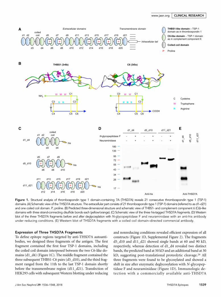
Expression of Three THSD7A FragmentsTo define epitope regions targeted by anti-THSD7A autoanti-bodies, we designed three fragments of the antigen. The firstfragment contained the first four TSP-1 domains, includingthe coiled coil domain interposed between the two C6-like do-mains (d1_d4) (Figure 1C). The middle fragment contained thethree subsequent THBS1-C6 pairs (d5_d10), and the third frag-ment ranged from the 11th to the last TSP-1 domain shortlybefore the transmembrane region (d11_d21). Transfection ofHEK293 cells with subsequentWestern blotting under reducing
and nonreducing conditions revealed efficient expression of allconstructs (Figure 1D, Supplemental Figure 2). The fragmentsd5_d10 and d11_d21 showed single bands at 60 and 90 kD,respectively, whereas detection of d1_d4 revealed two distinctbands, the predicted band at 50 kD and an additional band at 30kD, suggesting post-translational proteolytic cleavage.26 Allthree fragments were found to be glycosylated and showed ashift in size after enzymatic deglycosylation with N-glycopep-tidase F and neuraminidase (Figure 1D). Immunologic de-tection with a commercially available anti-THSD7A
Figure 1. Structural analysis of thrombospondin type 1 domain–containing 7A (THSD7A) reveals 21 consecutive thrombospondin type 1 (TSP-1)domains. (A) Schematic view of the THSD7A structure. The extracellular part consists of 21 thrombospondin type 1 (TSP-1) domains (referred to as d1–d21)and one coiled coil domain. P, proline. (B) Predicted three-dimensional structure and schematic view of THBS1- and complement component 6 (C6)–likedomains with three strand-connecting disulfide bonds each (yellow/orange). (C) Schematic view of the three his-tagged THSD7A fragments. (D) Westernblot of the three THSD7A fragments before and after deglycosylation with N-glycopeptidase F and neuraminidase with an anti-his antibodyunder reducing conditions. (E) Western blot of THSD7A fragments with a coiled coil domain–directed commercial antibody.
J Am Soc Nephrol 29: 1536–1548, 2018 THSD7A Epitopes 1539
www.jasn.org CLINICAL RESEARCH
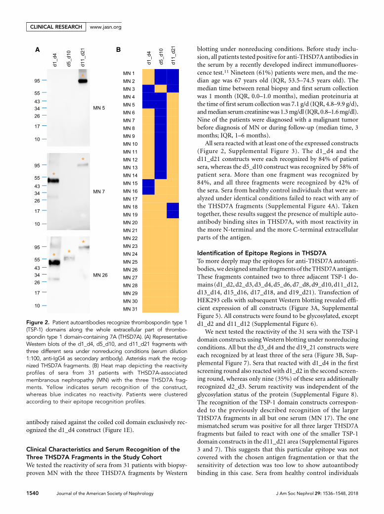
antibody raised against the coiled coil domain exclusively rec-ognized the d1_d4 construct (Figure 1E).
Clinical Characteristics and Serum Recognition of theThree THSD7A Fragments in the Study CohortWe tested the reactivity of sera from 31 patients with biopsy-proven MN with the three THSD7A fragments by Western
blotting under nonreducing conditions. Before study inclu-sion, all patients tested positive for anti-THSD7A antibodies inthe serum by a recently developed indirect immunofluores-cence test.11 Nineteen (61%) patients were men, and the me-dian age was 67 years old (IQR, 53.5–74.5 years old). Themedian time between renal biopsy and first serum collectionwas 1 month (IQR, 0.0–1.0 months), median proteinuria atthe time of first serum collectionwas 7.1 g/d (IQR, 4.8–9.9 g/d),andmedian serumcreatininewas 1.3mg/dl (IQR, 0.8–1.6mg/dl).Nine of the patients were diagnosed with a malignant tumorbefore diagnosis of MN or during follow-up (median time, 3months; IQR, 1–6 months).
All sera reacted with at least one of the expressed constructs(Figure 2, Supplemental Figure 3). The d1_d4 and thed11_d21 constructs were each recognized by 84% of patientsera, whereas the d5_d10 construct was recognized by 58% ofpatient sera. More than one fragment was recognized by84%, and all three fragments were recognized by 42% ofthe sera. Sera from healthy control individuals that were an-alyzed under identical conditions failed to react with any ofthe THSD7A fragments (Supplemental Figure 4A). Takentogether, these results suggest the presence of multiple auto-antibody binding sites in THSD7A, with most reactivity inthe more N-terminal and the more C-terminal extracellularparts of the antigen.
Identification of Epitope Regions in THSD7ATo more deeply map the epitopes for anti-THSD7A autoanti-bodies, we designed smaller fragments of theTHSD7Aantigen.These fragments contained two to three adjacent TSP-1 do-mains (d1_d2,d2_d3, d3_d4,d5_d6, d7_d8,d9_d10,d11_d12,d13_d14, d15_d16, d17_d18, and d19_d21). Transfection ofHEK293 cells with subsequent Western blotting revealed effi-cient expression of all constructs (Figure 3A, SupplementalFigure 5). All constructs were found to be glycosylated, exceptd1_d2 and d11_d12 (Supplemental Figure 6).
We next tested the reactivity of the 31 sera with the TSP-1domain constructs usingWestern blotting under nonreducingconditions. All but the d3_d4 and the d19_21 constructs wereeach recognized by at least three of the sera (Figure 3B, Sup-plemental Figure 7). Sera that reacted with d1_d4 in the firstscreening round also reacted with d1_d2 in the second screen-ing round, whereas only nine (35%) of these sera additionallyrecognized d2_d3. Serum reactivity was independent of theglycosylation status of the protein (Supplemental Figure 8).The recognition of the TSP-1 domain constructs correspon-ded to the previously described recognition of the largerTHSD7A fragments in all but one serum (MN 17). The onemismatched serum was positive for all three larger THSD7Afragments but failed to react with one of the smaller TSP-1domain constructs in the d11_d21 area (Supplemental Figures3 and 7). This suggests that this particular epitope was notcovered with the chosen antigen fragmentation or that thesensitivity of detection was too low to show autoantibodybinding in this case. Sera from healthy control individuals
Figure 2. Patient autoantibodies recognize thrombospondin type 1(TSP-1) domains along the whole extracellular part of thrombo-spondin type 1 domain-containing 7A (THSD7A). (A) RepresentativeWestern blots of the d1_d4, d5_d10, and d11_d21 fragments withthree different sera under nonreducing conditions (serum dilution1:100, anti-IgG4 as secondary antibody). Asterisks mark the recog-nized THSD7A fragments. (B) Heat map depicting the reactivityprofiles of sera from 31 patients with THSD7A-associatedmembranous nephropathy (MN) with the three THSD7A frag-ments. Yellow indicates serum recognition of the construct,whereas blue indicates no reactivity. Patients were clusteredaccording to their epitope recognition profiles.
1540 Journal of the American Society of Nephrology J Am Soc Nephrol 29: 1536–1548, 2018
CLINICAL RESEARCH www.jasn.org

failed to react with any of the TSP-1 domain constructs (Sup-plemental Figure 4B).
To investigate the presence of epitopes that are sensitive todenaturation by SDS, we applied native blotting (dot blot anal-ysis). All constructs were well recognized by an anti-his anti-body under native conditions (Figure 3C). Subsequently, wetested 30 of the previously tested 31 patients with availableserum. Twenty-one sera (70%) showed identical epitope pro-files in native and Western blotting. However, three sera didnot recognize a TSP-1 domain construct that was previouslyrecognized using Western blotting (Supplemental Figure 9A),and two sera did not show any reactivity in native blotting(data not shown). Interestingly, four sera recognized addi-tional TSP-1 domain constructs in native blotting, showingthe presence of denaturation-sensitive epitopes in these pa-tients (Figure 3C, Supplemental Figure 9B). Epitope recogni-tion profiles as deduced from Western blotting and nativeblotting are shown in Figure 3D. Altogether, 28 of 31 (90%)serum samples recognized multiple domains, and d1_d2(amino acids 48–192) was most frequently recognized (27 of31 patients; 87%) followed by d15_d16, d9_d10, and d13_d14,which were recognized by 61%, 52%, and 45% of the sera,respectively (Figure 3E).
All Western blot experiments were performed using anti-human IgG4 and anti-total human IgG as secondaryantibodies. Two sera recognized additional TSP-1 domainconstructs when tested with anti-total IgG compared withanti-IgG4, showing the presence of domain-specific autoanti-bodies different from IgG4 in these patients (Figure 4, A andB). Both patients did not have an associatedmalignant disease.Taken together, these data show that autoantibodies from pa-tients with THSD7A-associated MN preferentially bind to themost N-terminal region of THSD7A (amino acids 48–192)and that most sera recognize multiple epitope domains pre-sent in THSD7A.
Domain and Conformation Specificity of IdentifiedAutoantibodiesWe next investigated whether the identified autoantibodieswere indeed distinct domain-specific antibodies or whetherone clone of antibodies recognized several epitopes along theantigen due to sequence homology between the TSP-1 do-mains (Supplemental Figure 1A). We purified domain-specific IgG using recombinant d1_d3 and d9_d10 from apatient serum that recognized d1_d2, d7_d8, and d9_d10(Figure 4C). The d1_d3-purified antibodies exclusively rec-ognized d1_d2, whereas the d9_d10-purified antibodiesshowed exclusive reactivity with d9_d10 inWestern blot anal-ysis (Figure 4, D and E). The experiment was repeated withthree more patient sera with consistent results (not shown).These results indicate that the variety of domain recognitionin our patients is due to distinct autoantibodies against severalepitopes present in THSD7A.
Autoantibodies from patients with THSD7A-associatedMN have been reported to recognize THSD7A exclusively
under nonreducing conditions in Western blot analysis, sug-gesting autoantibody binding to a conformation-dependentepitope within the antigen.3 We tested nine of the previouslyanalyzed sera under both reducing and nonreducing condi-tions in a standardized side by side Western blot experiment.Sera were probed on full-length THSD7A; the d1_d4, d5_d10,and d11_d21 fragments; and the small TSP-1 domain con-structs. Under reducing conditions, all investigated sera com-pletely lost reactivity with full-length THSD7A; partially orcompletely lost reactivity with d1_d4, d5_d10, and d11_d21;and completely retained reactivity with the small TSP-1 do-main constructs (Figure 5) (not shown). Sera from healthycontrol individuals failed to react with the TSP-1 domainconstructs under reducing conditions (Supplemental Figure4). These experiments suggest that the small TSP-1 fragmentsare capable of refolding after loss of the reducing agent duringprotein transfer.
Association of Epitope Recognition Profiles withClinical Characteristics and Epitope Recognition duringFollow-UpWe next analyzed the association of the THSD7A epitope pro-files (Figure 3D) with clinical characteristics of the studycohort (Table 1). Patients whose sera recognized only oneor two TSP-1 domain constructs had significantly loweranti-THSD7A antibody levels and statistically nonsignificantlower proteinuria compared with patients whose sera recog-nized at least three TSP-1 domain constructs (P=0.02 andP=0.07, respectively). Remission of proteinuria was achievedby 86% of patients whose sera recognized one or two TSP-1domain constructs, but it was only achieved by 50% of pa-tients whose sera recognized three or more TSP-1 domains(P=0.18) (Table 1).
Follow-up sera were available from 16 of 31 patients in thestudy, and they were examined for changes in epitope recog-nition over time. In five patients, epitope profiles did notchange during follow-up (Supplemental Figure 10). All ofthese patients had stable anti-THSD7A antibody levels as mea-sured by immunofluorescence test and nephrotic-rangeproteinuria throughout the follow-up time. Sera from sevenpatients lost reactivity with one or more constructs (Supple-mental Figure 11). The anti-THSD7A antibody levelsdecreased in six of these patients, and five patients had a re-mission of proteinuria. The remaining four patients had achange in their epitope profile during follow-up (Supple-mental Figure 12). We found no specific patterns for theloss or gain of epitope recognition (i.e., preferential loss orgain of reactivity with one specific TSP-1 domain constructcompared with another) (Supplemental Table 1).
Immune Response against THSD7A after ActiveImmunizationWe next evaluated the epitope regions in human and mouseTHSD7A that are targeted by antibodies raised againstTHSD7A in three rabbits by coimmunization with human
J Am Soc Nephrol 29: 1536–1548, 2018 THSD7A Epitopes 1541
www.jasn.org CLINICAL RESEARCH

and mouse THSD7A cDNA. We have previously shown thatthese antibodies induce MN with severe nephrotic syndromewhen transferred into mice.21 A detailed sequence analysis of
mouse THSD7A showed that it shares over 90% of amino acidsequence homology with human THSD7A and that it is alsocomposed of 21 TSP-1 domains with the identical pattern of
Figure 3. Patient autoantibodies recognize multiple epitopes in thrombospondin type 1 domain–containing 7A (THSD7A) with apredominance for the most N-terminal region. (A) Western blot of HEK293 cell–expressed soluble thrombospondin type 1 (TSP-1)domain constructs with an anti-his antibody under reducing conditions. (B) Representative Western blots of the TSP-1 domain con-structs with three different sera under nonreducing conditions (serum dilution 1:100, anti-IgG4 as secondary antibody). Asterisks markthe recognized TSP-1 domain constructs. (C) Native blot (dot blot) analyses of the TSP-1 domain constructs with an anti-his antibody(row 1) and three representative sera (rows 2–4). Yellow asterisks mark the recognized TSP-1 domain constructs that were also rec-ognized using Western blotting, whereas the red asterisk marks an additional TSP-1 domain recognition (MN 5). (D) Heat map de-picting reactivity profiles of sera from all 31 patients with the small TSP-1 domain constructs as deduced from nonreduced Westernblotting and native blotting. Yellow indicates serum recognition of the construct, whereas blue indicates no reactivity. IFT, indirectimmunofluorescence test. (E) Recognition of the individual TSP-1 domains by patient sera in percentages.
1542 Journal of the American Society of Nephrology J Am Soc Nephrol 29: 1536–1548, 2018
CLINICAL RESEARCH www.jasn.org

THBS1- and C6-like domains (Supplemental Figure 13). Con-sequently, we used the same cloning and expression tech-niques for the mouse TSP-1 domains as described for thehuman TSP-1 domains. All mouse TSP-1 domain constructswere well expressed in HEK293 cells and secreted to the cul-ture medium (Figure 6A). We tested the three rabbit antiserafor their recognition of human and mouse TSP-1 domainsunder nonreducing conditions. Strikingly, all three rabbit an-tisera recognized the most N-terminal region d1_d2 of bothhuman andmouse THSD7A (Figure 6, B and C, SupplementalFigure 14), which was also the most frequently recognizedepitope region in our patient cohort (Figure 3E). There is nolower homology between the rabbit, mouse, and human pro-teins in the d1_d2 region compared with the other TSP-1domains (Supplemental Table 2). Therefore, a lack of homol-ogy in this region cannot be the reason for the preferred gen-eration of autoantibodies against the most N-terminal regionof THSD7A. Interestingly, the highly pathogenic rabbit
sera additionally recognized epitope domains in the moreC-terminal region of mouse THSD7A, also resembling thesituation found in our patient cohort.
In a similar approach, we immunized four mice by ballisticcDNA immunization with a mixture of human and mouseTHSD7A cDNA. As controls, four mice were mock immu-nized. THSD7A-immunized mice generated antibodies thatrecognized human but not mouse THSD7A (Figure 6D),showing that self-tolerance could not be overcome with thisparticular immunization technique. Accordingly,mice did notshow glomerular binding of mouse IgG in immunohisto-chemical analysis (Figure 6E) and did not develop proteinuria(not shown). Notably, sera from all THSD7A-immunizedmice also recognized d1_d2 (Figure 6, F and G). As for rabbitand human THSD7A, the amino acid sequence homology inthe N-terminal region of mouse and human THSD7A is com-parable (approximately 90% identity) with that of the otherTSP-1 domains (Supplemental Table 2), indicating that
Figure 4. Patient autoantibodies are IgG4-predominant and domain-specific. (A and B) Western blot under nonreducing conditions ofTSP-1 domain constructs with sera from two patients with thrombospondin type 1 domain–containing 7A (THSD7A)–associatedmembranous nephropathy (MN) using anti-IgG4 (upper panels) and anti-total IgG (lower panels) as secondary antibodies. Asterisksmark the recognized TSP-1 domain constructs. (C) Western blot under nonreducing conditions of TSP-1 domain constructs with serumfrom one patient with THSD7A-associated MN. (D and E) Antibodies that were purified using recombinant (D) d1_d3 and (E) d9_d10exclusively bound d1_d2 and d9_d10, respectively. Asterisks mark the recognized TSP-1 domain constructs.
J Am Soc Nephrol 29: 1536–1548, 2018 THSD7A Epitopes 1543
www.jasn.org CLINICAL RESEARCH

differences in the amino acid sequence are not the predomi-nant stimulus for the animals’ immune system to target thisregion. Taken together, these data show that induced rabbitand mouse anti-THSD7A antibodies, like patient autoanti-bodies, predominantly target the most N-terminal domainsof THSD7A.
DISCUSSION
This study identifies the epitope regions in THSD7A that aretargeted by autoantibodies from patients with MN. Usingcomprehensive sequence alignments, we characterized the
extracellular part of THSD7Aas a series of 21TSP-1 domains.On the basis of their amino acid sequence, these TSP-1 do-mains can be further classified as either THBS1-like or C6-like. THBS1 itself acts as an adhesive glycoprotein that caninteract with components of the extracellular matrix,27–30
cell receptors,31–37 and proteases,38,39 possibly leading toactivation of downstream signaling pathways, alterationsof protein localization, proteolytic processing, and proteininternalization.40 The homology of the THBS1-like domainsin THSD7A with THBS1 may give a hint on the presentlyunknown biologic functions of THSD7A on podocytes andallow speculation on potential pathomechanisms inTHSD7A-associated MN.
Table 1. Epitope recognition and clinical characteristics of the cohort
Clinical CharacteristicsSera with Recognition of One orTwo TSP-1 Domain Constructs
Sera with Recognition of More ThanTwo TSP-1 Domain Constructs
P Value
No. of patients (%) 10 (32) 21 (68) N/ANo. of epitopes, median (IQR) 2.0 (1.0–2.0) 4.0 (4.0–5.0) ,0.001Age, yr, median (IQR) 67.0 (52.0–76.5) 65.0 (54.0–69.0) 0.75Men (%) 5 (50) 14 (67) 0.45Proteinuria, g/d, median (IQR) 5.8 (3.4–7.4) 7.7 (5.3–10.7) 0.07Serum creatinine, mg/dl, median (IQR) 1.2 (0.9–1.6) 1.2 (0.8–1.8) 0.69Anti-THSD7A antibody level, median (IQR) 210 (32–320) 1000 (320–1000) 0.02Patients with malignancy (%) 3 (30) 6 (29) .0.99Patients with partial or complete remission ofproteinuria during follow-up (%)
6 of 7 (86) 8 of 16 (50) 0.18
Follow-up data on proteinuria were available for 23 of the patients included in our cohort. TSP-1, thrombospondin type 1; N/A, not applicable; IQR, interquartilerange; THSD7A, thrombospondin type 1 domain–containing 7A.
Figure 5. Patient autoantibodies recognize conformation-dependent epitopes in thrombospondin type 1 domain–containing 7A(THSD7A). Images depict Western blots of full-length THSD7A, the three THSD7A fragments, and the small thrombospondin type1 (TSP-1) domain constructs with one representative serum (MN 27) under nonreducing (upper panels) and reducing conditions(lower panels).
1544 Journal of the American Society of Nephrology J Am Soc Nephrol 29: 1536–1548, 2018
CLINICAL RESEARCH www.jasn.org

Figure 6. Rabbits and mice generate antibodies against the most N-terminal region of thrombospondin type 1 domain–containing 7A(THSD7A) after coimmunization with human and mouse THSD7A cDNA. (A) Western blot of HEK293 cell–expressed soluble mousethrombospondin type 1 (TSP-1) domain constructs with an anti-his antibody under reducing conditions. (B) Western blot under non-reducing conditions of human (upper panel) and mouse (lower panel) TSP-1 domain constructs with one representative rabbit anti-serum (serum dilution 1:100). Asterisks mark the recognized TSP-1 domain constructs. (C) Heat map depicting the reactivity profiles ofthree rabbit antisera with human and mouse TSP-1 domain constructs in Western blot analysis. Yellow indicates serum recognition ofthe construct, whereas blue indicates no reactivity. (D) Representative Western blot under nonreducing conditions of recombinanthuman and mouse THSD7A with serum from one of four mice that were immunized with a combination of human and mouse THSD7AcDNA. Four mice were mock immunized (empty control). (E) Representative immunohistochemical staining for mouse IgG in oneTHSD7A-immunized mouse and one control mouse. (F) Western blot under nonreducing conditions and (G) heat map analysis ofreactivity of four mouse antisera with the human TSP-1 domain constructs. Asterisks in F mark the recognized TSP-1 domain constructs.Yellow in G indicates serum recognition of the construct, whereas blue indicates no reactivity.
J Am Soc Nephrol 29: 1536–1548, 2018 THSD7A Epitopes 1545
www.jasn.org CLINICAL RESEARCH

In an unbiased approach to identify the epitope regions inTHSD7A, we designed three consecutive fragments of theantigen and tested for recognition by patient autoantibodies.Most patient sera reacted with several THSD7A fragmentswith a predominance for the more N-terminal and themore C-terminal part of the antigen. Additional epitopemap-ping revealed that the d1_d2 construct (amino acids 48–192)was recognized by 87% of the sera, indicating that the dom-inant epitope in THSD7A-associatedMN is located within themost N-terminal part of the antigen. In contrast, the reactivityin the more C-terminal part distributed over several epitopedomains. Interestingly, the preference of autoantibodies forthe most N-terminal region of the antigen is a similarity ofTHSD7A- and PLA2R1-associated MN. However, whereaspatient autoantibodies bind to the most N-terminal regionof PLA2R1 in all investigated patients,17,18 this is not the casefor THSD7A. Furthermore, most sera from patients withTHSD7A-associated MN recognize more than one antigendomain, and epitope profiles vary between the different pa-tients. This is in contrast to PLA2R1, for which only threeepitope regions have been identified so far.18 It is known fromother antibody-mediated autoimmune diseases, such asGrave disease, pemphigus vulgaris, and anti-brush border an-tibody disease, that autoantibodies target a great varietyof epitopes that distribute over the whole extracellular re-gions of the respective autoantigens but with an N-terminalpredominance.41–44
Compared with patients whose sera recognized only one ortwo TSP-1 domains, patients whose sera recognizedmore thantwo TSP-1 domains had higher anti-THSD7A antibody levels,tended to higher levels of proteinuria, and achieved a remissionof proteinuria in the observation period less often. Duringfollow-up, seven patients lost recognition of one or more epi-topes, which paralleled a decrease in anti-THSD7A antibodylevels and in most patients, a remission of proteinuria. Fromthese data it is not clear whether it is the individual epitopeprofile, the number of recognized epitopes, the antibody titeralone, or a combination of these factors that drive disease.Further studies are needed to investigate these issues.
The phenomenon of epitope spreading has been describedin human antibody-mediated autoimmune diseases45–49 anddifferent autoimmune animal models.50–53 In the experimen-tal MN model of Heymann nephritis, proteinuria increasedwith epitope spreading.54 This is in accordance with PLA2R1-associatedMN, where epitope spreading was reported to relatewith disease outcome.18,19 In our study, we did not find clearevidence for epitope spreading over time due to the smallnumber of patients with THSD7A-associated MN and sero-logic follow-up. In the future, larger cohorts of patients will beneeded to further dissect the relationship of epitope profiles atdiagnosis and the possible phenomenon of epitope spreadingwith disease activity and long-term clinical outcome.
Our study has several limitations. First, we did not use a quan-titative method, such as ELISA, to determine domain-specificautoantibody titers. Therefore, we cannot compare domain-
specific antibody levels between patients and correlate domain-specific titers with disease activity and outcome. Second, Westernblot experimentswere standardized regarding protein loading (onthe basis of anti-his reactivity under reducing conditions) andserum dilution (1:100) due to a limited amount of patient serum.It seems likely that a change in these parameters, such as higher orlower dilution of the used serum,may have led to slightly differentepitope profiles for some patients. Third, antibody binding siteswere mapped to peptides of 100–120 amino acids. Additionalwork is needed to define the genuine epitopes on the level often to 15 amino acids.
To investigate antibody generation against THSD7A, weanalyzed rabbits and mice that were immunized with a com-bination of human and mouse THSD7A cDNA. Remarkably,all animals raised an antibody response against the first twoN-terminal domains d1_d2 of both mouse and/or humanTHSD7A, andmost animals recognized several other domains,resembling the situation in our patient cohort. Thereby, theN-terminus of THSD7A does not predispose to antibodyformation due to a particularly low homology in this area be-tween the three species orthologs. This indicates that otherfeatures of the N-terminal region, such as epitope accessibility,might be responsible for the predominant antibody response.However, autoantibodies were induced against foreign pro-teins in these experiments, and importantly, the pathogenicityof antibodies targeting specific domains within the antigenremains to be investigated in future studies.
Identification of the epitopes that are targeted by autoanti-bodies from patients with THSD7A-associated MN is of highinterest for future investigations for the following reasons. (1)Autoimmunity may emerge as a result of molecular mimicrybetweenmicrobial or tumor antigens and host proteins.55,56 De-fining the precise antibody binding site(s) would allow align-ments with microbial proteins to uncover yet unknown linksbetween infections or tumors and primary MN. (2) The TSP-1domains in THSD7A might differentially interact with othermolecules and/or induce intracellular signaling.40 Interferenceof autoantibodies with these functions could represent a patho-mechanism in the mediation of podocyte damage in MN. (3)Epitope blocking/competition therapy or systemic autoantibodyextraction using the antibody binding fragment could constituteinnovative therapies for patients with antibody-mediated auto-immune diseases.57 Also, expression of epitope-containing do-mains on T cells in combination with intracellular signalingdomains can direct T cells to eliminate autoantibody-producingB cells through specific binding to the B cell receptor,58 repre-senting another potential therapeutic intervention for patientswith autoimmune primary MN that requires knowledge of theantibody binding domains.
In conclusion, our study shows that an autoimmuneprocesswith polyreactive IgG is part of the pathogenesis of THSD7A-associated MN. Additionally, we provide clinical and experi-mental evidence that the principal immune response targetsthe most N-terminal part of the protein, potentially enablingepitope-specific therapies in the future.
1546 Journal of the American Society of Nephrology J Am Soc Nephrol 29: 1536–1548, 2018
CLINICAL RESEARCH www.jasn.org

ACKNOWLEDGMENTS
The authors thank Eugen Kinzler and Daniela Bergleiter (III. Medi-
zinische Klinik, University Medical Center Hamburg-Eppendorf,
Hamburg, Germany) for technical assistance and Gudrun Dubberke,
Fabienne Seyfried, and Sarah Hewald (Insitute of Immunology,
University Medical Center Hamburg-Eppendorf, Hamburg, Germany)
for technical assistance with cDNA immunization.
This study was supported by grants from the Deutsche For-
schungsgemeinschaft as part of the Sonderforschungsbereich 1192
(project B1 to E.H. and R.A.K.S.; project B2 to G.Z., R.A.K.S., and
N.M.T.; and project B5 to F.K.-N.). E.H. is supported by the Else
Kröner-Fresenius Stiftung.
The following colleagues participated in the recruitment of the
patients for this study (in alphabetical order): Bahte S, Beckmann S,
Bokemeyer D, Born B, Boser M, Budde K, Dellanna F, Ferber J, Fielitz
JG, Floege J, Gerth J, Groll J, Grosser S, Hegner B, Hetzel GR,
Hollenbeck M, Hoyer J, Isbell LK, Jabs W, Jacobson J, Kidder D,
Koch C, Köhler S, Kortus-Götze B, Köstler F, Kresse S, Langer T,
Leidig B, Messtorff K, Möller J, Peitzmeier C, Rosenburg C, Rump C,
Sass C, SchmidtmannK, SchnegelsbergO, Tacuri-Strasser D, Thiele I,
Tiedeken P, Treiber W, Vitu J, Vosskühler A, Walz G, Weiner S, and
Worch P.
DISCLOSURESNone.
REFERENCES
1. Ruggenenti P, Fervenza FC, Remuzzi G: Treatment of membranousnephropathy: Time for a paradigm shift.Nat Rev Nephrol 13: 563–579,2017
2. Beck LH Jr., Bonegio RG, Lambeau G, Beck DM, Powell DW,Cummins TD, et al.: M-type phospholipase A2 receptor as targetantigen in idiopathic membranous nephropathy. N Engl J Med 361:11–21, 2009
3. Tomas NM, Beck LH Jr., Meyer-Schwesinger C, Seitz-Polski B, Ma H,Zahner G, et al.: Thrombospondin type-1 domain-containing 7A in id-iopathic membranous nephropathy. N Engl J Med 371: 2277–2287,2014
4. Hofstra JM, Beck LH Jr., Beck DM, Wetzels JF, Salant DJ: Anti-phospholipase A2 receptor antibodies correlate with clinical status inidiopathic membranous nephropathy. Clin J Am Soc Nephrol 6:1286–1291, 2011
5. Hoxha E, Harendza S, Pinnschmidt H, Panzer U, Stahl RA: PLA2Rantibody levels and clinical outcome in patients with membranousnephropathy and non-nephrotic range proteinuria under treatmentwith inhibitors of the renin-angiotensin system. PLoS One 9:e110681, 2014
6. Hoxha E, Harendza S, Pinnschmidt H, Panzer U, Stahl RA: M-typephospholipase A2 receptor autoantibodies and renal function in pa-tients with primary membranous nephropathy. Clin J Am Soc Nephrol9: 1883–1890, 2014
7. Beck LH Jr., Fervenza FC, Beck DM, Bonegio RG,Malik FA, Erickson SB,et al.: Rituximab-induced depletion of anti-PLA2R autoantibodiespredicts response in membranous nephropathy. J Am Soc Nephrol 22:1543–1550, 2011
8. Hofstra JM, Debiec H, Short CD, Pellé T, Kleta R, Mathieson PW, et al.:Antiphospholipase A2 receptor antibody titer and subclass in idiopathicmembranous nephropathy. J Am Soc Nephrol 23: 1735–1743, 2012
9. Stahl R, Hoxha E, Fechner K: PLA2R autoantibodies and recurrentmembranous nephropathy after transplantation. N Engl J Med 363:496–498, 2010
10. Gupta G, Fattah H, Ayalon R, Kidd J, Gehr T, Quintana LF, et al.: Pre-transplant phospholipase A2 receptor autoantibody concentration isassociated with clinically significant recurrence of membranous ne-phropathy post-kidney transplantation. Clin Transplant 30: 461–469,2016
11. Hoxha E, Beck LH Jr., Wiech T, Tomas NM, Probst C, Mindorf S, et al.:An indirect Immunofluorescence method facilitates detection ofthrombospondin type 1 domain-containing 7A-specific antibodies inmembranous nephropathy. J Am Soc Nephrol 28: 520–531, 2017
12. Hoxha E, Wiech T, Stahl PR, Zahner G, Tomas NM,Meyer-SchwesingerC, et al.: A mechanism for cancer-associated membranous nephropa-thy. N Engl J Med 374: 1995–1996, 2016
13. Wang J, Cui Z, Lu J, Probst C, Zhang YM, Wang X, et al.: Circulatingantibodies against thrombospondin type-I domain-containing 7A inChinese patients with idiopathic membranous nephropathy. Clin J AmSoc Nephrol 12: 1642–1651, 2017
14. Pedchenko V, Bondar O, Fogo AB, Vanacore R, Voziyan P, Kitching AR,et al.: Molecular architecture of the Goodpasture autoantigen in anti-GBM nephritis. N Engl J Med 363: 343–354, 2010
15. Roth AJ, Ooi JD, Hess JJ, van Timmeren MM, Berg EA, Poulton CE,et al.: Epitope specificity determines pathogenicity and detectability inANCA-associated vasculitis. J Clin Invest 123: 1773–1783, 2013
16. Kao L, Lam V, Waldman M, Glassock RJ, Zhu Q: Identification of theimmunodominant epitope region in phospholipase A2 receptor-mediating autoantibody binding in idiopathic membranous nephrop-athy. J Am Soc Nephrol 26: 291–301, 2015
17. Fresquet M, Jowitt TA, Gummadova J, Collins R, O’Cualain R,McKenzie EA, et al.: Identification of a major epitope recognized byPLA2R autoantibodies in primary membranous nephropathy. J Am SocNephrol 26: 302–313, 2015
18. Seitz-Polski B, Dolla G, Payré C, Girard CA, Polidori J, Zorzi K, et al.:Epitope spreading of autoantibody response to PLA2R associates withpoor prognosis in membranous nephropathy. J Am Soc Nephrol 27:1517–1533, 2016
19. Seitz-Polski B, Debiec H, Rousseau A, Dahan K, Zaghrini C, Payre C,et al.: Phospholipase A2 receptor 1 epitope spreading at baselinepredicts reduced likelihood of remission of membranous nephropathy.J Am Soc Nephrol 29: 401–408, 2018
20. Jäger V, Büssow K, Wagner A, Weber S, Hust M, Frenzel A, et al.: Highlevel transient production of recombinant antibodies and antibodyfusion proteins in HEK293 cells. BMC Biotechnol 13: 52, 2013
21. Tomas NM, Meyer-Schwesinger C, von Spiegel H, Kotb AM, Zahner G,Hoxha E, et al.: A heterologous model of thrombospondin type 1 do-main-containing 7A-associated membranous nephropathy. J Am SocNephrol 28: 3262–3277, 2017
22. Klenotic PA, Page RC, Misra S, Silverstein RL: Expression, purificationand structural characterization of functionally replete thrombospondin-1 type 1 repeats in a bacterial expression system. Protein Expr Purif 80:253–259, 2011
23. Aleshin AE, Schraufstatter IU, Stec B, Bankston LA, Liddington RC,DiScipio RG: Structure of complement C6 suggests a mechanism forinitiation and unidirectional, sequential assembly of membrane attackcomplex (MAC). J Biol Chem 287: 10210–10222, 2012
24. Pääkkönen K, Tossavainen H, Permi P, Rakkolainen H, Rauvala H, RauloE, et al.: Solution structures of the first and fourth TSR domains of F-spondin. Proteins 64: 665–672, 2006
25. Venkatesh B, Lee AP, Ravi V, Maurya AK, Lian MM, Swann JB, et al.:Elephant shark genome provides unique insights into gnathostomeevolution. Nature 505: 174–179, 2014
J Am Soc Nephrol 29: 1536–1548, 2018 THSD7A Epitopes 1547
www.jasn.org CLINICAL RESEARCH
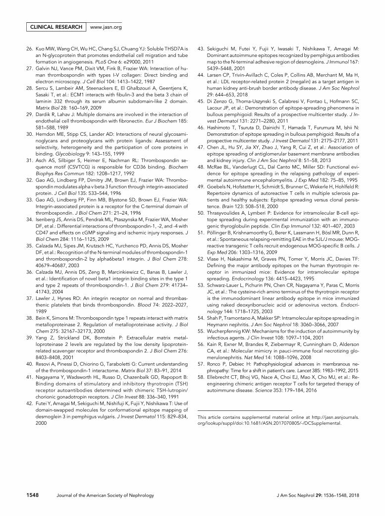
26. KuoMW,Wang CH,Wu HC, Chang SJ, Chuang YJ: Soluble THSD7A isan N-glycoprotein that promotes endothelial cell migration and tubeformation in angiogenesis. PLoS One 6: e29000, 2011
27. Galvin NJ, Vance PM, Dixit VM, Fink B, Frazier WA: Interaction of hu-man thrombospondin with types I-V collagen: Direct binding andelectron microscopy. J Cell Biol 104: 1413–1422, 1987
28. Sercu S, Lambeir AM, Steenackers E, El Ghalbzouri A, Geentjens K,Sasaki T, et al.: ECM1 interacts with fibulin-3 and the beta 3 chain oflaminin 332 through its serum albumin subdomain-like 2 domain.Matrix Biol 28: 160–169, 2009
29. Dardik R, Lahav J: Multiple domains are involved in the interaction ofendothelial cell thrombospondin with fibronectin. Eur J Biochem 185:581–588, 1989
30. Herndon ME, Stipp CS, Lander AD: Interactions of neural glycosami-noglycans and proteoglycans with protein ligands: Assessment ofselectivity, heterogeneity and the participation of core proteins inbinding. Glycobiology 9: 143–155, 1999
31. Asch AS, Silbiger S, Heimer E, Nachman RL: Thrombospondin se-quence motif (CSVTCG) is responsible for CD36 binding. BiochemBiophys Res Commun 182: 1208–1217, 1992
32. Gao AG, Lindberg FP, Dimitry JM, Brown EJ, Frazier WA: Thrombo-spondinmodulates alpha v beta 3 function through integrin-associatedprotein. J Cell Biol 135: 533–544, 1996
33. Gao AG, Lindberg FP, Finn MB, Blystone SD, Brown EJ, Frazier WA:Integrin-associated protein is a receptor for the C-terminal domain ofthrombospondin. J Biol Chem 271: 21–24, 1996
34. Isenberg JS, Annis DS, Pendrak ML, Ptaszynska M, Frazier WA, MosherDF, et al.: Differential interactions of thrombospondin-1, -2, and -4 withCD47 and effects on cGMP signaling and ischemic injury responses. JBiol Chem 284: 1116–1125, 2009
35. Calzada MJ, Sipes JM, Krutzsch HC, Yurchenco PD, Annis DS, MosherDF, et al.: Recognition of theN-terminal modules of thrombospondin-1and thrombospondin-2 by alpha6beta1 integrin. J Biol Chem 278:40679–40687, 2003
36. Calzada MJ, Annis DS, Zeng B, Marcinkiewicz C, Banas B, Lawler J,et al.: Identification of novel beta1 integrin binding sites in the type 1and type 2 repeats of thrombospondin-1. J Biol Chem 279: 41734–41743, 2004
37. Lawler J, Hynes RO: An integrin receptor on normal and thrombas-thenic platelets that binds thrombospondin. Blood 74: 2022–2027,1989
38. Bein K, Simons M: Thrombospondin type 1 repeats interact with matrixmetalloproteinase 2. Regulation of metalloproteinase activity. J BiolChem 275: 32167–32173, 2000
39. Yang Z, Strickland DK, Bornstein P: Extracellular matrix metal-loproteinase 2 levels are regulated by the low density lipoprotein-related scavenger receptor and thrombospondin 2. J Biol Chem 276:8403–8408, 2001
40. Resovi A, Pinessi D, Chiorino G, Taraboletti G: Current understandingof the thrombospondin-1 interactome. Matrix Biol 37: 83–91, 2014
41. Nagayama Y, Wadsworth HL, Russo D, Chazenbalk GD, Rapoport B:Binding domains of stimulatory and inhibitory thyrotropin (TSH)receptor autoantibodies determined with chimeric TSH-lutropin/chorionic gonadotropin receptors. J Clin Invest 88: 336–340, 1991
42. Futei Y, Amagai M, Sekiguchi M, Nishifuji K, Fujii Y, Nishikawa T: Use ofdomain-swapped molecules for conformational epitope mapping ofdesmoglein 3 in pemphigus vulgaris. J Invest Dermatol 115: 829–834,2000
43. Sekiguchi M, Futei Y, Fujii Y, Iwasaki T, Nishikawa T, Amagai M:Dominant autoimmune epitopes recognized by pemphigus antibodiesmap to theN-terminal adhesive region of desmogleins. J Immunol 167:5439–5448, 2001
44. Larsen CP, Trivin-Avillach C, Coles P, Collins AB, Merchant M, Ma H,et al.: LDL receptor-related protein 2 (megalin) as a target antigen inhuman kidney anti-brush border antibody disease. J Am Soc Nephrol29: 644–653, 2018
45. Di Zenzo G, Thoma-Uszynski S, Calabresi V, Fontao L, Hofmann SC,Lacour JP, et al.: Demonstration of epitope-spreading phenomena inbullous pemphigoid: Results of a prospective multicenter study. J In-vest Dermatol 131: 2271–2280, 2011
46. Hashimoto T, Tsuruta D, Dainichi T, Hamada T, Furumura M, Ishii N:Demonstration of epitope spreading in bullous pemphigoid: Results of aprospective multicenter study. J Invest Dermatol 131: 2175–2177, 2011
47. Chen JL, Hu SY, Jia XY, Zhao J, Yang R, Cui Z, et al.: Association ofepitope spreading of antiglomerular basement membrane antibodiesand kidney injury. Clin J Am Soc Nephrol 8: 51–58, 2013
48. McRae BL, Vanderlugt CL, Dal Canto MC, Miller SD: Functional evi-dence for epitope spreading in the relapsing pathology of experi-mental autoimmune encephalomyelitis. J Exp Med 182: 75–85, 1995
49. Goebels N, Hofstetter H, Schmidt S, Brunner C, Wekerle H, Hohlfeld R:Repertoire dynamics of autoreactive T cells in multiple sclerosis pa-tients and healthy subjects: Epitope spreading versus clonal persis-tence. Brain 123: 508–518, 2000
50. Thrasyvoulides A, Lymberi P: Evidence for intramolecular B-cell epi-tope spreading during experimental immunization with an immuno-genic thyroglobulin peptide. Clin Exp Immunol 132: 401–407, 2003
51. Pöllinger B, Krishnamoorthy G, Berer K, Lassmann H, Bösl MR, Dunn R,et al.: Spontaneous relapsing-remitting EAE in the SJL/J mouse: MOG-reactive transgenic T cells recruit endogenous MOG-specific B cells. JExp Med 206: 1303–1316, 2009
52. Vlase H, Nakashima M, Graves PN, Tomer Y, Morris JC, Davies TF:Defining the major antibody epitopes on the human thyrotropin re-ceptor in immunized mice: Evidence for intramolecular epitopespreading. Endocrinology 136: 4415–4423, 1995
53. Schwarz-Lauer L, Pichurin PN, Chen CR, Nagayama Y, Paras C, MorrisJC, et al.: The cysteine-rich amino terminus of the thyrotropin receptoris the immunodominant linear antibody epitope in mice immunizedusing naked deoxyribonucleic acid or adenovirus vectors. Endocri-nology 144: 1718–1725, 2003
54. Shah P, Tramontano A,Makker SP: Intramolecular epitope spreading inHeymann nephritis. J Am Soc Nephrol 18: 3060–3066, 2007
55. Wucherpfennig KW: Mechanisms for the induction of autoimmunity byinfectious agents. J Clin Invest 108: 1097–1104, 2001
56. Kain R, Exner M, Brandes R, Ziebermayr R, Cunningham D, AldersonCA, et al.: Molecular mimicry in pauci-immune focal necrotizing glo-merulonephritis. Nat Med 14: 1088–1096, 2008
57. Ronco P, Debiec H: Pathophysiological advances in membranous ne-phropathy: Time for a shift in patient’s care. Lancet 385: 1983–1992, 2015
58. Ellebrecht CT, Bhoj VG, Nace A, Choi EJ, Mao X, Cho MJ, et al.: Re-engineering chimeric antigen receptor T cells for targeted therapy ofautoimmune disease. Science 353: 179–184, 2016
This article contains supplemental material online at http://jasn.asnjournals.org/lookup/suppl/doi:10.1681/ASN.2017070805/-/DCSupplemental.
1548 Journal of the American Society of Nephrology J Am Soc Nephrol 29: 1536–1548, 2018
CLINICAL RESEARCH www.jasn.org
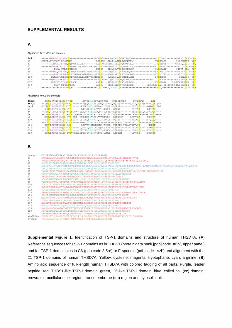
SUPPLEMENTAL RESULTS A Alignments for THBS1-like domains 3r6b ------INGGWGPWSPWDIC------------------SVTCGG---GVQKRSRLCNNPTPQFGGK----------------DCVGDVT---------ENQICNKQ-DCP
d1 QGEAEAPTLYWKT-GPWGRCMG-----------------DECGPG--GIQTRAVWCAHVEGWTTLH---------------TNCKQAER-------PNNQQNCFK--VCD
d2 -------LYDWRL-GPWNQCQPVISKSLEKP--------LECIKGEEGIQVREIACIQKDKDIPAE--------------DIICEYFEP------KPLLEQACLI--PCQ
d5 --------YGWRT-TEWTECRVDPLLSQQDKRRG--NQTALCGG---GIQTREVYCVQANENLLSQLSTHKNKEASKPMDLKLC-TGPI-------PNTTQLCHI--PCP
d7 ---------DWKA-VRLGNCEPDNG--------------KECGP---GTQVQEVVCINSDGEEVDR---------------QLCRDAIF-------PIPVA-CDA--PCP
d9 -------VYHWQT-GPWGQCIEDTSVSSFNTTTTWNGE-ASCSV---GMQTRKVICVRVNVGQVGP---------------KKCPESLR-------PETVRPCLL--PCK
d11 -------SYRWKT-HKWRRCQLVPWSVQQDSPGAQ----EGCGP---GRQARAITCRKQDGGQAGI---------------HECLQYAG-----PVPALTQACQI--PCQ
d13 -------KYNAQPVGNWSDCILPEGKVEVLLGMKVQGDIKECGQ---GYRYQAMACYDQNGRLVET---------------SRCNSHGY---------IEEACII--PCP
d15 -------QYLWVT-EPWSICKVTFVNM-----------RENCGE---GVQTRKVRCMQNTADGPSE-----------HVEDYLCDPEEM-------PLGSRVCKL--PCP
d17 -------HYDYNV-TDWSTCQLSEK--------------AVCGN---GIKTRMLDCVRSDGKSVDL---------------KYCEALGL----EKNWQMNTSCMV--ECP
d19 ---------RWQY-GQWSPCQVQE---------------AQCGE---GTRTRNISCVVSDGSADDF---------SKVVDEEFCADIELIIDGNKNMVLEESCSQ--PCP
d21 --EYKWMASAWKGSSRTVWCQRSDGINVT----------GGCLVM--SQPDADRSCNP------------------------PCSQP------------HSYCSETKTCH
Alignments for C6-like domains 3t5o1 --CFCDHYAWTQWTSCSK-TCN-------SGTQSRHRQIVVDKYYQE---NFCEQICSKQ-----ETRECNWQ-----RCP----
3t502 --INCLLGDFGPWSDCD--PCI--------EKQSKVRSVLRPSQFG---GQPCTAP-------LVAFQPCIPSK----LCKIE--
1szl GSETCIYSNWSPWSACSSSTC-------EKGKRMRQRMLKAQ--LDLSV--PCPDT--------QDFQPC-----MGPGCSDEDG
d3 --QDCIVSEFSAWSECS-KTC-------GSGLQHRTRHVVAPP-QFGGS--GCPNL--------TEFQVC-----QSSPCE----
d4 --KECQVSEWSEWSPCS-KTCHDMV--SPAGTRVRTRTIRQFP-IGSEK--ECPEF--------EEKEPCLSQGDGVVPCA----
d6 --TECEVSPWSAWGPCTYENCNDQQ--GKKGFKLRKRRITNEPTGGSGVTGNCPHL--------LEAIPC-----EEPACY----
d8 --KDCVLSTWSTWSSCS-HTC-SGK--TTEGKQIRARSILAYAGEEGGI--RCPNSSA-----LQEVRSC-----NEHPCT----
d10 --KDCIVTPYSDWTSCP-SSCKEGD--SSIRKQSRHRVIIQLP-ANGGR--DCTDP-------LYEEKAC----EAPQACQ----
d12 --DDCQLTSWSKFSSCN-GDC--------GAVRTRKRTLVGKS--KKKE--KCKNSHLYP---LIETQYC--------PCD----
d14 --SDCKLSEWSNWSRCS-KSC-------GSGVKVRSKWLREKP-YNGGR--PCPKLDHVNQAQVYEVVPC------HSDCN----
d16 --EDCVISEWGPWTQCV-LPC------NQSSFRQRSADPIRQP-ADEGR--SCPNA--------VEKEPC----NLNKNCY----
d18 --VNCQLSDWSSWSQCS-QTC------GLTGKMIRKRTVTQPF-QGDGR--PCPSLM-------DQSKPC-----PVKPCY----
d20 --GDCYLKDWSSWSLCQ-LTCVNGEDLGFGGIQVRSRPVIIQE-LENQH--LCPEQM-------LETKSC-----YDGQCY----
B leader MGLQARRWASGSRGAAGPRRGVLQLLPLPLPLPLLLLLLLRPGAGRA
d1 AAQGEAEAPTLYLWKTGPWGRCMGDECGPGGIQTRAVWCAHVEGWTTLHTNCKQAERPNNQQNCFKVCD
d2 WHKELYDWRLGPWNQCQPVISKSLEKPLECIKGEEGIQVREIACIQKDKDIPAEDIICEYFEPKPLLEQACLIPCQ
d3 QDCIVSEFSAWSECSKTCGSGLQHRTRHVVAPPQFGGSGCPNLTEFQVCQSSPCE
cc AEELRYSLHVGPWSTCSMPHSRQVRQARRRGKNKEREKDRSKGVKDPEARELIKKKRNRNRQNRQENKYWDIQIGYQTREVMCINKTGKAADLSFCQQEKLPMTFQSCVIT
d4 KECQVSEWSEWSPCSKTCHDMVSPAGTRVRTRTIRQFPIGSEKECPEFEEKEPCLSQGDGVVPCA
d5 TYGWRTTEWTECRVDPLLSQQDKRRGNQTALCGGGIQTREVYCVQANENLLSQLSTHKNKEASKPMDLKLCTGPIPNTTQLCHIPCP
d6 TECEVSPWSAWGPCTYENCNDQQGKKGFKLRKRRITNEPTGGSGVTGNCPHLLEAIPCEEPACY
d7 DWKAVRLGNCEPDNGKECGPGTQVQEVVCINSDGEEVDRQLCRDAIFPIPVACDAPCP
d8 KDCVLSTWSTWSSCSHTCSGKTTEGKQIRARSILAYAGEEGGIRCPNSSALQEVRSCNEHPCT
d9 VYHWQTGPWGQCIEDTSVSSFNTTTTWNGEASCSVGMQTRKVICVRVNVGQVGPKKCPESLRPETVRPCLLPCK
d10 KDCIVTPYSDWTSCPSSCKEGDSSIRKQSRHRVIIQLPANGGRDCTDPLYEEKACEAPQACQ
d11 SYRWKTHKWRRCQLVPWSVQQDSPGAQEGCGPGRQARAITCRKQDGGQAGIHECLQYAGPVPALTQACQIPCQ
d12 DDCQLTSWSKFSSCNGDCGAVRTRKRTLVGKSKKKEKCKNSHLYPLIETQYCPCD
d13 KYNAQPVGNWSDCILPEGKVEVLLGMKVQGDIKECGQGYRYQAMACYDQNGRLVETSRCNSHGYIEEACIIPCP
d14 SDCKLSEWSNWSRCSKSCGSGVKVRSKWLREKPYNGGRPCPKLDHVNQFQVYEVVPCHSDCN
d15 QYLWVTEPWSICKVTFVNMRENCGEGVQTRKVRCMQNTADGPSEHVEDYLCDPEEMPLGSRVCKLPCP
d16 EDCVISEWGPWTQCVLPCNQSSFRQRSADPIRQPADEGRSCPNAVEKEPCNLNKNCY
d17 HYDYNVTDWSTCQLSEKAVCGNGIKTRMLDCVRSDGKSVDLKYCEALGLEKNWQMNTSCMVECP
d18 VNCQLSDWSPWSECSQTCGLTGKMIRRRTVTQPFQGDGRPCPSLMDQSKPCPVKPCY
d19 RWQYGQWSPCQVQEAQCGEGTRTRNISCVVSDGSADDFSKVVDEEFCADIELIIDGNKNMVLEESCSQPCP
d20 GDCYLKDWSSWSLCQLTCVNGEDLGFGGIQVRSRPVIIQELENQHLCPEQMLETKSCYDGQCY
d21 EYKWMASAWKGSSRTVWCQRSDGINVTGGCLVMSQPDADRSCNPPCSQPHSYCSETKTCH
stalk/tm CEEGYTEVMSSNSTLEQCTLIPVVVLPTMEDKRGDVKTSRAVHPTQPSSNPAGRGRTWFLQPFGPDGRLKTW
cytosol VYGVAAGAFVLLIFIVSMIYLACKKPKKPQRRQNNRLKPLTLAYDGDADM
Supplemental Figure 1. Identification of TSP-1 domains and structure of human THSD7A. (A)
Reference sequences for TSP-1 domains as in THBS1 (protein data bank [pdb] code 3r6b1, upper panel)
and for TSP-1 domains as in C6 (pdb code 3t5o2) or F-spondin (pdb code 1szl3) and alignment with the
21 TSP-1 domains of human THSD7A. Yellow, cysteine; magenta, tryptophane; cyan, arginine. (B)
Amino acid sequence of full-length human THSD7A with colored tagging of all parts. Purple, leader
peptide; red, THBS1-like TSP-1 domain; green, C6-like TSP-1 domain; blue, coiled coil (cc) domain;
brown, extracellular stalk region, transmembrane (tm) region and cytosolic tail.
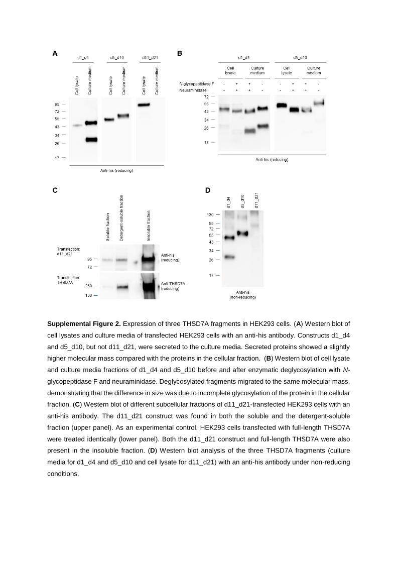
Supplemental Figure 2. Expression of three THSD7A fragments in HEK293 cells. (A) Western blot of
cell lysates and culture media of transfected HEK293 cells with an anti-his antibody. Constructs d1_d4
and d5_d10, but not d11_d21, were secreted to the culture media. Secreted proteins showed a slightly
higher molecular mass compared with the proteins in the cellular fraction. (B) Western blot of cell lysate
and culture media fractions of d1_d4 and d5_d10 before and after enzymatic deglycosylation with N-
glycopeptidase F and neuraminidase. Deglycosylated fragments migrated to the same molecular mass,
demonstrating that the difference in size was due to incomplete glycosylation of the protein in the cellular
fraction. (C) Western blot of different subcellular fractions of d11_d21-transfected HEK293 cells with an
anti-his antibody. The d11_d21 construct was found in both the soluble and the detergent-soluble
fraction (upper panel). As an experimental control, HEK293 cells transfected with full-length THSD7A
were treated identically (lower panel). Both the d11_d21 construct and full-length THSD7A were also
present in the insoluble fraction. (D) Western blot analysis of the three THSD7A fragments (culture
media for d1_d4 and d5_d10 and cell lysate for d11_d21) with an anti-his antibody under non-reducing
conditions.
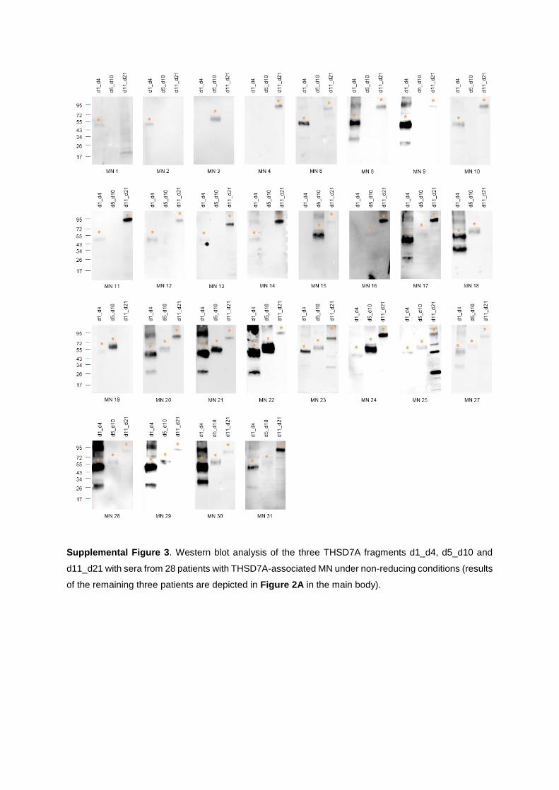
Supplemental Figure 3. Western blot analysis of the three THSD7A fragments d1_d4, d5_d10 and
d11_d21 with sera from 28 patients with THSD7A-associated MN under non-reducing conditions (results
of the remaining three patients are depicted in Figure 2A in the main body).
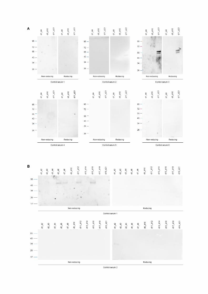
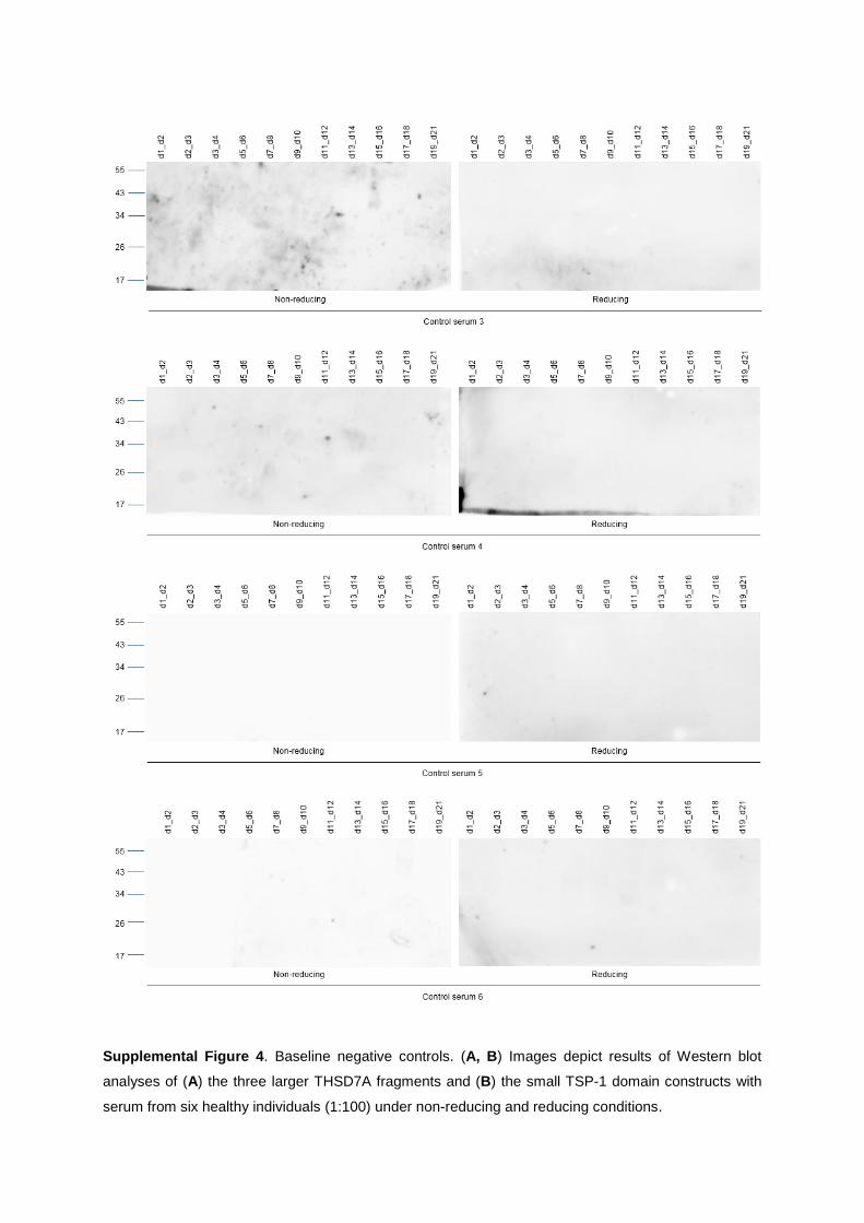
Supplemental Figure 4. Baseline negative controls. (A, B) Images depict results of Western blot
analyses of (A) the three larger THSD7A fragments and (B) the small TSP-1 domain constructs with
serum from six healthy individuals (1:100) under non-reducing and reducing conditions.
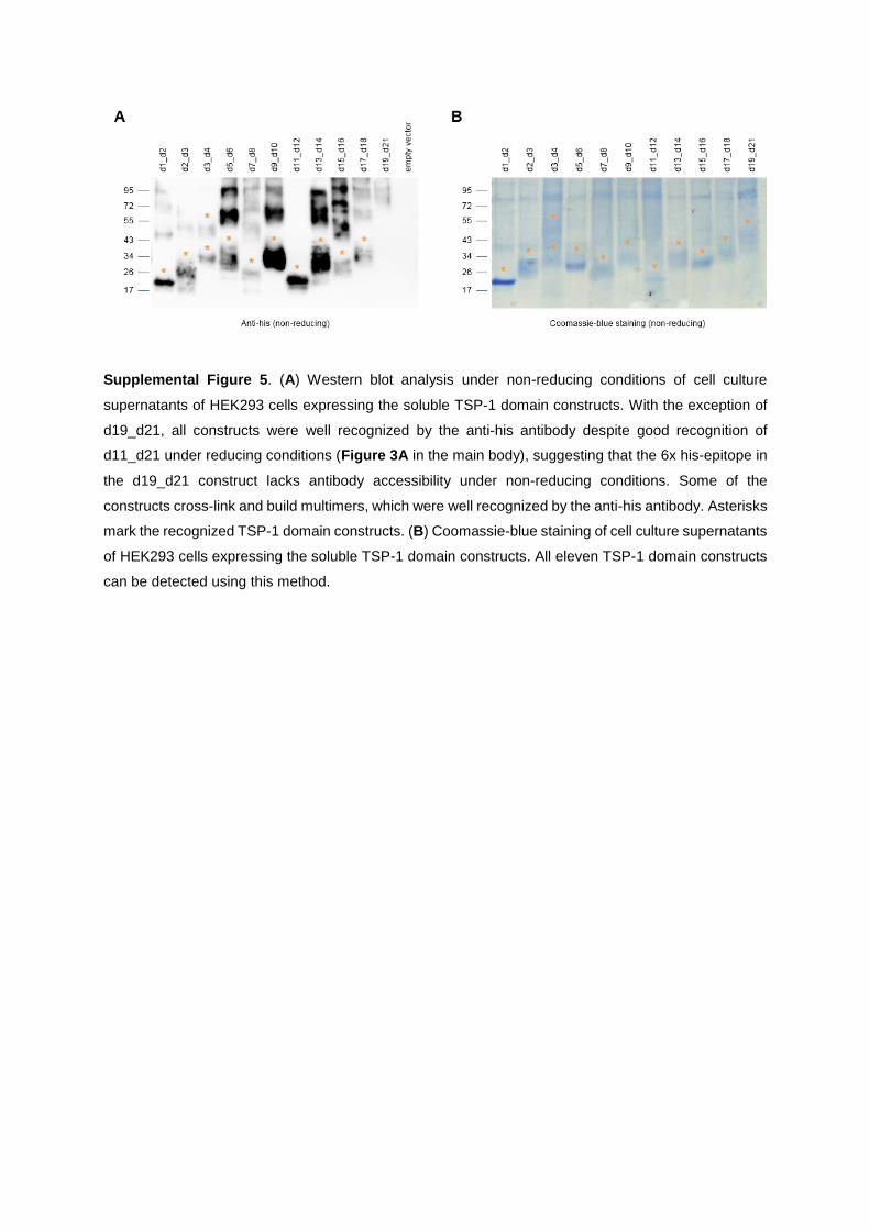
Supplemental Figure 5. (A) Western blot analysis under non-reducing conditions of cell culture
supernatants of HEK293 cells expressing the soluble TSP-1 domain constructs. With the exception of
d19_d21, all constructs were well recognized by the anti-his antibody despite good recognition of
d11_d21 under reducing conditions (Figure 3A in the main body), suggesting that the 6x his-epitope in
the d19_d21 construct lacks antibody accessibility under non-reducing conditions. Some of the
constructs cross-link and build multimers, which were well recognized by the anti-his antibody. Asterisks
mark the recognized TSP-1 domain constructs. (B) Coomassie-blue staining of cell culture supernatants
of HEK293 cells expressing the soluble TSP-1 domain constructs. All eleven TSP-1 domain constructs
can be detected using this method.

A
B
TSP-1 domain construct
Expected molecular mass
Molecular mass after deglycosylation
d1_d2 18 20
d2_d3 16 16
d3_d4 27 32
d5_d6 18 26
d7_d8 14 18
d9_d10 16 20
d11_d12 15 18
d13_d14 16 18
d15_d16 15 17
d17_d18 15 15
d19_d21 23 29
Supplemental figure 6. (A) Glycosylation of individual TSP-1 domain constructs. The TSP-1 domain
constructs were enzymatically deglycosylated using N-glycopeptidase F and neuraminidase. Shifts in
protein mass were evaluated by immunodetection with an anti-his antibody in Western blot analysis
under reducing conditions. (B) Predicted molecular mass according to
www.sciencegateway.org/tools/proteinmw.htm and molecular mass as estimated from migration in gel
electrophoresis after enzymatic deglycosylation. The fragments corresponded well to their expected
molecular masses or run slightly above.
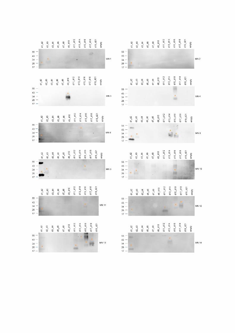
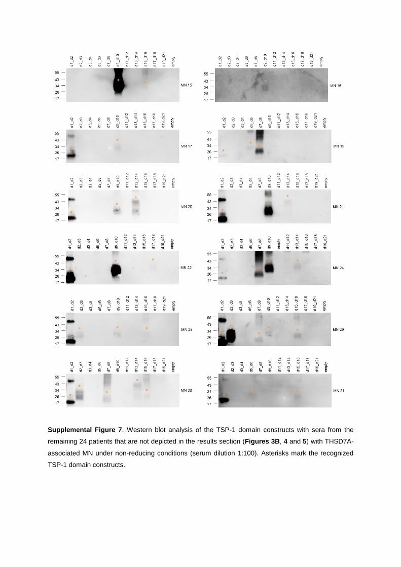
Supplemental Figure 7. Western blot analysis of the TSP-1 domain constructs with sera from the
remaining 24 patients that are not depicted in the results section (Figures 3B, 4 and 5) with THSD7A-
associated MN under non-reducing conditions (serum dilution 1:100). Asterisks mark the recognized
TSP-1 domain constructs.

Supplemental Figure 8. Serum recognition of deglycosylated TSP-1 domain constructs. The TSP-1
domain constructs were enzymatically deglycosylated using N-glycopeptidase F and neuraminidase.
Sera recognized both the glycosylated and deglycosylated forms in Western blot analysis under non-
reducing conditions. Asterisks mark the recognized TSP-1 domain constructs.

Supplemental Figure 9. Native blotting (dot blot analysis) of purified TSP-1 domain constructs with
sera from patients with THSD7A-associated MN. (A, B) Dot blot analysis of TSP-1 domain constructs
with sera that (A) recognized the same or less TSP-1 domain constructs or (B) showed additional
reactivity with TSP-1 domain constructs when compared with Western blotting. MN 9, MN 16 and MN
28 failed to recognize a TSP-1 domain construct that was previously recognized in Western blotting and
MN 2 and MN 12 failed to recognize any TSP-1 domain construct in native blotting (data not shown).
Yellow asterisks mark the recognized TSP-1 domain constructs that were also appreciated in Western
blotting while red asterisks mark additional TSP-1 domain recognition.

Supplemental Figure 10. Anti-THSD7A antibody levels and epitope profiles during follow-up in patients
with steady TSP-1 domain recognition. (A) In patient MN 4, epitope profiling showed serum recognition
of the d15_d16 construct, which remained unchanged during the observation period. IFT titer also
remained stable and proteinuria was constantly in the nephrotic range. The patient did not receive
immunosuppressive therapy. Lower panels depict the Western blot analyses of the TSP-1 domain
constructs with the first and last available serum from this patient. Asterisks mark the recognized TSP-
1 domain constructs. (B) Anti-THSD7A antibody levels and epitope profiles of four more patients with
stable TSP-1 domain recognition and persistent active disease. MN 21 and MN 28 received
immunosuppressive therapy during follow-up.
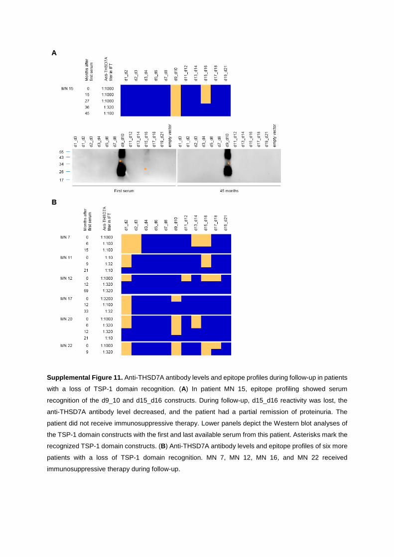
Supplemental Figure 11. Anti-THSD7A antibody levels and epitope profiles during follow-up in patients
with a loss of TSP-1 domain recognition. (A) In patient MN 15, epitope profiling showed serum
recognition of the d9_10 and d15_d16 constructs. During follow-up, d15_d16 reactivity was lost, the
anti-THSD7A antibody level decreased, and the patient had a partial remission of proteinuria. The
patient did not receive immunosuppressive therapy. Lower panels depict the Western blot analyses of
the TSP-1 domain constructs with the first and last available serum from this patient. Asterisks mark the
recognized TSP-1 domain constructs. (B) Anti-THSD7A antibody levels and epitope profiles of six more
patients with a loss of TSP-1 domain recognition. MN 7, MN 12, MN 16, and MN 22 received
immunosuppressive therapy during follow-up.
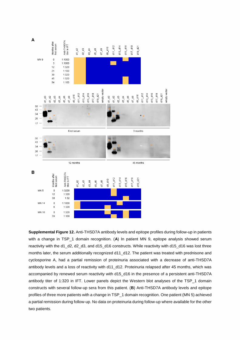
Supplemental Figure 12. Anti-THSD7A antibody levels and epitope profiles during follow-up in patients
with a change in TSP_1 domain recognition. (A) In patient MN 9, epitope analysis showed serum
reactivity with the d1_d2, d2_d3, and d15_d16 constructs. While reactivity with d15_d16 was lost three
months later, the serum additionally recognized d11_d12. The patient was treated with prednisone and
cyclosporine A, had a partial remission of proteinuria associated with a decrease of anti-THSD7A
antibody levels and a loss of reactivity with d11_d12. Proteinuria relapsed after 45 months, which was
accompanied by renewed serum reactivity with d15_d16 in the presence of a persistent anti-THSD7A
antibody titer of 1:320 in IFT. Lower panels depict the Western blot analyses of the TSP_1 domain
constructs with several follow-up sera from this patient. (B) Anti-THSD7A antibody levels and epitope
profiles of three more patients with a change in TSP_1 domain recognition. One patient (MN 5) achieved
a partial remission during follow-up. No data on proteinuria during follow-up where available for the other
two patients.
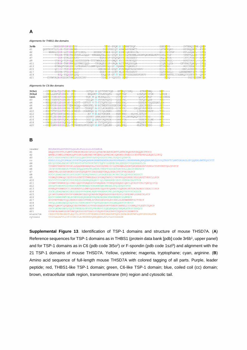
A Alignments for THBS1-like domains
3r6b -------INGGWGPWSPWDICSV------------------TCGG-GVQKRSRLCNNPTPQF----------------GGKDCVG---------DVTENQICNK-QDCP d1 QGDTEVPTLYLWK-TGPWGRCMG-----------------DDCGPGGIQTRAVWCAHVEGWTT---------------LHTNCKQAV-------RPSNQQNCF--KVCD
d2 ----WHKELYDWR-LGTWDRCQPVISKSL-----EKSRECVKGEE-GIQVREIMCIQKDKDIPA--------------EDIICEYFEP------KPLLEQACL--IPCQ
d5 --------TYGWR-TTEWTECHVDPLLSQQD--KRRANQTALCGG-GVQTREIYCIQTNDNMLSHGNTQKDKEASKPVDSKLCTGP--------VPNTTQLCH--VPCP
d7 ----------DWK-SVRLGDCEPD--------------NGKSCGP-GTQVQEVVCINSDGEEV---------------DRQLCRDA--------IFPIPVACD--APCP
d9 --------VYHWQ-TGPWGQCIEDTSVSSFN-TTTTWNGEASCSV-GMQTRKVICVRVNVGQV---------------GPKKCPESL-------RPETVRPCL--LPCR
d11 --------SYRWK-THKWRRCQLVPWSIQQD----VPGAQEGCGP-GRQARAITCRKQDGGQA---------------SIQECLQYA-----GPVPALTQACQ--IPCQ
d13 --------KYNAQPVGNWSDCILPEGKAEVLLGMKVQGDSKECGQ-GYRYQAMACYDQNGRLV---------------ETSRCNS---------HGYIEEACI--IPCP
d15 --------QYIWV-TEPWSVCKVTFV-----------DMRDNCGE-GVQTRKVRCMQNTADGPSEHV-----------EDYLCDPE-------DMPLGSRECK--LPCP
d17 --------HYDYN-VTDWSTCQLS--------------EKAVCGN-GIKTRMLDCVRSDGKSV---------------DLKYCEELG----LEKNWPMNTSCT--VECP
d19 ----------RWQ-YGQWSPCQVQ---------------EAQCGE-GTRTRNISCVVSDGSAEDFSKVV---------DEEFCANTELIIDGNKQIVLEETCT--QPCP
d21 ---EYKWVASAWKGSSRTVWCQRSDGINV----------TGGCLV-VQPDTDRSCNP------------------------PCSQ------------PHSYCSEMKTCR
Alignments for C6-like domains
3t5o1 --CFCDHYAWTQWTSCSK-TCN-------SGTQSRHRQIVVDKYYQE---NFCEQICSKQ-----ETRECNWQ-----RCP----
3t5o2 --INCLLGDFGPWSDCD--PCI--------EKQSKVRSVLRPSQFG---GQPCTAP-------LVAFQPCIPSK----LCKIE--
1szl GSETCIYSNWSPWSACSSSTCE-------KGKRMRQRMLKAQLDL----SVPCPDT--------QDFQPCMGP-----GCSDEDG
d3 --KDCIVSEFSPWSECSR-TCG-------SGLQHRTRHVVAPPQYG---GSGCPNL--------TEFQVCQSN-----PCE----
d4 --KECQVSEWLEWSPCSK-TCHDVT--SPTGTRVRTRTITQFPIGS---EKECPAL--------EEKEPCVSQGDGAVLCA----
d6 --IECEVSPWSAWGPCTYENCNDQQ--GKKGFKLRKRRITNEPTGGSGATGNCPHL--------LEAIPCEEP-----SCY----
d8 --KDCVLSAWSSWSSCSH-TCSGKT---TEGKQTRARSILAYAGEEG--GIRCPNI-----SALQEVRSCNEH-----PCT----
d10 --KDCVVTPYSDWTPCPS-SCREGD--SGARKQSRQRVIIQLPANG---GKECSDP-------LYEEKACEAPP----TCH----
d12 --DDCQFTSWSKFSSCNG-DCG--------AVRTRKRAIVGKSKK----KEKCKNS---HLYPLIETQYC--------PCD----
d14 --SDCKLSEWSNWSRCSK-SCG-------SGVKVRSKWLREKPYNG---GRPCPKLDHVNQAQVYEVVPCHS------DCN
d16 --EDCVISEWGPWTQCAL-PCNP------SGSRQRSADPIRQPADE---GRACPDA--------VEKEPCSLNK----NCY----
d18 --VNCQLSDWSSWSQCSQ-TCGL------TGKMIRKRTVTQPFQGD---GRPCPSL-------MEQSKPCPVK-----PCY----
d20 --GDCYLNDWSSWSLCQL-TCVNGEDLGFGGIQVRSRAVIIQELEN---QHLCPEQ-------MLETKSCDDG-----QCY----
B leader MGLRAGRLASPSRGVLQLLRLPLLLLLLLSSGARGA
d1 AAQGDTEVPTLYLWKTGPWGRCMGDDCGPGGIQTRAVWCAHVEGWTTLHTNCKQAVRPSNQQNCFKVCD
d2 WHKELYDWRLGTWDRCQPVISKSLEKSRECVKGEEGIQVREIMCIQKDKDIPAEDIICEYFEPKPLLEQACLIPCQ
d3 KDCIVSEFSPWSECSRTCGSGLQHRTRHVVAPPQYGGSGCPNLTEFQVCQSNPCE
cc EDESLYSLQVGPWSACSVPHTRQARQARRRGKNKEREKERGKAVKDPEARELIKKKRNRNRQNRQENRYWDIQIGYQTRDVTCLNRTGKSADLSFCQQERLPMTFQSCVIT
d4 KECQVSEWLEWSPCSKTCHDVTSPTGTRVRTRTITQFPIGSEKECPALEEKEPCVSQGDGAVLCA
d5 TYGWRTTEWTECHVDPLLSQQDKRRANQTALCGGGVQTREIYCIQTNDNMLSHGNTQKDKEASKPVDSKLCTGPVPNTTQLCHVPCP
d6 IECEVSPWSAWGPCTYENCNDQQGKKGFKLRKRRITNEPTGGSGATGNCPHLLEAIPCEEPSCY
d7 DWKSVRLGDCEPDNGKSCGPGTQVQEVVCINSDGEEVDRQLCRDAIFPIPVACDAPCP
d8 KDCVLSAWSSWSSCSHTCSGKTTEGKQTRARSILAYAGEEGGIRCPNISALQEVRSCNEHPCT
d9 VYHWQTGPWGQCIEDTSVSSFNTTTTWNGEASCSVGMQTRKVICVRVNVGQVGPKKCPESLRPETVRPCLLPCR
d10 KDCVVTPYSDWTPCPSSCREGDSGARKQSRQRVIIQLPANGGKECSDPLYEEKACEAPPTCH
d11 SYRWKTHKWRRCQLVPWSIQQDVPGAQEGCGPGRQARAITCRKQDGGQASIQECLQYAGPVPALTQACQIPCQ
d12 DDCQFTSWSKFSSCNGDCGAVRTRKRAIVGKSKKKEKCKNSHLYPLIETQYCPCD
d13 KYNAQPVGNWSDCILPEGKAEVLLGMKVQGDSKECGQGYRYQAMACYDQNGRLVETSRCNSHGYIEEACIIPCP
d14 SDCKLSEWSNWSRCSKSCGSGVKVRSKWLREKPYNGGRPCPKLDHVNQAQVYEVVPCHSDCN
d15 QYIWVTEPWSVCKVTFVDMRDNCGEGVQTRKVRCMQNTADGPSEHVEDYLCDPEDMPLGSRECKLPCP
d16 EDCVISEWGPWTCALPCNPSGSRQRSADPIRQPADEGRACPDAVEKEPCSLNKNCY
d17 HYDYNVTDWSTCQLSEKAVCGNGIKTRMLDCVRSDGKSVDLKYCEELGLEKNWPMNTSCTVECP
d18 VNCQLSDWSSWSQCSQTCGLTGKMIRKRTVTQPFQGDGRPCPSLMEQSKPCPVKPCY
d19 RWQYGQWSPCQVQEAQCGEGTRTRNISCVVSDGSAEDFSKVVDEEFCANTELIIDGNKQIVLEETCTQPCP
d20 GDCYLNDWSSWSLCQLTCVNGEDLGFGGIQVRSRAVIIQELENQHLCPEQMLETKSCDDGQCY
d21 EYKWVASAWKGSSRTVWCQRSDGINVTGGCLVVSQPDTDRSCNPPCSQPHSYCSEMKTCR
stalk/tm CEEGYTEVMSSNSTLEQCTLIPVVVIPTVEDKRGDVKTSRAVHPTQPSINPAGRGRTWFLQPFGPDGRLKTW
cytosol VYGVAAGAFVLLVFIVSMIYLACKKPKKPQRRQNNRLKPLTLAYDGDADM
Supplemental Figure 13. Identification of TSP-1 domains and structure of mouse THSD7A. (A)
Reference sequences for TSP-1 domains as in THBS1 (protein data bank [pdb] code 3r6b1, upper panel)
and for TSP-1 domains as in C6 (pdb code 3t5o2) or F-spondin (pdb code 1szl3) and alignment with the
21 TSP-1 domains of mouse THSD7A. Yellow, cysteine; magenta, tryptophane; cyan, arginine. (B)
Amino acid sequence of full-length mouse THSD7A with colored tagging of all parts. Purple, leader
peptide; red, THBS1-like TSP-1 domain; green, C6-like TSP-1 domain; blue, coiled coil (cc) domain;
brown, extracellular stalk region, transmembrane (tm) region and cytosolic tail.

Supplemental Figure 14. (A) Western blot analysis of reactivity of two different rabbit antisera with the
human and mouse TSP-1 domain constructs under non-reducing conditions. Asterisks mark the
recognized TSP-1 domain constructs.
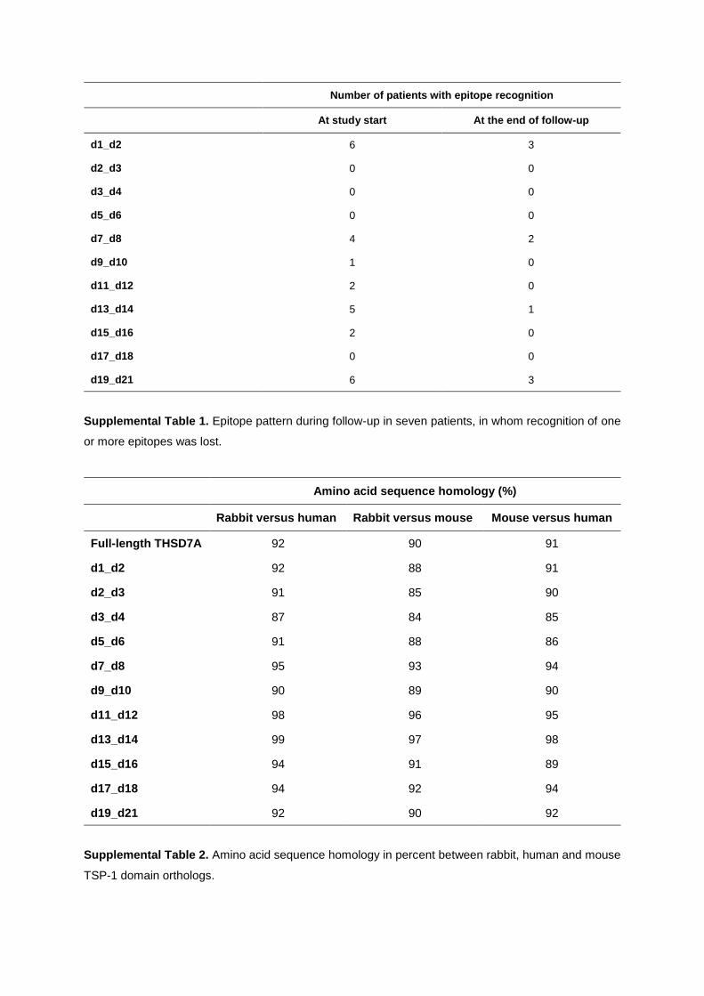
Number of patients with epitope recognition
At study start At the end of follow-up
d1_d2 6 3
d2_d3 0 0
d3_d4 0 0
d5_d6 0 0
d7_d8 4 2
d9_d10 1 0
d11_d12 2 0
d13_d14 5 1
d15_d16 2 0
d17_d18 0 0
d19_d21 6 3
Supplemental Table 1. Epitope pattern during follow-up in seven patients, in whom recognition of one
or more epitopes was lost.
Amino acid sequence homology (%)
Rabbit versus human Rabbit versus mouse Mouse versus human
Full-length THSD7A 92 90 91
d1_d2 92 88 91
d2_d3 91 85 90
d3_d4 87 84 85
d5_d6 91 88 86
d7_d8 95 93 94
d9_d10 90 89 90
d11_d12 98 96 95
d13_d14 99 97 98
d15_d16 94 91 89
d17_d18 94 92 94
d19_d21 92 90 92
Supplemental Table 2. Amino acid sequence homology in percent between rabbit, human and mouse
TSP-1 domain orthologs.

SUPPLEMENTAL METHODS
Cell culture, cell transfection and recombinant protein expression
Two lines of human embryonic kidney cells (HEK) were kept in culture for recombinant
expression of THSD7A fragments. HEK293-6E is a cell line that can be cultivated in the
absence of serum and is especially suitable for the expression of secreted proteins.4,5
HEK293T is a standard cell line for recombinant protein expression.6 HEK293-6E cells were
cultured in cell culture bottles with 10 ml of serum-free medium (Freestyle 293, Gibco by Life
Technologies, Grand Island, USA). HEK293T cells were cultured in 10 cm dishes with 10 ml
Dulbecco's Modified Eagle Medium (DMEM, Life Technologies, Grand Island, USA) with
addition of 10% fetal bovine serum (Thermo Scientific, Cramlington, United Kingdom) and 1%
Penicillin/Streptomycin (Life Technologies, Grand Island, USA).
All protein fragments were transfected to both cell types and expression efficiency was
evaluated. The d11_d21 construct was the only one not to be secreted into the culture medium
and expression was higher in HEK293T cells. Thus, we chose to express d11_d21 in HEK293T
cells for further experiments. All other constructs were well secreted into the culture medium
when expressed in HEK293-6E cells.
For transfection of HEK293-6E cells, a polythylenimine (PEI)-based method (Polyscience Inc.,
Warrington, USA) was used. For each approach, 126 µL (40 µg) of PEI was mixed with 124
µL water and 250 µL NaCl (300 mM) was added. 10 µg Plasmid DNA in 250 µL water was
mixed with the same amount of NaCl in a separate tube. Subsequently, solutions were slowly
mixed, vortexed and incubated for 30 min. The solution was then added to the cells. After 24
h the cells were fed with 250 µL feeding medium (Freestyle 293 medium supplemented with
20% tryptone). Five days later, cells were collected using centrifugation at 300 g and the
supernatant was centrifuged again at 14,000 g for 30 min.
For transfection of HEK293T cells, a calcium phosphate-based method was used.6 Ten µg of
plasmid DNA was mixed with 36 µL of 2 M CaCl2 and diluted with sterile water up to a volume
of 300 µL. This solution was gently mixed with an equal volume of 2x HEPES buffered saline
(HBS, 275 mM NaCl, 55 mM HEPES, pH 7.0) and incubated 30 min at room temperature. The
resulting solution was added drop wise to the cells. Medium was changed 24 h after
transfection. After 48 h, cells were scraped, centrifuged at 1,500 rpm for 5 minutes, washed
with PBS, and centrifuged again at 1,500 rpm for 5 minutes. Cells were then lysed in 50 mM
Tris pH 7.4, 1 mM EDTA, 150 mM NaCl, 1% Triton after addition of a protease inhibitor cocktail
(Roche Diagnostics, Indianapolis, USA), sonicated, and centrifuged at 14,000 g and 4 °C for
1 h. The supernatant was saved and protein concentration was determined using the Pierce
BCA protein assay kit according to the manufacturer’s instructions (Thermo Fisher, Waltham,
USA). All expressions (medium and cell lysate) were validated by Western blot and

immunological detection using an anti-his antibody (1:1,000, Thermo Scientific, Cramlington,
United Kingdom). For analyses with patient sera, the culture media of d1_d4- and d5_d10-
transfected cells and the cell lysate of d11_d21-transfected cells were used.
Protein fractionation and deglycosylation
To determine the subcellular location of the d11_d21 construct, cells were first lyzed in 50 mM
Tris pH 7.4, 0.32 M sucrose and 10 mM EDTA with a protease inhibitor cocktail (as above),
sonicated, rotated for 30 min at 4 °C, and centrifuged at 100,000 g for 1 h at 4 °C. The
supernatant was taken and labeled as the soluble (cytoplasmatic) fraction. The pellet was then
resuspended in 50 mM Tris pH 7.4, 150 mM NaCl, 0.1% SDS, 1% NP40 and 0.5% Na-DOC
with a protease inhibitor cocktail (as above), sonicated, rotated for 30 min at 4 °C, and
centrifuged at 100,000 g for 1 h at 4 °C. The supernatant was labeled as the detergent-soluble
(membrane) fraction. The cell pellet was then resuspended in 50 mM Tris pH 7.4, 10 mM DTT
and 8 M urea, sonicated, and labeled as the insoluble fraction. As a control experiment,
HEK293 cells transfected with full-length THSD7A were treated identically.
Deglycosylation experiments were performed using a combination of N-glycopeptidase F and
neuraminidase (both Roche Diagnsotics, Mannheim, Germany). Protein samples were
incubated overnight at 37 °C with deglycosylating enzymes or an equal volume of PBS (1 µl
per 100 µg of total protein).
Western blot and immunological detection
Protein samples were prepared for Western blot analysis by addition of 5x Laemmli buffer (1.5
M Tris-HCl pH 6.8, 50% glycerol, 10% SDS, 1% bromophenol blue) and subsequent heating
to 95 °C for 10 minutes. If reducing conditions were desired, 20% β-mercaptoethanol was
added to the 5x loading buffer. Proteins were separated by electrophoresis in 4-15% gradient
gels (Bio-Rad, Hercules, USA) in an electrophoresis chamber (Bio-Rad, Hercules, USA) in the
presence of a migration buffer (25 mM Tris, 192 mM glycine, 0.1% SDS; Amresco, Solon,
USA). Subsequently, proteins were transferred to methanol-soaked PVDF membranes
(Millipore, Billerica, USA) under semi-dry conditions in the presence of 25 mM Tris pH 8.5, 192
mM glycine, ethanol 20% using the Transblot Turbo system (Bio-Rad, Hercules, USA) at 25 V
constant for 35 min. Membranes were blocked in 5% dry milk with PBS plus Tween 0.05%
(PBS-T) for 2 h at room temperature followed by the incubation with the primary antibody (anti-
his antibody, 1:1,000, Thermo Scientific; anti-THSD7A antibody, 1:1,000, Atlas, #000923),
human serum, rabbit serum or mouse serum (all 1:100) in 0,5% dry milk in PBS-T. The next
day, membranes were washed three times with PBS-T the next day and incubated with the
secondary antibody in 5% dry milk in PBS-T for 2 h. HRP-conjugated mouse anti-human IgG4
and HRP-conjugated mouse anti-human IgG (both 1:20,000, SouthernBiotech, Birmingham,

USA), were used to analyze serum reactivity with the various THSD7A constructs. HRP-
conjugated goat anti-mouse IgG was used as secondary antibody for his detection and HRP-
conjugated goat anti-rabbit for THSD7A detection (both 1:20,000, SouthernBiotech,
Birmingham, USA). To visualize the protein bands, membranes were incubated in a
chemiluminescent substrate (SuperSignal West Pico and SuperSignal West Femto, Thermo
Scientific, Rockford, USA, used in a 4:1 ratio) followed by incremental luminescence detection
for up to 12 minutes with a Fujifilm LAS imager 3000. For testing human serum, protein loading
was standardized based on anti-his reactivity under reducing conditions and all sera were used
in a 1:100 dilution at all times.
Native blotting (dot blot analysis) and immunological detection
His-tagged TSP-1 domain constructs (d1_d2 to d19_d21) were purified under native conditions
using a Ni-NTA resin (His-Pur, Thermo Scientific, Rockford USA, #88221) applying the batch
method according to the manufacturer’s instructions. Equilibration, wash, and elution buffers
were PBS-based (pH 7.4) and supplemented with 10 mM imidazol, 25 mM imidazole, and 250
mM imidazole, respectively. Protein purification was validated by Coomassie-blue staining of
electrophoresed proteins. Purified proteins were dotted on nitrocellulose membranes (Protran,
Whatman, Dassel, Germany) and allowed to dry for 10 minutes at room temperature.
Membranes were then blocked in 4% dry milk in PBS plus Tween 0.08% (PBS-T) for 3 hours,
followed by incubation with human serum (1:100) or anti-his antibody (1:1000) in PBS-T with
0.5% dry milk. A mixture of HRP-conjugated anti-human IgG and anti-human IgG4 or anti-
mouse IgG (all 1:20,000, SouthernBiotech, Birmingham, USA) were used as secondary
antibodies and visualization of the protein signal was performed as described above.
Immunohistochemical analyses
1 µM paraffin sections of mouse kidneys were deparaffinized and rehydrated. Antigen retrieval
was obtained for rabbit IgG by digestion with protease XXIV (5 µg/mL, Sigma) for 15 min at
37°C. Nonspecific binding was blocked with 5% horse serum (Vector, Burlingame, USA) with
0.05% Triton X-100 (Sigma) in PBS for 30 min at room temperature prior to incubation at 4 °C
overnight with biotinylated anti-rabbit IgG (1:400) in blocking buffer. Staining was visualized
with the ZytochemPlus AP Polymer kit (Zytomed Systems, Berlin, Germany) according to the
manufacturer’s instruction with neufuchsin (Merck) as a color substrate. Nuclei were
counterstained with hemalaun (Merck) and sections were mounted with gum Arabic (Sigma,
St. Louis, USA). Negative controls were performed by omitting primary antibodies. Stainings
were evaluated with an Axioskop using the Axiovision software (all Zeiss).

SUPPLEMENTAL REFERENCES
1. Klenotic, PA, Page, RC, Misra, S, Silverstein, RL: Expression, purification and structural
characterization of functionally replete thrombospondin-1 type 1 repeats in a bacterial
expression system. Protein Expr Purif, 80: 253-259, 2011
2. Aleshin, AE, Schraufstatter, IU, Stec, B, Bankston, LA, Liddington, RC, DiScipio, RG: Structure of
complement C6 suggests a mechanism for initiation and unidirectional, sequential assembly of
membrane attack complex (MAC). J Biol Chem, 287: 10210-10222, 2012
3. Paakkonen, K, Tossavainen, H, Permi, P, Rakkolainen, H, Rauvala, H, Raulo, E, Kilpelainen, I,
Guntert, P: Solution structures of the first and fourth TSR domains of F-spondin. Proteins, 64:
665-672, 2006
4. Danquah, W, Meyer-Schwesinger, C, Rissiek, B, Pinto, C, Serracant-Prat, A, Amadi, M, Iacenda, D,
Knop, JH, Hammel, A, Bergmann, P, Schwarz, N, Assuncao, J, Rotthier, W, Haag, F, Tolosa,
E, Bannas, P, Boue-Grabot, E, Magnus, T, Laeremans, T, Stortelers, C, Koch-Nolte, F:
Nanobodies that block gating of the P2X7 ion channel ameliorate inflammation. Sci Transl Med,
8: 366ra162, 2016
5. Zhang, J, MacKenzie, R, Durocher, Y: Production of chimeric heavy-chain antibodies. Methods Mol
Biol, 525: 323-336, xv, 2009
6. Tomas, NM, Hoxha, E, Reinicke, AT, Fester, L, Helmchen, U, Gerth, J, Bachmann, F, Budde, K,
Koch-Nolte, F, Zahner, G, Rune, G, Lambeau, G, Meyer-Schwesinger, C, Stahl, RA:
Autoantibodies against thrombospondin type 1 domain-containing 7A induce membranous
nephropathy. J Clin Invest, 126: 2519-2532, 2016
www.kidney-international.org ba s i c re sea r ch
A novel mouse model of phospholipase A2receptor 1-associated membranous nephropathymimics podocyte injury in patients
Catherine Meyer-Schwesinger1,6, Nicola M. Tomas2,6, Silke Dehde2, Larissa Seifert2,Irm Hermans-Borgmeyer3, Thorsten Wiech4, Friedrich Koch-Nolte5, Tobias B. Huber2 andGunther Zahner21Institute of Cellular and Integrative Physiology, University Medical Center Hamburg-Eppendorf, Hamburg, Germany; 2III. Department ofMedicine, University Medical Center Hamburg-Eppendorf, Hamburg, Germany; 3Center of Molecular Neurobiology, University MedicalCenter Hamburg-Eppendorf, Hamburg, Germany; 4Institute of Pathology, University Medical Center Hamburg-Eppendorf, Hamburg,Germany; and 5Institute of Immunology, University Medical Center Hamburg-Eppendorf, Hamburg, Germany
Translational Statement
Over the last decade, the measurement of anti–phospholipase A2 receptor 1 (PLA2R1) antibodies forthe diagnosis and monitoring of patients with membra-nous nephropathy (MN) has been broadly implementedinto clinical practice worldwide. Despite this majoradvance, MN pathomechanisms are still incompletelyunderstood, and patient treatment relies on broad andunspecific immunosuppression. We envision that thismodel of PLA2R1-associated MN may help to dissect themolecular mechanisms that underlie podocyte injury inMN. The roles of the complement system and receptor-specific podocyte signaling programs represent subjectsof future interest that can be addressed using this model.Additionally, innovative therapeutic strategies targetingMN-specific pathomechanisms can be investigatedregarding their in vivo efficacy—a prerequisite for thetranslation to patient care in the future.
The phospholipase A2 receptor 1 (PLA2R1) is the majorautoantigen in patients suffering from membranousnephropathy. To date, the lack of endogenous glomerularexpression of PLA2R1 in mice and rats has impeded theestablishment of PLA2R1-dependent animal models of thisdisease. Here, we generated a transgenic mouse lineexpressing murine full-length PLA2R1 in podocytes.Furthermore, expression of murine PLA2R1 did not result inany morphological disturbance as high-resolution confocalmicroscopy demonstrated an intact nephrin distributionwith normal foot processes. Transfer of rabbit anti-mPLA2R1 antibodies to these mice induced nephroticrange proteinuria, hypercholesterolemia, andhistomorphological signs of membranous nephropathy.Immunohistochemical and immunofluorescence analysesrevealed enhanced staining for murine PLA2R1 in thepresence of unaffected staining for murinethrombospondin type-1 domain-containing 7A in thediseased mice, resembling what is classically found inpatients with PLA2R1-associated membranousnephropathy Thus, our mouse model of membranousnephropathy will allow investigation of PLA2R1-specificpathomechanisms and may help to develop and assessantigen-specific treatments in vivo.Kidney International (2019) -, -–-; https://doi.org/10.1016/
j.kint.2019.10.022
KEYWORDS: membranous nephropathy; mouse model; nephrotic syn-
drome; phospholipase A2 receptor 1 (PLA2R1); podocyte
Copyright ª 2019, International Society of Nephrology. Published by
Elsevier Inc. All rights reserved.
Correspondence: Gunther Zahner, III. Department of Medicine, UniversityMedical Center Hamburg-Eppendorf, Martinistraße 52, 20246 Hamburg,Germany. E-mail: [email protected] and NMT contributed equally.
Received 12 August 2019; revised 8 October 2019; accepted 24 October2019; published online 9 November 2019
Kidney International (2019) -, -–-
M embranous nephropathy (MN) is an autoimmuneglomerular disease and a frequent cause of nephroticsyndrome. Morphologically, the disease is character-
ized by granular deposition of IgG and complement systemcomponents along the glomerular filtration barrier as well as bysubepithelial electron-dense deposits and podocyte foot pro-cess effacement in electron microscopy.1,2 Circulatingautoantibodies against the podocyte membrane proteins phos-pholipase A2 receptor 1 (PLA2R1) and thrombospondin type-1domain-containing 7A (THSD7A) are found in around 75%–
80% of cases.3,4 Although recent animal studies demonstrateda pathogenic role of anti-THSD7A antibodies in the develop-ment of MN,5,6 the lack of endogenous PLA2R1 expressionin rodents7 precluded analogous antibody transfer studies.However, as the majority of MN cases are PLA2R1-associated, the development of a mouse model involving thisantigen is fundamental in order to experimentally investigatethe role of anti-PLA2R1 antibodies, understand the pathogenicroute through which anti-PLA2R1 autoantibodies induce
1
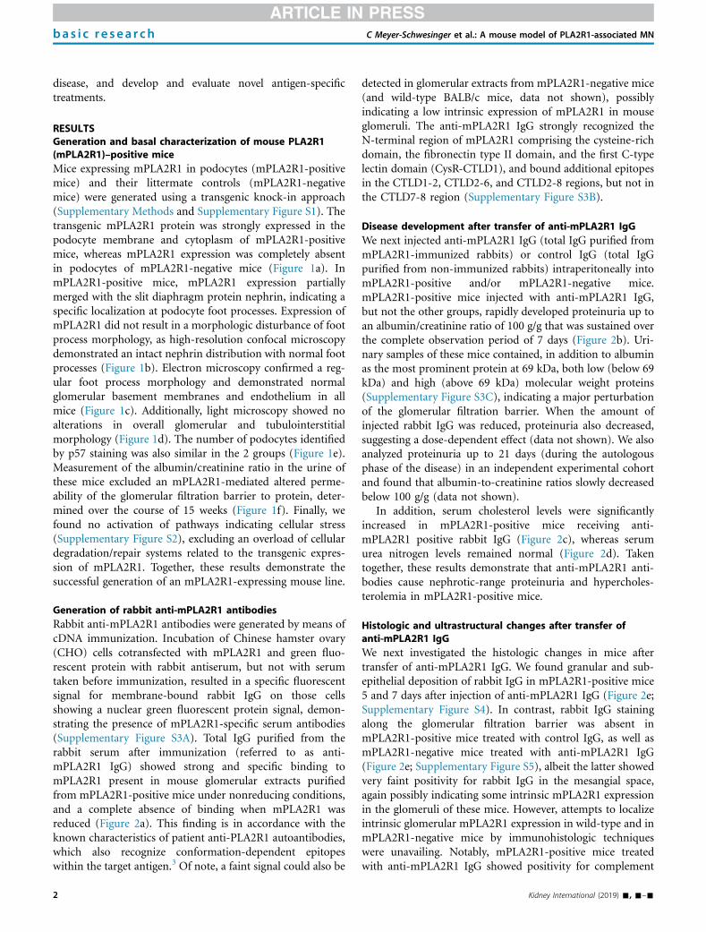
bas i c re sea r ch C Meyer-Schwesinger et al.: A mouse model of PLA2R1-associated MN
disease, and develop and evaluate novel antigen-specifictreatments.
RESULTSGeneration and basal characterization of mouse PLA2R1(mPLA2R1)–positive miceMice expressing mPLA2R1 in podocytes (mPLA2R1-positivemice) and their littermate controls (mPLA2R1-negativemice) were generated using a transgenic knock-in approach(Supplementary Methods and Supplementary Figure S1). Thetransgenic mPLA2R1 protein was strongly expressed in thepodocyte membrane and cytoplasm of mPLA2R1-positivemice, whereas mPLA2R1 expression was completely absentin podocytes of mPLA2R1-negative mice (Figure 1a). InmPLA2R1-positive mice, mPLA2R1 expression partiallymerged with the slit diaphragm protein nephrin, indicating aspecific localization at podocyte foot processes. Expression ofmPLA2R1 did not result in a morphologic disturbance of footprocess morphology, as high-resolution confocal microscopydemonstrated an intact nephrin distribution with normal footprocesses (Figure 1b). Electron microscopy confirmed a reg-ular foot process morphology and demonstrated normalglomerular basement membranes and endothelium in allmice (Figure 1c). Additionally, light microscopy showed noalterations in overall glomerular and tubulointerstitialmorphology (Figure 1d). The number of podocytes identifiedby p57 staining was also similar in the 2 groups (Figure 1e).Measurement of the albumin/creatinine ratio in the urine ofthese mice excluded an mPLA2R1-mediated altered perme-ability of the glomerular filtration barrier to protein, deter-mined over the course of 15 weeks (Figure 1f). Finally, wefound no activation of pathways indicating cellular stress(Supplementary Figure S2), excluding an overload of cellulardegradation/repair systems related to the transgenic expres-sion of mPLA2R1. Together, these results demonstrate thesuccessful generation of an mPLA2R1-expressing mouse line.
Generation of rabbit anti-mPLA2R1 antibodiesRabbit anti-mPLA2R1 antibodies were generated by means ofcDNA immunization. Incubation of Chinese hamster ovary(CHO) cells cotransfected with mPLA2R1 and green fluo-rescent protein with rabbit antiserum, but not with serumtaken before immunization, resulted in a specific fluorescentsignal for membrane-bound rabbit IgG on those cellsshowing a nuclear green fluorescent protein signal, demon-strating the presence of mPLA2R1-specific serum antibodies(Supplementary Figure S3A). Total IgG purified from therabbit serum after immunization (referred to as anti-mPLA2R1 IgG) showed strong and specific binding tomPLA2R1 present in mouse glomerular extracts purifiedfrom mPLA2R1-positive mice under nonreducing conditions,and a complete absence of binding when mPLA2R1 wasreduced (Figure 2a). This finding is in accordance with theknown characteristics of patient anti-PLA2R1 autoantibodies,which also recognize conformation-dependent epitopeswithin the target antigen.3 Of note, a faint signal could also be
2
detected in glomerular extracts from mPLA2R1-negative mice(and wild-type BALB/c mice, data not shown), possiblyindicating a low intrinsic expression of mPLA2R1 in mouseglomeruli. The anti-mPLA2R1 IgG strongly recognized theN-terminal region of mPLA2R1 comprising the cysteine-richdomain, the fibronectin type II domain, and the first C-typelectin domain (CysR-CTLD1), and bound additional epitopesin the CTLD1-2, CTLD2-6, and CTLD2-8 regions, but not inthe CTLD7-8 region (Supplementary Figure S3B).
Disease development after transfer of anti-mPLA2R1 IgGWe next injected anti-mPLA2R1 IgG (total IgG purified frommPLA2R1-immunized rabbits) or control IgG (total IgGpurified from non-immunized rabbits) intraperitoneally intomPLA2R1-positive and/or mPLA2R1-negative mice.mPLA2R1-positive mice injected with anti-mPLA2R1 IgG,but not the other groups, rapidly developed proteinuria up toan albumin/creatinine ratio of 100 g/g that was sustained overthe complete observation period of 7 days (Figure 2b). Uri-nary samples of these mice contained, in addition to albuminas the most prominent protein at 69 kDa, both low (below 69kDa) and high (above 69 kDa) molecular weight proteins(Supplementary Figure S3C), indicating a major perturbationof the glomerular filtration barrier. When the amount ofinjected rabbit IgG was reduced, proteinuria also decreased,suggesting a dose-dependent effect (data not shown). We alsoanalyzed proteinuria up to 21 days (during the autologousphase of the disease) in an independent experimental cohortand found that albumin-to-creatinine ratios slowly decreasedbelow 100 g/g (data not shown).
In addition, serum cholesterol levels were significantlyincreased in mPLA2R1-positive mice receiving anti-mPLA2R1 positive rabbit IgG (Figure 2c), whereas serumurea nitrogen levels remained normal (Figure 2d). Takentogether, these results demonstrate that anti-mPLA2R1 anti-bodies cause nephrotic-range proteinuria and hypercholes-terolemia in mPLA2R1-positive mice.
Histologic and ultrastructural changes after transfer ofanti-mPLA2R1 IgGWe next investigated the histologic changes in mice aftertransfer of anti-mPLA2R1 IgG. We found granular and sub-epithelial deposition of rabbit IgG in mPLA2R1-positive mice5 and 7 days after injection of anti-mPLA2R1 IgG (Figure 2e;Supplementary Figure S4). In contrast, rabbit IgG stainingalong the glomerular filtration barrier was absent inmPLA2R1-positive mice treated with control IgG, as well asmPLA2R1-negative mice treated with anti-mPLA2R1 IgG(Figure 2e; Supplementary Figure S5), albeit the latter showedvery faint positivity for rabbit IgG in the mesangial space,again possibly indicating some intrinsic mPLA2R1 expressionin the glomeruli of these mice. However, attempts to localizeintrinsic glomerular mPLA2R1 expression in wild-type and inmPLA2R1-negative mice by immunohistologic techniqueswere unavailing. Notably, mPLA2R1-positive mice treatedwith anti-mPLA2R1 IgG showed positivity for complement
Kidney International (2019) -, -–-
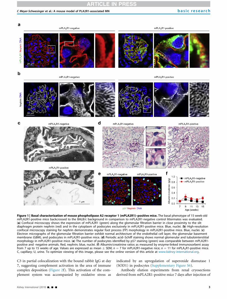
Figure 1 | Basal characterization of mouse phospholipase A2 receptor 1 (mPLA2R1)–positive mice. The basal phenotype of 15 week-oldmPLA2R1-positive mice backcrossed to the BALB/c background in comparison to mPLA2R1-negative control littermates was evaluated.(a) Confocal microscopy shows the expression of mPLA2R1 (green) along the glomerular filtration barrier in close proximity to the slitdiaphragm protein nephrin (red) and in the cytoplasm of podocytes exclusively in mPLA2R1-positive mice. Blue, nuclei. (b) High-resolutionconfocal microscopy staining for nephrin demonstrates regular foot process (FP) morphology in mPLA2R1-positive mice. Blue, nuclei. (c)Electron micrographs of the glomerular filtration barrier exhibit normal architecture of the endothelial cell layer, the glomerular basementmembrane (GBM), and podocytes in mPLA2R1-positive mice. (d) Periodic acid–Schiff staining shows normal glomerular and tubulointerstitialmorphology in mPLA2R1-positive mice. (e) The number of podocytes identified by p57 staining (green) was comparable between mPLA2R1-positive and -negative animals. Red, nephrin; blue, nuclei. (f) Albumin/creatinine ratios as measured by enzyme-linked immunosorbent assayfrom 7 up to 15 weeks of age. Values are expressed as mean � SEM; n ¼ 7 for mPLA2R1-negative nice; n ¼ 11 for mPLA2R1-positive mice.C, capillary; U, urine. To optimize viewing of this image, please see the online version of this article at www.kidney-international.org.
C Meyer-Schwesinger et al.: A mouse model of PLA2R1-associated MN bas i c re sea r ch
C3 in partial colocalization with the bound rabbit IgG at day7, suggesting complement activation in the area of immunecomplex deposition (Figure 2f). This activation of the com-plement system was accompanied by oxidative stress as
Kidney International (2019) -, -–-
indicated by an upregulation of superoxide dismutase 1(SOD1) in podocytes (Supplementary Figure S6).
Antibody elution experiments from renal cryosectionsderived from mPLA2R1-positive mice 7 days after injection of
3
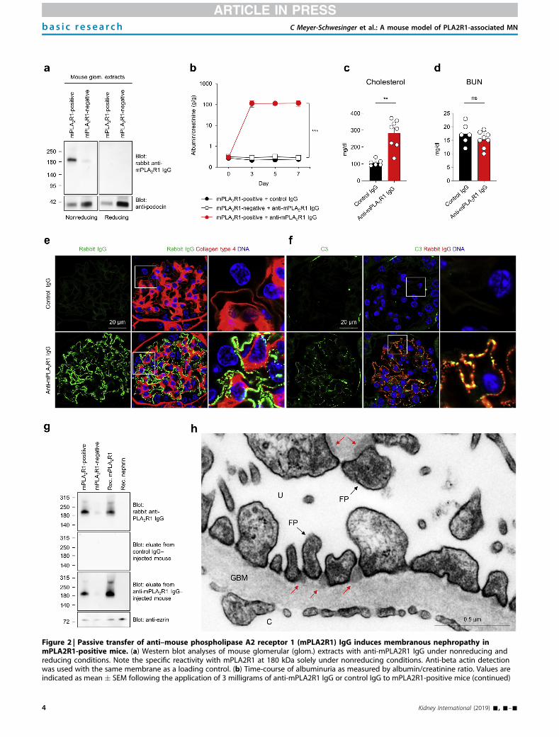
Figure 2 | Passive transfer of anti–mouse phospholipase A2 receptor 1 (mPLA2R1) IgG induces membranous nephropathy inmPLA2R1-positive mice. (a) Western blot analyses of mouse glomerular (glom.) extracts with anti-mPLA2R1 IgG under nonreducing andreducing conditions. Note the specific reactivity with mPLA2R1 at 180 kDa solely under nonreducing conditions. Anti-beta actin detectionwas used with the same membrane as a loading control. (b) Time-course of albuminuria as measured by albumin/creatinine ratio. Values areindicated as mean � SEM following the application of 3 milligrams of anti-mPLA2R1 IgG or control IgG to mPLA2R1-positive mice (continued)
bas i c re sea r ch C Meyer-Schwesinger et al.: A mouse model of PLA2R1-associated MN
4 Kidney International (2019) -, -–-
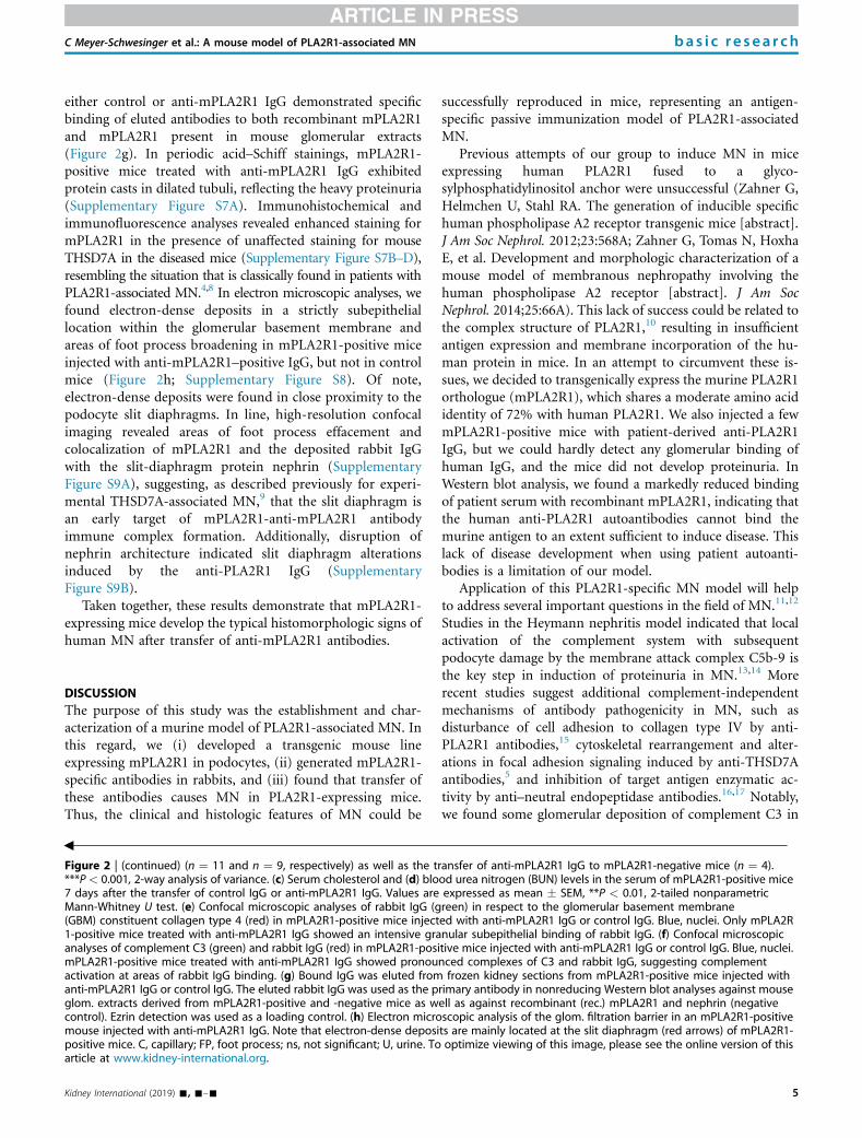
C Meyer-Schwesinger et al.: A mouse model of PLA2R1-associated MN bas i c re sea r ch
either control or anti-mPLA2R1 IgG demonstrated specificbinding of eluted antibodies to both recombinant mPLA2R1and mPLA2R1 present in mouse glomerular extracts(Figure 2g). In periodic acid–Schiff stainings, mPLA2R1-positive mice treated with anti-mPLA2R1 IgG exhibitedprotein casts in dilated tubuli, reflecting the heavy proteinuria(Supplementary Figure S7A). Immunohistochemical andimmunofluorescence analyses revealed enhanced staining formPLA2R1 in the presence of unaffected staining for mouseTHSD7A in the diseased mice (Supplementary Figure S7B–D),resembling the situation that is classically found in patients withPLA2R1-associated MN.4,8 In electron microscopic analyses, wefound electron-dense deposits in a strictly subepitheliallocation within the glomerular basement membrane andareas of foot process broadening in mPLA2R1-positive miceinjected with anti-mPLA2R1–positive IgG, but not in controlmice (Figure 2h; Supplementary Figure S8). Of note,electron-dense deposits were found in close proximity to thepodocyte slit diaphragms. In line, high-resolution confocalimaging revealed areas of foot process effacement andcolocalization of mPLA2R1 and the deposited rabbit IgGwith the slit-diaphragm protein nephrin (SupplementaryFigure S9A), suggesting, as described previously for experi-mental THSD7A-associated MN,9 that the slit diaphragm isan early target of mPLA2R1-anti-mPLA2R1 antibodyimmune complex formation. Additionally, disruption ofnephrin architecture indicated slit diaphragm alterationsinduced by the anti-PLA2R1 IgG (SupplementaryFigure S9B).
Taken together, these results demonstrate that mPLA2R1-expressing mice develop the typical histomorphologic signs ofhuman MN after transfer of anti-mPLA2R1 antibodies.
DISCUSSIONThe purpose of this study was the establishment and char-acterization of a murine model of PLA2R1-associated MN. Inthis regard, we (i) developed a transgenic mouse lineexpressing mPLA2R1 in podocytes, (ii) generated mPLA2R1-specific antibodies in rabbits, and (iii) found that transfer ofthese antibodies causes MN in PLA2R1-expressing mice.Thus, the clinical and histologic features of MN could be
=
Figure 2 | (continued) (n ¼ 11 and n ¼ 9, respectively) as well as the t***P < 0.001, 2-way analysis of variance. (c) Serum cholesterol and (d) blo7 days after the transfer of control IgG or anti-mPLA2R1 IgG. Values areMann-Whitney U test. (e) Confocal microscopic analyses of rabbit IgG (g(GBM) constituent collagen type 4 (red) in mPLA2R1-positive mice inject1-positive mice treated with anti-mPLA2R1 IgG showed an intensive graanalyses of complement C3 (green) and rabbit IgG (red) in mPLA2R1-posimPLA2R1-positive mice treated with anti-mPLA2R1 IgG showed pronouactivation at areas of rabbit IgG binding. (g) Bound IgG was eluted fromanti-mPLA2R1 IgG or control IgG. The eluted rabbit IgG was used as the pglom. extracts derived from mPLA2R1-positive and -negative mice as wcontrol). Ezrin detection was used as a loading control. (h) Electron micromouse injected with anti-mPLA2R1 IgG. Note that electron-dense deposipositive mice. C, capillary; FP, foot process; ns, not significant; U, urine. Toarticle at www.kidney-international.org.
Kidney International (2019) -, -–-
successfully reproduced in mice, representing an antigen-specific passive immunization model of PLA2R1-associatedMN.
Previous attempts of our group to induce MN in miceexpressing human PLA2R1 fused to a glyco-sylphosphatidylinositol anchor were unsuccessful (Zahner G,Helmchen U, Stahl RA. The generation of inducible specifichuman phospholipase A2 receptor transgenic mice [abstract].J Am Soc Nephrol. 2012;23:568A; Zahner G, Tomas N, HoxhaE, et al. Development and morphologic characterization of amouse model of membranous nephropathy involving thehuman phospholipase A2 receptor [abstract]. J Am SocNephrol. 2014;25:66A). This lack of success could be related tothe complex structure of PLA2R1,10 resulting in insufficientantigen expression and membrane incorporation of the hu-man protein in mice. In an attempt to circumvent these is-sues, we decided to transgenically express the murine PLA2R1orthologue (mPLA2R1), which shares a moderate amino acididentity of 72% with human PLA2R1. We also injected a fewmPLA2R1-positive mice with patient-derived anti-PLA2R1IgG, but we could hardly detect any glomerular binding ofhuman IgG, and the mice did not develop proteinuria. InWestern blot analysis, we found a markedly reduced bindingof patient serum with recombinant mPLA2R1, indicating thatthe human anti-PLA2R1 autoantibodies cannot bind themurine antigen to an extent sufficient to induce disease. Thislack of disease development when using patient autoanti-bodies is a limitation of our model.
Application of this PLA2R1-specific MN model will helpto address several important questions in the field of MN.11,12
Studies in the Heymann nephritis model indicated that localactivation of the complement system with subsequentpodocyte damage by the membrane attack complex C5b-9 isthe key step in induction of proteinuria in MN.13,14 Morerecent studies suggest additional complement-independentmechanisms of antibody pathogenicity in MN, such asdisturbance of cell adhesion to collagen type IV by anti-PLA2R1 antibodies,15 cytoskeletal rearrangement and alter-ations in focal adhesion signaling induced by anti-THSD7Aantibodies,5 and inhibition of target antigen enzymatic ac-tivity by anti–neutral endopeptidase antibodies.16,17 Notably,we found some glomerular deposition of complement C3 in
ransfer of anti-mPLA2R1 IgG to mPLA2R1-negative mice (n ¼ 4).od urea nitrogen (BUN) levels in the serum of mPLA2R1-positive miceexpressed as mean � SEM, **P < 0.01, 2-tailed nonparametricreen) in respect to the glomerular basement membraneed with anti-mPLA2R1 IgG or control IgG. Blue, nuclei. Only mPLA2Rnular subepithelial binding of rabbit IgG. (f) Confocal microscopictive mice injected with anti-mPLA2R1 IgG or control IgG. Blue, nuclei.nced complexes of C3 and rabbit IgG, suggesting complementfrozen kidney sections from mPLA2R1-positive mice injected with
rimary antibody in nonreducing Western blot analyses against mouseell as against recombinant (rec.) mPLA2R1 and nephrin (negativescopic analysis of the glom. filtration barrier in an mPLA2R1-positivets are mainly located at the slit diaphragm (red arrows) of mPLA2R1-optimize viewing of this image, please see the online version of this
5

bas i c re sea r ch C Meyer-Schwesinger et al.: A mouse model of PLA2R1-associated MN
our mouse model of PLA2R1-associated MN, in contrast toresults in the recently published mouse model of THSD7A-associated MN,6 in which C3 was barely detectable. Futurestudies are needed to investigate the pathogenic relevance ofglomerular complement activation and antibody-induced al-terations in antigen function and podocyte signaling inexperimental PLA2R1- and THSD7A-associated MN.
The identification of the antibody binding regions inPLA2R118–20 and THSD7A21 will give rise to the developmentof novel and individualized therapies. For example, antibodyclearance using epitope-specific immunoadsorption, trappingof pathogenic antibodies by antibody-binding fragments, andepitope blockage using specifically engineered antagonistsrepresent therapeutic approaches that can be evaluated fortheir in vivo efficacy in this experimental passive model ofPLA2R1-associated MN. The preclinical testing of innovativetreatments targeting the autoantibody-producing cells,22,23
however, would require an animal model involving the pro-duction of autoantibodies by members of the B-cell lineage.Our mPLA2R1-expressing mice may enable the generation ofsuch an active immunization model by means of immuni-zation with either the mPLA2R1 protein or mPLA2R1 cDNA.Interestingly, recognition of epitopes in PLA2R1 beyond thecysteine-rich domain has been found to be associated withhigher proteinuria, poor clinical outcome during follow-up,and reduced response to treatment with rituximab in pa-tients with MN.20,24,25 However, it is unclear whether thisdiversified antigen recognition results from differential auto-antibody repertoires already defined at disease initiation orfrom intramolecular epitope spreading over time. In Hey-mann nephritis, epitope spreading from the N-terminal to-ward the C-terminal region occurred when rats wereimmunized with a 236-mer N-terminal residue of megalin.26
Whether this immunologic phenomenon is also part of theimmune response against PLA2R1 could be investigated insuch an active disease model. Furthermore, how antibodybinding to single or multiple epitopes relates to diseaseseverity can be addressed using the herein described passivemodel of PLA2R1-associated MN by means of domain-specific antibodies.
In conclusion, we generated a murine PLA2R1-dependentMN mouse model that can be robustly induced by rabbitanti-PLA2R1 antibodies and replicates human PLA2R1-associated MN. This model opens new avenues to compre-hensive understanding of the molecular mechanisms thatunderlie podocyte injury in PLA2R1-associated MN and toinvestigation of innovative therapeutic strategies targetingMN-specific pathomechanisms regarding their in vivo efficacy—aprerequisite for translation to patient care in the future.
METHODSDetails on the transgenic generation of mPLA2R1-positive mice andrabbit anti-mPLA2R1 antibodies, antibody purification, conductionof animal experiments, histologic analyses, electron microscopystudies, cell culture, isolation of mouse glomeruli, antibody elution,and statistical analyses are presented in the Supplementary Methods.
6
DISCLOSURENMT holds a patent: “Methods and Kits for Monitoring MembranousNephropathy.” All the other authors declared no competing interests.
ACKNOWLEDGMENTSThis study was supported by grants from the DeutscheForschungsgemeinschaft (DFG) as part of the Collaborative ResearchCenter (CRC) 1192 (project B2 to GZ and NMT, project B3 to CMS,project B5 to FKN, and project B6 to TW). CMS holds a HeisenbergProfessorship Grant (ME 2108). NMT holds an Emmy Noether Grant(TO 1013) of the DFG. TBH was supported by the DFG (CRC1192,CRC1140, CRC992, HU 1016/8-2), by the Bundesministerium fürBildung und Forschung (BMBF) (STOP-FSGS 01GM1901C), by theEuropean Research Council (ERC grant 61689, DNCure) and by theH2020-IMI2 consortium BEAt-DKD (Innovative Medicines Initiative 2Joint Undertaking under grant agreement No. 115974).
The authors thank Ronja Steinmetz, Marlies Sachs, GudrunDubberke, Fabienne Seyfried, Sarah Hewald, and Ulrike Langbehn foroutstanding technical assistance.
SUPPLEMENTARY MATERIALSupplementary Methods and Supplementary References.Figure S1. Generation of mPLA2R1 full-length podocyte-specifictransgenic mice.Figure S2. Transgenic expression of mPLA2R1 does not result incellular/proteostatic stress.Figure S3. Generation of anti-PLA2R1 antibodies in rabbits andantibody transfer.Figure S4. mPLA2R1-positive mice treated with rabbit anti-mPLA2R1antibodies but not with control rabbit IgG deposit rabbit IgG at theglomerular filtration barrier on day 5.Figure S5. mPLA2R1-negative mice treated with rabbit anti-mPLA2R1antibodies do not deposit rabbit IgG nor complement at theglomerular filtration barrier in comparison to mPLA2R1-positive mice.Figure S6. Superoxide dismutase 1 (SOD1) is upregulated inmPLA2R1-positive mice treated with rabbit anti-mPLA2R1 antibodies.Figure S7. Light microscopic changes and mPLA2R1 staining inmPLA2R1-positive mice 7 days after the transfer of control IgG andanti-mPLA2R1 IgG.Figure S8. Electron microscopy studies.Figure S9. High-resolution confocal and electron microscopic eval-uations of immune complexes 7 days after the injection of rabbit anti-mPLA2R1 IgG to mPLA2R1-positive mice.Supplementary material is linked to the online version of the paper atwww.kidney-international.org.
REFERENCES1. Fogo AB, Lusco MA, Najafian B, et al. AJKD atlas of renal pathology:
membranous nephropathy. Am J Kidney Dis. 2015;66:e15–e17.2. Segawa Y, Hisano S, Matsushita M, et al. IgG subclasses and complement
pathway in segmental and global membranous nephropathy. PediatrNephrol. 2010;25:1091–1099.
3. Beck LH Jr, Bonegio RG, Lambeau G, et al. M-type phospholipase A2receptor as target antigen in idiopathic membranous nephropathy.N Engl J Med. 2009;361:11–21.
4. Tomas NM, Beck LH Jr, Meyer-Schwesinger C, et al. Thrombospondintype-1 domain-containing 7A in idiopathic membranous nephropathy.N Engl J Med. 2014;371:2277–2287.
5. Tomas NM, Hoxha E, Reinicke AT, et al. Autoantibodies againstthrombospondin type 1 domain-containing 7A induce membranousnephropathy. J Clin Invest. 2016;126:2519–2532.
6. Tomas NM, Meyer-Schwesinger C, von Spiegel H, et al.A heterologous model of thrombospondin type 1 domain-containing7A-associated membranous nephropathy. J Am Soc Nephrol. 2017;28:3262–3277.
Kidney International (2019) -, -–-

C Meyer-Schwesinger et al.: A mouse model of PLA2R1-associated MN bas i c re sea r ch
7. Rinschen MM, Godel M, Grahammer F, et al. A multi-layered quantitativein vivo expression atlas of the podocyte unravels kidney diseasecandidate genes. Cell Rep. 2018;23:2495–2508.
8. Hoxha E, Kneissler U, Stege G, et al. Enhanced expression of the M-typephospholipase A2 receptor in glomeruli correlates with serum receptorantibodies in primary membranous nephropathy. Kidney Int. 2012;82:797–804.
9. Herwig J, Skuza S, Sachs W, et al. Thrombospondin type 1 domain-containing 7A localizes to the slit diaphragm and stabilizes membranedynamics of fully differentiated podocytes. J Am Soc Nephrol. 2019;30:824–839.
10. Dong Y, Cao L, Tang H, et al. Structure of human M-type phospholipaseA2 receptor revealed by cryo-electron microscopy. J Mol Biol. 2017;429:3825–3835.
11. Ronco P, Debiec H. Pathophysiological advances in membranousnephropathy: time for a shift in patient’s care. Lancet. 2015;385:1983–1992.
12. Salant DJ. Unmet challenges in membranous nephropathy. Curr OpinNephrol Hypertens. 2019;28:70–76.
13. Salant DJ, Belok S, Madaio MP, et al. A new role for complement inexperimental membranous nephropathy in rats. J Clin Invest. 1980;66:1339–1350.
14. Baker PJ, Ochi RF, Schulze M, et al. Depletion of C6 preventsdevelopment of proteinuria in experimental membranous nephropathyin rats. Am J Pathol. 1989;135:185–194.
15. Skoberne A, Behnert A, Teng B, et al. Serum with phospholipase A2receptor autoantibodies interferes with podocyte adhesion to collagen.Eur J Clin Invest. 2014;44:753–765.
16. Debiec H, Guigonis V, Mougenot B, et al. Antenatal membranousglomerulonephritis due to anti-neutral endopeptidase antibodies. N EnglJ Med. 2002;346:2053–2060.
Kidney International (2019) -, -–-
17. Vivarelli M, Emma F, Pelle T, et al. Genetic homogeneity but IgG subclass-dependent clinical variability of alloimmune membranous nephropathywith anti-neutral endopeptidase antibodies. Kidney Int. 2015;87:602–609.
18. Kao L, Lam V, Waldman M, et al. Identification of the immunodominantepitope region in phospholipase A2 receptor-mediating autoantibodybinding in idiopathic membranous nephropathy. J Am Soc Nephrol.2015;26:291–301.
19. Fresquet M, Jowitt TA, Gummadova J, et al. Identification of a majorepitope recognized by PLA2R autoantibodies in primary membranousnephropathy. J Am Soc Nephrol. 2015;26:302–313.
20. Seitz-Polski B, Dolla G, Payre C, et al. Epitope spreading of autoantibodyresponse to PLA2R associates with poor prognosis in membranousnephropathy. J Am Soc Nephrol. 2016;27:1517–1533.
21. Seifert L, Hoxha E, Eichhoff AM, et al. The most N-terminal region ofTHSD7A is the predominant target for autoimmunity in THSD7A-associated membranous nephropathy. J Am Soc Nephrol. 2018;29:1536–1548.
22. Ellebrecht CT, Bhoj VG, Nace A, et al. Reengineering chimeric antigenreceptor T cells for targeted therapy of autoimmune disease. Science.2016;353:179–184.
23. Kansal R, Richardson N, Neeli I, et al. Sustained B cell depletion by CD19-targeted CAR T cells is a highly effective treatment for murine lupus. SciTransl Med. 2019;11.
24. Seitz-Polski B, Debiec H, Rousseau A, et al. Phospholipase A2 receptor 1epitope spreading at baseline predicts reduced likelihood of remission ofmembranous nephropathy. J Am Soc Nephrol. 2018;29:401–408.
25. Seitz-Polski B, Dahan K, Debiec H, et al. High-dose rituximab and earlyremission in PLA2R1-related membranous nephropathy. Clin J Am SocNephrol. 2019;14:1173–1182.
26. Shah P, Tramontano A, Makker SP. Intramolecular epitope spreading inHeymann nephritis. J Am Soc Nephrol. 2007;18:3060–3066.
7
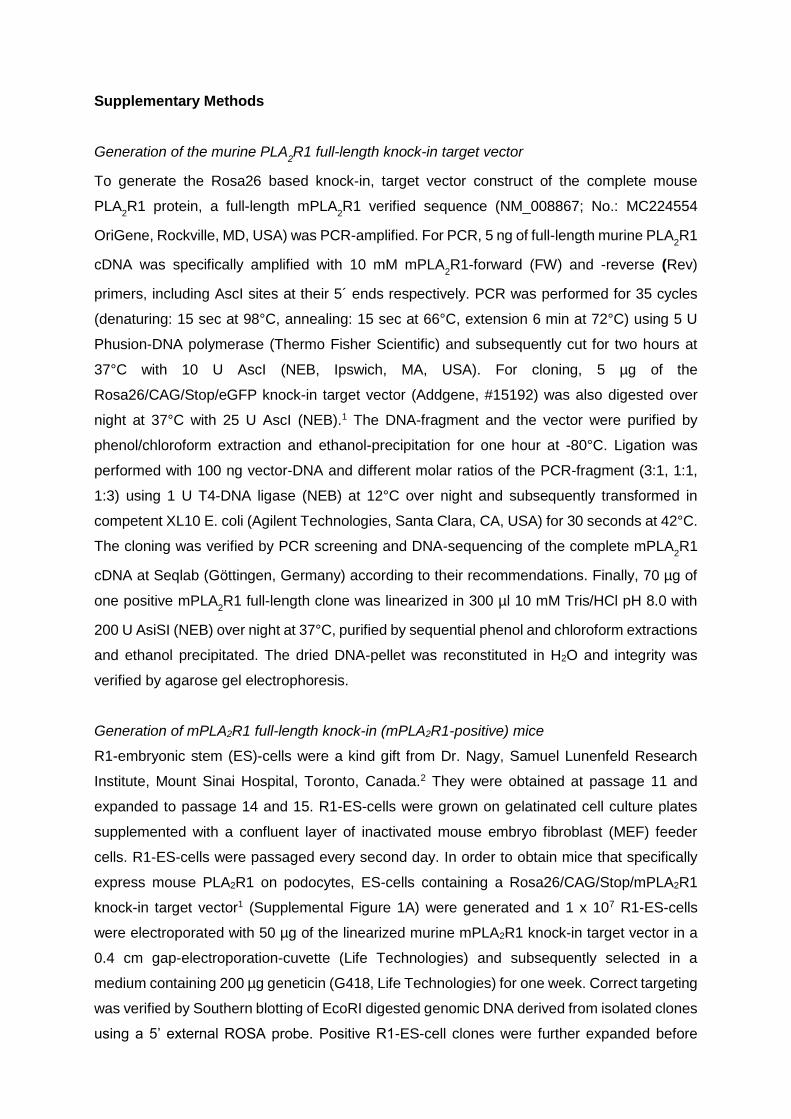
Supplementary Methods
Generation of the murine PLA2R1 full-length knock-in target vector
To generate the Rosa26 based knock-in, target vector construct of the complete mouse
PLA2R1 protein, a full-length mPLA
2R1 verified sequence (NM_008867; No.: MC224554
OriGene, Rockville, MD, USA) was PCR-amplified. For PCR, 5 ng of full-length murine PLA2R1
cDNA was specifically amplified with 10 mM mPLA2R1-forward (FW) and -reverse (Rev)
primers, including AscI sites at their 5´ ends respectively. PCR was performed for 35 cycles
(denaturing: 15 sec at 98°C, annealing: 15 sec at 66°C, extension 6 min at 72°C) using 5 U
Phusion-DNA polymerase (Thermo Fisher Scientific) and subsequently cut for two hours at
37°C with 10 U AscI (NEB, Ipswich, MA, USA). For cloning, 5 µg of the
Rosa26/CAG/Stop/eGFP knock-in target vector (Addgene, #15192) was also digested over
night at 37°C with 25 U AscI (NEB).1 The DNA-fragment and the vector were purified by
phenol/chloroform extraction and ethanol-precipitation for one hour at -80°C. Ligation was
performed with 100 ng vector-DNA and different molar ratios of the PCR-fragment (3:1, 1:1,
1:3) using 1 U T4-DNA ligase (NEB) at 12°C over night and subsequently transformed in
competent XL10 E. coli (Agilent Technologies, Santa Clara, CA, USA) for 30 seconds at 42°C.
The cloning was verified by PCR screening and DNA-sequencing of the complete mPLA2R1
cDNA at Seqlab (Göttingen, Germany) according to their recommendations. Finally, 70 µg of
one positive mPLA2R1 full-length clone was linearized in 300 µl 10 mM Tris/HCl pH 8.0 with
200 U AsiSI (NEB) over night at 37°C, purified by sequential phenol and chloroform extractions
and ethanol precipitated. The dried DNA-pellet was reconstituted in H2O and integrity was
verified by agarose gel electrophoresis.
Generation of mPLA2R1 full-length knock-in (mPLA2R1-positive) mice
R1-embryonic stem (ES)-cells were a kind gift from Dr. Nagy, Samuel Lunenfeld Research
Institute, Mount Sinai Hospital, Toronto, Canada.2 They were obtained at passage 11 and
expanded to passage 14 and 15. R1-ES-cells were grown on gelatinated cell culture plates
supplemented with a confluent layer of inactivated mouse embryo fibroblast (MEF) feeder
cells. R1-ES-cells were passaged every second day. In order to obtain mice that specifically
express mouse PLA2R1 on podocytes, ES-cells containing a Rosa26/CAG/Stop/mPLA2R1
knock-in target vector1 (Supplemental Figure 1A) were generated and 1 x 10⁷ R1-ES-cells
were electroporated with 50 µg of the linearized murine mPLA2R1 knock-in target vector in a
0.4 cm gap-electroporation-cuvette (Life Technologies) and subsequently selected in a
medium containing 200 µg geneticin (G418, Life Technologies) for one week. Correct targeting
was verified by Southern blotting of EcoRI digested genomic DNA derived from isolated clones
using a 5’ external ROSA probe. Positive R1-ES-cell clones were further expanded before

injection into blastocysts. For blastocyst injection, 15 trypsinized R1-ES-cells with clearly
visible, small, round nuclei without vacuoles were collected and injected into one blastocyst.
Eight of these blastocysts were implanted into the uterus of a female mouse mated with
vasectomized males at day 2.5 post coitum. The resulting chimeric males were mated with
wild-type BALB/c females. Littermates of the F1 generation were genotyped by PCR to obtain
offsprings that pass on the mPLA2R1 knock-in. These mice carried the transgenic information
of mPLA2R1 under the control of a floxed stop-cassette to prevent transactivation by the CAG-
enhancer. These animals are referred to as mPLA2R1-negative precursor mice and were
further used for breeding with NPHS2-Cre mice3 to obtain a podocyte-specific mPLA2R1
expression. Due to the podocyte-specific action of the NPHS2 (podocin) promoter-triggered
Cre-recombinase, the stop cassette was removed and mPLA2R1 expression on glomerular
podocytes was initiated, these mice are herein further referred to as mPLA2R1-positive mice.
mPLA2R-negative mice were backcrossed into the BALB/c background for 9 generations.
The last backcross was performed with heterozygous Podocine Cre BALB/c mice to obtain
mPLA2R-positive and-negative mice. Almost all mPLA2R1-positive animals express mPLA2R1
in podocytes in more than 90% of Glomeruli. The different genotypes were verified by three
PCRs (Supplemental Figure 1B) either showing the presence of Cre (PodoCre) and the
mPLA2R1 transgene and the allele-frequency of the transgene (R26 wt).
Generation of rabbit antibodies against mPLA2R1
Two rabbit antisera recognizing mPLA2R1 were produced by cDNA immunization essentially
as described previously.4-6 The expression construct encoding full-length mPLA2R1 (OriGene)
was conjugated to 1 µm gold particles (Bio-Rad Laboratories) and ballistically injected into two
rabbits at the antibody core unit of the University Medical Center Hamburg-Eppendorf. The
rabbits received four immunizations in 3-6 week intervals, each with 12 shots of plasmid-
conjugated gold particles (1 µg DNA/mg gold per shot). Serum was obtained 3 weeks after the
last DNA immunization and antiserum-specificity for native mPLA2R1 was verified using
immunofluorescence staining of CHO cells transiently transfected with mPLA2R1 for 24 hours
with serial dilutions of the antisera and using Western blotting on mouse glomerular extracts.
Bound antibodies were detected with phycoerythrin- or HRP-conjugated anti-rabbit IgG
antibodies (Dianova), respectively.
Animal Experiments
Fourteen week-old male mPLA2R1-positive or -negative mice were injected intraperitoneally
with 3 mg of purified rabbit IgG. To determine albuminuria, urine was collected using metabolic
cages. Urine albumin content was quantified using a commercially available ELISA system
(Bethyl) according to the manufacturer’s instructions. Albumin values were standardized

against urinary creatinine values (as determined according to Jaffé) of the same sample. BUN
and cholesterol levels were measured by standard procedures at the Department of Clinical
Chemistry at the University Medical Center Hamburg-Eppendorf.
Mice were euthanized after seven days for the final removal of blood, urine and kidneys. All
animals had free access to tap water and standard animal chow and were bred in the animal
facility of the University Medical Center Hamburg-Eppendorf according to national and
institutional animal care and ethical guidelines. Animal experiments were approved by the
veterinarian agency of Hamburg and the local animal care committee.
Immunofluorescence analyses
For immunolocalization of nephrin (guinea pig pAB, 1:200; Acris), murine PLA2R1 (rabbit pAB
K1115, 1:600 and rat mAB B16/2 1:50, both self-made), rabbit IgG (AF488-rbIgG H+L, 1:200;
Jackson ImmunoResearch Laboratories), C3 (FITC-goat pAB, 1:50; Cappel), THSD7A (goat
pAB, 1:200; Santa Cruz); pIRE1α (rabbit pAB, 1:100; Cell Signaling, LMP7 (rabbit pAB, 1:300
Abcam), Lamp2 (rabbit pAB, 1:500; Sigma), or LC3B (rabbit pAB, 1:50; Cell Signaling),
collagen-type 4 (goat pAB, 1:400; Southern Biotechnologies), laminin (rabbit pAB, 1:1000;
Sigma), p57 (rabbit pAB, 1:500; Santa Cruz) 3 µM paraffin sections of experimental mouse
kidneys were deparaffinized and rehydrated to water. Antigen retrieval was obtained by boiling
in citrate buffer at pH 6.1 (30-60 minutes at constant 98°C), or by digestion with protease XXIV
(5 µg/ml; Sigma-Aldrich) for 15 minutes at 37°C. Unspecific binding was blocked with 5% horse
serum (Vector Laboratories) with 0.05% Triton X-100 (Sigma-Aldrich) in PBS for 30 minutes
at room temperature before incubation at 4°C overnight with primary antibodies in blocking
buffer. Staining was visualized with fluorochrome-conjugated secondary antibodies (all affinity
purified from Jackson ImmunoResearch Laboratories; 1:400) for 30 minutes at room
temperature in 5% horse serum with 0.05% Triton X-100. To control for cross-reactivity of the
anti-rabbit secondary antibodies with the injected and bound rabbit anti-PLA2R1 IgG, intrinsic
IgG was destroyed by excessive boiling. Furthermore, TrueBlot anti-rabbit antibodies (1:200;
eBioscience) were used, which only bind to native rabbit IgG. Additionally, every individual
mouse was controlled by omitting the primary antibody. Nuclei were counterstained with
Hoechst (1:1000; Molecular Probes). Stainings were evaluated with a confocal LSM800 with
airyscan using the ZEN blue software (all Zeiss), or by an Axioskop using the Axiovision
software for light microscopy (all Zeiss). Fiji software was used for quantification of mPLA2R1
staining intensity in relation to glomerular capillary tuft area. For immunohistochemistry to
PLA2R1, THSD7A and rabbit IgG, stainings were visualized using the AP-Polymer kit
(Zytochem) following the manufacturer’s instructions. Neufuchsin (Sigma) was used for color
development, nuclei were counterstained with hemalaun (Sigma).
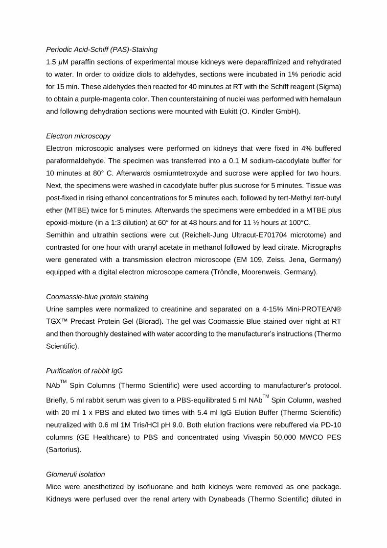
Periodic Acid-Schiff (PAS)-Staining
1.5 µM paraffin sections of experimental mouse kidneys were deparaffinized and rehydrated
to water. In order to oxidize diols to aldehydes, sections were incubated in 1% periodic acid
for 15 min. These aldehydes then reacted for 40 minutes at RT with the Schiff reagent (Sigma)
to obtain a purple-magenta color. Then counterstaining of nuclei was performed with hemalaun
and following dehydration sections were mounted with Eukitt (O. Kindler GmbH).
Electron microscopy
Electron microscopic analyses were performed on kidneys that were fixed in 4% buffered
paraformaldehyde. The specimen was transferred into a 0.1 M sodium-cacodylate buffer for
10 minutes at 80° C. Afterwards osmiumtetroxyde and sucrose were applied for two hours.
Next, the specimens were washed in cacodylate buffer plus sucrose for 5 minutes. Tissue was
post-fixed in rising ethanol concentrations for 5 minutes each, followed by tert-Methyl tert-butyl
ether (MTBE) twice for 5 minutes. Afterwards the specimens were embedded in a MTBE plus
epoxid-mixture (in a 1:3 dilution) at 60° for at 48 hours and for 11 ½ hours at 100°C.
Semithin and ultrathin sections were cut (Reichelt-Jung Ultracut-E701704 microtome) and
contrasted for one hour with uranyl acetate in methanol followed by lead citrate. Micrographs
were generated with a transmission electron microscope (EM 109, Zeiss, Jena, Germany)
equipped with a digital electron microscope camera (Tröndle, Moorenweis, Germany).
Coomassie-blue protein staining
Urine samples were normalized to creatinine and separated on a 4-15% Mini-PROTEAN®
TGX™ Precast Protein Gel (Biorad). The gel was Coomassie Blue stained over night at RT
and then thoroughly destained with water according to the manufacturer’s instructions (Thermo
Scientific).
Purification of rabbit IgG
NAbTM
Spin Columns (Thermo Scientific) were used according to manufacturer’s protocol.
Briefly, 5 ml rabbit serum was given to a PBS-equilibrated 5 ml NAbTM
Spin Column, washed
with 20 ml 1 x PBS and eluted two times with 5.4 ml IgG Elution Buffer (Thermo Scientific)
neutralized with 0.6 ml 1M Tris/HCl pH 9.0. Both elution fractions were rebuffered via PD-10
columns (GE Healthcare) to PBS and concentrated using Vivaspin 50,000 MWCO PES
(Sartorius).
Glomeruli isolation
Mice were anesthetized by isofluorane and both kidneys were removed as one package.
Kidneys were perfused over the renal artery with Dynabeads (Thermo Scientific) diluted in

HBSS. Then kidneys were unpacked, minced into small pieces, digested in collagenase
solution (1.2 mg/ ml collagenase 1A, 100U/ ml DNAseI in HBSS) at 37°C for 15 min with gentle
agitation. Collagenase-digested tissue was gently pressed through a 100 µm cell strainer
followed by a 10 ml wash with HBSS. This solution was passed through a new 100 µm cell
strainer followed by a 10 ml wash with HBSS. The glomerular suspension was then centrifuged
at 200x g for 5 min. The glomerular pellet was resuspended in 6 ml HBSS and transferred into
four 2 ml cups. Glomeruli containing Dynabeads were collected by a magnetic particle
concentrator and washed for three times with HBSS 0.5% BSA, combined with HBSS,
glomeruli were counted and centrifuged at 4000x g for 5 min. The glomerular pellet was snap
frozen and kept at -80°C. The entire procedure was performed at 4°C with the exception of the
collagenase digestion at 37°C. For the generation of mouse glomerular extracts, frozen pellets
were homogenized in 100 µl of P1 (100 mM Tris/HCl pH 8.0, 1 mM MgCl2) under liquid
nitrogen. Then, the powder was immediately lysed in 100 µl P2 (50 mM Tris/Cl pH 7.4, 150
mM NaCl, 0.1 SDS, 1% NP-40, 0.5% Sodium-Deoxycholate containing protease inhibitor
cocktail “Plus” (Roth, Karlsruhe, Germany) for 1 h at 4°C. The insoluble debris was pelleted at
20,000 x g for 10 minutes at 4°C and the supernatant was incubated over night at 4°C with 50
µl ProteinG resin (Thermo Fisher Scientific) to remove any mouse IgG. Finally, the ProteinG
resin was pelleted for 5 minutes at 20,000x g and 4°C before the supernatant was further used
for immunoblot analysis
HEK cell transfection
Full-length mPLA2R1 and all other mPLA2R1 sub-fragments used were cloned into the
pcDNA3.1 vector (Invitrogen) containing a C-terminal hexahistidine tag and HEK293T cells
were PEI-transfected using this construct. Three days post-transfection, cells were lysed in a
native lysis buffer (50 mM Tris/HCl pH 7.4, 150 mM NaCl, 1 mM EDTA, 1% Triton X100) to
obtain recombinant complete mPLA2R1.
Antibody Elution
100 to 120 (10 µM thick) cryo-sections from mouse renal tissue were thawed in 1 ml PBS and
centrifuged at low speed for 1 min. After washing three times in 1 ml PBS, the pellets were
resuspended in 150 µl of 25 mM citrate buffer pH 3.2 and incubated on ice for 20 min with
occasional shaking. Then, the samples were centrifuged for 5 min at 14,000 rpm and the
supernatants were mixed with an equal volume of 1 M Tris/HCl, pH 8.0. In a second elution
step, the pellets were resuspended in 25 mM citrate buffer, pH 2.5, incubated on ice, and
centrifuged again. Finally, the supernatants were added to the first elution and diluted in 2 ml
of 5% dry-milk in PBS-0.05% Tween 20. These samples were used as primary antibody source
in Western blot analysis.

Statistical analyses
Data are expressed as mean ± standard error of the mean (SEM). For statistical analyses we
performed the non-parametric Mann-Whitney U test to enable robust conclusions on effect
significance in case of departures from normality associated with small sample sizes or the
two-tailed Student’s t-test in parametric data sets. For comparison of more than two conditions,
non-parametric one-way ANOVA was performed, for paired observations the two-way ANOVA
was used.
Immunoblot analyses
Similar amounts of total protein obtained from mouse glomerular extracts were loaded on a 4-
15% Mini-PROTEAN® TGX™ Precast Protein Gel (Biorad ) and separated under non-
reducing conditions. Proteins were transferred to polyvinylidene difluoride membranes (Bio-
Rad) under semi-dry conditions using Trans-blot Turbo (Bio-Rad) at 25 V constant for 25 min.
After protein transfer, membranes were blocked for one hour at 4°C in 5% dry-milk, PBS-0.05%
Tween 20 and incubated overnight with rabbit sera at a 1:10.000 dilution. HRP-conjugated
goat anti-rabbit IgG 1:20.000 (Sigma-Aldrich) was used as the secondary antibody. Signals
were detected using a chemiluminescent substrate (ECL Clarity, Biorad) with a Luminescent
Detection Imager 600 (GE Healthcare).
Supplementary References
1. Thai, TH, Calado, DP, Casola, S, et al. Regulation of the germinal center response by microRNA-
155. Science. 2007;316:604-608.
2. Nagy, A, Rossant, J, Nagy, R, et al. Derivation of completely cell culture-derived mice from early-
passage embryonic stem cells. Proc Natl Acad Sci U S A. 1993;90:8424-8428.
3. Moeller, MJ, Sanden, SK, Soofi, A, et al. Podocyte-specific expression of cre recombinase in
transgenic mice. Genesis. 2003;35:39-42.
4. Adriouch, S, Dubberke, G, Diessenbacher, P, et al. Probing the expression and function of the
P2X7 purinoceptor with antibodies raised by genetic immunization. Cell Immunol.
2005;236:72-77.
5. Tomas, NM, Hoxha, E, Reinicke, AT, et al. Autoantibodies against thrombospondin type 1 domain-
containing 7A induce membranous nephropathy. J Clin Invest. 2016;126:2519-2532.
6. Moller, S, Jung, C, Adriouch, S, et al. Monitoring the expression of purinoceptors and nucleotide-
metabolizing ecto-enzymes with antibodies directed against proteins in native conformation.
Purinergic Signal. 2007;3:359-366.

Supplementary Figures
Supplementary Figure S1: Generation of mPLA2R1 full-length podocyte specific transgenic mice.
(a) Schematic depiction of the Rosa 26-based mPLA2R1 full-length specific knock-in target construct
including the transactivation by the CAG enhancer. After breeding with NPHS2 Cre deleter mice,
mPLA2R1-positive mice are obtained which express mPLA2R1 specifically in podocytes. (b) PCR-based
genotyping demonstrating the identification of the Cre-recombinase (268 bp), the mPLA2R1 transgene
(620 bp), as well as the determination of the mPLA2R1 allele frequency by R26 wt (233 bp).

Supplementary Figure S2: Transgenic expression of mPLA2R1 does not result in
cellular/proteostatic stress. Representative confocal images to marker proteins such as superoxide
dismutase (SOD2, oxidative stress), pIRE1a (endoplasmic reticulum stress), LMP7
(immunoproteasome subunit) and polyubiquitinated proteins (both markers for proteostatic stress in the
proteasomal system), and autophagosomal LC3B and lysosomal Lamp2 (both indicators of an
autophagosomal-lysosomal reaction) in conjunction with nephrin (red). Blue: Nuclei. The stress levels
of mPLA2R1-positive and -negative mice are similar.

Supplementary Figure S3: Generation of anti-PLA2R1 antibodies in rabbits and antibody
transfer. (a) CHO cells were co-transfected with nuclear GFP and mPLA2R1 and stained with either
rabbit preimmune or rabbit anti-mPLA2R1 immune serum at a dilution of 1:400. Rabbit IgG bound to
mPLA2R1 was detected with an AF586 anti-rabbit antibody (red). Note red fluorescence in CHO-cells
stained with rabbit anti-mPLA2R1 antibody positive serum but not with rabbit preimmune serum. (b)
Western blot analyses of lysates from HEK cells that were transiently transfected with flag-tagged full-
length mPLA2R1 and the his-tagged mPLA2R1 mutants CTLD2-8, CTLD2-6, CysR-CTLD1, CTLD7-8
and CTLD1-2 with rabbit anti-mPLA2R1 IgG diluted 1:10,000 under non-reducing conditions. Western
blot of the same lysates with an anti-his antibody under non-reducing conditions demonstrates
successful recombinant protein expression. Asterisks indicate specific bands. (c) Coomassie-blue
staining of urinary samples from a mPLA2R1-positive mouse injected with control IgG (left) and a
mPLA2R1-positive mouse injected with anti-mPLA2R1 IgG (right).
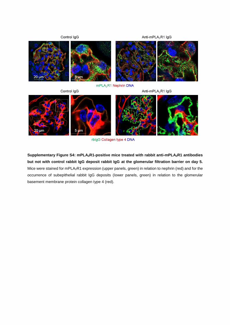
Supplementary Figure S4: mPLA2R1-positive mice treated with rabbit anti-mPLA2R1 antibodies
but not with control rabbit IgG deposit rabbit IgG at the glomerular filtration barrier on day 5.
Mice were stained for mPLA2R1 expression (upper panels, green) in relation to nephrin (red) and for the
occurrence of subepithelial rabbit IgG deposits (lower panels, green) in relation to the glomerular
basement membrane protein collagen type 4 (red).
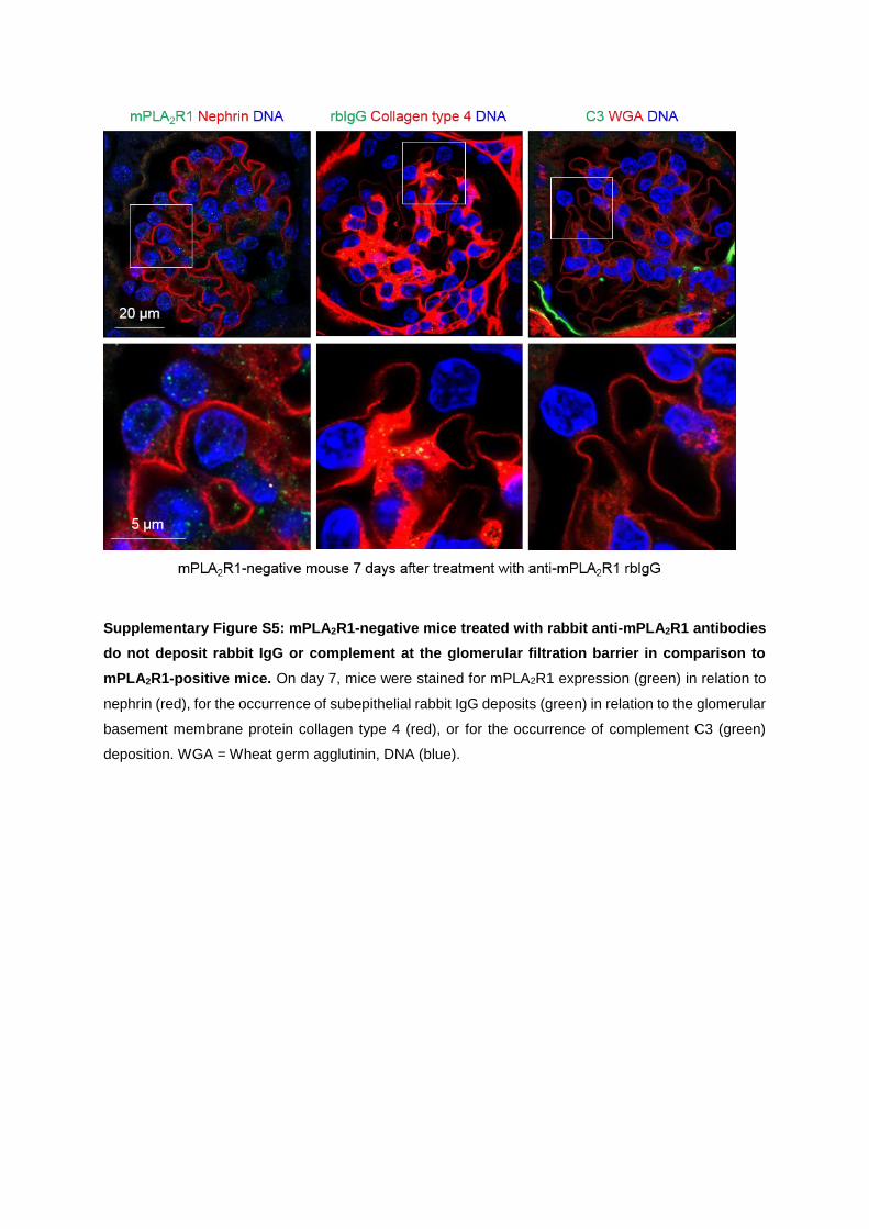
Supplementary Figure S5: mPLA2R1-negative mice treated with rabbit anti-mPLA2R1 antibodies
do not deposit rabbit IgG or complement at the glomerular filtration barrier in comparison to
mPLA2R1-positive mice. On day 7, mice were stained for mPLA2R1 expression (green) in relation to
nephrin (red), for the occurrence of subepithelial rabbit IgG deposits (green) in relation to the glomerular
basement membrane protein collagen type 4 (red), or for the occurrence of complement C3 (green)
deposition. WGA = Wheat germ agglutinin, DNA (blue).
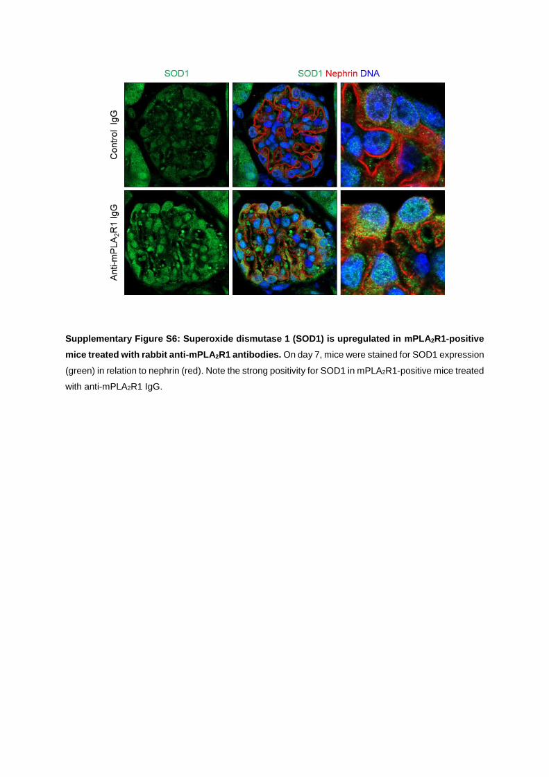
Supplementary Figure S6: Superoxide dismutase 1 (SOD1) is upregulated in mPLA2R1-positive
mice treated with rabbit anti-mPLA2R1 antibodies. On day 7, mice were stained for SOD1 expression
(green) in relation to nephrin (red). Note the strong positivity for SOD1 in mPLA2R1-positive mice treated
with anti-mPLA2R1 IgG.

Supplementary Figure S7: Light microscopic changes and mPLA2R1 staining in mPLA2R1-
positive mice seven days after transfer of control IgG and anti-mPLA2R1 IgG. (a) Evaluation of
renal cortex tissue integrity using PAS-staining. No major glomerular alterations (magnification) were
observed in mPLA2R1-positive mice treated with anti-mPLA2R1 IgG, while signs of heavy proteinuria,
such as protein casts (asterisks) in dilated tubuli, were visible under these conditions. (b)
Immunohistochemical staining for mPLA2R1 and mTHSD7A. Note the typical pathological picture of a
PLA2R1-associated MN with enhanced and granular PLA2R1 staining and unaltered linear THSD7A
expression. (c) Representative immunofluorescence staining for mPLA2R1 in mPLA2R1-positive mice
after transfer of control IgG or anti-mPLA2R1 IgG, demonstrating enhanced staining for mPLA2R1 in the
diseased mouse. (d) Quantification of mPLA2R1 staining from mPLA2R1-positive mice injected with
control IgG (n=6 mice analyzed) or anti-mPLA2R1 IgG (n=8 mice analyzed). MFI, mean fluorescence
intensity. ***P<0.001, two-tailed nonparametric Mann-Whitney U test.
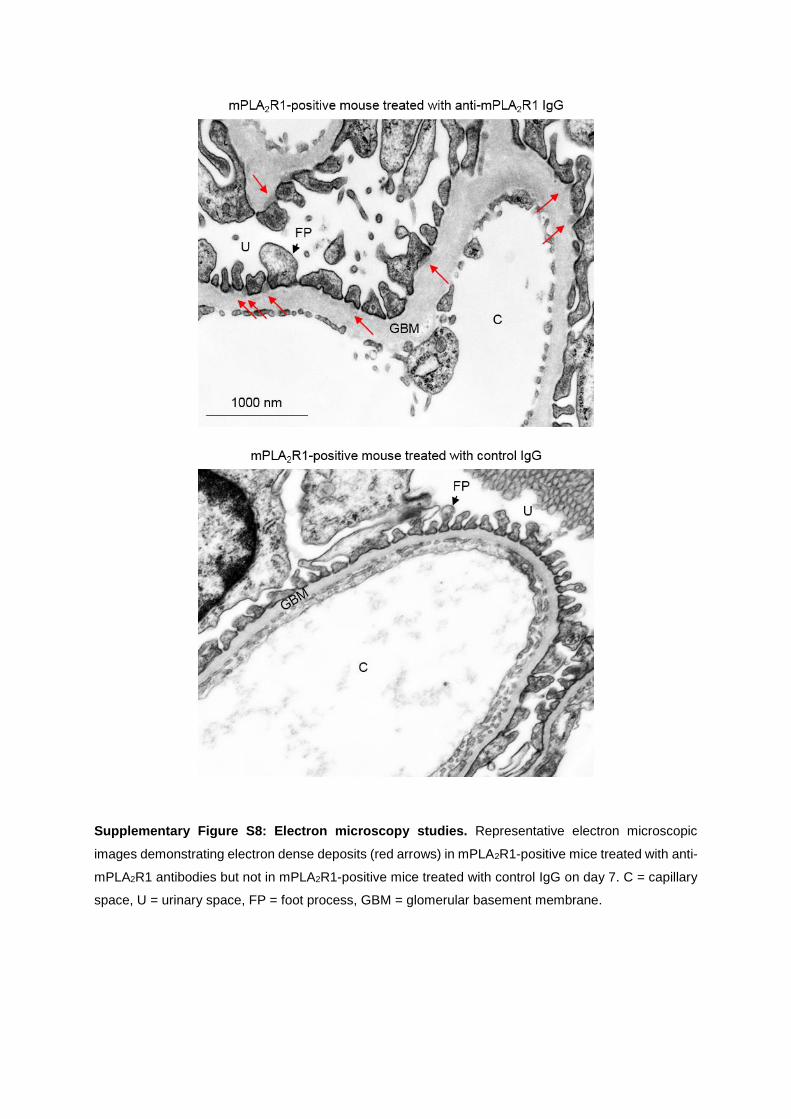
Supplementary Figure S8: Electron microscopy studies. Representative electron microscopic
images demonstrating electron dense deposits (red arrows) in mPLA2R1-positive mice treated with anti-
mPLA2R1 antibodies but not in mPLA2R1-positive mice treated with control IgG on day 7. C = capillary
space, U = urinary space, FP = foot process, GBM = glomerular basement membrane.

Supplementary Figure S9: High resolution confocal and electron microscopic evaluations of
immune complexes seven days after injection of rabbit anti-mPLA2R1 IgG to mPLA2R1-positive
mice. (a) High-resolution confocal analysis of rabbit IgG (red) and mPLA2R1 (green) in relation to the
slit diaphragm protein nephrin (light blue). Note in the magnified transversal plane (lower panels) of the
glomerular filtration barrier the co-localization of deposited rabbit IgG and mPLA2R1 with nephrin
(arrows). (b) High-resolution confocal microscopy for the slit diaphragm protein nephrin (white) revealed
focal foot process effacement in mPLA2R1-positive mice treated with anti-mPLA2R1 IgG on day seven,
but not in mice trated with control IgG. Blue, nuclei.