characterization of anhydrous silanization and antibody immobilization on silicon dioxide surface
DESCRIPTION
doi:10.1109/ISSMD.2004.1689550TRANSCRIPT

IEEE/EMBS International Summer School on Medical Devices and Biosensors (ISSS-MD)
Characterization of Anhydrous Silanization and Antibody Immobilization onSilicon dioxide Surface
Manoj Joshil, M.Goyal', R.Pinto2, S. Mukherjil*
'School of Biosciences and Bioengineering, Indian Institute of Technology Bombay, Mumbai, India.2 Department of Electrical Engineering, Indian Institute ofTechnology Bombay, Mumbai, India.
Abstract- Formation of uniform and cluster free silane The thicker silane layer on the sensor surface alters themonolayer is one of the fundamental prerequisite for affinity mechanical properties of the biosensor. It also inducescantflever based biosensors. We report anhydrous silanization stresses on the sensor surface which may leads to difficultiesprotocol for uniform silane monolayer on silicon dioxide (SiO2) s n the s ensor suracewicayi edo difficulTisurface using [3-(2-aminoethyl) aminopropyll-trimethoxysiane(APTES) and characterized by AFM, spectroscopic anticipate these problems silane monolayer with controlledellipsometry and FTIR. Silanization coverage is controled by thickness on biosensor is essential.availability of surface water. The roughness of the resulting In the conventional protocol of aqueous phasesilanized surface following our protocol is in the range of SiO2 silanization, the aminosilanes undergo hydrolysis andsurface roughness. Silanized SiO2surface is used to immobilize polymerization in the bulk phase before depositing andhuman immunoglobulin (HlgG) on it. FITC tagged goat forming bonds with the silicon dioxide surface [1]. However,antihuman IgG is allowed to react with HIgG. The immobilized this results in the formation of polysilane networks prior tosurface is further characterized using Fluorescent spectroscopy deposition. Thus polymerization of multifunctional silaneand Fluorescent Microscope. Characterization results obtained molecules is observed both parallel and perpendicular to thefrom anhydrous silanization protocol are compared with the solfces leadtoforatoofiane me cule teconventional aqueous silanization protocol. surface. This leads to formation of silane molecule clusters
and non-uniform silane layer thickness on sensor surface.Keywords - affinity cantilever, silanization, APTES, FITC In anhydrous silanization, trace water molecules on the
support surface are utilized to hydrolyze the silaneI. INTRODUCTION molecules to form siloxane bonds. The degree of
silanization depends on the concentration of waterChemical modification of SiO2 surface by molecules present on the surface when the aminosilane is
organofunctional silane is a well-known technique for applied. Reduced silane coverage can be obtained by heatingbiosensor applications. Most of the approaches however the surface at high temperature (to remove adsorbed waterproduce non-uniform silane thickness and clustering of molecules) prior to application ofthe aminosilane [2].silane molecules on the sensor surface which reduce the The main goal of this paper is to demonstrate thesensitivity of affinity cantilever. The sensitivity of the qualitative improvement of silanized surface in anhydrousaffinity cantilever biosensor depends on maximum silanization, which is a prerequisite for improving thedeflection of cantilever due to surface force generated by sensitivity of affinity based cantilever biosensors. Theimmobilized antibody on its surface as shown in Fig.l. surface morphology of the silanized surface is studied withMaximally reproducible surface force can be achieved only AFM and thickness measurement using spectroscopicif antibodies immobilized on cantilever are in uniform single ellipsometer. The orientation of silane molecule islayer. investigated using FTIR. It is shown that anhydrous
Cantilever Antiboies Surface Force silanization technique produce dense and uniform antibody)!y Xs.3N . <9ZY a <; t-> immobilization. Characterization results obtained from
anhydrous silanization protocol are compared with theconventional aqueous silanization protocol.
SIlIcon II. METHODOLOGY
Fig. I Cantilever surface with uniform antibody immobilization to develop Silicon wafer of resistivity 4-6 Q.cm and orientationsurface force <100> was selected and cleaned by RCA cleaning followed
*Co---------- author- ;9122 - -----by dry thermal oxidation at 1100°C for 1 hour to get oxide*Corresponding author- Tel. +91 22 2576-7767 tikeso 0 m PE a bandfo imEmail- mukherji(icc.iitb.ac.in thickness of 100 m. APTES was obtamed from Sigma-----__-- --Aldrich USA and HIgG/ FITC tagged goat antihuman IgGFinancial support from the Government of India under the National from Bangalore Genei. To generate silanol sites on oxidizedProgramme on Smart Materials is gratefully acknowledged. silicon, samples were dipped in sulphochromic solution
0-7803-8612-4/04/$20.00 © 2004 IEEE 7
Authorized licensed use limited to: IEEE Xplore. Downloaded on October 22, 2008 at 08:17 from IEEE Xplore. Restrictions apply.
(Iml DI water with 500ug K2Cr2O7 added with 20ml H2SO4) silane layer at different silane concentrations was measuredfor 10 minutes followed by DI water rinse. This removes with spectroscopic ellipsometer and the orientation of silaneany native carbon impurities and creates OH groups on SiO2 molecule on the SiO2 surface investigated with FTIR. Thesurface by opening siloxane bonds. Surface adsorbed water antibody immobilization on silanized SiO2 surface waswas removed by heating the sample at 2000C (1400C in studied with fluorescence spectroscopy and fluorescentaqueous protocol) for two hours under vacuum. microscope.
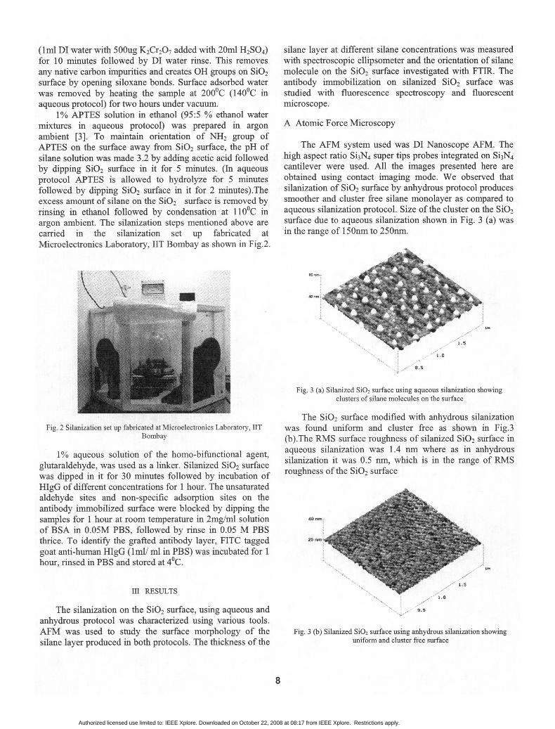
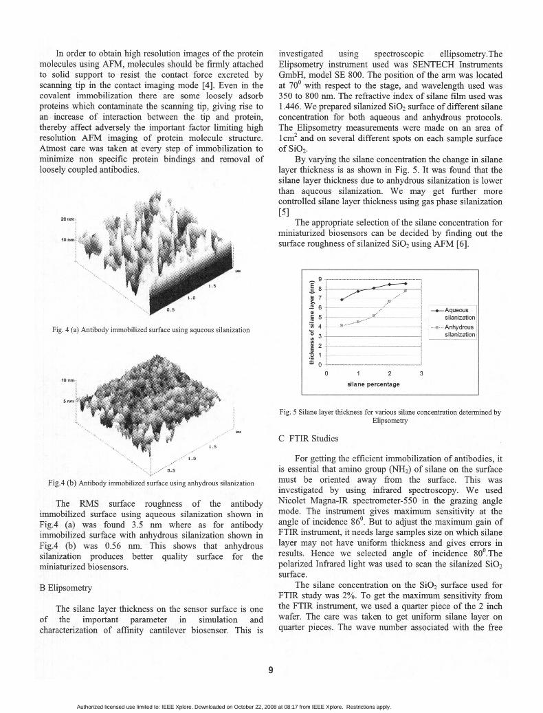
1% APTES solution in ethanol (95:5 % ethanol watermixtures in aqueous protocol) was prepared in argon A Atomic Force Microscopyambient [3]. To maintain orientation of NH2 group ofAPTES on the surface away from SiO2 surface, the pH of The AFM system used was DI Nanoscope AFM. Thesilane solution was made 3.2 by adding acetic acid followed high aspect ratio Si3N4 super tips probes integrated on Si3N4by dipping SiO2 surface in it for 5 minutes. (In aqueous cantilever were used. All the images presented here areprotocol APTES is allowed to hydrolyze for 5 minutes obtained using contact imaging mode. We observed thatfollowed by dipping SiO2 surface in it for 2 minutes).The silanization of SiO2 surface by anhydrous protocol producesexcess amount of silane on the SiO2 surface is removed by smoother and cluster free silane monolayer as compared torinsing in ethanol followed by condensation at 110°C in aqueous silanization protocol. Size of the cluster on theSiO2argon ambient. The silanization steps mentioned above are surface due to aqueous silanization shown in Fig. 3 (a) wascarried in the silanization set up fabricated at in the range of 150nm to 250nm.Microelectronics Laboratory, IIT Bombay as shown in Fig.2.
E 0+~~~~~~~~~~~~~~~~~~~~~~~~~~~~~~~~~~~.Fig. 3 (a) Silanized SiO2 surface using aqueous silanization showing
clusters of silane molecules on the surface
The SiO2 surface modified with anhydrous silanizationFig. 2 Silanization set up fabricated at Microelectronics Laboratory, IIT was found uniform and cluster free as shown in Fig.3
Bombay (b).The RMS surface roughness of silanized SiO2 surface in
1% aqueous solution of the homo-bifunctional agent, aqueous silanization was 1.4 nm where as in anhydrousglutaraldehyde, was used as a linker. Silanized SiO2 surface silanization it was 0.5 nm, which is in the range of RMSwas dipped in it for 30 minutes followed by incubation of roughness of the SiO2 surfaceHIgG of different concentrations for I hour. The unsaturatedaldehyde sites and non-specific adsorption sites on theantibody immobilized surface were blocked by dipping thesamples for 1 hour at room temperature in 2mg/ml solution 40
of BSA in 0.05M PBS, followed by rinse in 0.05 M PBSthrice. To identify the grafted antibody layer, FITC taggedgoat anti-human HIgG (lml/ ml in PBS) was incubated for 1hour, rinsed in PBS and stored at 4°C.
M RESULTS
The silanization on the SiO2 surface, using aqueous and y.sanhydrous protocol was characterized using various tools.AFM was used to study the surface morphology of the Fig. 3 (b) Silanized SiO2 surface using anhydrous silanization showing
silane layer produced in both protocols. The thickness of the uniform and cluster free surface
8
Authorized licensed use limited to: IEEE Xplore. Downloaded on October 22, 2008 at 08:17 from IEEE Xplore. Restrictions apply.
In order to obtain high resolution images of the protein investigated using spectroscopic ellipsometry.Themolecules using AFM, molecules should be firmly attached Elipsometry instrument used was SENTECH Instrumentsto solid support to resist the contact force excreted by GmbH, model SE 800. The position of the arm was locatedscanning tip in the contact imaging mode [4]. Even in the at 700 with respect to the stage, and wavelength used wascovalent immobilization there are some loosely adsorb 350 to 800 nm. The refractive index of silane film used wasproteins which contaminate the scanning tip, giving rise to 1.446. We prepared silanized SiO2 surface of different silanean increase of interaction between the tip and protein, concentration for both aqueous and anhydrous protocols.thereby affect adversely the important factor limiting high The Elipsometry measurements were made on an area ofresolution AFM imaging of protein molecule structure. lcm2 and on several different spots on each sample surfaceAtmost care was taken at every step of immobilization to of SiO2.minimize non specific protein bindings and removal of By varying the silane concentration the change in silaneloosely coupled antibodies. layer thickness is as shown in Fig. 5. It was found that the
silane layer thickness due to anhydrous silanization is lowerthan aqueous silanization. We may get further morecontrolled silane layer thickness using gas phase silanization[5]
The appropriate selection of the silane concentration forminiaturized biosensors can be decided by finding out thesurface roughness of silanized SiO2 using AFM [6].
. 76 . __Aqueous
ai 5, . . / - ~~~~~silanization
Fig. 4 (a) Antibody immobilized surface using aqueous silanization __ silanization
0-0 1 2 3
1o.n | | - - 1 i sdiane percentage
Fig. 5 Silane layer thickness for various silane concentration detemiined byElipsometry
C FTIR Studies
For getting the efficient immobilization of antibodies, it'.'.zD.S is essential that amino group (NH2) of silane on the surface
Fig.4 (b) Antibody immobilized surface using anbydrous silanization must be oriented away from the surface. This wasinvestigated by using infrared spectroscopy. We used
The RMS surface roughness of the antibody Nicolet Magna-IR spectrometer-550 in the grazing angleimmobilized surface using aqueous silanization shown in mode. The instrument gives maximum sensitivity at theFig.4 (a) was found 3.5 nm where as for antibody angle of incidence 860. But to adjust the maximum gain ofimmobilized surface with anhydrous silanization shown in FTIR instrument, it needs large samples size on which silaneFig.4 (b) was 0.56 nm. This shows that anhydrous layer may not have uniform thickness and gives errors insilanization produces better quality surface for the results. Hence we selected angle of incidence 800.Theminiaturized biosensors. polarized Infrared light was used to scan the silanized SiO2
surface.B Elipsometry The silane concentration on the SiO2 surface used for
FTIR study was 2%. To get the maximum sensitivity fromThe silane layer thickness on the sensor surface is one the FTIR instrument, we used a quarter piece of the 2 inch
of the important parameter in simulation and wafer. The care was taken to get uniform silane layer oncharacterization of affinity cantilever biosensor. This is quarter pieces. The wave number associated with the free
9
Authorized licensed use limited to: IEEE Xplore. Downloaded on October 22, 2008 at 08:17 from IEEE Xplore. Restrictions apply.

NH2 group is in the range of 3300 cmn to 3500 cm-' [7]. We In Fig. 7 antibody immobilized with aqueous silanizationfound the peaks of free NH2 group of APTES on the sio2 shows relatively higher fluorescent intensity due tosurface at 3361 cmn' as shown in Fig. 6. attachment of antibody on silane clusters on SiO2 the surface.
E Fluorescent Microscopy
Aqueous s.lanizatlon Fluorescence microscopy was used to compare thequality of antibody immobilized surface using both, aqueousand anhydrous silanization protocols. We used ZEISSAxioskope-2 MAT fluorescent microscope. Silanized SiO2
X . -Anhydrous silanization surfaces prepared with 1% silane solution in aqueous andanhydrous silanization protocol were immobilized usingHIgG (0.5mg/ml in PBS). To identify the grafted antibody
3600 3361 3000 layer, a drop ofFITC tagged goat antihuman IgG (mImlml inWave number (cm-1) PBS) is incubated on it.
Fig. 8 (a) and (b) shows the fluorescence images onFig. 6 FTIR of aqueous and anhydrous silanized SiO2 surface. The peaks SiO2 surface treated with aqueous and anhydrous
associated with free NH2 group on the silanized surface at 3361cm71. silanization protocols. We observed green fluorescence onthe area under the curve due to drop of FITC tagged goatThe amplitude ofNwa2 group peaks associated with antihuman IgG. We also observed dense and uniformn
aqueous silanization was found more than anhydrous fluorescence on the sample treated with anhydrous protocolsilanization compared with the sample treated with aqueous silanization.
D Fluorescence Spectroscopy
The amount of antibody immobilization on silanizedSi02 surface due to both aqueous and anhydrous protocolwas investigated using fluorescence spectroscopy. We usedPerkin-Elmer LS55 Luminescence spectrometer (PMT-R928). To find out the excitation wavelength of FITC ,tagged goat antihuman HIgG, UV spectroscopy was done. PBS) wuThe peak absorption was at 294 nm wavelength. Four Si02samples of each 1cm2 area were silanized at 1% silanesolution by both aqueous and anhydrous silanization. Onthese samples, HIgG of different concentration (0.2mg/ml to Fig. 8 (a) Fluorescence image of antibody immobilized SiO2 surface treated0.8mg/ml in PBS) followed by FITC tagged goat antihuman with aqueous silanization protocol.Area under the curve due to drop ofHIgG (Iml/ml in PBS) wasimmobilized. FITC tagged goat antihuman IgG shows the fluorescence emission
The peak intensity of emission was observed at higherwavelength of 440 n 2m.
Fluorescent Intensity at 440 nm'1O000
d800
j600 -Silanization400 __ aAqueous
S i ~~~~~~~~~~~Slianization~200j - _g0
U. 00.1 00.5 1 ~~~~~~~~~~~Fig.8 (b) Fluorescence image of antibody immobilized SiO2 surface treatedAnltbody concentration with anhydrous silanization protocol. Area under the curve due to drop of
(mg/ml) FITC tagged goat antihuman IgG shows the fluorescence emission
Fig. 7 Change in fluorescent intensity with antibody concentration. Theexcitation wavelength used was 294 mn and peak emission observed at 440
nm
10
Authorized licensed use limited to: IEEE Xplore. Downloaded on October 22, 2008 at 08:17 from IEEE Xplore. Restrictions apply.

IV. CONCLUSION [6] J. N. Volle, G. Chambon, A. Sayah, "Enhancedsensitivity detection of protein immobilization by
In order to achieve the controlled silane layer fluorescent interference on oxidized silicon,"thickness using anhydrous silanization on the biosensor Biosensors and Bioelectronics, vol 19, pp 457-464,surface, we need to control the surface adsorbed water. This 2003can be done by controlling the temperature of dehydrationprocess prior to silanization. The temperature varying from [7] Koji Nakanishi, "Infrared Absorption Spectroscopy,"1400C to 2000C can give variable silane coverage on the Holden-Day, Inc., San Francisco, 1962.SiO2 surface. It is reported in the literature that the loss ofhydroxyl group on the surface occurs at temperatures greaterthan 200°C. Another alternative for liquid phase silanizationcan be gas phase silanization. The main challenge associatedwith gas phase silanization is to maintain the orientation ofamino group.
We achieved cluster free and uniform silane monolayerusing anhydrous silanization. The RMS surface roughnessof the silane layer obtained from anhydrous silanizationprotocol was 0.5 nm which is in the range of SiO2 surfaceroughness. The minimum thickness of the silane layerachieved using anhydrous silanization protocol wasapproximately 4 nm. The layer of antibodies immobilized onsio2 surface using anhydrous silanization protocol,observed under fluorescent microscope, was found denseand uniform as compared to aqueous silanization protocol.
ACKNOWLEDGMENT
The authors thank Professors R. Lal and R. Rao fromElectrical Engineering Departnent, IIT Bombay, for theirhelpful discussion on the fabrication of silanization setupand experimentation.
REFERENCES
[1] Plueddemann E. P, "Silane Coupling Agents," 2ndEd, Plenum Press, New Yark, 1991.
[2] Wayne Yoshida, Robert P.Castro, "MultilayerAlkoxysilane Silylation of oxide surface," Langmuirvol. 17, pp 5882-5888, 2001.
[3] Dr. Bhagwati Prasad, "Capacitive Immunosensor forFibronectin," 'Ph.D. thesis' submitted to school ofBioscience and Bioengineering, IIT Bombay, 2000.
[4] Hong Xing You, Chirstopher R.Lowe, "AFM Studies ofProtein Adsorption," Journal of colloid and interfacescience, vol.182, pp 586-601, 1996.
[5] Ulf Jonsson, Goran Olofsson, "Chemical VapourDeposition of Silane," Thin Films, vol.124,pp1l7-1231985
11
Authorized licensed use limited to: IEEE Xplore. Downloaded on October 22, 2008 at 08:17 from IEEE Xplore. Restrictions apply.