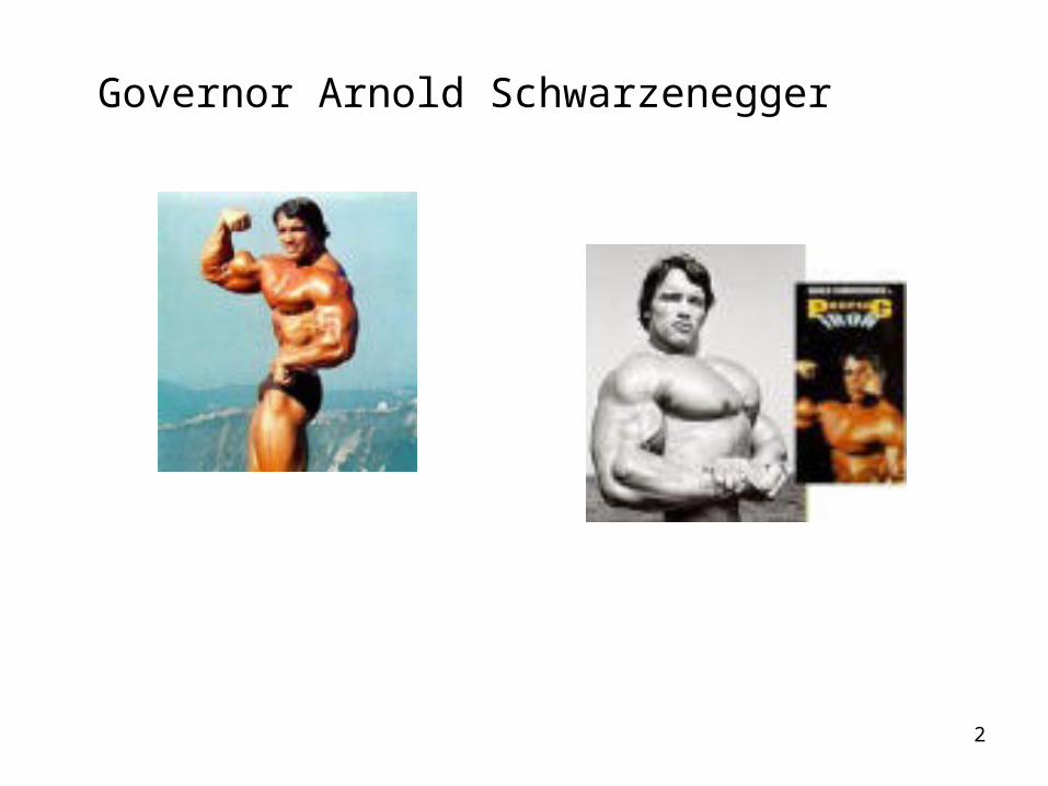
chapter 9: muscles and muscle tissue. 2 governor arnold schwarzenegger
TRANSCRIPT
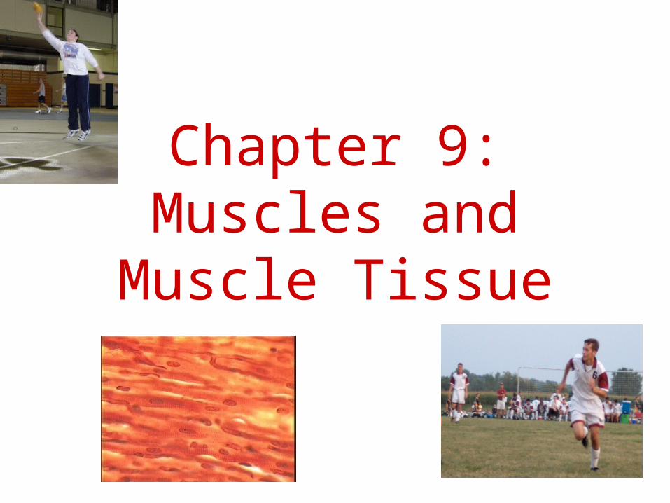
Chapter 9:Muscles and Muscle
Tissue
2
Governor Arnold Schwarzenegger

3
Muscle Functions• Producing Movement• Maintaining Posture• Stabilizing Joints• Generating Heat
4
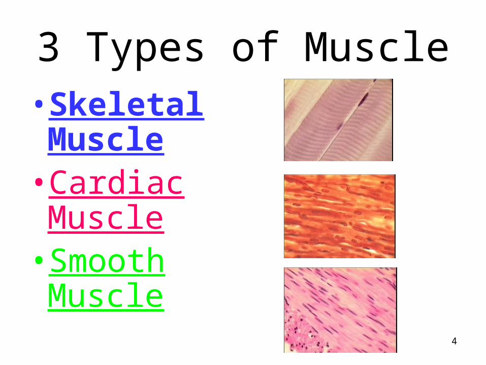
3 Types of Muscle•Skeletal Muscle
•Cardiac Muscle
•Smooth Muscle
5
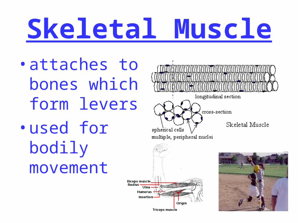
Skeletal Muscle•attaches to
bones which form levers
•used for bodily movement
6
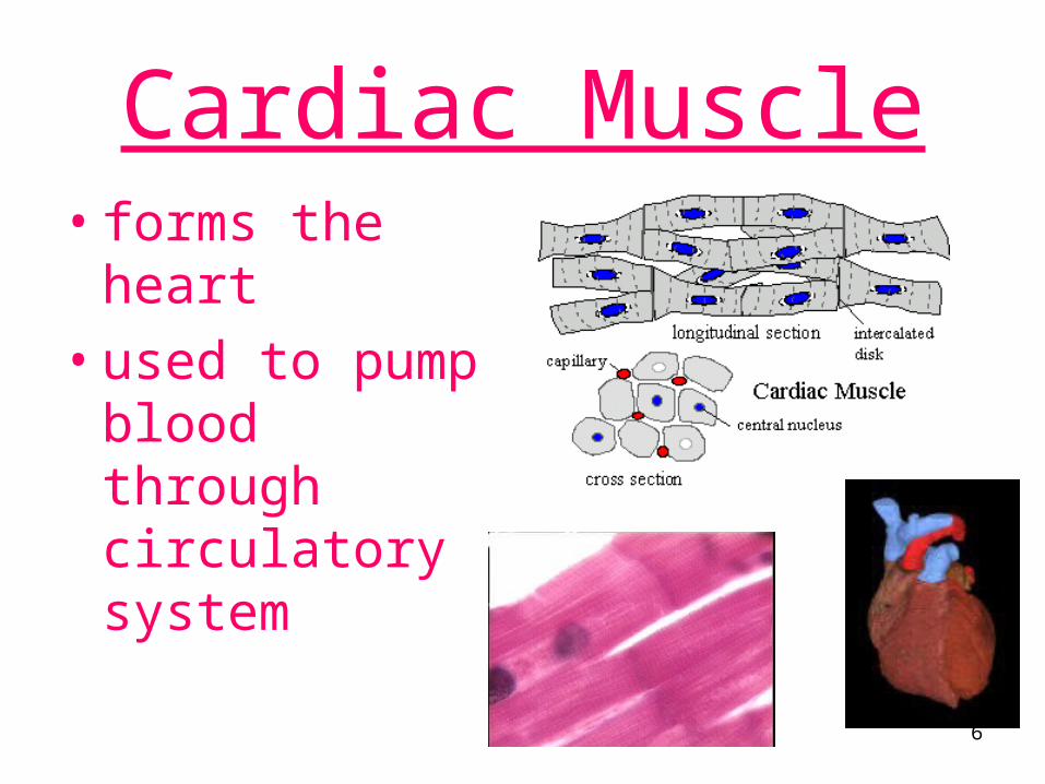
Cardiac Muscle• forms the
heart• used to pump
blood through circulatory system
7
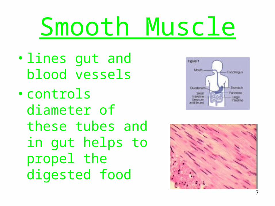
Smooth Muscle• lines gut and blood
vessels• controls diameter
of these tubes and in gut helps to propel the digested food
8
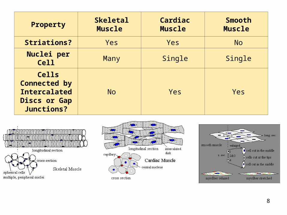
Property SkeletalMuscle
CardiacMuscle
SmoothMuscle
Striations? Yes Yes No
Nuclei per Cell Many Single Single
Cells Connected byIntercalated
Discs or Gap Junctions?
No Yes Yes
9
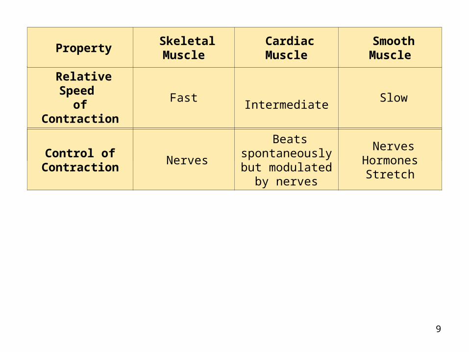
Relative Speed of Contraction
Fast Intermediate Slow
Voluntary Control?
Yes No No
Control of Contraction
Nerves
Beats spontaneously
but modulated by nerves
NervesHormones
Stretch
Property SkeletalMuscle
CardiacMuscle
SmoothMuscle

10
Muscle cells• Large, long cells called FIBERS • Contain two types of proteins--actin
and myosin• Excitable (irritable), contractile,
extensible, and elastic • Myo-, mys, or Sarco code for muscle
11
Skeletal Muscle Anatomy
•Rich blood supply to center of muscle, branch to capillaries sheathing cells
•Rich nerve supply, branching into each muscle, ending one branch to each muscle fiber
12
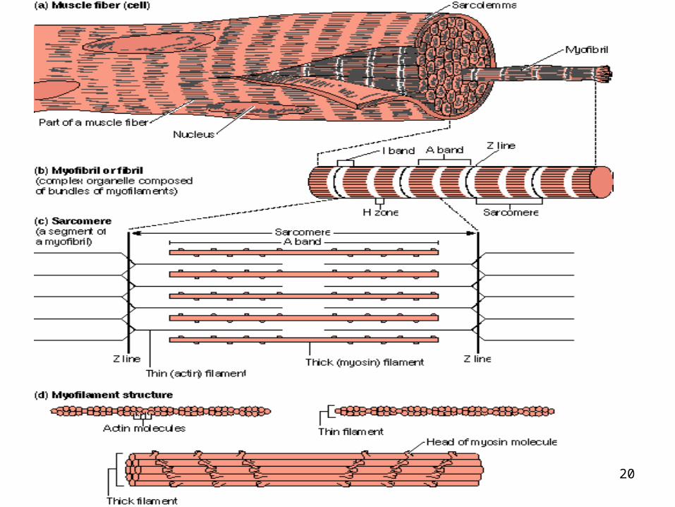
Structure of Skeletal Muscle
• Cigar shaped, multinucleate cells • Packed with myofilaments made
of actin and myosin creating visible bands (striations)
• Varied length, may be over a foot long

13
Structure cont.• Surrounded by dense, fibrous
Connective tissue (CT) sheaths• Endomysium around each fiber • Perimysium around bundles of
fibers (fascicles) • Epimysium around all fascicles
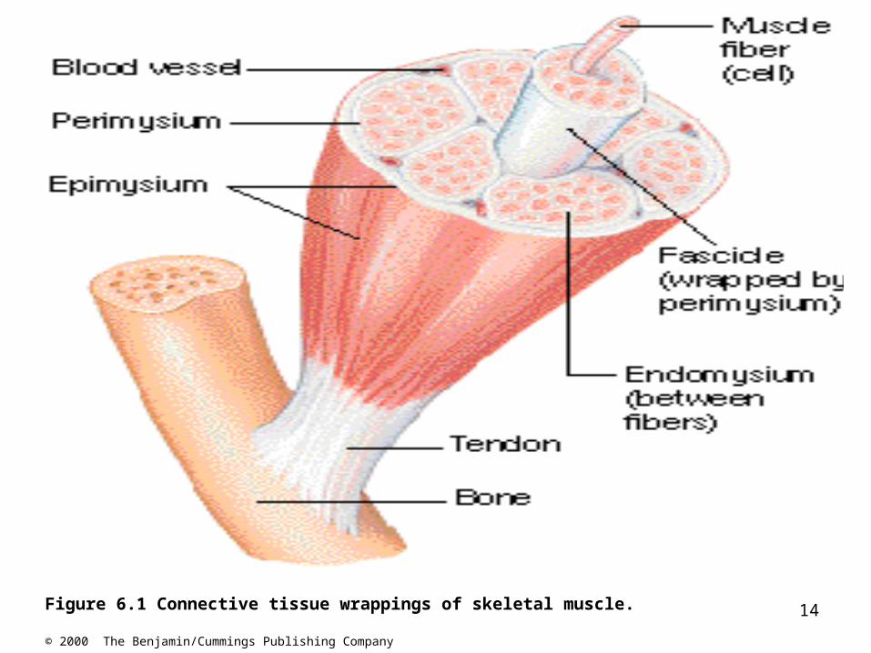
14Figure 6.1 Connective tissue wrappings of skeletal muscle.
© 2000 The Benjamin/Cummings Publishing Company
15
Microscopic structure• Muscle fibers filled with Sarcoplasm
– Glycogen and myoglobin
• Nuclei pushed to the edge of sarcolemma by long protein strands that run length of the cell, myofibrils
• Composed of contractile units, sarcomeres, made of myofilaments
16
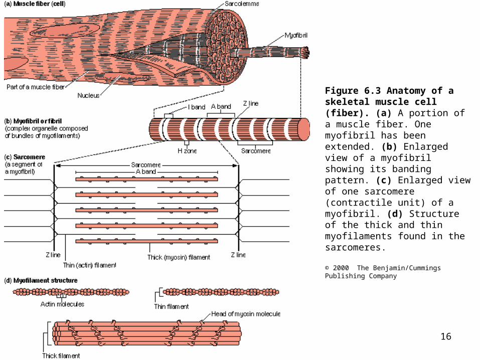
Figure 6.3 Anatomy of a skeletal muscle cell (fiber). (a) A portion of a muscle fiber. One myofibril has been extended. (b) Enlarged view of a myofibril showing its banding pattern. (c) Enlarged view of one sarcomere (contractile unit) of a myofibril. (d) Structure of the thick and thin myofilaments found in the sarcomeres.
© 2000 The Benjamin/Cummings Publishing Company
17
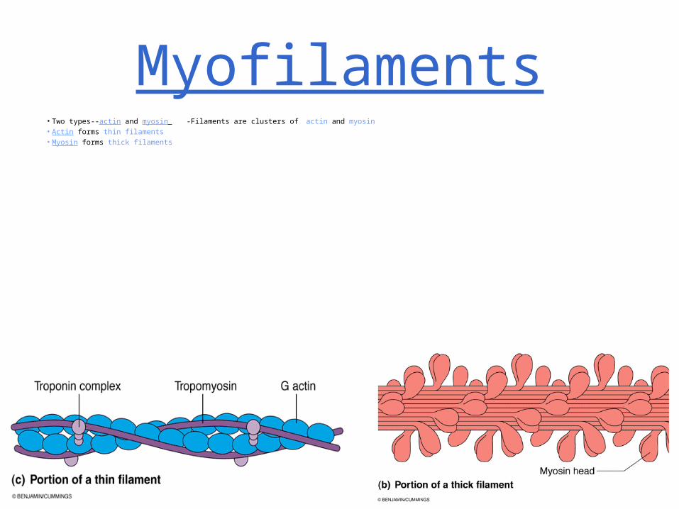
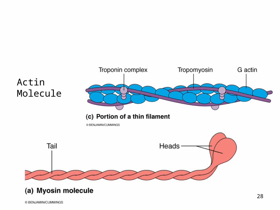
Myofilaments• Two types--actin and myosin -Filaments are clusters of actin and myosin • Actin forms thin filaments • Myosin forms thick filaments
18
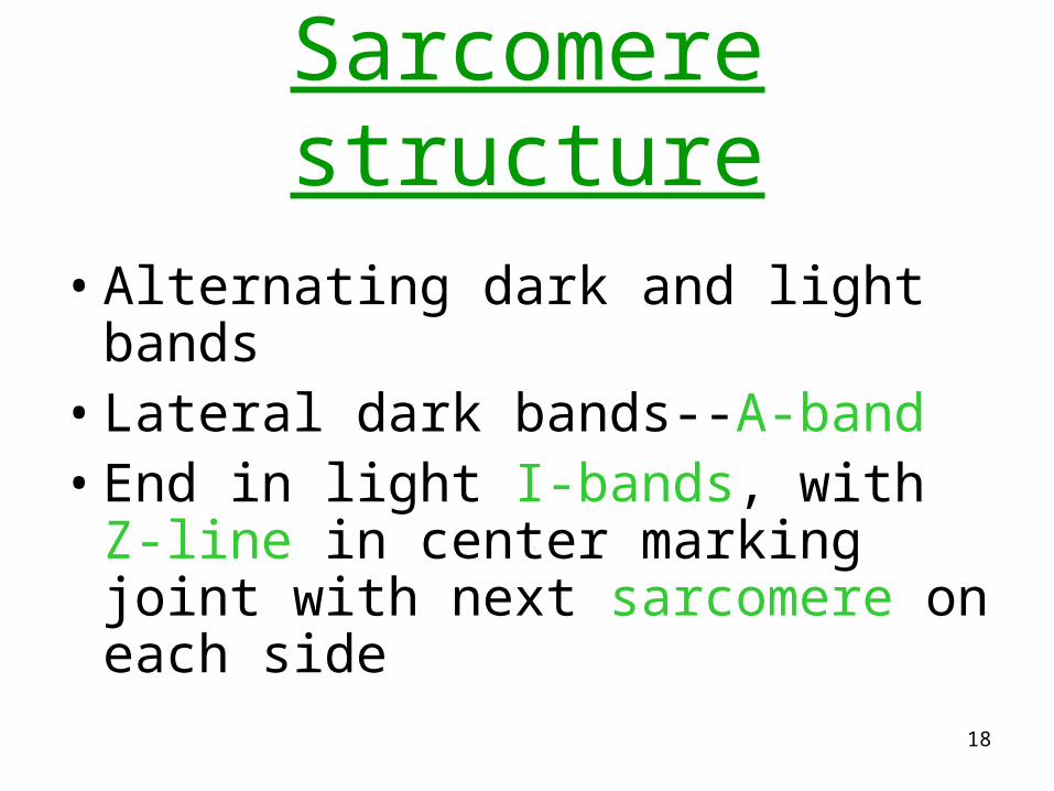
Sarcomere structure
• Alternating dark and light bands • Lateral dark bands--A-band • End in light I-bands, with Z-line in
center marking joint with next sarcomere on each side
19
20
21
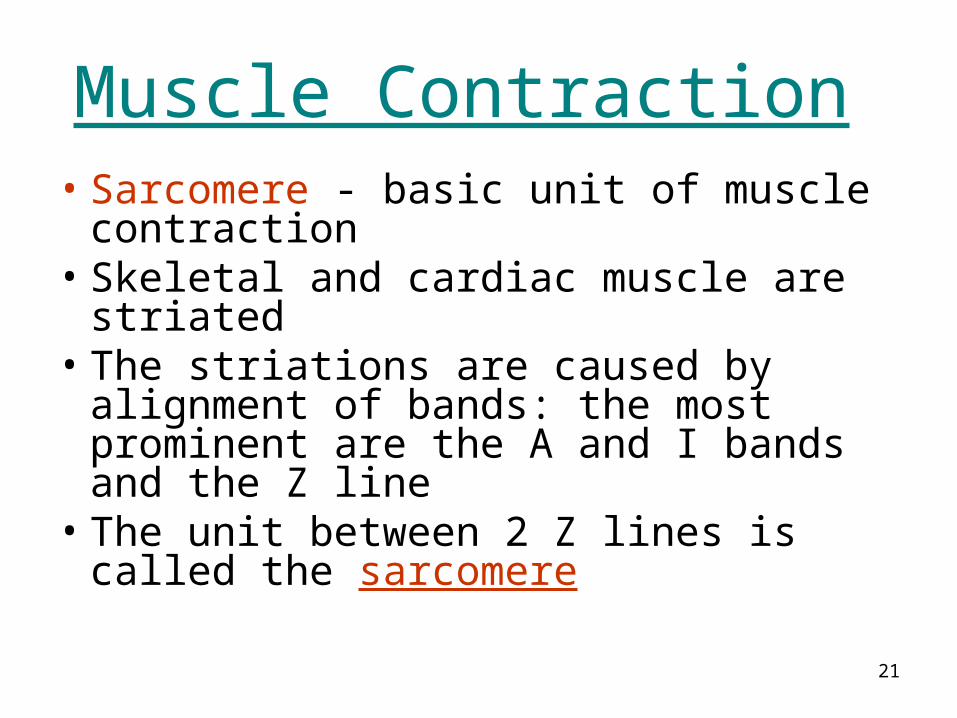
Muscle Contraction
• Sarcomere - basic unit of muscle contraction
• Skeletal and cardiac muscle are striated
• The striations are caused by alignment of bands: the most prominent are the A and I bands and the Z line
• The unit between 2 Z lines is called the sarcomere
22
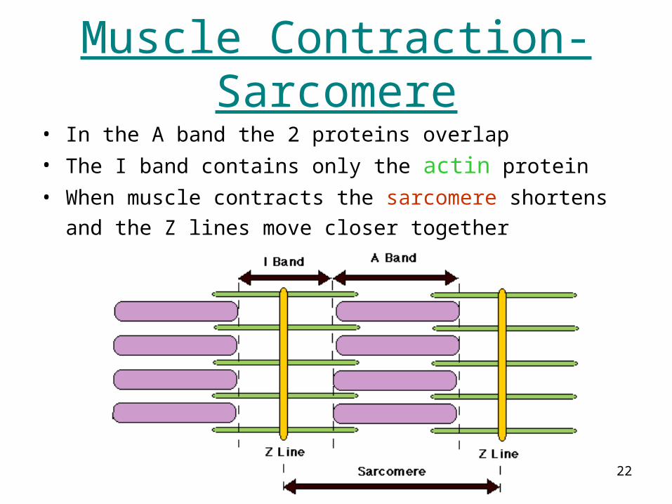
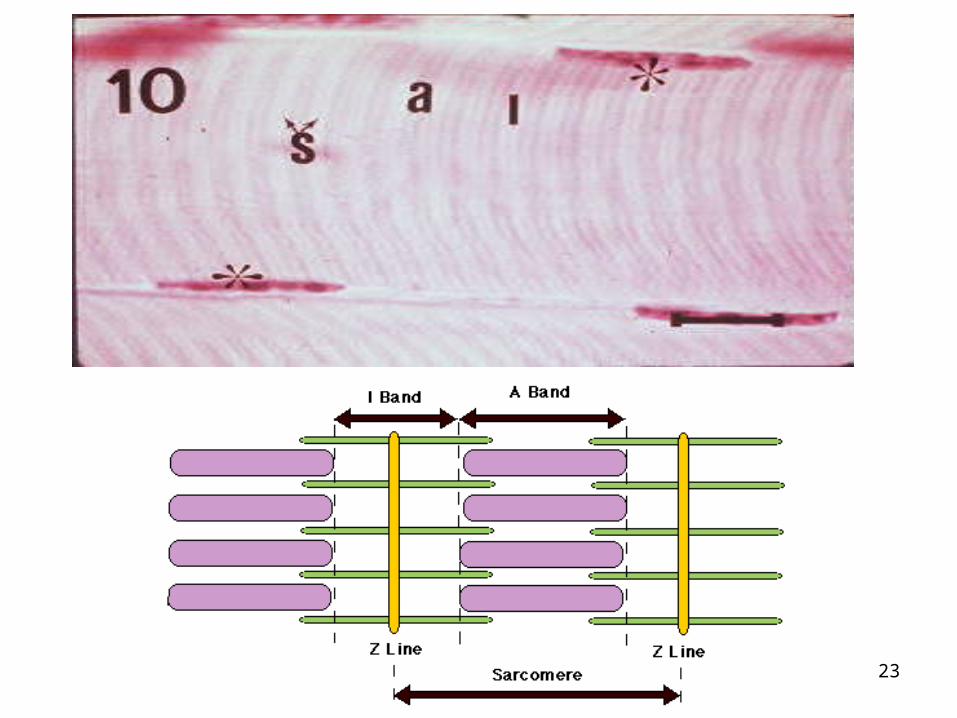
Muscle Contraction-Sarcomere
• In the A band the 2 proteins overlap • The I band contains only the actin protein
• When muscle contracts the sarcomere shortens and
the Z lines move closer together
23
24
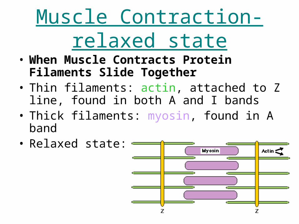
Muscle Contraction-relaxed state
• When Muscle Contracts Protein Filaments Slide Together
• Thin filaments: actin, attached to Z line, found in both A and I bands
• Thick filaments: myosin, found in A band • Relaxed state:
25
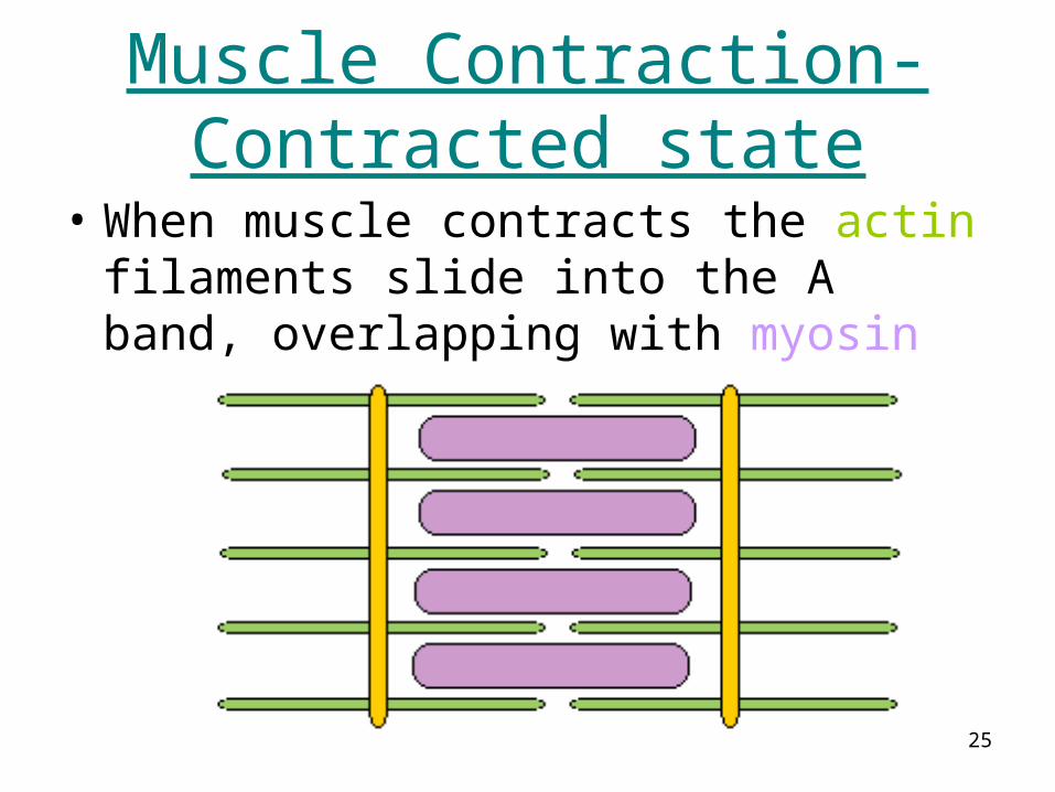
Muscle Contraction-Contracted state
• When muscle contracts the actin filaments slide into the A band, overlapping with myosin
26
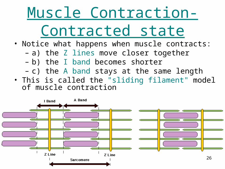
Muscle Contraction-Contracted state
• Notice what happens when muscle contracts: – a) the Z lines move closer together – b) the I band becomes shorter – c) the A band stays at the same length
• This is called the "sliding filament" model of muscle contraction
27
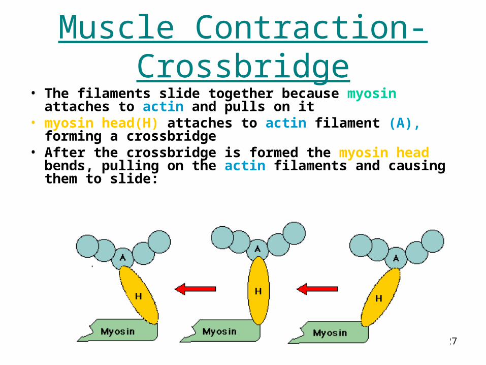
Muscle Contraction-Crossbridge
• The filaments slide together because myosin attaches to actin and pulls on it
• myosin head(H) attaches to actin filament (A), forming a crossbridge
• After the crossbridge is formed the myosin head bends, pulling on the actin filaments and causing them to slide:
28
Actin Molecule

29