chapter 6 - hox networks and the origins of motor neuron ... · and hox factors 191 6.3. hox/foxp1...
TRANSCRIPT
Author's personal copyAuthor's personal copy
C H A P T E R S I X
C
IS
*
{
urrent
SN 0
SmiloSchoDepaInstitu
Hox Networks and the Origins
of Motor Neuron Diversity
Jeremy S. Dasen* and Thomas M. Jessell†
Contents
1. In
Top
070
w Nol ortmete,
troduction
ics in Developmental Biology, Volume 88 # 2009
-2153, DOI: 10.1016/S0070-2153(09)88006-X All rig
euroscience Program, Department of Physiology and Neuroscience, New York Unf Medicine, New York, NY, USAnts of Neuroscience, and Biochemistry and Molecular Biophysics, Howard Hughes MKavli Institute for Brain Science, Columbia University, New York, NY, USA
Else
hts
iver
ed
170
2. S
pinal Motor Neuron Diversity 1712
.1. G eneration of generic motor neuron identity 1722
.2. A natomical organization of spinal motor neuron subtypes 1743. H
ox Expression in Developing Motor Neurons 1763
.1. R ostrocaudal positional information in spinalcord development
1763
.2. R egulation of Hox expression by FGF, Retinoid, Wnt,and TGFb signaling
1773
.3. T he emergence of definitive Hox patterns in motor neurons 1784. H
ox Proteins Determine Motor Neuron Columnar Identityand Connectivity
1794
.1. S pecification of segmentally restricted columnar subtypesby Hox genes
1794
.2. M echanisms of columnar Hox function in motorneuron connectivity
1825. H
ox Transcriptional Networks and the Specification of MotorPool Identities
1825
.1. A ssignment of motor pool identities by Hox genes 1845
.2. H ox genes control the specificity of motorneuron-muscle connectivity
1865
.3. E xtrinsic and intrinsic programming of motor pool identities 1876. R
estriction and Refinement of Hox Activities During MotorNeuron Differentiation
1886
.1. F oxP1: An accessory factor for Hox proteinsin motor columns and pools
189vier Inc.
reserved.
sity
ical
169
170 Jeremy S. Dasen and Thomas M. Jessell
Author's personal copy
6
Ab
Mo
en
act
ge
wit
be
mi
be
pro
cor
div
be
an
ap
the
inp
ne
ron
fac
cen
Th
pro
sp
.2.
str
tor
viro
ivit
neti
hin
hav
c pa
en
gra
d th
The
ers
twe
d m
prop
su
uts
uron
s a
tors
tra
e em
vid
ecifi
C
a
b
n
y.
c
io
t
s
m
a
e
e
o
b
f
n
,
l
e
c
oordinate control of motor axon targeting by FoxP1
and Hox factors
ct
ehaviors are the primary means by which animals interact
ment, forming the final output of most central nervous syst
The neural circuits that govern basic locomotor functions app
ally hard wired and are comprised of discrete groups of neuron
the spinal cord. These local microcircuits coordinate simple
rs in response to sensory stimuli and underlie the generation
terns of neural activity necessary for walking. In recent years t
ignificant advances in understanding the genetic and
s that determine the specificity of neural connections within
at are critical for the emergence of coordinate motor behavior
ssembly of circuits within the spinal cord requires the gen
cell types to accommodate the intricate sets of interco
n motor neurons, sensory neurons, interneurons, and muscle
st critical aspect of this process is that motor neurons se
riate muscle targets in the periphery with fidelity and precis
sequent steps in motor neuron connectivity, such as their de
rom higher brain centers, their circuits with sensory neurons
s are constrained by the early connections formed between m
d their muscle targets. The actions of a single family of tra
encoded by the chromosomally clustered Hox genes, appear
role in defining the specificity of motor neuron-muscle con
erging logic of Hox protein function in motor neuron specific
more general insights into the programs that determine
ity in other CNS regions.
wit
em
ea
s r
re
o
he
mo
the
s.
era
nn
. T
lec
ion
sc
an
ot
nsc
to
ne
atio
s
191
6
.3. H ox/FoxP1 interactions and the origins of motorneuron diversity
1926
.4. C oordinate regulation of neuronal and mesodermalHox programs
1937. C
onclusions 194Refe
rences 195h their
(CNS)
r to be
esiding
flexive
f rhyth-
re have
lecular
spinal
tion of
ections
he first
t their
. All of
ending
d inter-
or neu-
ription
have a
ctivity.
n may
ynaptic
1. Introduction
Much of the computational power of vertebrate nervous systems isdedicated to the goal of controlling movement (Sherrington, 1906), andmotor systems have necessarily evolved flexibility and adaptability torespond to the biomechanical challenges imposed by the outside world.Among the most sophisticated motor programs are those executed by thelimbs—from the orderly recruitment of flexor and extensor muscles during

Hox Networks in Motor Neuron Specification 171
Author's personal copy
locomotion to the higher level muscle synergies used in grasping and objectmanipulation. From a developmental perspective, the control of diversemotor behaviors presents a challenging problem in target specificity—demanding the coordinate activation of over a hundred distinct muscles,each by a dedicated set of spinal motor neurons. The selectivity with whichmotor neurons innervate their limb target muscles therefore represents anearly and critical element in the assembly of vertebrate sensory-motor circuits.
The stereotypic nature of developing motor axonal projections withinthe limb led to the idea that motor neurons possess subtype identities thatdefine their innervation patterns (Landmesser, 2001; Milner andLandmesser, 1999). The precision of nerve-muscle connectivity was arguedto have its origins in the ability of motor neuron subtypes to send their axonsinto nerve branches that bring them to different muscle targets. The conceptthat motor neurons possess intrinsic features that direct selective patterns ofaxonal projection and target innervation received further support fromstudies showing that motor neurons are able to redirect their axons alongnew trajectories and find their correct muscle targets when forced to enterthe limb from aberrant positions (Lance-Jones and Landmesser, 1980;Landmesser, 2001). Within the spinal cord, the cell bodies of motor neuronsthat project axons along a given peripheral pathway are grouped in discreteclusters—termed motor columns, divisions, and pools—and these neuronalsubtypes occupy fixed positions within the spinal cord. These motor neuronsubtypes appear to acquire an early identity that instructs their axons togrow along highly specific trajectories to their muscle targets.
Work over the past decade has begun to define the molecular programsthat operate during embryonic development to determine motor neuron fateand associated patterns of connectivity. One key insight into these molecularprograms is that core features of motor neuron identity that determinemigratory routes, settling positions, patterns of axonal projections, and selec-tion of synaptic targets are defined by the selective profile of transcriptionfactor expression. The actions of one diverse family of transcription factors,encoded by theHox genes, are central mediators of the intrinsic programs thatshape motor neuron subtype identity and target muscle specificity.
The focus of this chapter is to outline recent studies that have helped todefine the mechanisms by which a Hox-based transcriptional networkcontrols motor neuron identity and connectivity in the developing spinal cord.
2. Spinal Motor Neuron Diversity
Terrestrial vertebrates possess hundreds of anatomically distinct musclegroups. The motor neurons that innervate these diverse targets areorganized into discrete clusters within the spinal cord. The position that

172 Jeremy S. Dasen and Thomas M. Jessell
Author's personal copy
these groups of motor neurons occupy within the spinal cord is relativelyfixed from animal to animal, and thus cell body position is often predictiveof target innervation pattern (Hollyday and Jacobson, 1990; Landmesser,1978b). Understanding the genetic programs that contribute to the forma-tion of motor innervation maps has been a major challenge in the field.In this section we describe studies that have helped to define the earlyprograms which establish motor neurons as a class and the emergence ofmotor neuron topographic maps.
2.1. Generation of generic motor neuron identity
Motor neurons and several classes of interneurons are generated in responseto graded extrinsic signals acting along the dorsoventral axis of the neuraltube. These secreted signals include sonic hedgehog (Shh) from the noto-chord and floor plate, and fibroblast growth factors (FGFs) and retinoic acid(RA) by the paraxial mesoderm. The detailed mechanisms through whichShh, RA, and FGFs define progenitor identities will not be addressed heresince this topic has been the subject of several review articles (Dessaud et al.,2008; Jessell, 2000; Shirasaki and Pfaff, 2002). In brief, Shh, FGF, and RAsignaling induce the expression of distinct combinations of transcriptionfactors in neural progenitors. These initial patterns are subsequently refinedthrough the selective transcriptional cross-repressive interactions betweentranscription factors expressed at the boundaries between progenitordomains (Fig. 6.1A). Each progenitor domain in the neural tube expressesa unique profile of transcription factors, and these combinatorial patternsdefine progenitor fates (Briscoe et al., 2000). Progenitors that give to motorneurons depend on the activities of the bHLH protein Olig2 and thehomeodomain factors Pax6, Nkx6.1, and Nkx6.2 (Novitch et al., 2001;Vallstedt et al., 2001; Zhou and Anderson, 2002). Thus a major output ofthe dorsoventral signaling system is to define the identity of motor neuronsas opposed to ventral interneurons.
Soon after their generation, at about embryonic day (e) 9.5 in mouse,spinal motor neurons express a set of homeodomain transcription factors(notably Hb9, Lhx3, Isl1, and Isl2), that that control features common to allspinal motor neurons as well as those that are involved in later aspects ofsubtype diversification (Fig. 6.1A) (Arber et al., 1999; Pfaff et al., 1996;Sharma et al., 1998; Thaler et al., 1999, 2004). These generic motor neuronscharacteristics include the projections of axons outside the spinal cord andthe release of acetylcholine as the primary neurotransmitter. Althoughpatterning events mediated by Shh, RA, and FGF signaling define howmotor neurons as a class are specified, additional signaling pathways arepresumably necessary for the further diversification of motor neurons intodistinct subtypes.

[Shh]
Class I
Class II
MNs
Dorsoventral axis
Nkx6Olig2Pax6
Hb9Isl1/2Lhx3/4
pMN
Progenitors Postmitotic
[FGF8]
[Gdf11][RA]
Brachial Thoracic Lumbar
Hox5 Hox6 Hox7 Hox8 Hox9 Hox10
Hox5
Hox6
Hox8
Hox9
Hox10
Rostrocaudal axis
A
B
3� 5�
d
v
r c
Hox11
Figure 6.1 Patterning along the dorsoventral and rostrocaudal axes of the neural tube.(A) Motor neurons and ventral interneurons are generated along the dorsoventral (d-v)axis in response to the graded activities of sonic hedgehog (Shh) which induces thepatterned expression of transcription factors in progenitor cells. Class I factors areinduced by Shh while Class II transcription factors are repressed. Selective cross-repressive interactions between Class I and Class II transcription factor sharpen theboundaries between progenitor domains (Briscoe et al., 2000). Retinoic acid (RA)from the paraxial mesoderm and fibroblast growth factor (FGF) signaling also influencethe pattern of transcription factors in neural tube progenitors (not shown). (B) Alongthe rostrocaudal axis graded FGF signaling induces the expression of chromosomallylinked Hox genes in the neural tube. Hox genes located at one end of the cluster areexpressed more rostrally (r) while genes at the opposite end are expressed caudally (c)in response to higher levels of FGF. At more rostral levels Hox genes are regulated bygraded RA signaling while at more caudal levels Hox genes are regulated by gradedGdf11.
Hox Networks in Motor Neuron Specification 173
Author's personal copy

174 Jeremy S. Dasen and Thomas M. Jessell
Author's personal copy
2.2. Anatomical organization of spinal motorneuron subtypes
At the time of their birth, all motor neurons possess a set of core features thatdistinguish them from other classes of neurons, but rapidly diversify intosubtype identities that allow them to form selective connections with targetcells. Major distinctions in the identities of motor neurons have beendefined through studies of their position, axon trajectory, and pattern ofmuscle innervation. One level of organization is the allocation of motorneurons to columnar groups, each column occupying a defined positionalong the rostrocaudal axis of the spinal cord. Four major columnar classeshave been described, each innervating a unique set of peripheral targettissues (Fig. 6.2). The most prominent of these columnar groups are thelateral motor columns (LMCs) which are generated at limb levels of thespinal cord and innervate limb muscles. At thoracic levels visceral pregan-glionic column (PGC) motor neurons innervate sympathetic ganglia whilehypaxial motor column (HMC) neurons innervate intercostal and abdomi-nal wall musculature (Gutman et al., 1993; Prasad and Hollyday, 1991).In contrast to these segmentally restricted motor columns, motor neurons inthe median motor column (MMC) are present at all levels of the spinal cordand innervate dorsal axial musculature (Fetcho, 1987; Gutman et al., 1993).
Additional layers of anatomical organization are present within these maincolumns, notably the segregation of the LMC into ‘‘divisions’’ and ‘‘pools.’’At both forelimb and hindlimb levels of the spinal cord, the LMC is split intotwo divisions: a medial division which contains neurons projecting axonsventrally within the limb mesenchyme and a lateral division which containsneurons that project dorsally (Fig. 6.2B) (Landmesser, 1978a; Tosney andLandmesser, 1985a,b). These two divisional subgroups have been linked tothe particular mode of actions of their muscle targets: dorsally projectinglateral LMC axons frequently innervate extensor muscles, whereas medialLMC axons project ventrally and typically innervate flexor muscles. Thefunctional relevance of this anatomical segregation is still unclear.
A third level of motor neuron diversity is evident in the segregation ofmotor neurons into motor pools (Romanes, 1942). Motor pools occupydistinct positions within the LMC, each pool innervating a dedicated targetmuscle (Hollyday and Jacobson, 1990; Landmesser, 1978b). Whereas fore-limb and hindlimb motor neurons share similar columnar and divisionalproperties, the organization of motor pools between these two levels of thespinal cord is quite distinct, reflecting differences in the overall pattern ofmusculature between the forelimb and hindlimb. Nevertheless motor poolsinnervating forelimb and hindlimb form topographic maps which aresimilarly organized. At both levels of the spinal cord motor pools locatedmore rostrally tend to innervate muscles located rostral and proximal withinthe limb, while caudal pools project to more caudal and distal regions(Hollyday and Jacobson, 1990; Landmesser, 1978b).

MMC
HMC
PGC
LMC
Brachial Thoracic Lumbar
MMC
m
LMC
l
Brachial
Pools
A
B
CLMC
Limb
PGC
scg
HMC
Intercostal m.
MMC
Axial m.
PGC
HMCMMC
Thoracic
MMC
m
LMC
l
Lumbar
Figure 6.2 Motor neuron columnar, divisional and pool organization. (A) Motorcolumns and motor pools are generated at specific positions along the rostrocaudalaxis. The cell bodies of motor neurons that send axons to the limb are contained withinthe lateral motor column (LMC) at brachial and lumbar levels of the spinal cord.Preganglionic column (PGC) motor neurons and hypaxial motor column (HMC)neurons are found at thoracic levels. Motor neurons within the medial motor column(MMC) are generated at all rostrocaudal levels of the spinal cord. Motor pools aregenerated at specific rostrocaudal positions within the LMC. (B) Schematic of crosssections of brachial, thoracic, and lumbar spinal cord showing the position of motorcolumns and divisions. Medial (m) and lateral (l) divisions of the LMC are indicated.(C) Projection patterns of motor neuron columnar subtypes. LMC neurons project tothe limb, PGC neurons to sympathetic chain ganglia (scg), HMC neurons to intercostaland body wall muscles (m), MMC neurons to axial muscle.
Hox Networks in Motor Neuron Specification 175
Author's personal copy
176 Jeremy S. Dasen and Thomas M. Jessell
Author's personal copy
3. Hox Expression in Developing
Motor Neurons
The diversification of motor neurons into specific columnar, divisional,and pool subtypes relies on the acquisition of a positional identity alongthe rostrocaudal axis of the spinal cord. Like neuronal specification alongthe dorsoventral axis, motor neurons acquire positional information inresponse to graded signals which initiate transcriptional programs withinprogenitor and postmitotic cells. A critical output of the rostrocaudal signal-ing pathways acting on the neural tube is the establishment of selectivepatterns of Hox gene expression within specific motor neuron subtypes.
3.1. Rostrocaudal positional information in spinalcord development
Early insights into the role of rostrocaudal positional information in loco-motor circuit assembly came from embryological manipulations in the chickembryo. One of the most dramatic set of experiments was performed byVictor Hamburger and colleagues who demonstrated that if the region ofthe spinal cord responsible for synchronous activation of wing muscles isgrafted to the level of the hindlimb, chickens will synchronously activatemuscle in the legs (Narayanan and Hamburger, 1971). Conversely, graftinghindlimb-level spinal cord to wing levels causes the chick to alternate wingmovements, in a pattern similar to walking. These studies provide evidencethat the intrinsic properties of neurons generated at specific rostrocaudallevels have critical roles in establishing the local circuitries controlling basicbehavioral outputs.
More recent studies on rostrocaudal programming have demonstratedthat positional identities are regulated by signals derived from axial struc-tures, such as the node and notochord, as well as from paraxial tissues, thepresomitic mesoderm and somites (Ensini et al., 1998; Lance-Jones et al.,2001; Liu et al., 2001). Grafting experiments in chick indicated that region-ally restricted signals govern the specification of motor neuron columnarsubtypes. Transposition of thoracic and brachial levels of the neural tubeduring a critical period of development respecifies columnar fates—neuronsderived from the former thoracic region of the neural tube acquire LMCidentity, and conversely, motor neurons from the former brachial levelacquire a CT identity (Ensini et al., 1998; Shieh, 1951). The signalsfor columnar respecification derive, in part, from the adjacent paraxialmesoderm, since a similar switching of motor neuron columnar identitycan be elicited by transposition of brachial and thoracic paraxial mesoderm(Ensini et al., 1998).

Hox Networks in Motor Neuron Specification 177
Author's personal copy
3.2. Regulation of Hox expression by FGF, Retinoid, Wnt,and TGFb signaling
While grafting studies in chick indicated that the regional identities ofmotor neurons are controlled through mesodermal signals, the identity ofthese signals and the intrinsic mediators of their actions were not known.One class of transcription factors with an evolutionarily conserved role inestablishing differences in cell identity along the rostrocaudal axis aremembers of the chromosomally arrayed Hox gene family. The expressionof Hox genes within the spinal cord is closely aligned with their positionwithin the Hox cluster: genes located at the 30 end of the cluster areexpressed more anteriorly than genes at the 50 end (Fig. 6.1B) (Kmita andDuboule, 2003; Lemons and McGinnis, 2006). The precise mechanism bywhich spatial colinear expression of Hox genes emerges in the neural tube isunknown; although gradients of signaling molecules appear to impart theinitial profiles of Hox expression in most tissues where colinearity has beenexamined.
The expression of Hox genes within the CNS is controlled by multiplesignaling molecules including FGFs, retinoids, Wnts, and members of thetransforming growth factor (TGF) b superfamily (Bel-Vialar et al., 2002; Diezdel Corral and Storey, 2004; Liu, 2006; Liu et al., 2001; Nordstrom et al.,2006). Graded FGF signaling is involved in establishing the initial inductionof Hox gene expression at brachial, thoracic, and lumbar levels of the spinalcord (Bel-Vialar et al., 2002; Dasen et al., 2003; Liu et al., 2001). At the caudalend of the chick embryo, an organizing region called Hensen’s node and thepresomitic mesoderm are primary sources of FGF signals. As the tail budregresses caudally during axis extension, more posterior regions of the spinalcord are exposed to FGF in higher concentration and over longer periods oftime (Dubrulle and Pourquie, 2004; Liu et al., 2001). Both in vitro and in vivostudies have shown that Hox genes located at the 30 end of a cluster areinduced by low levels of FGF while those at the 50 end are induced byprogressively higher FGF levels (Bel-Vialar et al., 2002; Dasen et al., 2003;Liu et al., 2001). As a consequence,Hox4-Hox8 paralog genes are expressed atbrachial levels, Hox8-Hox9 genes at thoracic, and Hox10-Hox13 genesat lumbar levels of the spinal cord (Fig. 6.1B).
While graded FGF signals contribute to initial Hox patterns, other signal-ing systems participate in regulating subsets of Hox genes within a cluster.At more anterior levels RA signaling provided by paraxial mesoderm andsomites has been shown to regulate Hox expression at brachial levels(Liu et al., 2001). The action of retinoids is in part to antagonize the FGFgradient (Diez del Corral and Storey, 2004); although the mechanismsunderlying this process are not known. At more caudal levels, the TGFbfamily member Gdf11 has been shown to be essential both in vitro and in vivoin the regulation ofHox8-Hox10 paralogs at thoracic and lumbar levels of the

178 Jeremy S. Dasen and Thomas M. Jessell
Author's personal copy
spinal cord (Liu, 2006; McPherron et al., 1999), and this inductive factorappears to act in concert with high levels of FGF signaling (Liu et al., 2001).Thus Hox expression in motor neurons relies on the convergent actions ofmultiple signaling pathways (Fig. 6.1B).
3.3. The emergence of definitive Hox patternsin motor neurons
Although graded signals are necessary to establish the initial pattern of Hoxgene expression in motor neurons, the links between extrinsic signals andHox protein expression in motor neurons are still unclear. While RAresponse elements have been characterized in the Hox genes controllingregional identity in the hindbrain (Glover et al., 2006; Trainor andKrumlauf, 2000), similar control elements have yet to be described forHox genes expressed within the spinal cord. How the neural tube interpretsthe FGF gradient is also unknown; although a class of homeodomainproteins related to the Drosophila gene caudal has been implicated. Manipu-lation of vertebrate caudal homeobox (Cdx) activities produces phenotypeswhich are similar to manipulations of FGF signaling. Misexpression ofactivated forms of Cdx can shift the patterns of Hox expression in the spinalcord (Bel-Vialar et al., 2002), while in Zebrafish loss of Cdx activities dereg-ulates Hox expression and the spinal cord acquires a hindbrain-like character(Shimizu et al., 2006; Skromne et al., 2007). The mechanisms by which Cdxproteins regulate Hox expression are not known, but may involve directCdx binding to individual Hox regulatory elements or interaction withlocus control regions within the Hox clusters.
Another unresolved issue is the inordinate temporal delay between thetime of neural tube exposure to graded FGF signals and the emergence ofHox expression in postmitotic motor neurons. FGF signaling appears to acton neural progenitors, and manipulation of FGF signals can rapidly switchHox transcriptional profiles at early stages of development (Bel-Vialar et al.,2002; Dasen et al., 2003). However the expression of some Hox proteins bymotor neurons is detected only in postmitotic cells (Dasen et al., 2003).The mechanisms which introduce this apparent delay between Hox RNAand protein expression are not known; although Hox genes are under thecontrol of several layers of posttranscriptional regulation, including silencingby microRNAs (Chopra and Mishra, 2006). Another possibility is thatexpression of HoxB cluster genes in progenitors, which are generally notdetected in postmitotic motor neurons (Dasen et al., 2005), prefiguresprogenitors to express HoxA, HoxC, and HoxD genes.

Hox Networks in Motor Neuron Specification 179
Author's personal copy
4. Hox Proteins Determine Motor Neuron
Columnar Identity and Connectivity
Early insights into the role of Hox genes in CNS development camefrom studies in the vertebrate hindbrain. In the hindbrain the metamericorganization of the rhombomeres provides anatomical landmarks for defin-ing global aspects of Hox function, and several Hox gene mutants arecharacterized by homeotic transformation of rhombomeres identities andchanges in neuronal identity (reviewed in Guthrie (2007)). In contrast, therelative morphological homogeneity and lack of columnar and pool specificmolecular markers in the spinal cord created a challenge in linking Hoxfunction to the specification of motor neuron subtypes. Although it hadbeen recognized that different levels of the spinal cord express distinct Hoxgenes (Carpenter, 2002), the link to motor neuron columnar, divisional,and pool identities were unclear.
4.1. Specification of segmentally restricted columnarsubtypes by Hox genes
Certain motor neuron populations can be defined by the combinatorialexpression of LIM-homeodomain proteins (Tsuchida et al., 1994); althoughno single LIM-homeodomain protein is specific for a single neuronal class inthe spinal cord. The identification of molecular markers that are selectivelyexpressed in two segmentally restricted motor columns, LMC and PGCneurons, provided a means to explore the signaling pathways that specifymotor neuron subtypes. PGC neurons can be distinguished from otherthoracic-level spinal motor neurons by expression of BMP5, a TGFb familymember (William et al., 2003) as well as nuclear phospho-(p)-Smad1/5/8(Dasen et al., 2008). At limb levels LMC neurons can be defined by theirselective expression of retinaldehyde dehydrogenase-2 (RALDH2), a keyenzyme in retinoic acid synthesis (Sockanathan and Jessell, 1998).
Expression of Hox proteins is closely aligned with the position in whichmolecularly defined columnar subtypes are generated: expression of Hox6proteins segregates with brachial (forelimb) LMC neurons, Hox9 proteinswith thoracic PGC neurons, and Hox10 proteins with lumbar (hindlimb)LMC neurons (Fig. 6.3A) (Choe et al., 2006; Dasen et al., 2003; Lance-Jones et al., 2001; Liu et al., 2001). Consistent with the model where Hoxprotein expression is controlled by graded FGF signaling, elevation of FGFlevels at brachial levels of the spinal cord induces the expression of Hoxc9,a Hox gene normally restricted to thoracic levels (Dasen et al., 2003).

LMC
MCLMC
Forelimb
Hindlimb
Sympatheticchain ganglia
Hox6
Hox9
Hox10Brachial
Thoracic
Lumbar
PGC
Shh
FGF
Hb9
Hox6
RALDH2(RA)LMC
Lhx1 EphA4Lhx1EphA4D-V
R-C
Hox9
Hox6 LMC
PGC
FoxP1
Hox10 LMC
A
C
B
Isl1
Figure 6.3 Hox genes and the specification of segmentally restricted motor columns.(A) Hox6, Hox9, and Hoxd10 are expressed in motor neurons at distinct rostrocaudallevels of the spinal cord and direct motor neuron identity and peripheral target connec-tivity. Hox6 activities control brachial LMC identity, Hox9 control PGC identity, andHox10 lumbar LMC identity. (B) Regulatory interactions betweenHox genes in motorneuron columnar fates. Cross-repressive interactions between Hox6, Hox9, andHox10 proteins refine Hox profiles and Hox activator functions define LMC andPGC identities. (C) Role of LMC Hox genes in the program controlling motor neurondivisional identities and axonal projections along the dorsoventral axis of the limb. Theconvergent activities of motor neuron specific transcription factors (Hb9) and limb-level Hox genes (e.g., Hoxc6) direct expression of FoxP1 and RALDH2 in LMCneurons. RALDH2 creates a neuronal source of RA which leads to the induction ofLhx1 expression by lateral LMC neurons. Lhx1 directs expression of the guidancereceptor EphA4, and EphA4 directs motor axons toward the dorsal limb.
180 Jeremy S. Dasen and Thomas M. Jessell
Author's personal copy
This switch in Hox patterns is accompanied by the loss of brachial LMCneurons, characterized by the abolishment of Hoxc6 and RALDH2 expres-sion. Motor neurons are instead converted to a PGC cell fate, defined byexpression of BMP5 and pSmad. The effects of elevated FGF are notrestricted to changes in marker gene expression, but extend to multipleaspects of columnar identity including switches in the patterns of migration,peripheral connectivity, and the number of motor neurons generated atlimb and thoracic levels (Dasen et al., 2003).
Are the effects of elevating FGF signaling on motor neuron columnaridentity mediated by changes in Hox gene expression? Consistent with adirect role in columnar specification, misexpression of Hoxc9 at brachial

Hox Networks in Motor Neuron Specification 181
Author's personal copy
levels is sufficient to convert LMC to PGC motor neurons, while expres-sion of Hoxc6 or Hoxd10 at thoracic levels can convert PGC and HMC toLMC neurons (Dasen et al., 2003; Shah et al., 2004). These actions of Hoxproteins rely on their ability to cross-repress each others expression: Hox6and Hox10 protein can repress Hox9 expression while Hox9 can repressHox6 expression (Fig. 6.3B). Moreover, the actions of Hox proteins ininducing columnar fates and establishing Hox boundaries appear to beseparable, as expression of a constitutive repressor forms block columnarinducing activities but retain their cross-repressive functions (Dasen et al.,2003). Thus like many transcription factors Hox proteins possess intrinsicactivator and repressor functions, and these functional differences servedistinct but coherent roles in motor neuron subtype specification (Fig. 6.3B).
The principles of Hox protein function along the rostrocaudal axisparallel those that operate along the dorsoventral axis which specify motorneurons as a class. Along both axes, the initial graded activity of a secretedsignaling factor establishes broad domains of homeodomain protein expres-sion that are subsequently refined through selective cross-repressive inter-actions. However, these two programs of transcriptional cross-repressionappear to operate at different stages of neuronal specification. Dorsoven-trally, homeodomain cross-repressive interactions are evident within neuralprogenitor cells (Briscoe et al., 2000), whereas along the rostrocaudal axisHox cross-repression occurs within postmitotic neurons (Dasen et al.,2003). Nevertheless, the convergence of these two patterning programsensures that Hox-directed features of columnar differentiation are confinedto postmitotic motor neurons.
Aspects in the logic of Hox function in spinal motor neuron diversifica-tion are distinct from that used in rostrocaudal patterning of Drosophila larvaeand in the vertebrate hindbrain. In these two systems, the actions of posteri-orly expressed Hox genes typically dominate over those of more anteriorlyexpressed genes—a phenomenon termed posterior dominance (Duboule andMorata, 1994). The findings in the spinal cord argue against posterior preva-lence of Hox function in postmitotic motor neuron specification, sinceectopic caudal expression of Hoxc6 is as effective as ectopic rostral expressionof Hoxc9. Several exceptions to the posterior prevalence rule of Hox func-tion have been reported in both fly and vertebrate embryos (Duboule andMorata, 1994; Jegalian and De Robertis, 1992). Recent genetic analysis ofaxial skeleton patterning in Hox mutants provides further evidence against aglobal dominance of posterior Hox genes in somite derivatives (McIntyreet al., 2007). Thus tissue context may influence functional dominancesbetween Hox genes. In neural progenitors Hox genes are expressed in over-lapping and nested patterns, one possibility is that posterior dominanceoperates at early stages of spinal cord development to help determine thefinal Hox pattern in postmitotic neurons.

182 Jeremy S. Dasen and Thomas M. Jessell
Author's personal copy
4.2. Mechanisms of columnar Hox function in motorneuron connectivity
In addition to regulating the expression of columnar-specific genes, Hoxproteins can also direct the peripheral connectivity of LMC and PGCneurons. At forelimb levels of the spinal cord the switching of LMCneurons to a PGC fate forces motor neurons to project their axons to thenormal PGC targets, sympathetic chain ganglia. Conversely, expression ofHoxc6 or Hoxd10 at thoracic levels induces LMC fate and these neuronsproject into the limb (Dasen et al., 2005; Shah et al., 2004). Thus Hoxproteins not only influence columnar identity at the level of molecularmarker expression but also contribute to the initial specificity of motoraxon projections in the periphery.
How might the activities of Hox proteins determine columnar-specificpatterns of axonal innervation? Markers of newly generated LMC and PGCneurons, RALDH2 and BMP5/pSmad, are intimately involved in induc-tive signaling. Although the function of BMP5/pSmad signaling in PGCneurons is not known, RALDH2 activity appears to be required for theinitial specificity in which LMC neurons project into the limb (Fig. 6.3C).The RALDH2-dependent synthesis of retinoids by LMC neurons is neces-sary for the specification of lateral LMC neuronal identity, in order toinduce expression of Lhx1, a LIM-homeodomain protein (Kania et al.,2000). Lhx1 expression has been shown to direct the dorsal projection ofLMC motor axons in the developing limb through its ability to regulateEphA4 expression, a guidance receptor required for axons to avoid theventral limb mesenchyme (Eberhart et al., 2002; Kania and Jessell, 2003).
Thus an early step in the Hox-dependent specification of LMC identityis to direct RALDH2 expression, and trigger a series of downstreamsignaling events within postmitotic motor neurons that govern the patternof motor neuron connectivity in the developing limb. More generally thesefindings suggest that a key step in the organization of columnar differentia-tion at different segmental levels of the spinal cord is the induction ofsignaling factors in different columnar subtypes of motor neurons, whichin turn directs a molecular program for motor neuron connectivity.
5. Hox Transcriptional Networks and the
Specification of Motor Pool Identities
Within LMC divisions, motor neurons are further subdivided intomotor pools, each destined to innervate a single muscle target in the limb.A typical vertebrate limb contains over 50 muscle groups, requiring thegeneration of a diverse array of motor pool subtypes. Like columns, a motor

Hox Networks in Motor Neuron Specification 183
Author's personal copy
pools occupy stereotypic rostrocaudal positions within the spinal cord andmultiple pools can occupy a single segmental level (Fig. 6.4). As with thespecification of segmentally restricted motor columns, Hox transcriptionfactors appear to be critical determinants of pool identity and muscle targetspecificity.
The proposal that intrinsic motor pool identities direct target muscleconnectivity emerged first through embryological manipulations whichrevealed that the axons of specific LMC neurons project to their limbmuscle targets with high precision (Landmesser, 1978a), even when forcedto enter the limb from innapropriate positions (Landmesser, 2001). Classicalembryological studies have provided evidence that motor neurons within
Hoxc8
Hox5
Runx1
ScipPea3
Hox4
Hoxa7
Meis1
PecFCU
Hoxc8
Hoxc6
Rostral
Caudal
Transcriptional circuitMotor pool Hox profile
a7
a7c6a4c4
a4c4
M1
M1a7c6
Pec
FCUPea3
Scip
Hox6
A B
C
Pec
FCU
Pea3
Scip
Hox4
Hoxa7Meis1
Hoxc8
Hoxc6
Figure 6.4 A Hox transcriptional network controls motor pool identity and connec-tivity. (A) Hox genes determine the rostrocaudal position of motor pools within theLMC. At brachial levels of the spinal cord cross-repressive interactions between Hox5proteins and Hoxc8 establish the boundary between molecularly defined motor pools.Hox5 proteins (Hoxa5 and Hoxc5) are required to generate the motor pool thatexpresses the transcription factor Runx1 in rostral LMC neurons. Hoxc8 is requiredin caudal LMC neurons to generate the motor pools that express the transcriptionfactors Pea3 and Scip. (B) Intrasegmental specification of motor pool identity. At asingle segmental level of the spinal cord �6-10 pools are generated. Motor poolsprojecting to the pectoralis (Pec) and flexor carpi ulnaris (FCU) can be molecularlydefined by expression of the transcription factors Pea3 and Scip, respectively. Both Pecand FCU pools express unique profiles of Hox expression (M1:Meis1, Hoxc6 shown asc6, etc.). The patterns of Hox expression in the Pec and FCU pools are establishedthrough a transcriptional network which appears to be driven largely by Hox cross-repressive interactions. (C) Model for a Hox repressilator network in motor poolspecification. Individual motor neurons initially inherit expression of multiple Hoxgenes as a function of their position along the rostrocaudal axis. These patterns arerefined through repressive interactions on a cell by cell basis, giving rise to motorneurons with a specific Hox pattern (yellow, red, and green cells) that are scatteredthroughout the column. Subsequently, motor neurons cluster into discrete pools. Biasesin the strength of repression may favor expression of one Hox protein over anothergiving rise to pools of different sizes.

184 Jeremy S. Dasen and Thomas M. Jessell
Author's personal copy
the LMC acquire aspects of their pool identity as their axons first invade thelimb mesenchyme, well before approaching muscle targets (Hollyday,1980a,b, 1995; Landmesser, 1978a,b). As we discuss below Hox genesappear to impart the initial selectivity of motor neuron connectivity duringthis early intrinsic phase of motor neuron differentiation and regulate adiverse repertoire of downstream transcriptional programs that controlmultiple characteristics of motor pool identity.
5.1. Assignment of motor pool identities by Hox genes
The establishment of diverse motor pool subtypes presumably requires theactivities of a large number of transcriptional regulators. Systematic analysisof the expression of Hox gene expression in chick spinal cord revealed thatnearly two dozen are expressed by motor neurons, in a manner consistentwith a role in motor pool specification (Dasen et al., 2005). Roles for Hoxprotein activities in pool specification have been most thoroughly investi-gated in the brachial level motor neurons that innervate forelimb muscula-ture. Specific brachial motor pools can be molecularly defined bytranscription factor expression (e.g., Runx1, Pea3, Scip, and Nkx6 proteins,see Fig. 6.4), and the combinatorial expression of Hox4, Hox5, Hox6,Hox7, and Hox8 proteins appear to define motor neuron pool fate. Experi-mental manipulation of the pattern of Hox expression in motor neuronsleads to changes in motor pool identity, defined by a switch in the molecularprofile of pool-specific transcription factors and a change in the pattern ofperipheral connectivity of motor axons (Dasen et al., 2005).
Two Hox-dependent programs appear to operate within LMC neuronsto control pool fates, one assigning rostrocaudal motor pool position, anda second directing intrasegmental motor pool diversity (Fig. 6.4). Themechanisms by which the rostrocaudal positioning of motor pools is estab-lished by Hox proteins largely follow the strategy deployed in columnarspecification. Graded FGF and RA signaling determines the initial patternof Hox3-Hox8 expression by brachial LMC pools, and rostrocaudal motorpool boundaries are established through selective cross-repressive interac-tions between pairs of Hox protein. One set of Hox interactions, exempli-fied by the activities of the Hox5 and Hox8 proteins, constrains motor poolspecification to specific rostrocaudal levels of the LMC. At rostral levels ofthe brachial LMC twoHox5 paralogs (Hoxa5 andHoxc5) define the positionand identity of rostral motor pools whereas Hoxc8 defines caudal pools(Fig. 6.4A). Thus, the patterns of Hox expression that determine motorpool identity along the rostrocaudal axis of the LMC appear to be set by thesame extrinsic signals that establish columnar identities, and are subsequentlyreinforced through selective cross-repressive interactions.
Hox Networks in Motor Neuron Specification 185
Author's personal copy
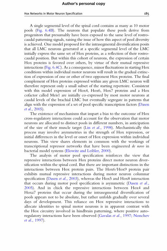
A single segmental level of the spinal cord contains as many as 10 motorpools (Fig. 6.4B). The neurons that populate these pools derive fromprogenitors that presumably have been exposed to the same level of rostro-caudal patterning signals, raising the issue of how this aspect of pool diversityis achieved. One model proposed for the intrasegmental diversification positsthat all LMC neurons generated at a specific segmental level of the LMCinitially express the same set of Hox proteins, as a reflection of their rostro-caudal position. But within this cohort of neurons, the expression of certainHox proteins is favored over others, by virtue of their mutual repressiveinteractions (Fig. 6.4C). As a consequence, minor fluctuations in starting Hoxconditions within individual motor neurons will result in the gradual extinc-tion of expression of one or other of two opponent Hox proteins. The finalcomplement of Hox proteins expressed within any given LMC neuron willtherefore represent only a small subset of the starting repertoire. Consistentwith this model expression of Hox4, Hox6, Hox7 proteins and a Hoxcofactor called Meis1 are initially co-expressed by most motor neurons atcaudal levels of the brachial LMC but eventually segregate in patterns thatalign with the expression of a set of pool specific transcription factors (Dasenet al., 2005).
The existence of mechanisms that impart a bias to the outcome of Hoxcross-regulatory interactions could account for the observation that motorneurons are allocated to distinct pools in different numbers, in anticipationof the size of their muscle target (Lin et al., 1998). Mechanistically thisprocess may involve asymmetries in the strength of Hox repression, orinitial differences in the level or onset of Hox expression within individualneurons. This view shares elements in common with the workings oftranscriptional repressor networks that have been engineered de novo inbacterial model systems (Elowitz and Leibler, 2000).
The analysis of motor pool specification reinforces the view thatrepressive interactions between Hox proteins direct motor neuron diver-sification within the spinal cord. But there are important distinctions in theinteractions between Hox protein pairs. The Hox6/Hox9 protein pairexhibits mutual repressive interactions during motor neuron columnarspecification (Dasen et al., 2003), whereas the Hox5 and Hox8 interactionthat occurs during motor pool specification is asymmetric (Dasen et al.,2005). And in chick the repressive interactions between Hox4 andHoxa7 proteins that occur during the intrasegmental diversification ofpools appears not to be absolute, but rather unfolds gradually over severaldays of development. This reliance on Hox repressive interactions toallocate identities to spinal motor neurons is in apparent contrast withthe Hox circuitry involved in hindbrain patterning, where positive auto-regulatory interactions have been observed (Gavalas et al., 1997; Nonchevet al., 1997).

186 Jeremy S. Dasen and Thomas M. Jessell
Author's personal copy
5.2. Hox genes control the specificity of motorneuron-muscle connectivity
Howmight Hox activities in motor pools coordinate motor axon trajectoryto specific muscle targets in the developing limb? On arriving at the base ofthe limb, the axons of LMC neurons select a ventral or dorsal trajectory inthe limb mesenchyme, and then establish specific anteroposterior andproximo-distal trajectories which take them to the position of newlycleaved muscle masses (Tosney and Landmesser, 1985a). As developingaxons project into the limb they navigate through a series of choice pointsen route to their synaptic targets. A major output of the Hox network inmotor neurons is the control of downstream transcription factor expression,some of which are necessary for motor axon guidance decisions (Fig. 6.4).To what extent are the patterns of motor axon innervation driven throughHox-regulated intermediate transcription factors and to what extent mightthey be controlled directly by Hox targets?
As described earlier, aspects of limb innervation pattern can be linked tothe program of columnar specification. The Hox6/10-activated program ofLMC specification appears to direct axons toward the limb and determine apattern of LIM-homeodomain protein expression that controls the dorsoven-tral trajectory of motor axons, through regulation of EphA4 expression(Kania and Jessell, 2003). The selection of certain muscle-specific nervetrajectories appears to be determined through activities of the Hox-inducedtranscription factors. Nkx6 homeodomain proteins are expressed by subsets ofLMC neurons in a pool-specific pattern that is controlled by Hox proteins(Dasen et al., 2003; De Marco Garcia and Jessell, 2008). In Nkx6.1 mutantmice these motor neuron pools fail to innervate their normal target andinvade foreign muscle targets (De Marco Garcia and Jessell, 2008). Anotherpool-specific Hox target, Pea3, is required for distinct aspects of motorneuron differentiation, including the clustering of neurons into pools,muscle-specific axonal arborization, and synaptic input onto motor neuronsfrom sensory neurons (Livet et al., 2002; Vrieseling and Arber, 2006). Thusmultiple facets of the Hox programming of motor pool identities areregulated through intermediate transcription factors.
Could Hox proteins exert a more direct role in the control of axonguidance decisions within the limb? After making their initial dorsoventralchoice at the base of the limb, motor axons follow cues that guide themalong the anteroposterior and proximo-distal axes (Stirling and Summerbell,1988). The basic pattern of muscle nerve branches is preserved in theabsence of the target muscle itself (Lewis et al., 1981; Phelan and Hollyday,1990, 1991), implicating the limb mesenchyme as a source of cues thatspecify motor axon trajectories. Hox proteins are also expressed by the limbmesenchyme (Izpisua-Belmonte and Duboule, 1992) and could contributeto the establishment of axonal trajectory by positioning guidance cues at

Hox Networks in Motor Neuron Specification 187
Author's personal copy
specialized decision regions (Tosney and Landmesser, 1985a). The existenceof a topographic relationship between cell body position and projectionsalong these axes raises the possibility that Hox proteins also exert direct rolesin the control of specific guidance receptors, or regulate other determinantsthat direct innervation specificity within the limb.
5.3. Extrinsic and intrinsic programming of motorpool identities
While many aspects of motor pool identity appear to be programmedthrough cell-intrinsic Hox transcriptional networks, the expression ofcertain pool specific transcription factors relies on the presence extrinsicsignals from the periphery. Expression of the ETS transcription factors Pea3and Er81 in pools depends on target-derived neurotrophic signals providedby the limb mesoderm and muscle targets (Haase et al., 2002; Lin et al., 1998).These signals appear to be permissive rather than instructive and not allmotor neurons are competent in their ability to respond to neurotrophins.In explants of spinal cord treated with glial-derived neurotrophic factor(GDNF), Pea3 is induced in a pattern approximating the normal numberin vivo and confined to the level of the spinal cord which normally expressesPea3 (Haase et al., 2002). Thus not all motor neurons are equivalent in theirability to respond to GDNF.
The competence of motor neurons to activate ETS genes appears to beconstrained by their pattern of Hox expression. Ectopic expression of Hoxc8in LMC neurons is sufficient to expand the domain of Pea3 expression (Dasenet al., 2005), suggesting the normal domain of Hoxc8 expression defines theregion of GDNF competence. Consistent with this hypothesis, in Hoxc8mutants motor neurons fail to fully activate Pea3 expression (Vermot et al.,2005), likely as a consequence of the inability of motor neurons to respond toGDNF. Hox proteins may therefore control the expression of targets thatendow motor neurons with the ability to respond to peripheral cues. Hoxtargets may include receptors for neurotrophic factors or other componentsnecessary for activation of the GDNF pathway.
These observations suggest that despite the importance of Hox-dependent steps in motor neuron specification, target-derived cues alsocontribute to the transcriptional programming of motor pools. Expressionof the target-induced factor Pea3 is critical for later aspects of motor pooldifferentiation such as clustering of motor neurons into pools, muscle-specific patterns of axonal innervation, and sensory-motor connectivity(Livet et al., 2002; Vrieseling and Arber, 2006). Thus, motor pool specifi-cation appears to unfold in two main phases: an early phase that confersaspects of motor neuron identity involved in the selection of target muscleconnectivity (Landmesser, 2001; Milner and Landmesser, 1999), and a laterphase, operating after motor axons have reached their muscle targets, that is

188 Jeremy S. Dasen and Thomas M. Jessell
Author's personal copy
associated with ETS gene expression and the clustering of motor neuronswithin the LMC (Livet et al., 2002; Price et al., 2002).
6. Restriction and Refinement of Hox Activities
During Motor Neuron Differentiation
Although Hox protein activities appear to be critical in the generationof diverse motor neuron subtypes, several lines of evidence suggest thatadditional factors are necessary to restrict their functions. First, Hox proteinsare broadly expressed throughout the embryo, and within the CNS they areexpressed by multiple classes of neurons including interneurons and sensoryneurons (Belting et al., 1998; Dasen et al., 2005; Ensini et al., 1998). Second,within a given spinal segment the same Hox protein can be expressed bymultiple columnar subtypes (Dasen et al., 2008). These observations suggestthe requirement for additional mechanisms to gate the actions of Hoxproteins in motor neurons. This gating function may be controlled throughtranscriptional cofactors or through motor neuron-specific targets of Hoxproteins.
Hox functions generally rely on interactions with a conserved family ofDNA binding cofactors that refine and constrain their activities (Mann andAffolter, 1998). Two classes of canonical Hox cofactors, Meis and Pbx/Prepproteins (vertebrates homologs of the Drosophila Extradenticle and Homo-thorax proteins), have pervasive roles as regulators of Hox activity (Mannand Affolter, 1998; and Selleri, 2006). Within the spinal cord Meis and Pbx/Prep proteins display broad patterns of expression (Dasen et al., 2005), andthus they are unlikely candidates as cell-type specific regulators of motorneuron Hox target specificities. Studies in Drosophila indicate that a distinctgroup of Hox accessory factors, the homeodomain protein Engrailed andthe forkhead homeodomain proteins Sloppy-paired 1/2, functions withExtradenticle and Homothorax to modify Hox activities (Gebelein et al.,2004). These factors have more restricted domains of expression andactivity: Engrailed regulates Hox activity in posterior compartment cellswhereas Slp1/2 regulate Hox activity in anterior cells (Gebelein et al., 2004).
The vertebrate counterparts of these ancillary Hox cofactors, Engrailedand Fox proteins are expressed by subsets of spinal neurons, and a vertebrateFoxP protein, FoxP1, is selectively expressed by spinal motor neurons(Saueressig et al., 1999; Shu et al., 2001; Tamura et al., 2003). Consistentwith a role in gating Hox activities, expression of FoxP1 is restricted to twoHox sensitive columns (PGC and LMC neurons) while it is excluded fromHox-independent populations (HMC and MMC neurons). This exclusionfrom HMC and MMC motor neurons appears to be a function of thepresence of inhibitory factors within these subtypes that prevent FoxP1

Hox Networks in Motor Neuron Specification 189
Author's personal copy
expression shortly after motor neurons are born (Dasen et al., 2008).As described below, recent studies indicate that FoxP1 is an essentialcofactor in the Hox-dependent program of differentiation in LMC andPGC neurons (Dasen et al., 2008; Rousso et al., 2008)
6.1. FoxP1: An accessory factor for Hox proteinsin motor columns and pools
Although FoxP1 is expressed by LMC and PGC neurons, the levels of FoxP1protein in these two columns differ dramatically: PGC neurons express lowlevels of FoxP1 and LMC neurons express high levels. These differences inFoxP1 levels appear to be set by the pattern of Hox expression as misexpres-sion of Hox determinants of LMC identities such as Hoxc6 and Hoxd10 canswitch HMC and PGC neurons to FoxP1 high LMC motor neurons atthoracic levels (Dasen et al., 2008). In turn, the levels of FoxP1 protein appearto be critical in the specification of columnar fates as misexpression of FoxP1in PGC and HMC neurons can convert these columns to LMC identities.These actions of FoxP1 appear to require continuous Hox function, sinceforced expression of FoxP1 is unable to induce LMC fates under conditionsin which Hox activity is repressed (Dasen et al., 2008). Thus FoxP1 appears toact jointly with Hox proteins rather than as a linear intermediary in thepathway of columnar specification (Fig. 6.5).
Consistent with a central role in the Hox-dependent steps of motorneuron differentiation, genetic inactivation of Foxp1 does not effect thegeneration of motor neurons, or Hox expression, but nevertheless thenumber of LMC and PGC neurons is dramatically reduced (Dasen et al.,2008; Rousso et al., 2008). Instead prospective LMC and PGC motorneurons acquire molecular features of HMC neurons and the spinal cordconsists almost entirely of two continuous columns (Fig. 6.5C). Strikingly allHox-dependent steps of motor neuron differentiation, included the colum-nar, divisional and pool identities, are effectively erased. Molecular features ofLMC divisional and pool identities are eroded in Foxp1mutants including theloss of specific LMC transcription factors (e.g., Lhx1, Pea3,Nkx6.1) as well asconnectivity and synaptic specificity determinants (EphA4, Sema3E, Cad20)(Dasen et al., 2008).
Although FoxP1 activities are essential for the transcriptional output ofHox proteins in LMC and PGC neurons, not all motor neuron Hoxfunctions depend on FoxP1. Expression of FoxP1 is induced by Hoxproteins, and thus this aspect of Hox function must be FoxP1 independent.In addition the repressive interactions between Hox paralogs are evident inMMC and HMC neurons (Dasen et al., 2003), even though they lackFoxP1 expression. The activities of Hox genes involved in hindbrainmotor neuron specification (Trainor and Krumlauf, 2000) also do notrequire FoxP1, since FoxP proteins are not expressed in motor neurons at

Hox6/10
FoxP1hi
Hox9
FoxP1lo
Hox
Hox
Hox Hox
RALDH2
pSmad, nNOS
Nkx6.1Pea3
Lhx1Hb9
Isl1
Pool
Column
MMC
Non-MMC
Tetrapod : thoracic
Tetrapod : limb
MMC
MMC
HMC
Divisional
LMC
HMC
Pools
MMC
HMCMMC
PGC
Wild type
Foxp1 -/-
FoxP1lo- PGC
FoxP1hi- LMC
LMC
PGC
Hox9
Hox6/10
A
B
FoxP1hi
FoxP1off
FoxP1loHox9
Hox6/10
FoxP1off
DC
MMC
MMCHMC
PGC
LMC
Figure 6.5 TheHox/FoxP1genenetwork and the emergenceofmotorneurondiversity.(A)Model for theemergenceofmotorneuroncolumnar subtypes.Thepresumedancestralstate ofmotor neurons is that ofMMCneurons. Early aquatic vertebrates appear to possesstwo columnar subtypes, dorsally projectingMMCandventrally projectingHMCneurons.In tetrapod vertebrates expression of FoxP1 at thoracic levels is under the control ofHox9proteins, allowing for the generation of PGC neurons. At limb levels expression of FoxP1is under the control of Hox6 and Hox10 proteins, allowing for the generation of LMCneurons and its resident motor pools. (B) The levels of FoxP1 are controlled by Hoxproteins in PGC and LMC neurons. HMC but not MMC neurons are competent torespond the patterning activities ofHox genes,which control the level of FoxP1 expressionand columnar fates. (C) Motor neuron columnar differentiation in wild type and Foxp1mutant mice, showing that FoxP1 controls the formation of PGC and LMC columns, aswell as the diversification of motor pools within the LMC. In the absence of FoxP1, twocontinuous columns (MMCandHMC) are generated. (D) Interactions of FoxP1 andHoxproteins during the transcriptional programmingofmotorneuroncolumnar andpool fates.FoxP1gates the activities of the twoHoxnetworks involved in the assignment of columnarand pool identities. The Hox/FoxP1 network controls a repertoire of downstream genesincluding transcription factors, signaling molecules, and guidance receptors.
190 Jeremy S. Dasen and Thomas M. Jessell
Author's personal copy
Hox Networks in Motor Neuron Specification 191
Author's personal copy
this level of the neuraxis. Meis and Pbx/Prep cofactors are expressed in thehindbrain, and Pbx proteins regulate cranial motor neuron identity (Moensand Selleri, 2006). Thus, the activities of FoxP1 as a mediator of Hox outputduring motor neuron differentiation may be exerted in a broader context ofMeis and Pbx/Prep activities. Meis and Pbx/Prep cofactors may impart afirst level of Hox specificity ( Joshi et al., 2007), with FoxP1 providingadditional filters on target gene activation.
6.2. Coordinate control of motor axon targeting by FoxP1and Hox factors
How does FoxP1/Hox network activity control the decisions taken bymotor axons as they project to their targets? At thoracic levels of the spinalcord, the switch from PGC to HMC fate in Foxp1 mutants is accompaniedby a redirection of motor axons from sympathetic ganglia to body walltargets (Dasen et al., 2008). In contrast, HMC-like motor neurons generatedat brachial and lumbar levels in Foxp1 mutants still project into the limb,following a path similar to that of LMC axons. This observation is consistentwith studies showing that after transplantation of thoracic spinal cord tolimb levels, motor axons are capable of innervating the limb (O’Brien et al.,1990; O’Brien and Oppenheim, 1990). Further evidence that this is thepredicted trajectory of HMC neurons generated at limb levels comes frommore recent studies showing that ectopic limbs induced adjacent to thoracicspinal cord are innervated selectively by axons of HMC neurons (Turneyet al., 2003). Thus, HMC and LMC neurons appear to be similar in theirinitial pursuit of a distal trajectory that takes them to body wall or limb targets.
In the limb, LMC axons establish stereotypic projections to individualmuscles (Landmesser, 2001). The establishment of these precise patterns ofconnectivity requires the actions of a multitude of Hox-dependent transcrip-tion factors, recognition molecules and guidance receptors. Despite the loss ofexpression of these factors in Foxp1 mutants, nerve branching and limbmuscle innervation patterns are remarkably well preserved (Dasen et al.,2008; Rousso et al., 2008). These findings fit best with the view that theoverall pattern of motor nerve branching within the limb is determinedby preestablished permissive or inhibitory domains within the limb mesen-chyme, with the FoxP1/Hox program providing LMC neurons withidentities that enable axons to respond to local cues that promote the selectionof just one of many available conduits. In this view, the trajectory of motorneurons deprived of FoxP1 activity will still be constrained by the existence ofpreordained paths, but without Hox-dependent intrinsic molecular program-ming, individual axons are reduced to choosing haphazardly among theirpotential options. Consistent with this model, in Foxp1 mutants the topo-graphic map of motor projections is lost, as motor neurons no longer projectinto the limb in an organized manner (Dasen et al., 2008).

192 Jeremy S. Dasen and Thomas M. Jessell
Author's personal copy
6.3. Hox/FoxP1 interactions and the origins of motorneuron diversity
Analysis of the Hox/FoxP1 transcriptional network has provided clues intothe possible mechanisms by which the tetrapod motor system emergedduring evolution. Aquatic vertebrates with behaviors driven by axial andhypaxial muscles lack PGC and LMC neurons but possess neurons that aresimilar to MMC and HMC neurons in character (Fetcho, 1992; Kusakabeand Kuratani, 2005). The appearance of LMC and PGC neurons is linked tothe formation of paired appendages (lateral fins and limbs) and a sympatheticnervous system—structures that emerged later in vertebrate evolution(Fetcho, 1992; Freitas et al., 2006; Funakoshi and Nakano, 2007). TheHox/FoxP1 dependent program of motor neuron diversification maytherefore have evolved to meet the demands of a new and diverse set ofperipheral target tissues, and to generate more elaborate set of motorbehaviors.
How did the Hox/FoxP1 transcriptional network emerge within motorneurons? The ancestral state of spinal motor neurons appears to be markedby expression of Lhx3, Isl1/2, and Hb9, a set of transcription factors con-served in the motor neurons of invertebrates (Landgraf and Thor, 2006).This transcriptional profile also defines early-born primary motor neuronsof Zebrafish and Xenopus embryos (Appel et al., 1995; Borodinsky et al.,2004), presumed counterparts of the motor neurons of jawless vertebrates(Fetcho, 1992), as well as the MMC neurons of birds and mammals ( Jessell,2000). The conserved transcriptional profile of this ancestral set of motorneurons may reflect common patterns of connectivity—the innervation ofsegmentally arrayed muscles involved in undulatory locomotor behaviors.The diversification of columnar subtypes from this ancestral group requiresrelief from the confining influence of the LIM-homeodomain factor Lhx3,as Lhx3 exerts a dominant activity in specifying MMC fates over othercolumnar subtypes (Sharma et al., 2000; William et al., 2003). This evasivestep may have involved a decrease in the strength of the Wnt signalingcomponent of the dorsoventral inductive pathway, since reducing Wnt4/5activity in mice promotes the generation of HMC neurons at the expense ofMMC neurons (Agalliu et al., 2009). Thus the basic spinal motor systeminduced by the dorsoventral signaling pathway comprises MMC and HMCneurons, arrayed in coextensive columns.
The induction of Foxp1 expression under the regulatory control of Hoxproteins may have permitted the twinned columnar organization of limblessvertebrates to diversify and generate PGC and LMC neurons. The formationof HMC neurons from an ancestral MMC-like group was a crucial step in thisdiversification program—creating a malleable population of Lhx3-negativemotor neurons to serve as the substrate for the FoxP1/Hox programof columnar and pool specification. Prospective HMC neurons, freed from
Hox Networks in Motor Neuron Specification 193
Author's personal copy
the dominant actions of Lhx3, became competent to respond to the rostro-caudal patterning activities of Hox proteins, allowing them to acquire PGC orLMC fates. FoxP1, through its dose-dependent inductive activity, is a keymediator of the Hox program of columnar specification (Figure 6.5). Underthe influence of Hox9 activity, newly generated Lhx3-negative thoracicmotor neurons acquire the capacity for low-level FoxP1 expression andappear to resolve their bi-potential HMC or PGC fate through mutualrepressive interactions between FoxP1 and Hb9 (Dasen et al., 2008). In thelimb-level domains of Hox6/10 expression, FoxP1 is induced at high levelswhich allow motor neurons to acquire an LMC fate.
The mechanisms by which the FoxP1/Hox network was recruited tothe task of motor neuron columnar diversification remains unclear. Theabsence of PGC and LMC neurons from early vertebrates could have itsbasis in changes in the cis-regulatory elements that control Foxp1 expression(Prud’homme et al., 2007; Shubin et al., 1997), such that Hox-sensitiveelements responsible for expression in spinal motor neurons were config-ured only at the time of formation of the sympathetic nervous system andpaired appendages. Alternatively, the rostrocaudal pattern of expression ofHox genes in the spinal cord of jawless vertebrates may differ from that inbirds and mammals (Force et al., 2002; Takio et al., 2007), and thus may failto produce a productive Hox code capable of activating FoxP1 expression.Further investigation into the regulatory elements controlling the Foxp1and Hox genes and their targets in motor neurons may provide insights intohow columnar subtypes arose in diverse vertebrate species.
6.4. Coordinate regulation of neuronal and mesodermalHox programs
The same extrinsic signaling systems which pattern Hox profiles in the CNSalso control Hox expression within the mesoderm, raising the possibility thatregulation of Hox expression in these distinct tissues may be involved incoordinating the position of motor neuron subtypes with the location andidentity of their peripheral target fields. One aspect of this program is theestablishment of a register between the rostrocaudal positionsmotor columnsand their targets. Application of FGFs to thoracic mesoderm has been shownto induce the formation of an ectopic limb in place of body wall mesoderm(Cohn et al., 1995), and the regulation of Hox9 gene paralog expression inlateral plate mesoderm in response to FGFs has been implicated to determinelimb bud position (Cohn et al., 1997). These observations support the ideathat exposure of neural and lateral platemesodermal cells to a common sourceof FGFs establishes distinct profiles of Hox gene expression in these twotissues, profiles that in turn control the alignment of LMC and limb position.
Similarly, the muscle targets of LMC motor neurons within the limbappear to acquire aspects of their identity throughHox-dependent programs.

194 Jeremy S. Dasen and Thomas M. Jessell
Author's personal copy
Limb musculature is derived from a population of migratory muscle pre-cursors that is generated selectively at limb levels through the actions ofthe LIM-homeodomain protein Lbx1 (Brohmann et al., 2000; Gross et al.,2000). Interestingly, a Hox determinant of LMC identity, Hoxa10, hasbeen shown to be sufficient to induce Lbx1 expression in muscle precursorsat thoracic levels and to reprogram nonmigratory myoblasts into migratorymuscle cells (Alvares et al., 2003). Moreover, Hox genes control patterningwithin the limb mesenchyme and may therefore exert additional roles in theguidance of axons along the anterior-posterior and proximal-distal axes ofthe limb (Izpisua-Belmonte andDuboule, 1992), prior to establishment of themature pattern of musculature. Thus, the coordinate regulation of Hoxexpression patterns during vertebrate evolution may provide a plausiblebasis for linking the formation and diversification of LMC motor neuronsubtypes to the appearance and patterning of paired appendages.
7. Conclusions
The task of specifying hundreds distinct motor neuron subtypes, eachprojecting to a specific target cell group, appears to have been met bydeploying a regulatory network of nearly two dozen Hox genes. The selec-tive connections formed between motor neurons and muscle are howeverjust one aspect of circuit assembly in the spinal cord. Additional componentsof motor neuron connectivity, such as the specificity of synaptic inputsfrom sensory neurons and interneurons, may also rely on the actions ofthe FoxP1/Hox transcriptional network. Hox and Fox proteins areexpressed by other classes of spinal neurons, suggesting these transcriptionfactor families could have amore extensive role in the assembly of locomotorcircuits. The self-organizing features inherent in the FoxP1/Hox networkmay have therefore helped endow developing motor circuits with theirapparent high degree of genetic determination.
Studies on the transcriptional programs that control diversity and synap-tic specificity in the spinal cord have provided a valuable model systemfor understanding the mechanisms of neuronal specification throughout theCNS. Emerging work on retinal and cortical neurons suggest an equivalenthigh degree of subtype diversity, even within a single class of neurons(Klausberger and Somogyi, 2008; Kong et al., 2005). Although retinal andcortical neurons are devoid of chromosomally clustered Hox gene expres-sion, it is likely that similar cell-intrinsic transcriptional networks controlneuronal specification throughout the CNS. Whether other CNS circuitsutilize coherent gene families to generate highly diverse neuronal subtypesremains to be determined.

Hox Networks in Motor Neuron Specification 195
Author's personal copy
REFERENCES
Agalliu, D., Takada, S., Agalliu, I., McMahon, A. P., and Jessell, T. M. (2009). Motor neuronswith axial muscle projections specified by Wnt4/5 signaling. Neuron. 61, 708–720.
Alvares, L. E., Schubert, F. R., Thorpe, C., Mootoosamy, R. C., Cheng, L., Parkyn, G.,Lumsden, A., and Dietrich, S. (2003). Intrinsic, Hox-dependent cues determine the fateof skeletal muscle precursors. Dev. Cell 5, 379–390.
Appel, B., Korzh, V., Glasgow, E., Thor, S., Edlund, T., Dawid, I. B., and Eisen, J. S.(1995). Motoneuron fate specification revealed by patterned LIM homeobox geneexpression in embryonic zebrafish. Development 121, 4117–4125.
Arber, S., Han, B., Mendelsohn, M., Smith, M., Jessell, T. M., and Sockanathan, S. (1999).Requirement for the homeobox gene Hb9 in the consolidation of motor neuronidentity. Neuron 23, 659–674.
Belting, H. G., Shashikant, C. S., and Ruddle, F. H. (1998). Multiple phases of expressionand regulation of mouse Hoxc8 during early embryogenesis. J. Exp. Zool. 282, 196–222.
Bel-Vialar, S., Itasaki, N., and Krumlauf, R. (2002). Initiating Hox gene expression: In theearly chick neural tube differential sensitivity to FGF and RA signaling subdividesthe HoxB genes in two distinct groups. Development 129, 5103–5115.
Borodinsky, L. N., Root, C. M., Cronin, J. A., Sann, S. B., Gu, X., and Spitzer, N. C. (2004).Activity-dependent homeostatic specification of transmitter expression in embryonicneurons. Nature 429, 523–530.
Briscoe, J., Pierani, A., Jessell, T. M., and Ericson, J. (2000). A homeodomain protein codespecifies progenitor cell identity and neuronal fate in the ventral neural tube. Cell 101,435–445.
Brohmann, H., Jagla, K., and Birchmeier, C. (2000). The role of Lbx1 in migration ofmuscle precursor cells. Development 127, 437–445.
Carpenter, E. M. (2002). Hox genes and spinal cord development. Dev. Neurosci. 24, 24–34.Choe, A., Phun, H. Q., Tieu, D. D., Hu, Y. H., and Carpenter, E. M. (2006). Expression
patterns of Hox10 paralogous genes during lumbar spinal cord development. Gene Expr.Patterns 6, 730–737.
Chopra, V. S., and Mishra, R. K. (2006). ‘‘Mir’’acles in Hox gene regulation. Bioessays 28,445–448.
Cohn, M. J., Izpisua-Belmonte, J. C., Abud, H., Heath, J. K., and Tickle, C. (1995).Fibroblast growth factors induce additional limb development from the flank of chickembryos. Cell 80, 739–746.
Cohn, M. J., Patel, K., Krumlauf, R., Wilkinson, D. G., Clarke, J. D., and Tickle, C. (1997).Hox9 genes and vertebrate limb specification. Nature 387, 97–101.
Dasen, J. S., Liu, J. P., and Jessell, T. M. (2003). Motor neuron columnar fate imposed bysequential phases of Hox-c activity. Nature 425, 926–933.
Dasen, J. S., Tice, B. C., Brenner-Morton, S., and Jessell, T. M. (2005). A Hox regulatorynetwork establishes motor neuron pool identity and target-muscle connectivity. Cell123, 477–491.
Dasen, J. S., De Camilli, A., Wang, B., Tucker, P. W., and Jessell, T. M. (2008). Hoxrepertoires for motor neuron diversity and connectivity gated by a single accessory factor,FoxP1. Cell 134, 304–316.
De Marco Garcia, N. V., and Jessell, T. M. (2008). Early motor neuron pool identity andmuscle nerve trajectory defined by postmitotic restrictions in Nkx6.1 activity.Neuron 57,217–231.
Dessaud, E., McMahon, A. P., and Briscoe, J. (2008). Pattern formation in the vertebrateneural tube: A sonic hedgehog morphogen-regulated transcriptional network. Development135, 2489–2503.
196 Jeremy S. Dasen and Thomas M. Jessell
Author's personal copy
Diez del Corral, R., and Storey, K. G. (2004). Opposing FGF and retinoid pathways:A signalling switch that controls differentiation and patterning onset in the extendingvertebrate body axis. Bioessays 26, 857–869.
Duboule, D., and Morata, G. (1994). Colinearity and functional hierarchy among genes ofthe homeotic complexes. Trends Genet. 10, 358–364.
Dubrulle, J., and Pourquie, O. (2004). fgf8 mRNA decay establishes a gradient that couplesaxial elongation to patterning in the vertebrate embryo. Nature 427, 419–422.
Eberhart, J., Swartz, M. E., Koblar, S. A., Pasquale, E. B., and Krull, C. E. (2002). EphA4constitutes a population-specific guidance cue for motor neurons. Dev. Biol. 247,89–101.
Elowitz, M. B., and Leibler, S. (2000). A synthetic oscillatory network of transcriptionalregulators. Nature 403, 335–338.
Ensini, M., Tsuchida, T. N., Belting, H. G., and Jessell, T. M. (1998). The control ofrostrocaudal pattern in the developing spinal cord: Specification of motor neuron subtypeidentity is initiated by signals from paraxial mesoderm. Development 125, 969–982.
Fetcho, J. R. (1987). A review of the organization and evolution of motoneurons innervatingthe axial musculature of vertebrates. Brain Res. 434, 243–280.
Fetcho, J. R. (1992). The spinal motor system in early vertebrates and some of its evolutionarychanges. Brain Behav. Evol. 40, 82–97.
Force, A., Amores, A., and Postlethwait, J. H. (2002). Hox cluster organization in the jawlessvertebrate Petromyzon marinus. J. Exp. Zool. 294, 30–46.
Freitas, R., Zhang, G., and Cohn, M. J. (2006). Evidence that mechanisms of fin developmentevolved in the midline of early vertebrates. Nature 442, 1033–1037.
Funakoshi, K., and Nakano, M. (2007). The sympathetic nervous system of anamniotes.Brain Behav. Evol. 69, 105–113.
Gavalas, A., Davenne, M., Lumsden, A., Chambon, P., and Rijli, F. M. (1997). Role ofHoxa-2 in axon pathfinding and rostral hindbrain patterning. Development 124,3693–3702.
Gebelein, B., McKay, D. J., and Mann, R. S. (2004). Direct integration of Hox andsegmentation gene inputs during Drosophila development. Nature 431, 653–659.
Glover, J. C., Renaud, J. S., and Rijli, F. M. (2006). Retinoic acid and hindbrain patterning.J. Neurobiol. 66, 705–725.
Gross, M. K., Moran-Rivard, L., Velasquez, T., Nakatsu, M. N., Jagla, K., andGoulding, M. (2000). Lbx1 is required for muscle precursor migration along a lateralpathway into the limb. Development 127, 413–424.
Guthrie, S. (2007). Patterning and axon guidance of cranial motor neurons. Nat. Rev.Neurosci. 8, 859–871.
Gutman, C. R., Ajmera, M. K., and Hollyday, M. (1993). Organization of motor poolssupplying axial muscles in the chicken. Brain Res. 609, 129–136.
Haase, G., Dessaud, E., Garces, A., de Bovis, B., Birling, M., Filippi, P., Schmalbruch, H.,Arber, S., and deLapeyriere, O. (2002). GDNF acts through PEA3 to regulate cell bodypositioning and muscle innervation of specific motor neuron pools. Neuron 35, 893–905.
Hollyday, M. (1980a). Motoneuron histogenesis and the development of limb innervation.Curr. Top. Dev. Biol. 15, 181–215.
Hollyday, M. (1980b). Organization of motor pools in the chick lumbar lateral motorcolumn. J. Comp. Neurol. 194, 143–170.
Hollyday, M. (1995). Chick wing innervation. I. Time course of innervation and earlydifferentiation of the peripheral nerve pattern. J. Comp. Neurol. 357, 242–253.
Hollyday, M., and Jacobson, R. D. (1990). Location of motor pools innervating chick wing.J. Comp. Neurol. 302, 575–588.
Izpisua-Belmonte, J. C., and Duboule, D. (1992). Homeobox genes and pattern formationin the vertebrate limb. Dev. Biol. 152, 26–36.

Hox Networks in Motor Neuron Specification 197
Author's personal copy
Jegalian, B. G., and De Robertis, E. M. (1992). Homeotic transformations in the mouseinduced by overexpression of a human Hox3.3 transgene. Cell 71, 901–910.
Jessell, T. M. (2000). Neuronal specification in the spinal cord: Inductive signals andtranscriptional codes. Nat. Rev. Genet. 1, 20–29.
Joshi, R., Passner, J. M., Rohs, R., Jain, R., Sosinsky, A., Crickmore, M. A., Jacob, V.,Aggarwal, A. K., Honig, B., and Mann, R. S. (2007). Functional specificity of a Hoxprotein mediated by the recognition of minor groove structure. Cell 131, 530–543.
Kania, A., and Jessell, T. M. (2003). Topographic motor projections in the limb imposed byLIM homeodomain protein regulation of ephrin-A:EphA interactions. Neuron 38,581–596.
Kania, A., Johnson, R. L., and Jessell, T. M. (2000). Coordinate roles for LIM homeoboxgenes in directing the dorsoventral trajectory of motor axons in the vertebrate limb. Cell102, 161–173.
Klausberger, T., and Somogyi, P. (2008). Neuronal diversity and temporal dynamics:The unity of hippocampal circuit operations. Science 321, 53–57.
Kmita, M., and Duboule, D. (2003). Organizing axes in time and space; 25 years of colineartinkering. Science 301, 331–333.
Kong, J. H., Fish, D. R., Rockhill, R. L., and Masland, R. H. (2005). Diversity of ganglioncells in the mouse retina: Unsupervised morphological classification and its limits.J. Comp. Neurol. 489, 293–310.
Kusakabe, R., and Kuratani, S. (2005). Evolution and developmental patterning of thevertebrate skeletal muscles: Perspectives from the lamprey. Dev. Dyn. 234, 824–834.
Lance-Jones, C., and Landmesser, L. (1980). Motoneurone projection patterns in embryonicchick limbs following partial deletions of the spinal cord. J. Physiol. 302, 559–580.
Lance-Jones, C., Omelchenko, N., Bailis, A., Lynch, S., and Sharma, K. (2001). Hoxd10induction and regionalization in the developing lumbosacral spinal cord. Development128, 2255–2268.
Landgraf, M., and Thor, S. (2006). Development ofDrosophila motoneurons: Specification andmorphology. Semin. Cell Dev. Biol. 17, 3–11.
Landmesser, L. (1978a). The development of motor projection patterns in the chick hindlimb. J. Physiol. 284, 391–414.
Landmesser, L. (1978b). The distribution of motoneurones supplying chick hind limbmuscles. J. Physiol. 284, 371–389.
Landmesser, L. T. (2001). The acquisition of motoneuron subtype identity and motor circuitformation. Int. J. Dev. Neurosci. 19, 175–182.
Lemons, D., and McGinnis, W. (2006). Genomic evolution of Hox gene clusters. Science313, 1918–1922.
Lewis, J., Chevallier, A., Kieny, M., and Wolpert, L. (1981). Muscle nerve branches do notdevelop in chick wings devoid of muscle. J. Embryol. Exp. Morphol. 64, 211–232.
Lin, J. H., Saito, T., Anderson, D. J., Lance-Jones, C., Jessell, T. M., and Arber, S. (1998).Functionally related motor neuron pool and muscle sensory afferent subtypes defined bycoordinate ETS gene expression. Cell 95, 393–407.
Liu, J. P. (2006). The function of growth/differentiation factor 11 (Gdf11) in rostrocaudalpatterning of the developing spinal cord. Development 133, 2865–2874.
Liu, J. P., Laufer, E., and Jessell, T. M. (2001). Assigning the positional identity of spinalmotor neurons: Rostrocaudal patterning of Hox-c expression by FGFs, Gdf11, andretinoids. Neuron 32, 997–1012.
Livet, J., Sigrist, M., Stroebel, S., De Paola, V., Price, S. R., Henderson, C. E., Jessell, T. M.,and Arber, S. (2002). ETS gene Pea3 controls the central position and terminal arborizationof specific motor neuron pools. Neuron 35, 877–892.
Mann, R. S., and Affolter, M. (1998). Hox proteins meet more partners. Curr. Opin. Genet.Dev. 8, 423–429.
198 Jeremy S. Dasen and Thomas M. Jessell
Author's personal copy
McIntyre, D. C., Rakshit, S., Yallowitz, A. R., Loken, L., Jeannotte, L., Capecchi, M. R.,and Wellik, D. M. (2007). Hox patterning of the vertebrate rib cage. Development 134,2981–2989.
McPherron, A. C., Lawler, A. M., and Lee, S. J. (1999). Regulation of anterior/posteriorpatterning of the axial skeleton by growth/differentiation factor 11. Nat. Genet. 22,260–264.
Milner, L. D., and Landmesser, L. T. (1999). Cholinergic and GABAergic inputs drivepatterned spontaneous motoneuron activity before target contact. J. Neurosci. 19,3007–3022.
Moens, C. B., and Selleri, L. (2006). Hox cofactors in vertebrate development. Dev. Biol.291, 193–206.
Narayanan, C. H., and Hamburger, V. (1971). Motility in chick embryos with substitutionof lumbosacral by brachial and brachial by lumbosacral spinal cord segments. J. Exp. Zool.178, 415–431.
Nonchev, S., Maconochie, M., Gould, A., Morrison, A., and Krumlauf, R. (1997). Cross-regulatory interactions betweenHox genes and the control of segmental expression in thevertebrate central nervous system. Cold Spring Harb. Symp. Quant. Biol. 62, 313–323.
Nordstrom, U., Maier, E., Jessell, T. M., and Edlund, T. (2006). An early role for WNTsignaling in specifying neural patterns of Cdx andHox gene expression and motor neuronsubtype identity. PLoS Biol. 4, e252.
Novitch, B. G., Chen, A. I., and Jessell, T. M. (2001). Coordinate regulation of motorneuron subtype identity and pan-neuronal properties by the bHLH repressor Olig2.Neuron 31, 773–789.
O’Brien, M. K., and Oppenheim, R. W. (1990). Development and survival of thoracicmotoneurons and hindlimb musculature following transplantation of the thoracic neuraltube to the lumbar region in the chick embryo: Anatomical aspects. J. Neurobiol. 21,313–340.
O’Brien, M. K., Landmesser, L., and Oppenheim, R. W. (1990). Development and survivalof thoracic motoneurons and hindlimb musculature following transplantation of thethoracic neural tube to the lumbar region in the chick embryo: Functional aspects.J. Neurobiol. 21, 341–355.
Pfaff, S. L., Mendelsohn, M., Stewart, C. L., Edlund, T., and Jessell, T. M. (1996). Require-ment for LIM homeobox gene Isl1 in motor neuron generation reveals a motorneuron-dependent step in interneuron differentiation. Cell 84, 309–320.
Phelan, K. A., and Hollyday, M. (1990). Axon guidance in muscleless chick wings: The roleof muscle cells in motoneuronal pathway selection and muscle nerve formation.J. Neurosci. 10, 2699–2716.
Phelan, K. A., and Hollyday, M. (1991). Embryonic development and survival of brachialmotoneurons projecting to muscleless chick wings. J. Comp. Neurol. 311, 313–320.
Prasad, A., and Hollyday, M. (1991). Development and migration of avian sympatheticpreganglionic neurons. J. Comp. Neurol. 307, 237–258.
Price, S. R., De Marco Garcia, N. V., Ranscht, B., and Jessell, T. M. (2002). Regulation ofmotor neuron pool sorting by differential expression of type II cadherins.Cell 109, 205–216.
Prud’homme, B., Gompel, N., and Carroll, S. B. (2007). Emerging principles of regulatoryevolution. Proc. Natl. Acad. Sci. USA 104(Suppl 1), 8605–8612.
Romanes, G. J. (1942). The development and significance of the cell columns in the ventralhorn of the cervical and upper thoracic spinal cord of the rabbit. J. Anat. Lond. 76,112–130.
Rousso, D. L., Gaber, Z. B., Wellik, D., Morrisey, E. E., and Novitch, B. G. (2008).Coordinated actions of the forkhead protein Foxp1 and Hox proteins in the columnarorganization of spinal motor neurons. Neuron 59, 226–240.

Hox Networks in Motor Neuron Specification 199
Author's personal copy
Saueressig, H., Burrill, J., and Goulding, M. (1999). Engrailed-1 and netrin-1 regulate axonpathfinding by association interneurons that project to motor neurons. Development 126,4201–4212.
Shah, V., Drill, E., and Lance-Jones, C. (2004). Ectopic expression of Hoxd10 in thoracicspinal segments induces motoneurons with a lumbosacral molecular profile and axonprojections to the limb. Dev. Dyn. 231, 43–56.
Sharma, K., Sheng, H. Z., Lettieri, K., Li, H., Karavanov, A., Potter, S., Westphal, H., andPfaff, S. L. (1998). LIM homeodomain factors Lhx3 and Lhx4 assign subtype identities formotor neurons. Cell 95, 817–828.
Sharma, K., Leonard, A. E., Lettieri, K., and Pfaff, S. L. (2000). Genetic and epigeneticmechanisms contribute to motor neuron pathfinding. Nature 406, 515–519.
Sherrington, C. S. (1906). ‘‘The Integrative Action of the Nervous System.’’ C. Scribner’ssons, New York.
Shieh, P. (1951). The neofomation of cells of preganglionic type in the cervical spinal cord ofthe chick embryo following its transplantation to the thoracic level. J. Exp. Zool. 117,359–395.
Shimizu, T., Bae, Y. K., and Hibi, M. (2006). Cdx-Hox code controls competence forresponding to Fgfs and retinoic acid in zebrafish neural tissue.Development 133, 4709–4719.
Shirasaki, R., and Pfaff, S. L. (2002). Transcriptional codes and the control of neuronalidentity. Annu. Rev. Neurosci. 25, 251–281.
Shu, W., Yang, H., Zhang, L., Lu, M. M., and Morrisey, E. E. (2001). Characterization of anew subfamily of winged-helix/forkhead (Fox) genes that are expressed in the lung andact as transcriptional repressors. J. Biol. Chem. 276, 27488–27497.
Shubin, N., Tabin, C., and Carroll, S. (1997). Fossils, genes and the evolution of animallimbs. Nature 388, 639–648.
Skromne, I., Thorsen, D., Hale, M., Prince, V. E., and Ho, R. K. (2007). Repression of thehindbrain developmental program by Cdx factors is required for the specification of thevertebrate spinal cord. Development 134, 2147–2158.
Sockanathan, S., and Jessell, T. M. (1998). Motor neuron-derived retinoid signaling specifiesthe subtype identity of spinal motor neurons. Cell 94, 503–514.
Stirling, R. V., and Summerbell, D. (1988). Specific guidance of motor axons to duplicatedmuscles in the developing amniote limb. Development 103, 97–110.
Takio, Y., Kuraku, S., Murakami, Y., Pasqualetti, M., Rijli, F. M., Narita, Y., Kuratani, S.,and Kusakabe, R. (2007). Hox gene expression patterns in Lethenteron japonicumembryos-insights into the evolution of the vertebrate Hox code. Dev. Biol. 308, 606–620.
Tamura, S., Morikawa, Y., Iwanishi, H., Hisaoka, T., and Senba, E. (2003). Expressionpattern of the winged-helix/forkhead transcription factor Foxp1 in the developingcentral nervous system. Gene Expr. Patterns 3, 193–197.
Thaler, J., Harrison, K., Sharma, K., Lettieri, K., Kehrl, J., and Pfaff, S. L. (1999). Activesuppression of interneuron programs within developing motor neurons revealed byanalysis of homeodomain factor HB9. Neuron 23, 675–687.
Thaler, J. P., Koo, S. J., Kania, A., Lettieri, K., Andrews, S., Cox, C., Jessell, T. M., andPfaff, S. L. (2004). A postmitotic role for Isl-class LIM homeodomain proteins in theassignment of visceral spinal motor neuron identity. Neuron 41, 337–350.
Tosney, K. W., and Landmesser, L. T. (1985a). Development of the major pathways forneurite outgrowth in the chick hindlimb. Dev. Biol. 109, 193–214.
Tosney, K. W., and Landmesser, L. T. (1985b). Growth cone morphology and trajectory inthe lumbosacral region of the chick embryo. J. Neurosci. 5, 2345–2358.
Trainor, P. A., and Krumlauf, R. (2000). Patterning the cranial neural crest: Hindbrainsegmentation and Hox gene plasticity. Nat. Rev. Neurosci. 1, 116–124.
200 Jeremy S. Dasen and Thomas M. Jessell
Author's personal copy
Tsuchida, T., Ensini, M., Morton, S. B., Baldassare, M., Edlund, T., Jessell, T. M., andPfaff, S. L. (1994). Topographic organization of embryonic motor neurons definedby expression of LIM homeobox genes. Cell 79, 957–970.
Turney, B. W., Rowan-Hull, A. M., and Brown, J. M. (2003). The innervation ofFGF-induced additional limbs in the chick embryo. J. Anat. 202, 83–92.
Vallstedt, A., Muhr, J., Pattyn, A., Pierani, A., Mendelsohn, M., Sander, M., Jessell, T. M.,and Ericson, J. (2001). Different levels of repressor activity assign redundant and specificroles to Nkx6 genes in motor neuron and interneuron specification.Neuron 31, 743–755.
Vermot, J., Schuhbaur, B., Le Mouellic, H., McCaffery, P., Garnier, J. M., Hentsch, D.,Brulet, P., Niederreither, K., Chambon, P., and Dolle, P. (2005). Retinaldehydedehydrogenase 2 and Hoxc8 are required in the murine brachial spinal cord for thespecification of Lim1þ motoneurons and the correct distribution of Islet1þ motoneurons.Development 132, 1611–1621.
Vrieseling, E., and Arber, S. (2006). Target-induced transcriptional control of dendriticpatterning and connectivity in motor neurons by the ETS gene Pea3. Cell 127,1439–1452.
William, C. M., Tanabe, Y., and Jessell, T. M. (2003). Regulation of motor neuron subtypeidentity by repressor activity of Mnx class homeodomain proteins. Development 130,1523–1536.
Zhou, Q., and Anderson, D. J. (2002). The bHLH transcription factors OLIG2 and OLIG1couple neuronal and glial subtype specification. Cell 109, 61–73.