chapter 5 morpho-anatomy of marijuana cannabis sativa l.)
TRANSCRIPT

Chapter 5Morpho-Anatomy of Marijuana(Cannabis sativa L.)
Vijayasankar Raman, Hemant Lata, Suman Chandra, Ikhlas A. Khanand Mahmoud A. ElSohly
Abstract Cannabis sativa is a complex species with highly variable morphologicalfeatures. The present chapter provides detailed descriptions of morphological andanatomical characters of various parts of C. sativa plant and illustrated withbright-field and scanning electron micrographs. Male and female flowers occur inseparate plants. Three types of glandular trichomes namely, glandular stalked,glandular sessile and bulbous glandular trichomes are found. Of these, glandularstalked trichomes are restricted to the floral bracts in pistillate plants and anthers instaminate plants. The other two types of glandular trichomes are found in variousparts including bracts, leaves, stems and petioles. Two types of non-glandulartrichomes namely, cystolith trichomes and slender covering trichomes, are present.Cystolith trichomes are primarily found on the adaxial leaf surface while thecovering trichomes are commonly present on the abaxial leaf surface, stems,petioles and tepals. Cystolith crystals of calcium carbonate and cluster crystals ofcalcium oxalate are observed in the leaves. Anatomical features of various parts ofthe plant are described and illustrated.
V. Raman (&) � H. Lata � S. Chandra � I.A. Khan � M.A. ElSohlyNational Center for Natural Products Research, School of Pharmacy,University of Mississippi, University, MS 38677, USAe-mail: [email protected]
I.A. KhanDepartment of Pharmacognosy, School of Pharmacy,University of Mississippi, University, MS 38677, USA
M.A. ElSohlyDepartment of Pharmaceutics, School of Pharmacy,University of Mississippi, University, MS 38677, USA
© Springer International Publishing AG 2017S. Chandra et al. (eds.), Cannabis sativa L. - Botany and Biotechnology,DOI 10.1007/978-3-319-54564-6_5
123
5.1 Introduction
The genus Cannabis L. belongs to the flowering plant family Cannabaceae. There iscontroversy in the number of species in the genus Cannabis. Some authors considerthat the genus is polyspecific, consisting of two to three species namely C. sativa,C. indica and C. ruderalis while some others have recognized different varietieswithin the species C. sativa, such as var. mexicana, var. Americana, var. sativa andvar. indica. However, the majority of authors regard the genus as representing onlyone highly polymorphic species C. sativa L. (Bouquet 1950; Gilmore et al. 2003;Klimko 1980; Miller 1970; Small 1975; Small and Cronquist 1976; Wu et al.2003). The latter monotypic species concept is followed in the present work.
Cannabis sativa (Fig. 5.1) is widely considered to be indigenous to Central Asia,confined to an area that stretches from Turkestan in the west, to Pakistan in the east,and from South China in the north to the Himalayas in the south (Wills 1998).Being one of the earliest domesticated plants in the history of mankind, and withlong history of cultivation, the original distribution of C. sativa is unclear (Wu et al.2003).
5.2 Morphology
The plants of Cannabis sativa are erect, annual herbs, which are mostly dioecious,rarely monoecious, growing up to 1–6 m in height (Miller 1970; Wu et al. 2003).The stems are green, hollow, cylindrical and longitudinally ridged. The extent ofbranching is variable; secondary branches vary from opposite to alternate. Leafarrangement varies from decussate at lower branches to alternate at terminal ones.Petioles are up to 7 cm long, cylindrical with a median groove along the upper side,and covered with non-glandular and glandular trichomes (Fig. 5.2e, f); petiolulesare 0.5–1.5 cm long.
The leaves (Fig. 5.1a–e) are palmately 3-9-lobed, showing actinodromousvenation (Jiang et al. 2006); the youngest leaves are sometimes unlobed. The lobesare narrowly oblong-lanceolate, 3–20 cm long, up to 1.8 cm wide, dark greenabove, paler beneath, attenuate at base, caudate-acuminate at apex, and serratealong the margins. The serrations along the margins are prominent, curved andpointed towards the tips of the leaf blades. Each lobe has a primary midrib andseveral secondary veins at either side. Each of the secondary veins run out obliquelyfrom the midrib and enters into a serration of the margin. The veins are prominentlyraised forming ridges on the abaxial side whereas they are impressed on the adaxialside forming grooves. The lowest pair of lobes is usually much smaller than theothers and pointing backwards (Fig. 5.1e). In seedlings, the first pair of leaves is1-foliolate and the second and third pairs are three and five-foliolate, respectively(Potter 2009).
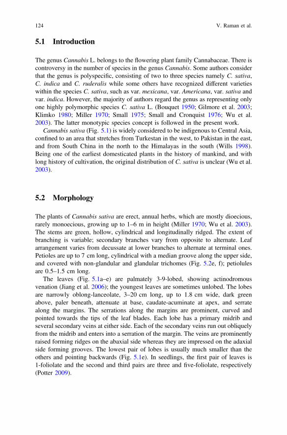
124 V. Raman et al.

Male flowers are pale green, borne on axillary laxly branched cymose panicles.Flowers in the panicles occur solitarily, in clusters, or in 3-flowered cymules. Eachflower consists of five tepals, five stamens and a slender pedicel. The tepals areovate-oblong, 2–4 cm long, yellowish- or whitish-green, spreading, and minutelyhairy. The stamens are drooping and consist of slender filaments and oblong,greenish anthers. Pollen grains are liberated through terminal pores in the anthers(UNODC 2009).
Female flowers (Fig. 5.1a–c; 5.2a) are dark green, subsessile and are borne inpairs. The flowers are closely aggregated at the apex of short spike inflorescences,which are densely formed in the upper axils of branches. Each flower consists of
Fig. 5.1 Morphology of C. sativa. a–c Twigs with female inflorescences; d A twig of a maleplant; e Leaves showing variation in the number of lobes; f Seeds
5 Morpho-Anatomy of Marijuana (Cannabis sativa L.) 125

ovary with a style that ends in a pair of long slender feathery stigmas at apex(Fig. 5.2a), a membranous perianth surrounding the ovary, and a bract. Thestyle-stigma portion of the pistil in wild-growing plants generally measures about3 mm long and the styles are usually 2-branched. However, Small and Naraine(2016a) have observed that in illicitly grown Cannabis cultivars, which are pro-tected against pollen, the style-stigma portion averages over 8 mm long and the
Fig. 5.2 Micro-morphology of different parts of C. sativa a, c, e, f Scanning Electron Microscopy(SEM); b, d Light microscopy (LM)]. a Female flowers; b Outer surface of a bract showingnumerous glandular trichomes with yellow contents; c Portions of bract and young leaves withvarious types of trichomes; d A portion of bract; e Petiole surface covered with nonglandular andglandular trichomes; f Lower surface of leaf base and a portion of petiole showing branching ofmajor veins to leaf lobes
126 V. Raman et al.

styles are often 3-branched. The perianth is transparent, smooth or slightly fringedalong the margin, at maturity covers about two-thirds of the ovary. The bracts(Fig. 5.2a–d) are green, scabrous, with overlapping edges, enclosing the femaleflower except the exserted stigmas, acuminate at apex. The fruit is an achene, ovoid,ellipsoid or subglobose, about 4–6 mm long and 3–4 mm in diameter, smooth,somewhat compressed, brownish grey and mottled, containing a single seed with ahard shell (Fig. 5.1f). Sometimes, the Cannabis “seed” of commerce is actually thefruit still enclosed in its hooded floral bract (Hayward 1938; UNODC 2009).
Male and female flowers occur in separate plants; they generally bloom duringJuly-August. Male plants are usually taller and the female plants are usually morerobust than male plants. Several cultivars with varying features occur in cultivation.Morphological characteristics of Cannabis plants are influenced by the seed strainas well as by environmental factors such as soil type, light, water, nutrients andspace (UNODC 2009).
5.3 Trichomes
The trichomes of Cannabis have been well studied in the past. Briosi and Tognini(1894) published one of the earliest works that provided detailed descriptions andillustrations of Cannabis trichomes. Most of the earlier workers, however, descri-bed only some aspects of mature trichomes, except Ram and Nath (1964), whostudied the ontogeny of the trichomes. Several papers published on Cannabis in the1960s and 1970s included characterization of trichomes using light or scanningelectron microscopes and were primarily aimed to aid in the forensic identificationof illicit Cannabis products (Dayanandan and Kaufman 1976; Hammond andMahlberg 1973; 1977; Ledbetter and Krikorian 1975; Mitosinka et al. 1972;Nakamura 1969; Shimomura et al. 1967; Thornton and Nakamura 1972; Turneret al. 1977).
Almost all aerial parts of the Cannabis plants are covered with trichomes. Twomajor types of trichomes are present in C. sativa: (A) glandular trichomes and(B) non-glandular trichomes.
5.3.1 Glandular Trichomes
Glandular trichomes are the primary structures for synthesis and storage ofcannabinoids in C. sativa. Three types of glandular trichomes, namelycapitate-stalked, capitate-sessile and bulbous, are present in Cannabis (Hammondand Mahlberg 1973, 1977).
1. Capitate-stalked glandular trichome (Fig. 5.3a–e): This type of trichomesposses a large globular head generally measuring 50–70 µm in diameter and a
5 Morpho-Anatomy of Marijuana (Cannabis sativa L.) 127

128 V. Raman et al.

robust multicellular stalk of 100–200 µm. High-THC strains have larger glan-dular heads {up to 119 µm (Small and Naraine, 2016b)}. The length of the stalkis highly variable; some of the glands have shorter stalks, some appear to bealmost sessile. These glands are particularly abundant on bracts of pistillateplants and become the most conspicuous feature of older bracts (Hammond andMahlberg 1977).
2. Capitate-sessile glandular trichome (Fig. 5.3a, f, g): This is the most con-spicuous type during early stages of bract development. It is mainly found in theabaxial leaf surfaces, petioles and young stems. The gland consists of a largeglobose head measuring about 30–50 µm in diameter (Hammond and Mahlberg1973). Although appearing as stalkless, these trichomes possess a very shortstalk of one-cell high but 2–4 cells thick (Dayanandan and Kaufman 1976).
3. Bulbous glandular trichome (Fig. 5.3e): This is the smallest type of glandulartrichome found in C. sativa. These trichomes possess a 1-2-celled stalk and a1-4-celled head. These trichomes vary in their sizes and are generally about 10–20 µm in diameter and 15–30 µm in height (Dayanandan and Kaufman 1976;Hammond and Mahlberg 1973).
The capitate-stalked and capitate-sessile glandular trichomes are similar in allrespects except the former type trichomes have a massive, multi-cellular stalk. Theglobular head in both types of glands is made up of eight cells developed fromepidermal initials. These cells form a cellular disc which is about 30 µm in diameterand about 15 µm in height. Due to the accumulation of the resinous secretionbetween the outer surface of the disc and the extended cuticular membrane, theglandular head becomes spherical in shape.
5.3.2 Non-glandular Trichomes
These are unicellular covering trichomes found on stems, leaves, petioles, stipules,bracts and tepals. The non-glandular trichomes are of the following two kinds:
JFig. 5.3 Glandular and non-glandular trichomes in C. sativa [C- LM; all others SEM; A, B,G- colorized SEM images]. a Portion of bract displaying glandular and non-glandular trichomes;b–d Capitate stalked glandular trichomes (note an ‘eyespot’ on the glandular head in image B; theglandular disc and cuticular membrane in c; and a slightly broken ‘neck’ of glandular headshowing 4-cell arrangement, in image d); e A capitate stalked glandular trichome and two of thebulbous glandular trichomes; f A group of capitate sessile glandular trichomes on a young leaf; asessile glandular trichome on abaxial leaf surface (note the presence of stomata); h Morphology ofconical cystolith trichomes on adaxial leaf surface; i Cystolith trichomes in sectional view showinglarge cystolith crystals. Gh glandular head, Gt-1 capitate stalked glandular trichome, Gt-2 capitatesessile glandular trichome, Gt-3 bulbous glandular trichome, Ng non-glandular trichome, Nk neck,Sk stalk, St stomata
5 Morpho-Anatomy of Marijuana (Cannabis sativa L.) 129

1. The shorter and larger cystolith-containing conical trichomes (Fig. 5.3h, i),which are about 50–125 µm long with a large base measuring about 60–140 µmin diameter. These trichomes are found mainly on the adaxial surface of theleaves. About 15–20 epidermal cells form a circle around the base of the tri-chomes. These trichomes, with their enlarged base and shortly pointed tip,appear like a ‘claw’.
2. The longer and slender trichomes (Figs. 5.2e, f; 5.3a), which are about 250–370 µm long and are abundantly distributed on the abaxial leaf surfaces, stems,petioles and tepals.
The non-glandular trichomes are generally pointed towards the apices of leavesor stems. The trichomes located on or near the major veins have a warty surfacewhereas those occurring between the veins have slightly warty or smooth surface(Jiang et al. 2006). Silica (SiO2. nH2O) is reported to be distributed more or lessevenly all over these trichomes (Dayanandan and Kaufman 1976). The enlargedbasal part of the cystolith trichome contains large crystal of calcium carbonate(CaCO3) (Fig. 5.3i). They are prominent in the trichomes found on the adaxial leafsurface. Few trichomes containing cystolith crystals are also found on the abaxialleaf surface, stem and petiole. Calcium (Ca) is mainly deposited in the form ofCaCO3 in the cystolith, but small amount of Ca may also be present throughout theinner cavity of the trichomes (Dayanandan and Kaufman 1976).
Both glandular and non-glandular trichomes are present in both pistillate andstaminate plants and they are found in Cannabis plants from the early seedling stageto maturity. The capitate-stalked glandular trichomes are found only in the bracts ofpistillate plants and anthers in the staminate plants (Dayanandan and Kaufman1976). Bulbous and capitate-sessile glandular trichomes occur on all parts ofvegetative and flowering shoots except for the hypocotyl and cotyledons whereascapitate-stalked glands are restricted to flowering regions of the plants. Bracts havethe highest concentration of glandular trichomes than any other part on pistillateplants (Hammond and Mahlberg 1973). The capitate-stalked glands are found onlyin the flowering bracts in pistillate plants. In staminate plants, this trichome type isrestricted only to longitudinal rows along the inner surfaces of anthers (Dayanandanand Kaufman 1976).
5.4 Anatomy
Cannabis has been associated with human since ancient times, however, little isknown about its comparative anatomy (Anderson 1974). Tippo (1938) made fewgeneral comments on the wood of C. sativa, and Nassonov (1940) discussed aboutstem shape and leaf trace number in transections in his work on geographicalraces of hemp. He stated that wild and cultivated forms of hemp could not bedifferentiated clearly based on anatomy of stem and bast fibers. Hayward (1938)studied general morphology, seedling anatomy and floral structure of hemp.
130 V. Raman et al.

Metcalfe and Chalk (1950) compiled anatomical data available at that time.Shimomura et al. (1967) differentiated between C. sativa and C. indica based onleaf and bract anatomy with emphasis on trichomes. Anderson (1974) studied woodanatomy of Cannabis and found significant anatomical differences between C.sativa and C. indica (Fig. 5.4) .
In transection, the leaf of C. sativa shows thin lamina and major veins, which aredepressed above and prominently raised beneath (Fig. 5.5a, b). Each of the upperand lower epidermis is unilayered. In surface view, the epidermal cells show
Fig. 5.4 Leaf micro-morphology of C. sativa [C and F- LM; all others SEM]. a Adaxial leafsurface; b, c Adaxial leaf epidermis; d, e Abaxial leaf surface; f, g Abaxial leaf epidermis showingstomata. Cc cystolith trichome, Cu cuticle striations, Gt-2 capitate sessile glandular trichome, Gt-3bulbous glandular trichome, Ngt non-glandular trichome, St stomata
5 Morpho-Anatomy of Marijuana (Cannabis sativa L.) 131

slightly undulate anticlinal walls. Upper epidermis (Fig. 5.4a–c) shows the char-acteristic cystolith trichomes with an enlarged base containing large cystolithcrystal. Numerous nonglandular and glandular trichomes are present on the lowerepidermis (Fig. 5.4d–g). Stomata (Fig. 5.4f, g) are numerous on the lowerepidermis and are not observed in the upper epidermis. The mesophyll consists of
Fig. 5.5 Anatomy of C. sativa [A and C- LM; all others SEM]. a, b Transection (TS) of leafthrough midrib; c, d TS of leaf through lamina; e TS of stem, with a portion enlarged (f). Chchlorenchyma, Co collenchyma, Ct cystolith trichome, Fu furrows, Gt-2 capitate sessile glandulartrichome, Gt-3 bulbous glandular trichome, La lamina, Ld laticifer duct, Le lower epidermis, Mrmidrib, Ngt non-glandular trichome, Pa palisade, Pf pericyclic fibers, Ph phloem, Pi pith, Riridges, Sp spongy tissue, Up upper epidermis, Xy xylem
132 V. Raman et al.

palisade and spongy tissue. Palisade is unilayered, consists of thin columnar cells,and occupying more than half thickness of the lamina. Spongy cells are looselyarranged with large air spaces leading to stomatal cavities (Fig. 5.5c, d).Transection of midrib (Fig. 5.5a, b) shows a single collateral vascular bundle. Smallgroups of collenchyma cells are present beneath the upper epidermis and inside thelower epidermis. A few laticifer ducts with yellow-brown secretions are found inthe phloem (Evert 2006). Cluster crystals of calcium oxalate (Fig. 5.6e, f) arecommonly found in the mesophyll, and phloem parenchyma of the veins (Hayward1938).
The petiole is more or less triangular in cross section showing a groove at theadaxial side (Fig. 5.6a). The epidermis is unilayered and produces numerousnonglandular and glandular trichomes. A ring of collenchyma is located adjacent tothe epidermis, which is narrow near the groove and much wider at the abaxial andlateral sides. The vascular bundle is collateral with xylem above and phloem below.The vessel elements are arranged in radial rows. A few laticifer ducts are found inthe phloem. The space surrounding the midrib vascular bundle is filled withchlorenchyma (Hayward 1938).
The stem has a wavy outline in transection due to ridges and furrows (Fig. 5.5e).The epidermis is unilayered and produces numerous nonglandular and glandulartrichomes. This is followed by a unilayered hypodermis and a few layers ofchlorenchyma, which is lined by the endodermis layer. The pericycle is wide,consisting of numerous pericyclic fibers distributed among large polygonal par-enchyma cells. These fibers have thickened and lignified walls and narrow lumina,and measure about 5–20 µm in diameter. The secondary phloem forms a narrowring, and consists of bast (phloem) fibers, parenchyma and a few laticifer ductsfilled with yellow-brown contents. Cambium is wide, made up of several layers ofradially arranged cells. The xylem comprises of large vessel elements, which areabout 30–100 µm in diameter, circular or angular in cross section and occur soli-tarily or a few arranged in radial rows. The xylem fibers have thickened andlignified walls and are arranged in radial rows (Hayward 1938) (Fig. 5.5e, f).
Transection of a primary root (Fig. 5.6c, d) shows a unilayered epidermis and alayer of hypodermis. The cortex is wide and parenchymatous. The endodermis isunilayered and the pericycle is multilayered. The vascular bundle consists of adiarch xylem and two groups of primary phloem (Hayward 1938).
In cross section, the pericarp of the fruit (Fig. 5.6b) shows the following tissuearrangement: the outermost layer, the epicarp, is made up of thick-walled scle-renchyma cells showing sinuous anticlinal walls in surface view. The hypodermisconsists of one or more layers of loosely arranged spongy parenchyma cells.Numerous vascular bundles traverse this region. The third zone consists of a layerof brown cells with thick walls. This is followed by a narrow region of colorless,collapsed cells with thin, sinuous radial walls. The innermost layer of the pericarp ismade up of palisade cells with heavily thickened walls and narrow lumina(Hayward 1938; Winton 1906).
5 Morpho-Anatomy of Marijuana (Cannabis sativa L.) 133

The seed coat (Fig. 5.6b) is two-layered in transection. The outermost layerconsists of tube cells, and the inner layer is made up of spongy parenchyma cells.The seed coat is followed by perisperm and endosperm, each one-cell layeredin thickness. The cells of endosperm contain aleurone grains. The embryo isU-shaped, consists of two cotyledons enclosing an epicotyl, a hypocytyl andprimary root (Hayward 1938).
Fig. 5.6 Anatomy of C. sativa [E- LM; all others SEM, F- colorized SEM]. a TS of petiole; b TSof root; c, d TS of root; e, f cluster crystals of calcium oxalate in the leaf midrib. Br bract, Cccluster crystals, Co cortex, Co cotyledon, Gr adaxial groove, Pe pericarp, Ph phloem, Vb vascularbundle, Xy xylem
134 V. Raman et al.

5.5 Conclusion
Study of morphological features of plants is crucial for species identification. Theutilization of anatomical and microscopic characters of plants has become a stan-dard practice especially in plants that exhibit variable morphological features.Cannabis sativa is one such highly complex taxa, exhibiting a wide range ofvariations in its morphological features such as habit and size of plants, size andarrangement of leaves and the shape, size and number of lobes, indumentums, sizeand branching of stems, and number and arrangement of flowers. This is possiblydue to the long history of domestication, extensive hybridization, and excessiveselection of preferred phenotypes and chemotypes. Thus, the taxonomy ofCannabis is confusing. The original geographical distribution of the taxon is vague,and no purely wild populations exist. As a result, the genus has been treated indifferent ways by different authors. Several botanists have proposed that Cannabisis a polyspecific genus including three different species. Whereas, many others haveopined that it is a monospecific genus with a single species, C. sativa. Some authorshave recognized different varieties and subspecies within the species C. sativa.Several authors have studied the morphology of the species; however, most of themhave focused on the trichome characteristics and their usage in forensic identifi-cation of the plant material. Further studies of detailed comparative morphologicaland anatomical characteristics of the taxon involving a wide range of plant materialsfrom various parts of its presumed original distribution could yield better under-standing of the taxonomy of the species as well as the extent of its morphologicaland anatomical variations.
Acknowledgements This work was supported in part by the National Institute on Drug Abuse,National Institute of Health, Department of Health and Human Services, ContractNo. N01DA-10-7773.
References
Anderson LC (1974) A study of systematic wood anatomy in Cannabis. Botanical MuseumLeaflets, Harvard University 24:29–36
Bouquet RJ (1950) Cannabis. Bull Narc 2:14–30Briosi G, Tognini F (1894) Intorno alla anatomia della canapa (Cannabis sativa L.). Atti Ist Bot
Pavia 3:91–209Dayanandan P, Kaufman PB (1976) Trichomes of Cannabis sativa L. (Cannabaceae). Am J Bot
63:578–591Evert RF (2006) Esau’s plant anatomy: meristems, cells, and tissues of the plant body: their
structure, function, and development. John Wiley, Hoboken, New JerseyGilmore S, Peakall R, Robertson J (2003) Short tandem repeat (STR) DNA markers are
hypervariable and informative in Cannabis sativa: implications for forensic investigations.Forensic Sci Int 131:65–74
Hammond CT, Mahlberg PG (1973) Morphology of glandular hairs of Cannabis sativa fromscanning electron microscopy. Am J Bot 60:524–528
5 Morpho-Anatomy of Marijuana (Cannabis sativa L.) 135

Hammond CT, Mahlberg PG (1977) Morphogenesis of capitate glandular hairs of Cannabis sativa(Cannabaceae). Am J Bot 64:1023–1031
Hayward HE (1938) The structure of economic plants. Macmillan Co., New YorkJiang HE, Li X, Zhao YX, Ferguson DK, Hueber F, Bera S, Wang YF, Zhao LC, Liu CJ, Li CS
(2006) A new insight into Cannabis sativa (Cannabaceae) utilization from 2500-year-oldYanghai Tombs, Xinjiang, China. J Ethnopharmacol 108:414–422
Klimko M (1980) Morphological variability of Cannabis sativa L. Bull Soc Amis Sci Lett Poznan.D 20:127–134
Ledbetter MC, Krikorian A (1975) Trichomes of Cannabis sativa as viewed with scanningelectron microscope. Phytomorphology 25:166–176
Metcalfe CR, Chalk L (1950) Anatomy of the dicotyledons. Clarenden Press, OxfordMiller NG (1970) The genera of Cannabaceae in the southeastern United States. J Arnold
Arboretum 51:185–203Mitosinka GT, Thornton JI, Hayes TL (1972) The examination of cystolithic hairs of Cannabis
and other plants by means of the scanning electron microscope. J Forensic Sci Soc 12:521–529Nakamura GR (1969) Forensic aspects of cystolith hairs of Cannabis and other plants. J Ass
Official analyt Chem 52:5–16Nassonov V (1940) Anatomical characteristics of the geographical races of hemp [in Russian].
Vestnik Sotsialisticheskogo Rastenievodstva 4:107–120Potter D (2009) The propagation, characterisation and optimisation of Cannabis sativa as a
phytopharmaceutical (PhD Thesis). Department of Pharmaceutical Science Research. King’sCollege, London
Ram HM, Nath R (1964) The morphology and embryology of Cannabis sativa Linn. Phytomorph14:414–429
Shimomura H, Shigehiro M, Kuriyama E, Fujita M (1967) Studies on Cannabis. I. Microscopicalcharacters of their internal morphology and spodogram (Japanese). Yakugaku Zasshi: Journalof the Pharmaceutical Society of Japan 87:1334–1341
Small E (1975) American law and the species problem in Cannabis: Science and Semantics. BullNarc 27:1–20
Small E, Cronquist A (1976) A practical and natural taxonomy for Cannabis. Taxon: 405–435Small E, Naraine SGU (2016a) Expansion of female sex organs in response to prolonged virginity
in Cannabis sativa (marijuana). Genet Resour Crop Evol 63:339–348Small E, Naraine SGU (2016b) Size matters: evolution of large drug-secreting resin glands in elite
pharmaceutical strains of Cannabis sativa (marijuana). Genet Resour Crop Evol 63:349–359Thornton JI, Nakamura GR (1972) The identification of marijuana. J Forensic Sci Soc 12:461–519Tippo O (1938) Comparative anatomy of the Moraceae and their presumed allies. Bot Gaz 100:1–
99Turner JC, Hemphill JK, Mahlberg PG (1977) Gland distribution and cannabinoid content in
clones of Cannabis sativa L. Am J Bot 64:687–693UNODC (2009) Recommended methods for the identification and analysis of Cannabis and
Cannabis products (Manual). United Nations, New YorkWills S (1998) Cannabis use and abuse by man: an historical perspective. Harwood Academic,
AmsterdamWinton AL (1906) The microscopy of vegetable foods. John Wiley & Sons, LondonWu Z, Zhou Z-K, Bartholomew B (2003) Cannabaceae. In: Wu Z, Raven PH (eds) Flora of China.
Science Press, Beijing
136 V. Raman et al.