chapter 3 experimental 3.1 materials and …archive.lib.cmu.ac.th/full/t/2011/envs20811sh_ch3.pdf44...
TRANSCRIPT
CHAPTER 3
EXPERIMENTAL
3.1 Materials and Chemicals
3.1.1The materials and chemicals for hapten synthesis
3.1.1.1 Synthesis of DDT-haptens
1) 2,2′-Bis(4-chlorophenyl)ethanol (DDOH), 188883, Aldrich, Sigma-
Aldrich Co. USA.
2) Dichloro benzhydrol (DCBH), TCI, Japan (kindly given by Prof. Dr.
Takahiko Takatori, School of Medicine, University of Tokyo)
3) Succinic anhydride, 14089, Fluka, Sigma-Aldrich Co. USA.
4) Glutaric anhydride, 49670, Fluka, Sigma-Aldrich Co. USA.
5) 4-Dimethyl amino pyridine (DMAP), 39405, Fluka, Sigma-Aldrich,
Germany.
6) Hydrochloric acid (HCl), 37%, proanalysis, K405566517, Merck,
Germany.
7) Sodium chloride (NaCl), K41042404 021, Merck, Germany.
8) Magnesium sulphate (MgSO4), 63136, Fluka, Sigma-Aldrich,
Germany.
9) Thin-layer chromatography (TLC) silica gel 60F254 20 x 20 cm
aluminium sheet, Merck, Germany.
10) Silica gel 60F254, Merck, Germany.
40
3.1.1.2 Synthesis of cypermethrin-haptens
1) Methyl-3-(2,2-dichlorovinyl)-2,2-dimethyl-(1-cyclopropane)
carboxylate, CAS no. 61898-95-1, Acros, Germany.
2) β-alanine methyl ester hydrochloride, 05210, Fluka, Germany.
3) 1-[3-(dimethylamino) propyl]-3-ethylcarbodiimide hydrochloride
(EDC), 03450, Fluka, Sigma-Aldrich, Germany.
4) Sodium hydroxide (NaOH), K2433898 735, Merck, Germany.
5) Thin-layer chromatography (TLC) silica gel 60F254 20 x 20 cm
aluminium sheet, Merck, Germany.
6) Silica gel 60F254, Merck, Germany.
3.1.1.3 The materials and chemicals for preparation of immunogens and
coating antigens
1) Bovine serum albumin (BSA), A-7906, Sigma Chemical Co.,
Germany.
2) Albumin from hen egg white (egg albumin; OVA), 05440, Fluka,
Sigma-Aldrich Chemie GmbH, Germany.
3) Dimethylformamide (DMF), Merck, Germany.
4) 4- dimethylaminopyridine, 39405, Fluka, Sigma-Aldrich Co. USA.
5) N, N – dicyclohexylcarbodiimide, 36650, Fluka, Sigma-Aldrich Co.
USA.
6) N- hydroxysuccinimide, 56480, Fluka, Sigma-Aldrich Co. USA.
7) 1-[3-(dimethylamino)propyl]-3-ethylcarbodiimide hydrochloride
(EDC), 03450, Fluka, Sigma-Aldrich, Germany.

41
8) Spectra/Por
membrane tubing, Spectrum, Spectrum Laboratory, Inc
9) Bio-Rad protein assay, 500-0006, Bio-Rad laboratories,Inc.
3.1.1.4 Organic solvents
1) Toluene, 9460-03, Actual analysis, J.T. Baker, USA.
2) Ethyl acetate, 9280-03, Actual analysis, J.T. Baker, USA.
3) Petroleum ether, 9268-05, Actual analysis, J.T. Baker, USA
4) Acetic acid, K30802863 224, Pro-analysis, Merck, Germany.
5) Pyridine, EC-No. 203809-9, BDH, England.
6) Sulfuric acid (H2SO4), K31004031, Pro-analysis, Merck, Germany.
7) Hexane, 9309-03, Actual analysis, J.T. Baker, USA.
8) Methanol, 9093-68, Actual analysis, J.T. Baker, USA.
9) Dimethyl sulfoxide (DMSO), D/4121/PB17, Analytical grade, Fisher
Scientific, United Kingdom.
10) Tetrahydrofuran (THF), 9731, Merck, Germany.
3.1.1.5 Standard insecticides
1) o,p’-DDE, ch-10311, 99.8%, Laboratory of Dr. Ehrenstorfer, Augburg,
Gemany.
2) o,p’-DDT, c-12081000, 98.0%, Laboratory of Dr. Ehrenstorfer,
Augburg, Gemany.
3) o,p’-DDD, c-120820, 99.3%, Laboratory of Dr. Ehrenstorfer, Augburg,
Gemany.

42
4) p,p’-DDE, PS-696, 99.5%, Chem Service Inc., West Chester, PA,
USA.
5) p,p’-DDT, PS-699, 99%, Chem Service Inc., West Chester, PA, USA.
6) p,p’-DDD, 49009, 98.5%, Supelco, Bellelofente, PA, USA.
7) p,p’-DDA, 35484, 99.9%, Riedel-de Haen, Sigma-Aldrich Co. USA.
8) p,p’-DDM, 35488, 99.1%, Riedel-de Haen, Sigma-Aldrich Co. USA.
9) p,p’-DBP, 45421, 99.7%, Riedel-de Haen, Sigma-Aldrich Co. USA.
10) Dicofol, 36677, 96.9%, Riedel-de Hean, Sigma-Aldrich Co. USA.
11) Cypermethrin, c11890000, 91.0%, Laboratory of Dr. Ehrenstorfer,
Augburg, Gemany.
12) Permethrin, c15990000, 94.0%, Laboratory of Dr. Ehrenstorfer,
Augburg, Gemany.
13) Cyfluthrin, c11850000, 99%, Laboratory of Dr. Ehrenstorfer, Augburg,
Gemany.
14) Deltamethrin, c12120000, 99%, Laboratory of Dr. Ehrenstorfer,
Augburg, Gemany.
3.1.1.6 Immunological materials
1) Immuno plate Maxisorp 96F, 442404, NUNC, Denmark.
2) HRP- Conjugate anti - mouse Ig G antibody (H+L), 81-6520, Zymed
Laboratories Invitrogen immuno detection, USA.
3) Ortho-phenyleneline diamine, P-1526, Sigma Chemical Co., USA.
4) Tween
20, 63158, Sigma, United Kingdom.
43
5) Complete Freund’s adjuvant (CFA), F-5881, Sigma Chemical Co.,
USA.
6) Incomplete Freund’s adjuvant (IFA), F-5506, Sigma Chemical Co.,
USA
3.1.1.7 Equipments
1) UV-2101 PC UV/vis spectrophotometer, Shimadzu Scientific
Instrument, Inc. Japan.
2) Micro plate spectrophotometer, Sunrise, TECAN, Austria.
3) Gas chromatograph 5890, Hewlett Packard, USA.
4) Gas chromatograph 7890A, Agilent Technologies, USA.
5) Mass-spectro detector (MSD), 5975C, Agilent Technologies, USA.
6) 1H and
13C NMR, Bruker, Bruker analytic GmbH, Germany.
7) VACELUTE, SPS24, Varian, USA.
8) Bond elute-C18, Part no. 12102028, Varian, USA.
3.2 Methods
3.2.1 Synthesis of haptens
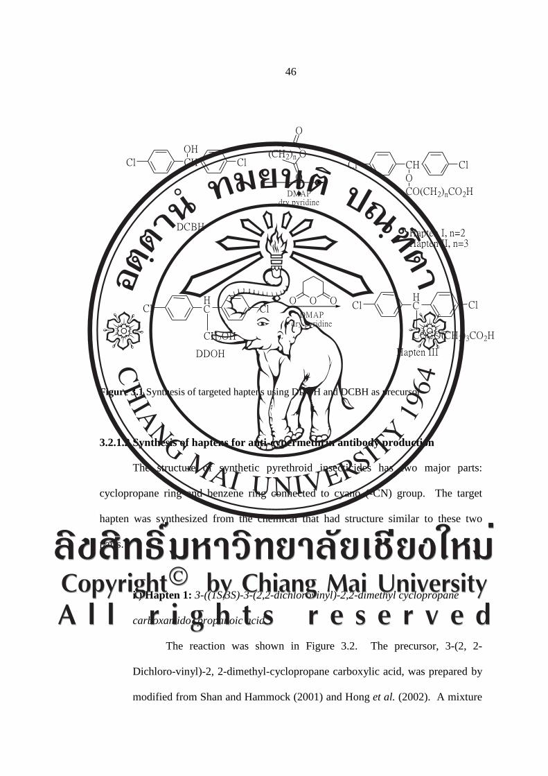
3.2.1.1 Synthesis of haptens for anti-DDT antibody production
Haptens with chemical structure resembling DDT were designed and
modified structures were produced by spacer arm attachment. Two kinds of
hapten were synthesized with modified from Beasley et al. (1998)
(Figure 3.1), hapten I and hapten II were synthesized by attaching hydroxyl
(-OH) to DCBH. Haptens were synthesized by linking hydroxyl group of

44
DDOH with glutaric anhydride and DCBH with succinic acid anhydride by
esterification in dry pyridine. These substitutes act as spacer arm joining the
two aromatic rings through the carbon atom of carboxylic acid. Structures of
haptens were confirmed by 1H and
13C Nuclear Magnetic Resonance
Spectroscopy (NMR) and gas chromatography/Mass- spectrometry (GC/MS).
The synthesis of the two haptens is described in details below.
i) Hapten I: 4-(bis(4-chlorophenyl)methoxy)-4-oxobutanoic acid.
Hapten I was synthesized according the previous reported
procedures (Hongsibsong et al., 2010). A mixture of 200 mg (0.78
mmol) of DCBH, 750 mg (7.5 mmol) of succinic anhydride, and 10
mg (0.08 mmol) of dimethylaminopyridine (DMAP) in 10 ml dry
pyridine was stirred overnight at room temperature. Twenty milliliters
of water was then added and the mixture was evaporated dry. Crude
was rinsed twice with 10 ml toluene and dissolved in 10 ml ethyl
acetate. Crude solution was washed once with 10 ml of cool HCl,
twice with 10 ml water, and twice with 10 ml saturated sodium
chloride, respectively. The product was dry over magnesium sulphate
(MgSO4). Purity of the product was confirmed by TLC. Crude product
was purified by column chromatography on silica gel using ethyl
acetate: hexane (40:60 v/v) solvent. Single-compound fraction was
collected and evaporated dry. The structure of the compound was
confirmed by 1H and
13C NMR, and GC/MS.
45
ii) Hapten II: 5-(bis(4-chlorophenyl)methoxy)-5-oxopentanoic acid was
synthesized using the same procedure as hapten I but glutaric anhydride
was substituted for succinic anhydride.
iii) Hapten III: Pentanedioic acid mono-[2,2-bis-(4-chloro-phenyl)-
ethyl] ester:
This hapten was synthesized according to diagram shown in
Figure 3.1. 2,2′-Bis(4-chlorophenyl) ethanol (DDOH, 100 mg,
0.37 mmol) was reacted with glutaric anhydride (375 mg, 3.74 mmol)
in 5 mL of dry pyridine with 5 mg of Dimethyl aminopyridine
overnight at room temperature. Twenty milliliters of water was then
added to the mixture and evaporated to remove pyridine. The crude
compound was rinsed with toluene and solvent was removed by
evaporation and then dissolved in ethyl acetate. The miture was
washed with 1 M HCl, water, and brine, before drying over MgSO4.
Crude product was purified by column chromatography on silica gel
using ethyl acetate: petroleum ether (40:60 v/v) with 0.01% acetic acid
as mobile solvent. Single-compound fraction was collected and
evaporated to dryness. The structure was confirmed by 1H-,
13C-NMR
and GC/MS.
46
DCBH
CH
OH
ClClO
O
OCH ClCl
O
CO(CH2)nCO2HDMAPdry pyridine
Hapten I, n=2Hapten II, n=3
O OO
DMAPdry pyridine
HC
CH2OH
Cl ClHC
COCO(CH2)3CO2H
Cl Cl
DDOH Hapten III
(CH2)n
Figure 3.1 Synthesis of targeted haptens using DDOH and DCBH as precursor
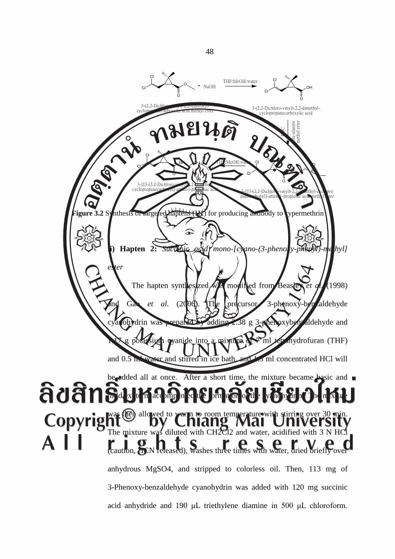
3.2.1.2 Synthesis of haptens for anti-cypermethrin antibody production
The structure of synthetic pyrethroid insecticides has two major parts:
cyclopropane ring and benzene ring connected to cyano (-CN) group. The target
hapten was synthesized from the chemical that had structure similar to these two
parts.
i ) Hapten 1: 3-((1S,3S)-3-(2,2-dichlorovinyl)-2,2-dimethyl cyclopropane
carboxamido) propanoic acid
The reaction was shown in Figure 3.2. The precursor, 3-(2, 2-
Dichloro-vinyl)-2, 2-dimethyl-cyclopropane carboxylic acid, was prepared by
modified from Shan and Hammock (2001) and Hong et al. (2002). A mixture

47
of 200 mg 3-(2, 2-Dichloro-vinyl)-2, 2-dimethyl-cyclopropanecarboxylic acid
methyl ester and 62.0 mg NaOH in 5 mL THF:MeOH:water (3:2:1, v/v/v) was
stirred for 4 hours at room temperature. The crude compound was purified by
thin layer chromatography on silica gel using ethyl acetate and hexane (40:60,
v/v). Then, a mixture of 50 mg beta alanine methyl ester and 65 mg EDC in
3 ml THF was stirred for 10 minutes. The 59 mg 3-(2, 2-dichlorophenyl) 2, 2-
dimethyl–(1-cyclopropane) carboxylic acid was slowly added to mixture
solution on ice bath (0C). The mixture solution was stirred for 10 hour in
heating box (50C) and evaporated to dryness. The crude was redissolved in
dichloromethane and then washed twice with 10 mL of cool HCl and twice
with 10mL water. The product was dry over magnesium sulphate. The target
hapten was confirmed by thin layer chromatography (TLC) and then purified
by column chromatography on silica gel using ethyl acetate and hexane
(10:1, v/v) solvents. Single compound fraction was collected and evaporated
to dryness. The structure and molecular mass of the obtained compound was
confirmed by 1H and
13C nuclear magnetic resonance (NMR) spectrometry
and GC-MS, respectively. Hapten obtained was a white-yellow powder
compound with Rf value of 0.26 (ethyl acetate:hexane:acetic acid, 1:10:0.01,
v/v).
48
Cl
Cl
O
O
Cl
Cl
O
OH
Cl
Cl
O
NHO
O
Cl
Cl
O
NHO
OH
NaOHTHF:MeOH:water
beta
-ala
nine
m
ethy
l es
ter
ED
C
THF:MeOH:water
3-(2,2-Dichloro-vinyl)-2,2-dimethyl-cyclopropanecarboxylic acid methyl ester
3-(2,2-Dichloro-vinyl)-2,2-dimethyl-cyclopropanecarboxylic acid
3-{[3-(2,2-Dichloro-vinyl)-2,2-dimethyl-cyclopropanecarbonyl]-amino}-propionic acid methyl ester
3-{[3-(2,2-Dichloro-vinyl)-2,2-dimethyl-cyclopropanecarbonyl]-amino}-propionic acid
Figure 3.2 Synthesis of targeted haptens (H1) for producing antibody to cypermethrin
ii) Hapten 2: Succinic acid mono-[cyano-(3-phenoxy-phenyl)-methyl]
ester
The hapten synthesized was modified from Beasley et al. (1998)
and Gao et al. (2006). The precursor, 3-phenoxy-benzaldehyde
cyanohydrin was prepared by adding 2.38 g 3-phenoxybenzaldehyde and
1.17 g potassium cyanide into a mixture of 7 ml tetrahydrofuran (THF)
and 0.5 ml water and stirred in ice bath, and 1.3 ml concentrated HCl will
be added all at once. After a short time, the mixture became basic and
mild exoterm accompanied the formation of the cyanohydrin. The mixture
was then allowed to warm to room temperature with stirring over 30 min.
The mixture was diluted with CH2Cl2 and water, acidified with 3 N HCl
(caution, HCN released), washes three times with water, dried briefly over
anhydrous MgSO4, and stripped to colorless oil. Then, 113 mg of
3-Phenoxy-benzaldehyde cyanohydrin was added with 120 mg succinic
acid anhydride and 190 μL triethylene diamine in 500 μL chloroform.
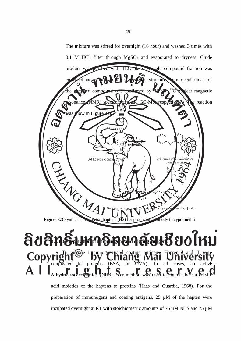
49
The mixture was stirred for overnight (16 hour) and washed 3 times with
0.1 M HCl, filter through MgSO4 and evaporated to dryness. Crude
product was purified with TLC plate. Single compound fraction was
collected and evaporated to dryness. The structure and molecular mass of
the obtained compound was confirmed by 1H and
13C nuclear magnetic
resonance (NMR) spectrometry and GC-MS, respectively. The reaction
was show in Figure 3.3.
OO
KCN
OOCO
CO
OH
N
DM
AP
OH
N
O
O
O
O
pyri
di n
e
THF/water
HCl
3-Phenoxy-benzaldehyde 3-Phenoxy-benzaldehyde cyanohydrin
Succinic acid mono-[cyano-(3-phenoxy-phenyl)-methyl] ester
H
Figure 3.3 Synthesis of targeted haptens (H2) for producing antibody to cypermethrin
3.2.1.3 Preparation of immunogens and coating antigens
To prepare immunogens and coating antigens, Hapten I and II were
conjugated to proteins (BSA, or OVA). In all cases, an active
N-hydroxyscuccinimide (NHS) ester method was used to couple the carboxylic
acid moieties of the haptens to proteins (Haas and Guardia, 1968). For the
preparation of immunogens and coating antigens, 25 µM of the hapten were
incubated overnight at RT with stoichiometric amounts of 75 µM NHS and 75 µM
50
dicyclohexylcarbodiimide (DCC) in 0.5 mL of dimethylformamide (DMF).
The mixture was then centrifuged at 10,000 rpm for 10 min. Four hundred
microliters of supernatant containing the active NHS ester of hapten was collected
and then slowly added to 2 mL of 15 mg/mL BSA or OVA in 50 mM carbonate
buffer, pH 9.6. The mixture was allowed to react at RT for 4 h with stirring.
The mixture was dialyzed overnight in PBS, pH 7.2. Concentrations of protein
were determined by Bradford protein assay.
i) Bradford protein assay
Dye reagent was prepared by diluting 1 part of concentrate dye
reagent (BioRad) with 4 parts of water. The working reagent was filtered
through Whatman paper #1 to remove particles. Standard BSA range
from 0.1 mg/mL to 0.5 mg/mL (0.1, 0.2, 0.3, 0.4, 0.5 mg/mL) was
prepared. Protein solutions were analyzed in duplicate. Ten microliters
of various dilutions of standard proteins or test samples were added to
a microtiter plate in the presence of 300 μL diluted dye reagent.
The mixture was mixed and incubated at RT for 5 min. Absorbance at
595 nm was read on ELISA plate reader. Protein concentration was
calculated from a standard curve.
ii) Hapten density assay
Ten micrograms per milliliter of DCBH and 100 μg/ml of BSA,
KLH, OVA, and immunogen were prepared in 1% methanol-PBS,
pH 7.5. The hapten-protein conjugates were examined individually by
51
UV/Vis spectrophotometer to confirm coupling. The spectra of
conjugates, free hapten, and native proteins were compared.
The absorbance of DCBH was measured at various
wavelengths to determine the wavelength of maximum absorbance
(λmax). The absorbance of other solutions was read at this wavelength,
and the hapten density was calculated according to Beer’s law.
A = εbC
A = Absorbance at λ max
ε = Molar absorbtivity
b = Path length of radiation
C = analyte concentration
UV/Vis spectral data were used to confirm the structures of
the final conjugates. Assuming that the molar absorptivity of haptens
was the same for the free and conjugated forms, the hapten densities
(the number of hapten molecules per molecule of protein) of the
conjugates were estimated directly by the mole absorbance ε (Moreno
et al., 2001)
Formula for hapten density calculation:
Hapten density =
εconjugation – ε protein
ε hapten

52
3.2.1.4 Antibody production
Antibody were produced and characterized by the following procedures.
BALB/c mice (8-10 weeks old) were immunized with immunogen. The first
doses for one mouse consisted of 30 µg protein of immunogen emulsified in
complete Freund’s adjuvant injected subcutaneously. Mice were given the
subsequent injections with immunogen emulsified in incomplete Freund’s
adjuvant at 3 week intervals. Blood was collected from mouse tail vein at 7 days
interval and sera were stored at -20oC
3.2.1.5 Detection of antibody by non-competitive indirect ELISA
Non-competitive indirect ELISA was performed similarly to previously
described protocol (Kramer et al., 2004). One hundred microliters of coating
antigen in 50 mM carbonate-bicarbonate buffer (pH 9.6) were added to Maxisorp
Immunoplate (Nunc). The assay plate was incubated overnight at room
temperature. On the following day, plate was washed with washing buffer (0.05%
Tween-PBS) and blocked by incubation with 200 µL/well of 1% gelatin in PBS at
37oC for 1 h. The blocked buffer was discarded and 100 µL of 1:10,000 serum in
washing buffer were added in duplicate wells of the assay plate. The plate was
incubated at 37oC for 1 h and was then washed 4 times with washing buffer, and
100 μL of 1: 5,000 horseradish peroxidase (HRP) conjugated goat-anti-mouse
immunoglobulin G (H+L) in washing buffer were added to each well. The plate
was incubated at 37oC for 1 h, washed 4 times, and 100 μL of the OPD substrate
solution were then added to each well. The plate was incubated in the dark at RT
53
for 30 min and then 50 μL of 2 N H2SO4 stop solution were added to each well.
Absorbance before and after stopping reaction were measured at 492 nm.
3.2.1.6 Development of indirect competitive ELISA (ic-ELISA) to detect p,p’-
DDE in human milk samples
The pAb was detected by non-competitive indirect ELISA and were
tested for specificity to cypermethrin by competitive indirect ELISA. In brief, 96
well Maxisorp Immunoplates were coated with coating antigen in 50 mM
carbonate-bicarbonate buffer (pH 9.6) at 4oC overnight. The wells were washed 4
times by using 0.05% Tween20/PBS, pH7.2 as washing buffer and then were
blocked by incubation with 200 µL/well of 1% gelatin in PBS. Fifty microlitters
of competitors having similar structure to DDE at different concentration and
50µL/well 1:2,500 diluted antibody 1:5,000 final dilution was dispensed into each
well, and the wells were then incubated at room temperature (25oC) for 1 hour 30
mins. The wells were washed and 100 µL of 1:5,000 HRP-conjugated goat anti-
mouse IgG in washing buffer were added to each well. After incubation at 25oC
for 1 hour, the plates were washed and 100 μL of OPD solution were added to the
wells. The reaction was allowed to continue for 30 min and was stopped by
adding 50 µL of 2 N H2SO4. The absorbance was read at 492 nm. Antibody from
a mouse that gave the best sensitivity and specificity to the target compounds was
selected for further study. The sensitivity of pAb was detected and calculated
inhibition concentrations at 50% (IC50) were fit to a four parameter logistic
equation.
54
The relative cross-reactivity (CR) was calculated by the following equation:
%CR = (IC50 of target compound /IC50 of related compound) x 100
i) The effect of coating antigen and organic solvent on activity of antibody
Effect of coating antigen: Two types of coating antigens, s-DCBH-
OVA and g-DDOH-OVA, were compared by ic-ELISA. p, p’-DDE was used
as a competitor.
Effect of organic solvent: DMSO is often used as an organic modifier
of the buffer formulation in the extraction of less hydrophilic analytes from
samples. To determine optimal concentrations of DMSO in the buffer, non
competitive indirect ELISA was carried out using various solutions of DMSO
(10%, 20%, 30%, 40%, and 50%) with 0.05% Tween-20 (v/v). DMSO at the
concentration that had no effect to antibody reaction was used as diluent
composition. The standard p,p’-DDE was diluted at the optimal concentration
of DMSO in PBS with or without 0.05% Tween-20 (v/v). The assays were
done in duplicate wells.
ii) Recovery and precision assay
Recovery assay was carried out by spiking three concentrations of
p,p’- DDE (10, 50 and 100 ng/mL) and three triplicate. After extracted with
C18 solid phase extraction, the residue was re-dissolved in DMSO.
The developed ELISA was performed to determine p,p’-DDE against standard
curve of p,p’-DDE. Recovery was reported in percent and precision would be
55
reported in %CV. Variations day to day were done by using the same
extracted milk samples from recovery assay for 3 days continue.
iii) Analysis of DDT and its metabolites in human milk samples
Human milk samples
Human milk samples were part of samples collected from health care
clinics of the Shoklo Malaria Research Unit (SMRU) in Maela camp, 50 km
north of Mae Sot district (at the Thai-Myanmar Border), Tak province,
northern Thailand, in 2004 to 2008. The study was approved by the Ethics
Committee of Faculty of Tropical Medicine, Mahidol University, Bangkok,
Thailand and the Oxford Tropical Research Ethics Committee, University of
Oxford, the United Kingdom. In brief, the purpose and methods of the survey
were explained to all participants in their own language, mostly Tai ethnic
group. Breast-milk samples were collected by manual expression into glass
tubes. The samples were mixed and aliquoted into Eppendorf tubes, and then
kept frozen at -20 C in Maela camp and transported to the SMRU office in
Mae Sot district. The collected samples were stored at -80 C in the freezer.
Samples were shipped frozen on dry ice to Toxicology lab of the Research
Institute for Health Sciences, Chiang Mai University for the analysis of DDT
and its metabolites using GC-ECD.
56
GC-ECD analysis
Human milk samples were extracted using the method described by
Prapamontol and Stevenson (Prapamontol and Stevenson, 1991) with slight
modification. In brief, 2 mL human milk was extracted with 10 mL (ethyl
acetate: methanol: acetone =1:2:2) and clean up with C18 solid phase
extraction (Bondelute, Varian, USA). Lipid residue in the eluate was treated
with 300 μL concentrated sulfuric acid. One microlitter of clean eluate was
injected for analysis on GC-ECD-63
Ni (HP 5890 A series II, USA) equipped
with an automatic sampler (HP 7673, USA), and a fused silica capillary
column (Ultra 2: 25 m x 0.32 mm i.d., with 0.52 μm film thickness) for
separation. Spiked skimmed cow milk was used for standard calibration curve
construction. The calibration curve for p,p’-DDE was constructed at 3,
6, 9, 12, 15 and 30 μg L-1
. The high concentration of p,p’-DDE in milk
samples will be diluted and repeated run again. The recovery of p,p’- DDE at
concentrations of 6 and 21 μg L-1
were 98.2 0.2 %and 89.1 ± 0.4 %,
respectively. The limit of detection (LOD) as described by signal to noise
ratio of 10:1 was 0.04 μg L-1
. As a part of quality assurance of analysis,
besides internal quality control, Environmental Research Group, Research
Institute for Health Sciences, Chiang Mai University, Chiang Mai, Thailand
participated in the German External Quality control (G-EQUAS, program 40
in 2007), University of Erlengen-Nuremberg, Erlangen, Germany and
obtained qualified results.
57
3.3 Data analysis and statistics
3.3.1 Calculation of antibody response as sensitivity to target compound were
analyzed by 4-parametric logistic regression and reported in IC50 by using
Graphpad prism version 4.0 software.
3.3.2 Correlation between p,p’-DDE data obtained from ic-ELISA and GC-
ECD were conducted with SPSS for WINDOWS (SPSS Inc., Chicago, IL;
Version 11.5)