chapter 20: nanotechnology for engineering cellular ...fanyanggroup.com/pdf/24 lai book...
TRANSCRIPT

453
20 Nanotechnology for Engineering Cellular Microenvironment and Gene Delivery
Janice H. Lai, Anusuya Ramasubramanian, Shaheen Jeeawoody, and Fan Yang
20.1 INTRODUCTION
Tissue engineering aims at repairing and restoring lost tissue structure and functions caused by disease processes or traumatic events (Langer and Vacanti 1993). Driven by clinical demands, extensive research efforts have been dedicated toward regen-erating a broad range of tissues, from the skin, cartilage, bone, and blood vessels to more complex organ-level systems such as the bladder. Most tissue engineering strat-egies involve using one or a combination of three key components: (1) cells; (2) scaf-folds; and (3) biological signals. Cells are the building blocks of biological tissues and cell-based therapy involves delivering cells directly into the body to help regenerate
CONTENTS
20.1 Introduction .................................................................................................. 45320.1.1 The Multifactorial Cellular Microenvironment ................................ 45420.1.2 Designing Scaffolds with Nanoscale Features ................................. 45520.1.3 Biological Signals to Enhance Tissue Regeneration ......................... 459
20.2 Gene Delivery ...............................................................................................46020.2.1 Considerations for Effective Gene Delivery .....................................460
20.2.1.1 Barriers to Gene Delivery ..................................................46020.2.2 Methods for Gene Delivery .............................................................. 461
20.2.2.1 Viral Methods .................................................................... 46120.2.2.2 Nonviral Approaches ......................................................... 462
20.3 High-Throughput Approach to Discovering Novel Materials for Gene Delivery ........................................................................................................46320.3.1 The Development of PBAEs for DNA Delivery ...............................46320.3.2 Combinatorial Approaches to siRNA Delivery Systems .................465
20.4 A Combinatorial Approach to Identify Synergistic Genetic Signals ...........46620.5 Conclusion .................................................................................................... 467References ..............................................................................................................468
454 Tissue Engineering and Regenerative Medicine
the lost tissue. For instance, autologous chondrocytes transplantation is a cell-based therapy for cartilage repair that involves harvesting healthy cartilage biopsy and then transplanting expanded cartilage cells back to a defected cartilage site in the same patient (Brittberg et al. 1994). Bone marrow transplantation is another example of cell-based therapy and has been widely used for a few decades to treat diseases such as leukemia. In addition to fully differentiated cell types, stem cells have gained tremendous attention as promising cell sources for tissue regeneration because of their ability to self-renew and differentiate into multiple types of cells in our body.
The success of tissue engineering is dependent on the ability to promote the desired cellular processes. Before stem cells can be broadly used for clinical applications, meth-ods must be developed to control their lineage-specific differentiation with functional stability. Scaffolds and biological signals can be used collectively with cells to promote tissue repair and regeneration. Given the intricate processes involved in tissue develop-ment and regeneration, it is crucial to understand how microenvironmental cues regulate cell behavior. Such knowledge can then guide the rational design of tissue engineering strategies to promote the desired cellular processes and tissue formation.
20.1.1 The MulTifacTorial cellular MicroenvironMenT
In the natural extracellular matrix (ECM), cell behavior is regulated by a complex network of microenvironmental cues including biochemical and biophysical signals. Biochemical signals can arise from cell–cell interactions, soluble signaling, or insoluble ECM. Such biochemical signals play an important role in influencing cellular processes such as adhesion, migration, proliferation, and differentiation. Tissue development and remodeling are also tightly regulated by these signals. For example, in wound healing, the ECM orchestrates a cascade of biological events such as cell migration and prolifer-ation, matrix synthesis, and angiogenesis. The ECM also serves as a reservoir for many growth factors, such as vascular endothelial growth factor (VEGF), basic fibroblast growth factors, and transforming growth factor β (TGF-β), sequestering and protecting these growth factors from degradation (Schultz and Wysocki 2009). After tissue injury, the ECM modulates the release of these growth factors through proteolytic mechanisms and facilitates the wound healing process (Ferrara 2010).
Biophysical signals can regulate cell behavior via either extrinsic mechanical forces or intrinsic matrix stiffness. Fluid shear stresses in blood vessels can directly influence endothelial cell gene expression and biosynthetic activities, which, in turn, regulate blood vessel remodeling processes. Fluid shear and hydrostatic compression in bone tissue facilitate bone cell mechanical adaptation and tissue remodeling (Papachroni et al. 2009). In addition to extrinsic mechanical forces, intrinsic matrix stiffness has also been shown to play a critical role in regulating cell fate. Mesenchymal stem cells (MSC) grown on two-dimensional substrates with various elasticities demonstrated preferential differentiation toward tissue lineages with similar elasticities (Engler et al. 2006). Similar cellular behavior in response to matrix stiffness has been recently reported in three-dimensions as well (Banerjee et al. 2009; Huebsch et al. 2010; Pek et al. 2010).
Aside from macroscopic biophysical cues, microstructures and nanostructures of the ECM, such as surface topography and organization of different ECM components, also regulate cell behavior. Microscale and nanoscale topological features, such as grooves,
455Nanotechnology for Engineering Cellular Microenvironment
ridges, pores, and pits, are present in many tissues. For example, collagen fibers, an abun-dant component in connective tissue, possess hierarchical features on the microscale and nanoscale level (Buehler 2006). In the tendon, 20–280 nm diameter collagen fibrils are organized into fiber bundles of 1–300 µm diameter. These collagen fiber bundles are further aligned and organized into larger structural units, fassicles, to provide maximum tensile strength (Silver et al. 2003). Bone is a composite material that possesses hierarchi-cal structures at the macroscale (cortical and cancellous bone), microscale (osteons and lamallae), and nanoscale (collagen fibrils, collagen molecules, and bone crystals) level (Rho et al. 1998). Not only do these multiscale hierarchical features confer mechanical and structural integrity to connective tissues, but they also interact with cells and bind to other ECM components (Kolácná et al. 2007).
Advances in nanotechnology facilitate better understanding of cell–matrix interac-tions at the nanoscale and inspire new strategies to control cell behavior. Numerous studies have demonstrated the effects of nanotopographical cues on cellular pro-cesses. When cultured on nanopatterned surfaces, both the cytoskeleton and nuclei of smooth muscle cells aligned to nanoscale gratings; proliferation was reduced and polarization pattern was altered in wound healing assays (Yim et al. 2005). Lamers and coworkers showed that the adhesion, morphology, and motility of osteoblasts are regulated by nanotopographical cues. Specifically, surface anisotropy regulated osteo-blast morphology whereas spacing (ridge-to-groove ratio) of nanotopographical cues influenced osteoblast motility (Lamers et al. 2010). In addition to cell adhesion, mor-phology, motility, and proliferation, nanotopographical cues may also regulate the dif-ferentiation of stem cells. By controlling scaffold nanotopographical features, MSCs have been shown to undergo osteogenesis without osteogenic supplements (Dalby et al. 2007; Oh et al. 2009). Dalby et al. (2007) showed that a semidisorder array of nano-pits of 120 nm diameter and 100 nm deep induced MSC osteogenesis, with enhanced osteoblastic gene expression and mineral deposition. In another study, human MSCs cultured on vertically oriented nanotubes of 70–100 nm diameter, but not less than 30 nm, differentiated into osteoblasts even in the absence of osteogenic supplement (Oh et al. 2009). All these findings highlighted the importance of nanostructures on influenc-ing cell behavior. As such, designing scaffolds with optimized nanoscale features may provide a powerful tool to direct cell fate for tissue engineering applications.
20.1.2 Designing scaffolDs wiTh nanoscale feaTures
Scaffold design is an important tissue engineering strategy to control cellular microen-vironment and promote tissue regeneration. Important criteria to consider for designing scaffolds include mechanical and physical properties, biocompatibility, and biodegrad-ability. In contrast to delivering cells alone, scaffolds provide a three-dimensional envi-ronment in which the cells can proliferate, migrate, and differentiate. Scaffolds also allow the incorporation of biochemical or physical cues that may promote the desired cellular processes and tissue formation (Lutolf and Hubbell 2005; Shin et al. 2003). Cell adhe-sion peptides conjugated to scaffolds can enhance cell attachment in three-dimensions, and incorporating protease-sensitive peptide into hydrogel network allows cell-mediated matrix remodeling. Biological signals, such as growth factors, can also be conjugated to the scaffold and released through cell-mediated mechanisms.

456 Tissue Engineering and Regenerative Medicine
Recapitulating microenvironmental cues at the nanometer scale has gained increasing attention as a way to better mimic cell–matrix interactions. Designing scaffolds with more refined control of scaffold functionalities in both a spatial and temporal fashion would provide a powerful tool to guide the desired tissue devel-opment processes. Different types of nanomaterials have been reported for a wide range of tissue engineering-related applications (Figure 20.1). Natural biomateri-als and synthetic polymers can form nanofibers through electrospinning and can be used as scaffolds for tissue engineering applications for bone, skin, and blood vessels (Kumbar et al. 2008; Ma et al. 2005; Matthews et al. 2002). For example, electrospun poly(ε-caprolactone) nanofiber scaffolds have been shown to support the osteogenesis of MSCs with increased mineralization and type I collagen production (Yoshimoto et al. 2003). Nanofibrous scaffolds based on collagen I with a coating of ECM molecules facilitated cell adhesion of human keratinocytes and accelerated early-stage wound healing in a rat model (Rho et al. 2006). Aside from forming nanofibers via electrospinning, amphiphilic peptides and polymeric dendrimers can self-assemble into nanofibers (Figure 20.2; Ma et al. 2005). These nanofibers have
500 µm
1 µm 20 nm
10 µm
5 nm
(a) (b)
(c) (d)
FIGURE 20.1 Examples of nanomaterials used in tissue engineering. (a) Poly(l-lactic acid) nanofibrous scaffold with interconnected spherical macropores. (b) Electrospun composite nanofibrous scaffold consists of synthetic biodegradable poly(ε-caprolactone), hydroxyapa-tite, and natural polymer gelatin (c) Vertically aligned, single-walled carbon nanotube forest. (d) Fe3O4 nanoparticles. (Reprinted from Zhang L. and Webster T. J. Nano Today 4(1), 2009. With permission.)

457Nanotechnology for Engineering Cellular Microenvironment
HN
HN
HN
HN
HN
HNN
HNH
NH
NH
NH
O
SH SH
SHSH
NH
NHH2N
OH
OH
O O O O
OPHO OH
O O
OOOOOOO
200 nm 100 nm
(a)
(b)
(c)
(d) (e)
FIGURE 20.2 Self-assembled nanofiber. (a) Chemical structure and (b) molecular model of peptide amphiphile highlighting the hydrophobic tail and peptide region functionalized with cell-adhesive ligand RGD. (c) Schematic of the self-assembly process of peptide amphiphile molecules into a nanofiber. (d) Transmission electron microscopic image of self-assembled nanofibers with 7.6 ± 1 nm diameter. (e) Self-assembled nanofibers after oxidative cross- linking. (Adapted from Hartgerink J. D. et al. Science 294(5547), 1684–1688, 2001. With permission.)
458 Tissue Engineering and Regenerative Medicine
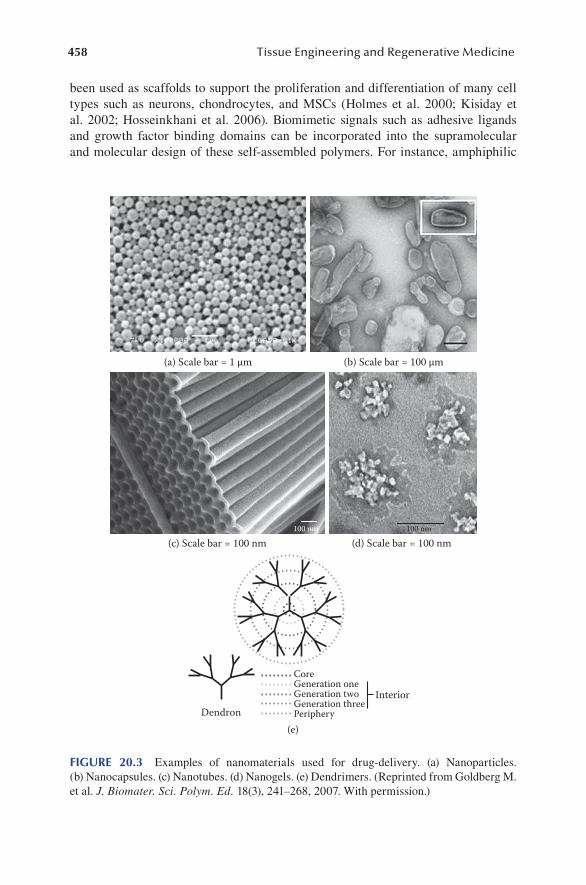
been used as scaffolds to support the proliferation and differentiation of many cell types such as neurons, chondrocytes, and MSCs (Holmes et al. 2000; Kisiday et al. 2002; Hosseinkhani et al. 2006). Biomimetic signals such as adhesive ligands and growth factor binding domains can be incorporated into the supramolecular and molecular design of these self-assembled polymers. For instance, amphiphilic
(a) Scale bar = 1 µm (b) Scale bar = 100 µm
(c) Scale bar = 100 nm
(e)Dendron
Interior
CoreGeneration oneGeneration twoGeneration threePeriphery
(d) Scale bar = 100 nm100 nm 100 nm
FIGURE 20.3 Examples of nanomaterials used for drug-delivery. (a) Nanoparticles. (b) Nanocapsules. (c) Nanotubes. (d) Nanogels. (e) Dendrimers. (Reprinted from Goldberg M.et al. J. Biomater. Sci. Polym. Ed. 18(3), 241–268, 2007. With permission.)

459Nanotechnology for Engineering Cellular Microenvironment
peptides with TGF-binding domains were shown to promote chondrogenic differen-tiation of human MSCs and enhance cartilage tissue regeneration in a full-thickness chondral defect rabbit model (Shah et al. 2010).
20.1.3 Biological signals To enhance Tissue regeneraTion
Biological signals, such as growth factors and nucleic acids, are often used to pro-mote specific cellular processes and tissue regeneration. Scaffolds can serve as a
+
Cell-surfaceattachmentEndocytosis
Exocytosis
Escapefromendosome
Escapefromlysosome
NuclearimportNucleusCytosol
Transportthrough
cytoplasm Microtubule
(b)(a)
(c)
FIGURE 20.4 Gene delivery with cationic material. (a) Schematic of spontaneous poly-plex formation by the electrostatic interaction between polycations (e.g., cationic lipids and polymers) and DNA. (b) Transmission electron microscopy (TEM) image of nanoparticles formed by plasmid DNA and PEI (scale bar = 200 nm). (c) Intracellular barriers to gene deliv-ery. After attaching to the targeted cell surface, the polyplex must be internalized (e.g., by endocytosis), escape from the endosome, transport through the cytoplasm, move toward the nucleus and across the nuclear membrane, and unpackage. (Adapted from Pack D. W. et al. Nat. Rev. Drug Discov. 4(7), 581–593, 2005. With permission.)

460 Tissue Engineering and Regenerative Medicine
delivery depot for bioactive molecules via various methods such as surface adsorp-tion or covalent modification (Babensee et al. 2000; Tessmar and Göpferich 2007). Nanomaterials such as nanoparticles, nanocapsules, nanotubes, nanogels, and den-drimers have also been explored as delivery vehicles to enhance the controlled release and cellular uptake for various types of biomolecules (Figure 20.3; Goldberg et al. 2007). Advances in gene therapy provide a powerful tool to promote lineage-specific differentiation via directly regulating the intrinsic signals of stem cells. Today, technology is being developed with the potential to either “turn on” a target gene, through DNA delivery, or “turn off” a gene by small interfering RNA (siRNA) delivery. Current challenges in gene therapy include the need to identify optimal therapeutic gene targets as well as the lack of safe and efficient delivery methods. Cationic polymers can self-assemble with DNA to form nanoparticles via electro-static interactions (Figure 20.4a and b), and different polymer designs have been used to overcome the hurdles involved in different steps during gene delivery. In this chapter, we will discuss recent advances in applying gene delivery as a tool to direct stem cell fate, with special focus on polymer-based gene delivery strategies using a nanomaterials approach.
20.2 GENE DELIVERY
20.2.1 consiDeraTions for effecTive gene Delivery
Successful gene therapy requires a safe and effective delivery system to transport the DNA to the target cell nuclei. The basic considerations for developing carriers for gene delivery include safety, biocompatibility, stability, and the ability to transfer therapeutic genes to the target cells at high efficiency (Pack et al. 2005). To facilitate smooth translation into clinical practice, the ease of large-scale manufacture, admin-istration, and cost-effectiveness must also be considered.
20.2.1.1 Barriers to Gene DeliveryTo develop highly efficient gene delivery systems, it is crucial to first understand the barriers that need to be overcome during the transport process (Figure 20.4c). In transit to the target cells, the genetic material must be protected from degradation and preserve its properties for effective transfection (Papisov 1998). It also needs to navigate through the extracellular space and arrive at the target cells. To achieve cell-specific targeting, receptor-mediated mechanisms can be incorporated into the deliv-ery vehicle (Ferkol et al. 1996; Wu et al. 1991; Wu and Wu 1988). Cellular uptake usually occurs via endocytosis, which leads the carrier/gene complexes into an acidic intracellular vesicle called an endosome. The genetic material needs to escape from the endosome, continue to transport through the cytoplasm, and finally transport into the nucleus and get expressed. During this multistep transport process, the genetic material continues to encounter barriers such as the possibility of exocytosis, degra-dation in the endosome, or the presence of cytosolic nucleases (Pollard et al. 2001). Finally, in the cases in which a delivery system, such as a synthetic polymer, is used, the delivery vehicle must release the genetic material and permit transcription once the genetic material–carrier complex is delivered to the nucleus.

461Nanotechnology for Engineering Cellular Microenvironment
20.2.2 MeThoDs for gene Delivery
Gene delivery methods can be broadly divided into two categories, viral and non-viral approaches. The viral approach uses viruses as the delivery vehicle and takes advantage of the ability of viruses to efficiently transfer genetic information into cells. Despite its high gene transfer efficiency, clinical translation of the viral-based approach is limited by safety concerns such as pathogenesis and potential immuno-genicity (Pack et al. 2005). Nonviral methods include the direct injection of naked genetic material, physical methods, and delivery with a gene transfer carrier such as synthetic polymers and lipids. Nonviral methods are potentially much safer, but often suffer from significantly lower transfection efficiency.
20.2.2.1 Viral MethodsIn viral methods, a replication-deficient viral vector can be formed by replacing the coding region of the viral genome with a therapeutic gene, turning the virus into a gene delivery vehicle. Viral vectors can generally be classified as integrating or non-integrating vectors. Retrovirus and adeno-associated viruses (AAV) can integrate their genome into the DNA of the host cell. They can achieve stable expressions in dividing cells, and are suitable for the treatment of chronic diseases, in which long-term gene expression is needed (Pack et al. 2005). Nonintegrating viruses, such as adenovirus and herpes simplex virus type I, transfer their genomes into the nucleus of the target cell as episomes without integration.
Retroviruses are enveloped RNA viruses that are present in all vertebrates. The three main classes of retroviruses are oncoretrovirus, lentivirus, and spumavirus. Oncoretrovirus was the first viral vector developed and has been widely used in clinical trials to treat diseases such as severe combined immunodeficiency (Heilbronn and Weger 2010; Thomas et al. 2003). A major limitation of using oncoretrovirus for gene delivery is that the virus can only be used to transduce dividing cells. Lentivirus, on the other hand, can enter the nucleus of both dividing and nondividing cells. Lentivirus has been shown to successfully deliver short hairpin RNAs into a wide range of dividing and nondividing cell types, including primary T cells, stem cells, and single-cell embryos, and achieve stable gene silencing (Rubinson et al. 2003). Lentiviral vectors have also been used to reprogram human somatic cells into pluripotent stem cells (Yu et al. 2007). The AAV is another commonly used vector for gene delivery due to their nonpathogenic nature and inability to self-replicate. The major limitation of AAV vector is that it can only accom-modate small gene products of up to 5 kb (Kootstra and Verma 2003).
Unlike integrating viral vectors, the genome of nonintegrating vectors remains episomal, eliminating the possibility of insertional mutagenesis. The capacity to carry large genetic materials, the ability to transfect nondividing cells, and high gene transfer efficiency in a variety of tissues make adenovirus vectors and herpes simplex virus type I promising gene delivery vehicles (Kay et al. 2001; Thomas et al. 2003). However, high immunogenicity and transient transgene expression limit the clinical potential of these nonintegrating viral vectors as gene delivery vehicles.
Naturally evolved as effective delivery vehicles, viruses have been shown to deliver therapeutic genes at high efficiency in many in vitro and in vivo studies (Goldberg et al. 2008; Pack et al. 2005; Thomas et al. 2003). Although more than

462 Tissue Engineering and Regenerative Medicine
70% of gene therapy-based clinical trials (as of July 2007) used virus-mediated gene delivery, there is still no FDA-approved viral vector-based gene therapy on the market (Check 2005; Green et al. 2008; Hollon 2000). Acute toxicity, oncogenesis, mutagenesis, and carcinogenicity are among the safety risks associated with viral gene therapy (Goldberg et al. 2007; Green et al. 2008; Pack et al. 2005; Thomas et al. 2003). High manufacturing costs, low cargo capacity, low quality control, and resistance to repeated infection are some of the additional characteristics of viral gene therapy that makes it an undesirable gene delivery method.
20.2.2.2 Nonviral Approaches20.2.2.2.1 Physical MethodsThe ability of DNA alone to transfect cells is very poor due to low cellular uptake and its susceptibility to nuclease degradation (Herweijer and Wolff 2003). Several physical methods have been developed to enhance DNA delivery efficiency including electroporation, pressurized intravascular delivery, sonoporation, laser irradiation, and magnetofection (Mehier-Humbert and Guy 2005). Electroporation increases the permeability of cell membranes to plasmid DNA by exposing the target cells to a series of electrical pulses. Pressurized intravascular delivery has been used to successfully transfect cells in a variety of tissue types including liver and skeletal muscle. Sonoporation, which usually involves the use of a low-dose ultrasound, is another physical method to enhance gene transfer efficiency. The application of ultrasound or laser irradiation leads to transient formation of small pores on the cell membrane and enhances permeability (Taniyama et al. 2002). Magnetofection involves the application of a magnetic field to enhance the uptake of plasmid DNA coupled with magnetic nanoparticles (Scherer et al. 2002). Most of these physical methods enhance the entry of DNA into cells by overcoming barriers posed by the cell membrane, but are often associated with significant cytotoxicity. Furthermore, the challenges associated with intracellular transport to the nucleus remain to be addressed (Geng et al. 2011; Guo and Huang 2011; Rychahou and Evers 2010).
20.2.2.2.2 Cationic Biomaterials for Nonviral Gene DeliveryBiomaterials, such as cationic polymers and lipids, are capable of condensing DNA into nanoparticles for enhanced delivery into target cells (Nguyen et al. 2009). Because of their lipophilic nature, cationic lipids can penetrate cell membranes and deliver DNA into cells, and have been widely used for nonviral-based gene deliv-ery in vitro. However, clinical applications of cationic lipids for gene therapy are impeded by their unfavorable biodistribution, as they tend to accumulate in the lung and liver when delivered in vivo. Furthermore, they may cause high levels of cyto-toxicity by disrupting the cell membrane.
Cationic polymers are promising gene delivery carriers due to their relative ease of manufacture and enhanced safety compared with viruses (Goldberg et al. 2008). However, polymer-mediated gene delivery is typically much less efficient than viral vectors due to the multistep barriers they need to overcome to achieve efficient expression (Green et al. 2008; Nguyen et al. 2009). The polymer must initially bind to, condense, or encapsulate the DNA in the form of a nanoparticle to protect it from

463Nanotechnology for Engineering Cellular Microenvironment
degradation. Once the nanoparticles get trafficked into the cell via endocytosis, they must be capable of escaping the endosome into the cytoplasm before endosomal deg-radation. Finally, the nanoparticles need to travel through the nuclear membrane and release the DNA cargo for gene expression. Two cationic polymers that have been widely used for gene delivery are polyethylenimine (PEI) and poly(l-lysine) (PLL; Goldberg et al. 2008; Green et al. 2008). However, PEI is nondegradable and has high cytotoxicity effects and a low transfection efficiency in comparison to viral vectors and high cytotoxicity effects (Green et al. 2008; Moghimi et al. 2005). PLL is biodegrad-able and can be conjugated with bioactive ligands to enhance target-specific delivery (Luo and Saltzman 2000). However, like PEI, PLL suffers from low transfection effi-ciency and high cytotoxicity (Brazeau et al. 1998). Because of the complex multistep barriers of the gene delivery process, it is very difficult to develop a polymer with high efficiency using the conventional design-driven, iterative synthesis approach.
20.3 HIGH-THROUGHPUT APPROACH TO DISCOVERING NOVEL MATERIALS FOR GENE DELIVERY
To accelerate the discovery of novel biomaterials for effective gene delivery, high-throughput screening has emerged as a novel approach to help better understand the structure–function relationships that govern polymer-mediated gene delivery. In a high-throughput screening approach, a large library of polymers with diverse chemi-cal structures can be synthesized in a combinatorial approach, and the polymer/DNA nanoparticles can then be screened for their transfection efficiency and cytotoxicity using high-throughput screening assays. The versatility of polymer chemistry, com-bined with computational tools for predicting structure–function relationships, can be used in combinatorial library synthesis to generate a broad range of cationic polymer vectors with specific properties (Goldberg et al. 2008). Compared with the conven-tional, iterative approach, such a high-throughput approach greatly accelerated the development of novel polymers with enhanced gene delivery efficiency (Akinc et al. 2003a,b; Anderson et al. 2003; Green et al. 2008; Thomas et al. 2007). The results of such screening studies yield valuable information on structure–function relation-ships, which can guide the design of the next generation of polymer libraries synthesis for further performance improvement. Poly(β-amino esters) (PBAEs) are an excellent example of biodegradable polymers for gene delivery that have arisen from such com-binatorial synthesis and high-throughput screening approaches (Green et al. 2008).
20.3.1 The DevelopMenT of pBaes for Dna Delivery
PBAEs constitute a particularly diverse class of cationic polymers for gene deliv-ery. PBAE has several advantages including its biodegradability via hydrolytically degradable ester groups, reduced toxicity, capacity for structural diversity, and abil-ity to trigger endosomal escape (Lynn and Langer 2000). Most importantly, PBAEs can be synthesized via a one-step reaction with conjugate addition between diacry-late and amine monomers, which makes it feasible for facile combinatorial synthesis (Figure 20.5a and b; Green et al. 2008). The first high-throughput effort to probe the

464 Tissue Engineering and Regenerative Medicine
O O O O
OO
O O
O O
N
N N
RR
R n
n
+
+
O O
OO
OO
NH2
R
∆
∆NHHN
RR
BU
6 61
70
75
80
86
87
93
94
8
17
20242528323660
AA
II
JJ
LL
C
D
E
F
O
M
O OOO OOO
OOO
OO O
O
OO
O O
OOO
OO
OOHHO
HOHO
HOHOHOHO H
N
HN
NH
NH
NH2
NH2
NH2
NH2
NH2
NH2
NH2NHHN
HN
HN
N
N
N
N
N
HO
HOHO
HONH2
NH2NH2
NH2NH2
NH2
O
OO
OO
OO
O
OOO O
O
O
O
OO
O OO
OOOO O
O OOO
OO O
OO
O
O O O
O
O
1.40
0
C32
JJ28
C28
AA
28U
28A
A24
AA
20JJ2
0A
A28
JJ32
D94
AA
24U
32D
60D
24E2
0O
20F3
2F2
8O
24A
A36
C36
E28
U94
O28
D61
D36
AA
60D
70D
61JJ9
4U
87D
60C
94F9
4U
80E8
6D
86D
87U
94LL
8E2
4M
17LL
6D
25E3
2D
32LL
8JJ3
6D
28U
36E8
0E3
6C
94JJ8
0E9
4A
A94
D93
B17
C86
U75
JJ86
C86
C75
C20
C80
F86
AA
61U
93U
86II
36II
28JJ2
4C
25U
25II
32
1.20
01.
050
1.00
00.
950
0.80
0
1.4001.3001.2001.1001.0501.0251.0000.9750.9500.9000.8000.600
100
10
1
0.1
Polymer composition
Gen
e ex
pres
sion
Amine/acrylatemonomer ratio
(a)
(b)
(c)
NH2
NH2

465Nanotechnology for Engineering Cellular Microenvironment
structure–function relationship of PBAEs synthesized a combinatorial library of 140 PBAEs composed of 20 amine monomers and 7 diacrylate monomers (Akinc et al. 2003a). Results from this screening showed that polymer structures can significantly influence the size and charge of the polymer/DNA nanoparticles, which has a sig-nificant impact on transfection efficiency. Lead polymers formed complexes smaller than 250 nm and had a positive ζ-potential, and demonstrated four to eight times higher transfection efficiency than PEI (Akinc et al. 2003b). Two leading PBAEs were further optimized by varying other parameters, including chemical structures of the polymer end groups, molecular weight, and polymer to DNA ratios (Akinc et al. 2003b). Using a semiautomated, parallel synthesis and screening process, Anderson et al. created a library of 2350 structurally unique, degradable PBAEs (Anderson et al. 2003). This combinatorial library was synthesized by diluting monomers in dimethyl sulfoxide, a low-viscosity medium, and using a fluid-handling robot and 12-channel micropipette to automate monomer mixing and thereby simultaneously setting up all 2350 reactions (Anderson et al. 2003). The library was later screened for its ability to bind DNA and transfect COS-7 cells, an easy to transfect cell line, under serum-free conditions. The screening identified a subset of 46 polymers with performance superior to the PEI control (Anderson et al. 2003; Green et al. 2008). These studies found that lead polymers can condense DNA into nanoparticles with smaller sizes and more positive surface charge, and such lead polymers also showed structural similarities (Figure 20.5c). Among the top nine polymers, all were formed from a conjugate addition of an amino alcohol and a hydrophobic diacrylate mol-ecule (Anderson et al. 2005; Green et al. 2008). Moreover, the three top-performing polymers (i.e., C28, C32, and JJ28) all had converging structures (Anderson et al. 2005). These high-throughput studies highlight the importance of combinatorial synthesis and screening platforms in uncovering structure–function relationships and thereby optimizing polymeric gene delivery systems.
20.3.2 coMBinaTorial approaches To sirna Delivery sysTeMs
Recent advances in RNA interference (RNAi) have provided another powerful tool for regulating cell behavior via gene silencing. RNAi is a gene-silencing mechanism that involves double-stranded RNA-mediated sequence-specific mRNA degradation and is a powerful mechanism for controlling cell behavior. However, the success of RNAi-based therapeutics requires an efficient delivery system to transport the 21–25 nucleotide double-stranded siRNAs into the target cells. Chemically synthesized siRNA often degrades rapidly in vivo and direct injection of naked siRNA often
FIGURE 20.5 (a) High-throughput synthesis and screening of PBAEs for gene delivery. PBAE synthesis by the conjugate addition of amines to diacrylate groups. (b) Acrylate and amino monomers used for the synthesis of the PBAE library. (c) Transfection proficiency of the PBAE polymer library. COS-7 cells were transfected with PBAE/DNA nanoparticles. PBAE polymers with different amine/acrylate monomer ratios were screened. (Adapted from Anderson D. G. et al. Mol. Ther. 11(3), 426–434, 2005 and Green J. J. et al. Acc. Chem. Res. 41(6), 749–759, 2008. With permission.)

466 Tissue Engineering and Regenerative Medicine
leads to poor gene silencing (Urban-Klein et al. 2005). The low success rate of early studies highlighted the importance of developing delivery systems that would pro-tect siRNA from degradation and facilitate its cellular uptake. Earlier trials showed that PEI can complex synthetic siRNAs into nanoparticles and help preserve the bioactivity of these molecules (Urban-Klein et al. 2005); however, PEI/siRNA com-plexes have significant toxicity effects (Akinc et al. 2008). In an effort to develop novel biomaterials for safe and efficient siRNA delivery, Akinc et al. synthesized a combinatorial library of lipid-like materials (lipidoids; Akinc et al. 2008). From a large library of more than 1200 structurally diverse lipidoids, Akinc and colleagues identified lipidoids that facilitate high levels of specific siRNA-mediated silencing of endogenous gene transcripts. Top-performing lipidoids also share common struc-tures including amide linkages, more than two alkyl tails of eight to twelve car-bons and a secondary amide (Akinc et al. 2008). Such information provides valuable guidelines for further optimizing siRNA delivery vectors.
20.4 A COMBINATORIAL APPROACH TO IDENTIFY SYNERGISTIC GENETIC SIGNALS
Gene therapy offers a promising approach for promoting desired cellular processes and tissue development by upregulating inductive genes via DNA delivery or down-regulating inhibitory genes via RNAi delivery. Codelivery of multiple genetic signals may act synergistically to accelerate the desired cellular processes. Although exten-sive work has been performed on delivering single genetic signals for gene therapy, efforts using synergistic genetic signals are only beginning to emerge. The cellular microenvironment is highly dynamic and multifactorial. Many signaling pathways are interconnected, affecting cell fate in a synergistic or antagonistic manner. For example, bone morphogenic protein-6 (BMP-6) or insulin-like growth factor-1 (IGF-1) alone could not induce chondrogenesis, whereas on codelivery, such factors with TGF-β3 can synergistically promote chondrogenic differentiation (Indrawattana et al. 2004). Likewise, most tissue morphogenesis processes are tightly regulated by interactive signals that either turn on an activator gene, or turn off an inhibitor gene. Identifying synergistic genetic signals that promote the desired cellular processes would provide a powerful tool for directing tissue regeneration.
High-throughput screening has been explored to facilitate the discovery of potent gene targets in regulating stem cell differentiation. For example, Zhao et al. screened a synthetic siRNA library targeting 5000 human genes, which yielded 12 candidate suppressors for osteogenic specification in human MSCs (Zhao and Ding 2007). A recent study examined the effects of codelivering two genes, BMP2 and core-binding factor α-1 (Cbfa1), on osteogenic differentiation of adipose-derived stem cells (ADSCs; Lee et al. 2010). BMP2- and Cbfa1-transduced ADSCs showed an upregulation of osteogenic markers and increased mineralization. Codelivery of BMP2 and dexamethasone, a small molecule activator of osteogenesis, also led to increased expression of early-stage osteogenic markers in mouse embryonic stem cells (Blum et al. 2004). BMP-transduced, mouse muscle-derived stem cells also showed increased angiogenesis and mineralization when cotransduced with VEGF (Peng et al. 2002). These studies demonstrated that codelivering multiple genetic

467Nanotechnology for Engineering Cellular Microenvironment
signals may synergistically promote lineage-specific differentiation. A recent study has also explored the potential benefits of codelivering multiple inductive and sup-pressive genes on osteogenic differentiation (Ramasubramanian et al. 2011). The gene expression of three target genes, BMP2, an osteogenic inducer, as well as GNAS and Noggin, osteogenic suppressors, were modulated in a combinatorial manner using biomaterials-mediated gene delivery. Compared with BMP2 DNA delivery alone, codelivery of BMP2 DNA and either siGNAS or siNoggin significantly accelerated osteogenic differentiation in human ADSCs, with enhanced osteogenic gene expres-sion and mineralization. These results suggest that inductive or suppressive genetic switches interact in a complex, nonlinear manner. Given the complex interactions among various genetic signals, it is highly desirable to develop high-throughput screening assays to facilitate the identification of synergistic genetic signals for gene therapy (Nguyen et al. 2009; Ramasubramanian et al. 2011).
20.5 CONCLUSION
Advances in nanotechnology have provided powerful tools to control cell fate and tissue regeneration. These strategies can be broadly classified into two categories, the “outside–in” or “inside–out” approaches. The outside–in approach focuses on engineering the cellular microenvironment via recapitulating the sophisticated struc-ture and biological functionalities found in the native ECM. Nanotechnology facili-tates the incorporation of nanoscale features into scaffold design to better mimic native tissues, which helps elucidate the complex interactions between cells and their surrounding microenvironment. The inside–out approach influences cell behavior via directly regulating genetic signals using biomaterials-mediated gene delivery. Toward this end, the use of cationic lipids or polymers in gene delivery systems offers great versatility in chemical modification to circumvent barriers in gene deliv-ery. High-throughput synthesis and screening approaches have demonstrated great promise in accelerating the rapid discovery of novel biomaterials that can condense nucleic acids into nanoparticles for efficient gene delivery. Identifying potential syn-ergistic signals using high-throughput screening would also provide potentially more powerful gene targets for promoting desired tissue formation. Finally, validating the efficacy of these strategies in appropriate animal models will be crucial for the suc-cessful translation of their final applications in the clinical setting.
Although significant advances have been made in applying nanotechnology for advancing tissue regeneration, challenges remain before such therapies can be suc-cessfully translated from bench-to-bedside. For example, extensive efforts have been dedicated to understanding individual types of microenvironmental cues (e.g., nano-topography), but how complex interactive cues regulate cell fate and tissue devel-opment remains largely unknown. Moreover, many of the molecular mechanisms underlying these effects have yet to be discovered. As for the application of nanoma-terials for gene delivery, there is an impending need to develop technology that facil-itates efficient targeting to specific cell types or tissues in situ. Biomaterial-based vectors allow the possibility of conjugating biofunctional moieties such as antibodies that target specific cells or tissues. In sum, nanotechnology will continue to provide a powerful tool for both fundamental and applied research in tissue regeneration.

468 Tissue Engineering and Regenerative Medicine
The contributions of nanotechnology to the tissue engineering field will also become even more prominent as the field continues to evolve and better integrate with multi-ple disciplines, such as molecular and cell biology, materials science, computational biology, and medicine.
REFERENCES
Akinc, A., D. G. Anderson, D. M. Lynn, and R. Langer. 2003a. Synthesis of poly(beta-amino ester)s optimized for highly effective gene delivery. Bioconjugate Chemistry 14(5), 979–988.
Akinc, A., D. M. Lynn, D. G. Anderson, and R. Langer. 2003b. Parallel synthesis and bio-physical characterization of a degradable polymer library for gene delivery. Journal of the American Chemical Society 125(18), 5316–5323.
Akinc, A., A. Zumbuehl, M. Goldberg, E. S. Leshchiner, V. Busini, N. Hossain et al. 2008. A combinatorial library of lipid-like materials for delivery of RNAi therapeutics. Nature Biotechnology 26(5), 561–569.
Anderson, D. G., A. Akinc, N. Hossain, and R. Langer. 2005. Structure/property studies of polymeric gene delivery using a library of poly(beta-amino esters). Molecular Therapy 11(3), 426–434.
Anderson, D. G., D. M. Lynn, and R. Langer. 2003. Semi-automated synthesis and screening of a large library of degradable cationic polymers for gene delivery. Angewandte Chemie (International ed. in English) 42(27), 3153–3158.
Babensee, J. E., L. V. McIntire, and A. G. Mikos. 2000. Growth factor delivery for tissue engi-neering. Pharmaceutical Research 17(5), 497–504.
Banerjee, A., M. Arha, S. Choudhary, R. S. Ashton, S. R. Bhatia, D. V. Schaffer et al. 2009. The influence of hydrogel modulus on the proliferation and differentiation of encapsu-lated neural stem cells. Biomaterials 30(27), 4695–4699.
Blum, J. S., M. B. Parrott, A. G. Mikos, and M. A. Barry. 2004. Early osteoblastic differentia-tion induced by dexamethasone enhances adenoviral gene delivery to marrow stromal cells. Journal of Orthopaedic Research 22(2), 411–416.
Brazeau, G. A., S. Attia, S. Poxon, and J. A. Hughes. 1998. In vitro myotoxicity of selected cat-ionic macromolecules used in non-viral gene delivery. Pharmaceutical Research 15(5), 680–684.
Brittberg, M., A. Lindahl, A. Nilsson, C. Ohlsson, O. Isaksson, and L. Peterson. 1994. Treatment of deep cartilage defects in the knee with autologous chondrocyte transplan-tation. New England Journal of Medicine 331(14), 889–895.
Buehler, M. J. 2006. Nature designs tough collagen: Explaining the nanostructure of collagen fibrils. Proceedings of the National Academy of Sciences of the United States of America 103(33), 12285–12290.
Check, E. 2005. Gene therapy put on hold as third child develops cancer. Nature 433, 561.Dalby, M. J., N. Gadegaard, R. Tare, A. Andar, M. O. Riehle, P. Herzyk et al. 2007. The con-
trol of human mesenchymal cell differentiation using nanoscale symmetry and disorder. Nature Materials 6(12), 997–1003.
Engler, A. J., S. Sen, H. L. Sweeney, and D. E. Discher. 2006. Matrix elasticity directs stem cell lineage specification. Cell 126(4), 677–689.
Ferkol, T., J. C. Perales, F. Mularo, and R. W. Hanson. 1996. Receptor-mediated gene trans-fer into macrophages. Proceedings of the National Academy of Sciences of the United States of America 93(1), 101–105.
Ferrara, N. 2010. Binding to the extracellular matrix and proteolytic processing: Two key mechanisms regulating vascular endothelial growth factor action. Molecular Biology of the Cell 21(5), 687–690.

469Nanotechnology for Engineering Cellular Microenvironment
Geng, T., Y. Zhan, J. Wang, and C. Lu. 2011. Transfection of cells using flow-through electro-poration based on constant voltage. Nature Protocols 6(8), 1192–1208.
Goldberg, M., R. Langer, and X. Jia. 2007. Nanostructured materials for applications in drug delivery and tissue engineering. Journal of Biomaterials Science. Polymer Edition 18(3), 241–268.
Goldberg, M., K. Mahon, and D. Anderson. 2008. Combinatorial and rational approaches to polymer synthesis for medicine. Advanced Drug Delivery Reviews 60(9), 971–978.
Green, J. J., R. Langer, and D. G. Anderson. 2008. A combinatorial polymer library approach yields insight into nonviral gene delivery. Accounts of Chemical Research 41(6), 749–759.
Guo, X., and Huang, L. 2011. Recent advances in nonviral vectors for gene delivery. Accounts of Chemical Research. DOI: 10.1021/ar200151m.
Hartgerink, J. D., E. Beniash, and S. I. Stupp. 2001. Self-assembly and mineralization of peptide–amphiphile nanofibers. Science 294(5547), 1684–1688.
Heilbronn, R., and S. Weger. 2010. Viral vectors for gene transfer: Current status of gene thera-peutics. Handbook of Experimental Pharmacology 197, 143–170.
Herweijer, H., and J. A. Wolff. 2003. Progress and prospects: Naked DNA gene transfer and therapy. Gene Therapy 10(6), 453–458.
Hollon, T. 2000. Researchers and regulators reflect on first gene therapy death. Nature Medicine 6, 6.
Holmes, T. C., S. de Lacalle, X. Su, G. Liu, A. Rich, and S. Zhang. 2000. Extensive neurite out-growth and active synapse formation on self-assembling peptide scaffolds. Proceedings of the National Academy of Sciences of the United States of America 97(12), 6728–6733.
Hosseinkhani, H., M. Hosseinkhani, F. Tian, H. Kobayashi, and Y. Tabata. 2006. Osteogenic differentiation of mesenchymal stem cells in self-assembled peptide–amphiphile nano-fibers. Biomaterials 27(22), 4079–4086.
Huebsch, N., P. R. Arany, A. S. Mao, D. Shvartsman, O. A. Ali, S. A. Bencherif et al. 2010. Harnessing traction-mediated manipulation of the cell/matrix interface to control stem-cell fate. Nature Materials 9(6), 518–526.
Indrawattana, N., G. Chen, M. Tadokoro, L. H. Shann, H. Ohgushi, T. Tateishi et al. 2004. Growth factor combination for chondrogenic induction from human mesenchymal stem cell. Biochemical and Biophysical Research Communications 320(3), 914–919.
Kay, M. A., J. C. Glorioso, and L. Naldini. 2001. Viral vectors for gene therapy: The art of turning infectious agents into vehicles of therapeutics. Nature Medicine 7(1), 33–40.
Kisiday, J., M. Jin, B. Kurz, H. Hung, C. Semino, S. Zhang et al. 2002. Self-assembling peptide hydrogel fosters chondrocyte extracellular matrix production and cell divi-sion: Implications for cartilage tissue repair. Proceedings of the National Academy of Sciences of the United States of America 99(15), 9996–10001.
Kolácná, L., J. Bakesová, F. Varga, E. Kostáková, L. Plánka, A. Necas et al. 2007. Biochemical and biophysical aspects of collagen nanostructure in the extracellular matrix. Physiological Research 56, Suppl 1, S51–60.
Kootstra, N. A., and I. M. Verma. 2003. Gene therapy with viral vectors. Annual Review of Pharmacology and Toxicology 43, 413–439.
Kumbar, S. G., R. James, S. P. Nukavarapu, and C. T. Laurencin. 2008. Electrospun nanofiber scaffolds: Engineering soft tissues. Biomedical Materials 3(3), 034002.
Lamers, E., R. van Horssen, J. te Riet, F. C. van Delft, R. Luttge, X. F. Walboomers et al. 2010. The influence of nanoscale topographical cues on initial osteoblast morphology and migration. European Cells and Materials 20, 329–343.
Langer, R., and J. P. Vacanti. 1993. Tissue engineering. Science 260(5110), 920–926.Lee, S. J., S. W. Kang, H. J. Do, I. Han, D. A. Shin, J. H. Kim et al. 2010. Enhancement of bone
regeneration by gene delivery of BMP2/Runx2 bicistronic vector into adipose-derived stromal cells. Biomaterials 31(21), 5652–5659.

470 Tissue Engineering and Regenerative Medicine
Luo, D., and W. M. Saltzman. 2000. Synthetic DNA delivery systems. Nature Biotechnology 18(1), 33–37.
Lutolf, M. P., and J. A. Hubbell. 2005. Synthetic biomaterials as instructive extracellular microenvironments for morphogenesis in tissue engineering. Nature Biotechnology 23(1), 47–55.
Lynn, D. M., and R. Langer. 2000. Degradable poly(beta-amino esters): Synthesis, charac-terization, and self-assembly with plasmid DNA. Journal of the American Chemical Society 122(44), 10761–10768.
Ma, Z., M. Kotaki, R. Inai, and S. Ramakrishna. 2005. Potential of nanofiber matrix as tissue-engineering scaffolds. Tissue Engineering 11(1–2), 101–109.
Matthews, J. A., G. E. Wnek, D. G. Simpson, and G. L. Bowlin. 2002. Electrospinning of col-lagen nanofibers. Biomacromolecules 3(2), 232–238.
Mehier-Humbert, S., and R. H. Guy. 2005. Physical methods for gene transfer: Improving the kinetics of gene delivery into cells. Advanced Drug Delivery Reviews 57(5), 733–753.
Moghimi, S. M., P. Symonds, J. C. Murray, A.C. Hunter, G. Debska, and A. Szewczyk. 2005. A two-stage poly(ethylenimine)-mediated cytotoxicity: Implications for gene transfer/therapy. Molecular Therapy 11, 990–995.
Nguyen, D. N., J. J. Green, J. M. Chan, R. Langer, and D. G. Anderson. 2009. Polymeric materials for gene delivery and DNA vacinnation. Advanced Materials 21(8), 847–867.
Oh, S., K. S. Brammer, Y. S. Li, D. Teng, A. J. Engler, S. Chien et al. 2009. Stem cell fate dictated solely by altered nanotube dimension. Proceedings of the National Academy of Sciences of the United States of America 106(7), 2130–2135.
Pack, D. W., A. S. Hoffman, S. Pun, and P. S. Stayton. 2005. Design and development of poly-mers for gene delivery. Nature Reviews Drug Discovery 4(7), 581–593.
Papachroni, K. K., D. N. Karatzas, K. A. Papavassiliou, E. K. Basdra, and A. G. Papavassiliou. 2009. Mechanotransduction in osteoblast regulation and bone disease. Trends in Molecular Medicine 15(5), 208–216.
Papisov, M. I. 1998. Theoretical considerations of RES-avoiding liposomes: Molecular mechan-ics and chemistry of liposome interactions. Advanced Drug Delivery Reviews 32(1–2), 119–138.
Pek, Y. S., A. C., Wan, and J. Y. Ying. 2010. The effect of matrix stiffness on mesenchymal stem cell differentiation in a 3D thixotropic gel. Biomaterials 31(3), 385–391.
Peng, H., V. Wright, A. Usas, B. Gearhart, H. C. Shen, J. Cummins et al. 2002. Synergistic enhancement of bone formation and healing by stem cell-expressed VEGF and bone morphogenetic protein-4. Journal of Clinical Investigation 110(6), 751–759.
Pollard, H., G. Toumaniantz, J. L. Amos, H. Avet-Loiseau, G. Guihard, J. P. Behr et al. 2001. Ca2+-sensitive cytosolic nucleases prevent efficient delivery to the nucleus of injected plasmids. Journal of Gene Medicine 3(2), 153–164.
Ramasubramanian, A., S. Shiigi, G. K. Lee, and F. Yang. 2011. Non-viral delivery of induc-tive and suppressive genes to adipose-derived stem cells for osteogenic differentiation. Pharmaceutical Research 28(6), 1328–1337.
Rho, J. Y., L. Kuhn-Spearing, and P. Zioupos. 1998. Mechanical properties and the hierarchi-cal structure of bone. Medical Engineering and Physics 20(2), 92–102.
Rho, K. S., L. Jeong, G. Lee, B. M. Seo, Y. J. Park, S. D. Hong et al. 2006. Electrospinning of collagen nanofibers: Effects on the behavior of normal human keratinocytes and early-stage wound healing. Biomaterials 27(8), 1452–1461.
Rubinson, D. A., C. P. Dillon, A. V. Kwiatkowski, C. Sievers, L. Yang, J. Kopinja et al. 2003. A lentivirus-based system to functionally silence genes in primary mammalian cells, stem cells and transgenic mice by RNA interference. Nature Genetics 33(3), 401–406.
Rychahou, P. G., and B. M. Evers. 2010. Hydrodynamic delivery protocols. Methods in Molecular Biology 623, 189–195.
471Nanotechnology for Engineering Cellular Microenvironment
Scherer, F., M. Anton, U. Schillinger, J. Henke, C. Bergemann, A. Krüger et al. 2002. Magnetofection: Enhancing and targeting gene delivery by magnetic force in vitro and in vivo. Gene Therapy 9(2), 102–109.
Schultz, G. S., and A. Wysocki. 2009. Interactions between extracellular matrix and growth factors in wound healing. Wound Repair and Regeneration 17(2), 153–162.
Shah, R. N., N. A. Shah, M. M. Del Rosario Lim, C. Hsieh, G. Nuber, and S. I. Stupp. 2010. Supramolecular design of self-assembling nanofibers for cartilage regenera-tion. Proceedings of the National Academy of Sciences of the United States of America 107(8), 3293–3298.
Shin, H., S. Jo, and A. G. Mikos. 2003. Biomimetic materials for tissue engineering. Biomaterials 24(24), 4353–4364.
Silver, F. H., J. W. Freeman, and G. P. Seehra. 2003. Collagen self-assembly and the develop-ment of tendon mechanical properties. Journal of Biomechanics 36(10), 1529–1553.
Taniyama, Y., K. Tachibana, K. Hiraoka, M. Aoki, S. Yamamoto, K. Matsumoto et al. 2002. Development of safe and efficient novel nonviral gene transfer using ultrasound: Enhancement of transfection efficiency of naked plasmid DNA in skeletal muscle. Gene Therapy 9(6), 372–380.
Tessmar, J. K., and A. M. Göpferich. 2007. Matrices and scaffolds for protein delivery in tissue engineering. Advanced Drug Delivery Reviews 59(4–5), 274–291.
Thomas, C. E., A. Ehrhardt, and M. A. Kay. 2003. Progress and problems with the use of viral vectors for gene therapy. Nature Reviews Genetics 4(5), 346–358.
Thomas, M., J. J. Lu, C. Zhang, J. Chen, and A. M. Klibanov. 2007. Identification of novel superior polycationic vectors for gene delivery by high-throughput synthesis and screen-ing of a combinatorial library. Pharmaceutical Research 24(8), 1564–1571.
Urban-Klein, B., S. Werth, S. Abuharbeid, F. Czubayko, and A. Aigner. 2005. RNAi-mediated gene-targeting through systemic application of polyethylenimine (PEI)-complexed siRNA in vivo. Gene Therapy 12(5), 461–466.
Wu, G. Y., and C. H. Wu. 1988. Receptor-mediated gene delivery and expression in vivo. Journal of Biological Chemistry 263(29), 14621–14624.
Wu, G. Y., J. M. Wilson, F. Shalaby, M. Grossman, D. A. Shafritz, and C. H. Wu. 1991. Receptor-mediated gene delivery in vivo. Partial correction of genetic analbuminemia in Nagase rats. Journal of Biological Chemistry 266(22), 14338–14342.
Yim, E. K., R. M. Reano, S. W. Pang, A. F. Yee, C. S. Chen, and K. W. Leong. 2005. Nanopattern-induced changes in morphology and motility of smooth muscle cells. Biomaterials 26(26), 5405–5413.
Yoshimoto, H., Y. M. Shin, H. Terai, and J. P. Vacanti. 2003. A biodegradable nanofiber scaf-fold by electrospinning and its potential for bone tissue engineering. Biomaterials 24(12), 2077–2082.
Yu, J. Y., M. A. Vodyanik, K. Smuga-Otto, J. Antosiewicz-Bourget, J. L. Frane, S. Tian et al. 2007. Induced pluripotent stem cell lines derived from human somatic cells. Science 318(5858), 1917–1920.
Zhang, L., and T. J. Webster. 2009. Nanotechnology and nanomaterials: Promises for improved tissue regeneration. Nano Today 4(1).
Zhao, Y., and S. Ding. 2007. A high-throughput siRNA library screen identifies osteogenic suppressors in human mesenchymal stem cells. Proceedings of the National Academy of Sciences of the United States of America 104(23), 9673–9678.
