chapter 15 feeling jumpy: the nervous...
TRANSCRIPT

Chapter 15
Feeling Jumpy: The Nervous SystemIn This Chapter! Breaking down the structure of nerves
! Centering with the central nervous system
! Branching out with the peripheral nervous system
! Taking a hands-off approach to the autonomic nervous system
! Examining the senses
Throughout this book, you look at the human body from head to toe, exploring how itcollects and distributes the molecules it needs to grow and thrive, how it reproduces
itself, and even how it gets rid of life’s nastier byproducts. In this chapter, however, you lookat the living computer that choreographs the whole show, the one system that contributesthe most to making us who we are as humans.
The nervous system is the communications network that goes into nearly every part of thebody, enervating your muscles, pricking your pain sensors, and letting you reach beyondyourself into the larger world. More than 80 major nerves make up this intricate network,and each nerve contains somewhere around 1 million neurons (individual nerve cells).It’s through this complex network that you respond both to external and internal stimuli,demonstrating a characteristic called irritability (the capacity to respond to stimuli, not thetendency to yell at annoying people).
There are three functional types of cells in the nervous system: receptor cells that receive astimulus (sensing); conductor cells that transmit impulses (integrating); and effector cells, ormotor neurons, which bring about a response such as contracting a muscle. Put another way,there are three functions of the human nervous system as a whole: orientation, or the abilityto generate nerve impulses in response to changes in the external and internal environments(this also can be referred to as perception); coordination, or the ability to receive, sort, anddirect those signals to channels for response (this also can be referred to as integration); andconceptual thought, or the capacity to record, store, and relate information received and toform plans for future reactions to environmental change (which includes specific action).
In this chapter, you get a feel for how the nervous system is put together. You practice identi-fying the parts and functions of nerves and the brain itself as well as the structure and activi-ties of the Big Three parts of the whole nervous system: the central, the peripheral, and theautonomic systems. In addition, we touch on the sensory organs that bring information intothe human body.
Building from Basics: Neurons, Nerves,Impulses, Synapses
Before trying to study the system as a whole, it’s best to break it down into buildingblocks first.
NeuronsThe basic unit that makes up nerve tissue is the neuron (also called a nerve cell). Itsproperties include that marvelous irritability that we speak of in the chapter introduc-tion as well as conductivity, otherwise known as the ability to transmit a nerve impulse.
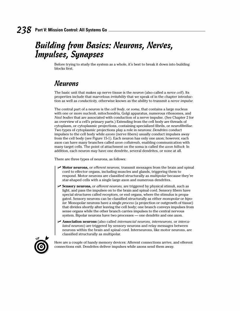
The central part of a neuron is the cell body, or soma, that contains a large nucleuswith one or more nucleoli, mitochondria, Golgi apparatus, numerous ribosomes, andNissl bodies that are associated with conduction of a nerve impulse. (See Chapter 2 foran overview of a cell’s primary parts.) Extending from the cell body are threads ofcytoplasm, or cytoplasmic projections, containing specialized fibrils, or neurofibrillae.Two types of cytoplasmic projections play a role in neurons: Dendrites conductimpulses to the cell body while axons (nerve fibers) usually conduct impulses awayfrom the cell body (see Figure 15-1). Each neuron has only one axon; however, eachaxon can have many branches called axon collaterals, enabling communication withmany target cells. The point of attachment on the soma is called the axon hillock. Inaddition, each neuron may have one dendrite, several dendrites, or none at all.
There are three types of neurons, as follows:
! Motor neurons, or efferent neurons, transmit messages from the brain and spinalcord to effector organs, including muscles and glands, triggering them torespond. Motor neurons are classified structurally as multipolar because they’restar-shaped cells with a single large axon and numerous dendrites.
! Sensory neurons, or afferent neurons, are triggered by physical stimuli, such aslight, and pass the impulses on to the brain and spinal cord. Sensory fibers havespecial structures called receptors, or end organs, where the stimulus is propa-gated. Sensory neurons can be classified structurally as either monopolar or bipo-lar. Monopolar neurons have a single process (a projection or outgrowth of tissue)that divides shortly after leaving the cell body; one branch conveys impulses fromsense organs while the other branch carries impulses to the central nervoussystem. Bipolar neurons have two processes — one dendrite and one axon.
! Association neurons (also called internuncial neurons, interneurons, or interca-lated neurons) are triggered by sensory neurons and relay messages betweenneurons within the brain and spinal cord. Interneurons, like motor neurons, areclassified structurally as multipolar.
Here are a couple of handy memory devices: Afferent connections arrive, and efferentconnections exit. Dendrites deliver impulses while axons send them away.
238 Part V: Mission Control: All Systems Go

NervesWhereas neurons are the basic unit of the nervous system, nerves are the cable-likebundles of axons that weave together the peripheral nervous system. There are threetypes of nerves:
! Afferent nerves are composed of sensory nerve fibers (axons) grouped togetherto carry impulses from receptors to the central nervous system.
! Efferent nerves are composed of motor nerve fibers carrying impulses from thecentral nervous system to effector organs, such as muscles or glands.
! Mixed nerves are composed of both afferent and efferent nerve fibers.
The diameter of individual axons (nerve fibers) tends to be microscopically small —many are no more than a micron, or one-millionth of a meter. But these same axonsextend to lengths of 1 millimeter and up. The longest axons in the human body runfrom the base of the spine to the big toe of each foot, meaning that these single-cellfibers may be 1 meter or more in length.
Each axon is swathed in myelin, a white fatty material made up of concentric layers ofSchwann cells in peripheral nerves. Oligodendrocytes in the central nervous system arealso associated with myelinated nerve fibers. The result is a structure referred to as amyelin sheath. Gaps in the sheath called nodes of Ranvier give the underlying nervefiber access to extracellular fluid, to speed up propagation of the nerve impulse.Nonmyelinated nerve fibers lie within body organs and therefore don’t need protectivemyelin sheaths to help them transmit impulses. Many peripheral nerve cell fibers alsoare protected by a neurilemmal sheath, a membrane that surrounds both the nervefiber and its myelin sheath.
Dendrites
Cell body
A. Motor Neuron B. Sensory Neuron
Cell body
NucleolusNucleus
Nucleolus
Impu
lse
to C
NS
Impu
lse
NucleusNucleus of Schwann cell
Axon
Schwann cellNode of Ranvier
Synaptic bouton
Impu
lse
from
CN
S
Axon
Figure 15-1:The motorneuron on
the left andsensory
neuron onthe right
show thecell struc-tures and
the paths ofimpulses.
239Chapter 15: Feeling Jumpy: The Nervous System

From the inside out, nerves are composed of the following:
! Axon: The impulse-conducting process of a neuron
! Myelin sheath: An insulating envelope that protects the nerve fiber and facilitatestransmission of nerve impulses
! Neurolemma (or neurilemma): A thin membrane present in many peripheralnerves that surrounds the nerve fiber and the myelin sheath
! Endoneurium: Loose, or areolar, connective tissue surrounding individual fibers
! Fasciculi: Bundles of fibers within a nerve
! Perineurium: The same kind of connective tissue as endoneurium; surrounds abundle of fibers
! Epineurium: The same kind of connective tissue as endoneurium and perineurium;surrounds several bundles of fibers
There also is a class of cells called neuroglia, or simply glia, that act as the supportivecells of the nervous system, providing neurons with nutrients and otherwise protectingthem. Glia include oligodendrocytes that support the myelin sheath within the centralnervous system; star-shaped cells called astrocytes that both support nerve tissue andcontribute to repairs when needed; and microgliacytes, cells that remove dead or dyingparts of tissue (this type of cell is called a phagocyte, which literally translates from theGreek words for “cell that eats”).
ImpulsesNeuron membranes are semi-permeable (meaning that certain small molecules likeions can move in and out but larger molecules can’t), and they’re electrically polarized(meaning that positively charged ions called cations rest around the outside mem-brane surface while negatively charged ions called anions line the inner surface; youcan find more about ions in Chapter 1).
A neuron that isn’t busy transmitting an impulse is said to be at its resting potential. Butthe nerve impulse theory, or membrane theory, says that things switch around when astimulus — a nerve impulse, or action potential — moves along the neuron. A stimuluschanges the specific permeability of the fiber membrane and causes a depolarizationdue to a reshuffling of the cations and anions. This change spreads along the nervefiber and constitutes the nerve impulse. It’s called an all-or-none response becauseeach neuron has a specific threshold of excitation. Once that threshold is exceeded, thenerve fiber responds with a fixed impulse. After depolarization, repolarization occursfollowed by a refractory period, during which no further impulses occur, even if thestimuli’s intensity increases.
Intensity of sensation, however, depends on the frequency with which one nerveimpulse follows another and the rate at which the impulse travels. That rate is deter-mined by the diameter of the impacted fiber and tends to be more rapid in large nervefibers. It’s also more rapid in myelinated fibers than nonmyelinated fibers. The cyto-plasm of the axon or nerve fiber is electrically conductive and the myelin decreasesthe capacitance to prevent charge leakage through the membrane. Depolarization atone node of Ranvier is sufficient to trigger regeneration of the voltage at the next node.Therefore, in myelinated nerve fibers the action potential does not move as a wave butrecurs at successive nodes, traveling faster than in nonmyelinated fibers. This isreferred to as saltatory conduction (from the Latin word saltare, which means “to hopor leap”).
240 Part V: Mission Control: All Systems Go

SynapsesNeurons don’t touch, which means that when a nerve impulse reaches the end of aneuron, it needs to cross a gap to the next neuron or to the gland or muscle cell forwhich the message is intended. That gap is called a synapse, or synaptic cleft. An electricsynapse — generally found in organs and glial cells — uses channels known as gap junc-tions to permit direct transmission of signals between neurons. But in other parts of thebody, chemical changes occur to let the impulse make the leap. The end branches of anaxon each form a terminal knob or bulb called a bouton terminal (that first word’s pro-nounced boo-taw), beyond which there is a space between it and the next nerve path-way. When an impulse reaches the bouton terminal, the following happens:
1. Synaptic vesicles in the knob release a transmitter called acetylcholine thatflows across the gap and increases the permeability of the next cell mem-brane in the chain.
2. An enzyme called cholinesterase breaks the transmitter down into acetyl andcholine, which then diffuse back across the gap.
3. An enzyme called choline acetylase in the synaptic vesicles reunites theacetyl and choline, prepping the bouton terminal to do its job again whenthe next impulse rolls through.
Nervous about getting all this right? Try some practice questions:
1.–5. Match the term to its description.
1. _____ Irritability
2. _____ Conductivity
3. _____ Orientation
4. _____ Coordination
5. _____ Conceptual thought
6. The brain and spinal cord are called the
a. Central nervous system
b. Visceral afferent system
c. Autonomic nervous system
d. Peripheral nervous system
7. The functional unit of the nervous system is the
a. Axon
b. Nephron
c. Dendron
d. Neuron
241Chapter 15: Feeling Jumpy: The Nervous System
a. Tissue’s ability to respond to stimulation
b. Ability to receive impulses and direct them to channelsfor favorable response
c. Sense organs’ capacity to generate nerve impulse tostimulation
d. Spreading of the nerve impulse
e. Capacity to record, store, and relate information to be used to determine future action

8. The terminal structure of the cytoplasmic projection of the neuron cannot be a(n)
a. Node of Ranvier
b. End organ
c. Effector
d. End bulb
e. Receptor
9. The afferent fiber that carries impulses to a neuron’s cell body is called a
a. Nissl body
b. Neuron
c. Dendrite
d. Axon
e. Mitochondria
10. The membrane surrounding the axon (nerve fiber) is the
a. Sarcolemma
b. Neurilemma
c. Perineurium
d. Epineurium
11.–15. Match the term to its description.
11. _____ Astrocytes
12. _____ Microgliacytes
13. _____ Oligodendrocytes
14. _____ Axons
15. _____ Dendrites
16. The neuroglia cells are important as
a. Sensory tissue
b. Supporting tissue
c. Irritable tissue
d. Conducting tissue
17. Axons tend to consist of
a. Single processes
b. Several synapses
c. Multiple processes
d. None of the above
e. Both a and c
242 Part V: Mission Control: All Systems Go
a. Cytoplasmic projections carrying impulses to the cell body
b. Cells that form and preserve myelin sheaths
c. Cytoplasmic projections carrying impulses from the cell body
d. Cells that are phagocytic
e. Cells that contribute to the repair process of the centralnervous system

18. A synapse between neurons is best described as the
a. Transmission of a continuous impulse
b. Transmission of an electrical impulse
c. Transmission of an impulse through a chemical and physical change
d. Transmission of an impulse through a physical change
e. Transmission of an impulse through a chemical change
19.–23. Match the term to its description.
19. _____ Endoneurium a. Fatty layer around an axon fiber
20. _____ Neurilemma b. Outer thin membrane around an axon fiber
21. _____ Schwann cell c. Cell in the sheath of an axon
22. _____ Node of Ranvier d. Depression in the sheath around a fiber
23. _____ Myelin sheath e. Connective tissue surrounding individual fibers ina nerve
24.–28. Match the term to its description.
24. _____ All-or-none response
25. _____ Cation
26. _____ Anion
27. _____ Polarization
28. _____ Depolarization
29.–33. Match the term to its description.
29. _____ Cholinesterase
30. _____ Choline acetylase
31. _____ Terminal bulb
32. _____ Acetylcholine
33. _____ Synapse
Minding the Central Nervous System and the Brain
Together, the brain and spinal cord make up the central nervous system. The spinalcord, which forms very early in the embryonic spinal canal, extends down into the tailportion of the vertebral column. But because bone grows much faster than nervetissue, the end of the cord soon is too short to extend into the lowest reaches of thespinal canal. In an adult, the 18-inch spinal cord ends between the first and secondlumbar vertebrae, roughly where the last ribs attach. Its tapered end is called the conusmedullaris. The cord continues as separate strands below that point and is referred toas the cauda equina (horse tail). A thread of fibrous tissue called the filum terminaleextends to the base of the coccyx (tailbone) and is attached by the coccygeal ligament.
243Chapter 15: Feeling Jumpy: The Nervous System
a. Impermeability of cell membrane
b. Negatively charged ion on the inner surface of the cellmembrane
c. Threshold of excitation determines ability to respond
d. Positively charged ion on the outer surface of the cellmembrane
e. Reshuffling of cell membrane ions; permeability ofcell membrane
a. Excitatory chemical necessary for continualnerve pathway
b. Enzyme for breakdown of excitatory chemical
c. Enzyme for reformation of excitatory chemical
d. Space between neurons
e. Contains storage vesicles for excitatory chemical
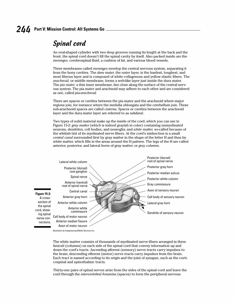
Spinal cordAn oval-shaped cylinder with two deep grooves running its length at the back and thefront, the spinal cord doesn’t fill the spinal cavity by itself. Also packed inside are themeninges, cerebrospinal fluid, a cushion of fat, and various blood vessels.
Three membranes called meninges envelop the central nervous system, separating itfrom the bony cavities. The dura mater, the outer layer, is the hardest, toughest, andmost fibrous layer and is composed of white collagenous and yellow elastic fibers. Thearachnoid, or middle membrane, forms a web-like layer just inside the dura mater.The pia mater, a thin inner membrane, lies close along the surface of the central nerv-ous system. The pia mater and arachnoid may adhere to each other and are consideredas one, called pia-arachnoid.
There are spaces or cavities between the pia mater and the arachnoid where majorregions join, for instance where the medulla oblongata and the cerebellum join. Thesesub-arachnoid spaces are called cisterna. Spaces or cavities between the arachnoidlayer and the dura mater layer are referred to as subdural.
Two types of solid material make up the inside of the cord, which you can see inFigure 15-2: gray matter (which is indeed grayish in color) containing unmyelinatedneurons, dendrites, cell bodies, and neuroglia; and white matter, so-called because ofthe whitish tint of its myelinated nerve fibers. At the cord’s midsection is a smallcentral canal surrounded first by gray matter in the shape of the letter H and then bywhite matter, which fills in the areas around the H pattern. The legs of the H are calledanterior, posterior, and lateral horns of gray matter, or gray columns.
Illustration by Imagineering Media Services Inc.
The white matter consists of thousands of myelinated nerve fibers arranged in threefuniculi (columns) on each side of the spinal cord that convey information up anddown the cord’s tracts. Ascending afferent (sensory) nerve tracts carry impulses tothe brain; descending efferent (motor) nerve tracts carry impulses from the brain.Each tract is named according to its origin and the joint of synapse, such as the corti-cospinal and spinothalmic tracts.
Thirty-one pairs of spinal nerves arise from the sides of the spinal cord and leave thecord through the intervertebral foramina (spaces) to form the peripheral nervous
Spinal nerve
Lateral white column
Anterior (ventral)root of spinal nerve
Anterior gray horn
Central canal
Anterior white column
Anterior whitecommissure
Anterior median fissure
Cell body of motor neuron
Axon of motor neuron
Posterior (dorsal)root of spinal nerve
Posterior gray horn
Posterior median sulcus
Posterior white column
Gray commissure
Axon of sensory neuron
Lateral gray horn
Cell body of sensory neuron
Dendrite of sensory neuron
Posterior (dorsal)root ganglion
Figure 15-2:A cross-
section ofthe spinal
cord, show-ing spinal
nerve con-nections.
244 Part V: Mission Control: All Systems Go

system, which we discuss in the later section “Taking Side Streets: The PeripheralNervous System.”
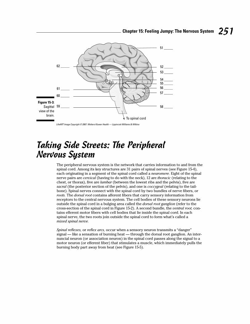
BrainOne of the largest organs in the adult human body, the brain tips the scales at 3 poundsand packs roughly 100 billion neurons (yes, that’s billion with a “b”) and 900 billionsupporting neuroglia cells. In this section, we review six major divisions of the brainfrom the bottom up (see Figure 15-3): medulla oblongata, pons, midbrain, cerebellum,diencephalon, and cerebrum.
Medulla oblongataThe spinal cord meets the brain at the medulla oblongata, or brainstem, just below theright and left cerebellar hemispheres of the brain. In fact, the medulla oblongata is con-tinuous with the spinal cord at its base (inferiorly) and back (dorsally) and locatedanteriorly and superiorly to the pons. All the afferent and efferent tracts of the cordcan be found in the brainstem as part of two bulges of white matter forming an areareferred to as the pyramids. Many of the tracts cross from one side to the other at thepyramids, which explains why the right side of the brain controls the left side of thebody and vice versa.
Along with the pons, the medulla oblongata also forms a network of gray and whitematter called the reticular formation, the upper part of the so-called extrapyramidalpathway. With its capacity to arouse the brain to wakefulness, it keeps the brain alert,directs messages in the form of impulses, monitors stimuli entering the sense recep-tors (accepting some and rejecting others it deems to be irrelevant), refines bodymovements, and effects higher mental processes such as attention, introspection, andreasoning. Although the cortex of the cerebrum is the actual powerhouse of thought, itmust be stimulated into action by signals from the reticular formation.
Nerve cells in the brainstem are grouped together to form nerve centers (nuclei) thatcontrol bodily functions, including cardiac activities, and respiration as well as reflexactivities such as sneezing, coughing, vomiting, and alimentary tract movements. Themedulla oblongata affects these reactions through the vagus, also referred to as cra-nial nerve X or the 10th cranial nerve. Three other cranial nerves also originate fromthis area: the 9th (IX) or glossopharyngeal, 11th (XI) or accessory, and 12th (XII) orhypoglossal.
PonsThe pons (literally “bridge”) does exactly as its name implies: It connects the cerebel-lum through a structure called the middle peduncle, the cerebrum by the superiorpeduncle, and the medulla oblongata by the inferior peduncle. It also unites the cere-bellar hemispheres, coordinates muscles on both sides of the body, controls facialmuscles (including those used to chew), and regulates the first stage of respiration.Oh, and it contains the nuclei for the following cranial nerves: the 5th (V) or trigemi-nal, the 6th (VI) or abducens, 7th (VII) or facial, and 8th (VIII) or vestibulocochlear.
MidbrainBetween the pons and the diencephalon lies the mesencephalon, or midbrain. It con-tains the corpora quadrigemina, which correlates optical and tactile impulses as wellas regulates muscle tone, body posture, and equilibrium through reflex centers in thesuperior colliculus. The inferior colliculus contains auditory reflex centers and isbelieved to be responsible for the detection of musical pitch. The midbrain contains
245Chapter 15: Feeling Jumpy: The Nervous System

the cerebral aqueduct, which connects the third ventricle of the thalamus with thefourth ventricle of the medulla oblongata (see the section “Ventricles” later in thischapter for more). The mesencephalon contains nuclei for the 3rd (III) or oculomotorcranial nerve and the 4th (IV) or trochlear cranial nerve. The red nucleus that containsfibers of the rubrospinal tract, a motor tract that acts as a relay station for impulsesfrom the cerebellum and higher brain centers, also lies within the midbrain, constitut-ing the superior cerebellar peduncle.
CerebellumThe cerebellum also is known as the little brain or small brain. The second-largest divi-sion of the brain, it’s just above and overhangs the medulla oblongata and lies justbeneath the rear portion of the cerebrum. Inside, the cerebellum resembles a tree calledthe arbor vitae, or “tree of life.” A central body called the vermis connects the two lateralmasses called the cerebellar hemispheres and assists in motor coordination and refine-ment of muscular movement, aiding equilibrium and muscle tone. The cerebellar cortexor gray matter contains Purkinje neurons with pear-shaped cell bodies, a multitude ofdendrites, and a single axon. It sends impulses to the white matter of the cerebellum andto other deeper nuclei in the cerebellum, and then to the brainstem. The cerebellarcortex has parallel ridges called the folia cerebelli, which are separated by deep sulci.
DiencephalonThe diencephalon, a region between the mesencephalon and the cerebrum, containsseparate brain structures called the thalamus, epithalamus, subthalamus, and hypothal-amus. The region where the two sides of the thalami come in contact and join forces iscalled the intermediate mass. The thalamus is a primitive receptive center throughwhich the sensory impulses travel on their way to the cerebral cortex. Here, nervefibers from the spinal cord and lower parts of the brain synapse with neurons leadingto the sensory areas of the cortex of the cerebrum. The thalamus is the great integrat-ing center of the brain with the ability to correlate the impulses from tactile, pain,olfactory, and gustatory (taste) senses with motor reactions.
The epithalamus contains the choroid plexus, a vascular structure that produces spinalfluid. The pineal body and olfactory centers also lie within the epithalamus, whichforms the roof of the third ventricle. The subthalamus is located below the thalamusand regulates the muscles of emotional expression.
The hypothalamus contains the centers for sexual reflexes; body temperature; water,carbohydrate, and fat metabolism; and emotions that affect the heartbeat and bloodpressure. It also has the optic chiasm (connecting the optic nerves to the optic tract),the posterior lobe of the pituitary gland, and a funnel-shaped region called theinfundibulum that forms the stalk of the pituitary gland.
CerebrumThe cerebrum, or forebrain, is often called the true brain. It has two cerebral hemi-spheres — the right and the left. A thin outer layer of gray matter called the cerebralcortex features folds or convolutions called gyri; furrows and grooves are referred to assulci, and deeper grooves are called fissures. A longitudinal fissure separates the cere-brum. The transverse fissure separates the cerebrum and the cerebellum. Each hemi-sphere has a set of controls for sensory and motor activities of the body. Interestingly,it’s not just right-side/left-side controls that are reversed in the cerebrum; the upperareas of the cerebral cortex control the lower body activities while the lower areas ofthe cortex control upper-body activities in a reversal called “little man upside down.”
Commissural fibers, a tract of nerves running from one side of the brain to the other,coordinate activities between the right and left hemispheres. The corpus callosum
246 Part V: Mission Control: All Systems Go
physically unites the two hemispheres and is the largest and densest mass of commis-sural fibers. A smaller mass called the fornix also plays a role.
Different functional areas of the cerebral cortex are divided into lobes:
! Frontal lobe: The seat of intelligence, memory, and idea association
! Parietal lobe: Functions in the sensations of temperature, touch, and sense ofposition and movement as well as the perception of size, shape, and weight
! Temporal lobe: Is responsible for perception and correlation of acoustical stimuli
! Occipital lobe: Handles visual perception
MedullaThe medulla, the region interior to the cortex, is composed of white matter that con-sists of three groups of fibers. Projection fibers carry impulses afferently from the brainstem to the cortex and efferently from the cortex to the lower parts of the central nerv-ous system. Association fibers originate in the cortical cells and carry impulses to theother areas of the cortex on the same hemisphere. Commissural fibers connect the twocerebral hemispheres.
VentriclesThe brain’s four ventricles are cavities and canals filled with cerebrospinal fluid. Twolateral ventricles are separated by the septum pellucidum. The lateral ventricles com-municate with the third ventricle through the foramen of Monro. The third ventricle isconnected by the cerebral aqueduct to the fourth ventricle, which is continuous withthe central canal of the spinal cord and contains openings to the meninges. The fourthventricle has openings that allow fluid to enter into the subarachnoid spaces.
Lining the ventricles is a thin layer of epithelial cells known as ependyma, or theependymal layer. Along with a network of capillaries from the pia mater, the ependymaand capillaries form the choroid plexus, which is the source of cerebrospinal fluid. Thechoroid plexus of each lateral ventricle produces the greatest amount of fluid. Fluidformed by the choroid plexus filters out by osmosis (refer to Chapter 2) and circulatesthrough the ventricles. Fluid is returned to the blood through the arachnoid villi,finger-like projections of the arachnoid meninx, which absorbs the fluid.
Twelve pairs of cranial nerves connect to the central nervous system via the brain (asopposed to the 31 pairs that connect via the spinal cord). Cranial nerves are identifiedby Roman numerals I through XII, and memorizing them is a classic test of anatomicalknowledge. Check out Table 15-1 for a listing of all the nerves, and then read on for amemory tool.
Table 15-1 Cranial Nerves
Number Name Type Function
I Olfactory Sensory Smell
II Optic Sensory Vision
III Oculomotor Mixed nerve Eyeball muscles
IV Trochlear Mixed nerve Eyeball muscles
(continued)
247Chapter 15: Feeling Jumpy: The Nervous System

Table 15-1 (continued)
Number Name Type Function
V Trigeminal Tri means “three,” so the three Skin; mastication types of trigeminal nerves are (chewing)1) Opthalmic nerve: sensory nerve; skin and mucous membranes of face and head; 2) Maxillary nerve: mixed nerve; mastication; 3) Mandibular nerve: mixed nerve; mastication
VI Abducens Mixed nerve Eye movements
VII Facial Mixed nerve Facial expression; salivary secretion;taste
VIII Vestibulocochlear Sensory Auditory nerve forhearing and equilibrium
IX Glossopharyngeal Mixed nerve Taste; swallowingmuscles of pharynx
X Vagus Mixed nerve Controls most internalorgans (viscera) fromhead and neck totransverse colon
XI Accessory Mixed nerve Swallowing andphonation
XII Hypoglossal Motor nerve Tongue movements
The first letters of each of these nerve names, in order, are OOOTTAFVGVAH. That’s amouthful, but students have come up with a number of memory tools to rememberthem. Our favorite is: Old Opera Organs Trill Terrific Arias For Various Grand VictoriesAbout History.
Put your knowledge of the central nervous system to the test:
248 Part V: Mission Control: All Systems Go
Q. The meninges’ functions areprimarily
a. Immunological
b. Supportive
c. Protective
d. Both a and b
e. Both b and c
A. The correct answer is supportiveand protective. Yes, meninges havetwo functions.

34. The cerebrum consists of two major halves called
a. Cerebellar hemispheres
b. Cerebral spheres
c. Cerebellar spheres
d. Cerebral hemispheres
35. The cerebrum is divided into two major halves by the
a. Lateral fissure
b. Transverse fissure
c. Longitudinal fissure
d. Fissure of Sylvius
e. Central sulcus
36. In the cerebrum, the
a. Right side tends to control the left side of the body and vice versa
b. Upper area controls lower-body activity
c. Lower area controls upper-body activity
d. None of the above are correct
e. A, b, and c are correct
37. The functions of the occipital lobe of the cerebrum pertain principally to
a. Visual activity
b. Autonomic control
c. Associative reasoning
d. Motor coordination
e. Auditory control
38. The cerebellum functions primarily as a center of
a. Visual activity
b. Associative reasoning
c. Auditory activity
d. Autonomic coordination
e. Motor control
39.–43. Match the term to its description.
39. _____ White matter
40. _____ Reticular formation
41. _____ Funiculus
42. _____ Dorsal root ganglion
43. _____ Gray matter
249Chapter 15: Feeling Jumpy: The Nervous System
a. Has the capacity to arouse the brain to wakefulness
b. Myelinated fibers
c. Bundles of nerve fibers arranged in tracts
d. Collection of cell bodies outside of the central nervoussystem
e. Unmyelinated fibers, cell bodies, and neuroglia

44.-48.Match the term to its description.
44. _____ Pons
45. _____ Cerebellum
46. _____ Medulla oblongata
47. _____ Cerebrum
48. _____ Mesencephalon
49. The largest quantity of cerebrospinal fluid originates from the
a. Foramen of Monro
b. Arachnoid villi
c. Lateral ventricle
d. Optic chiasm
e. Foramen of Luschka
50. The part of the brain that contains the thalamus, pituitary gland, and the optic chiasm is the
a. Diencephalon
b. Mesencephalon
c. Myelencephalon
d. Telencephalon
e. Metencephalon
51.–62. Use the terms that follow to identify the parts of the brain shown in Figure 15-3.
a. Pons
b. Thalamus
c. Cerebellum
d. Corpus callosum
e. Third ventricle
f. Hypothalamus
g. Cerebrum
h. Cerebral aqueduct
i. Midbrain
j. Pituitary gland
k. Medulla oblongata
l. Fourth ventricle
250 Part V: Mission Control: All Systems Go
a. Bridge connecting the medulla oblongata andcerebellum
b. Contains the centers that control cardiac, respiratory,and vasomotor functions
c. Contains the corpora quadrigemina and nuclei for theoculomotor and trochlear nerves
d. Controls motor coordination and refinement ofmuscular movement
e. Controls sensory and motor activity of the body
LifeART Image Copyright © 2007. Wolters Kluwer Health — Lippincott Williams & Wilkins
Taking Side Streets: The PeripheralNervous System
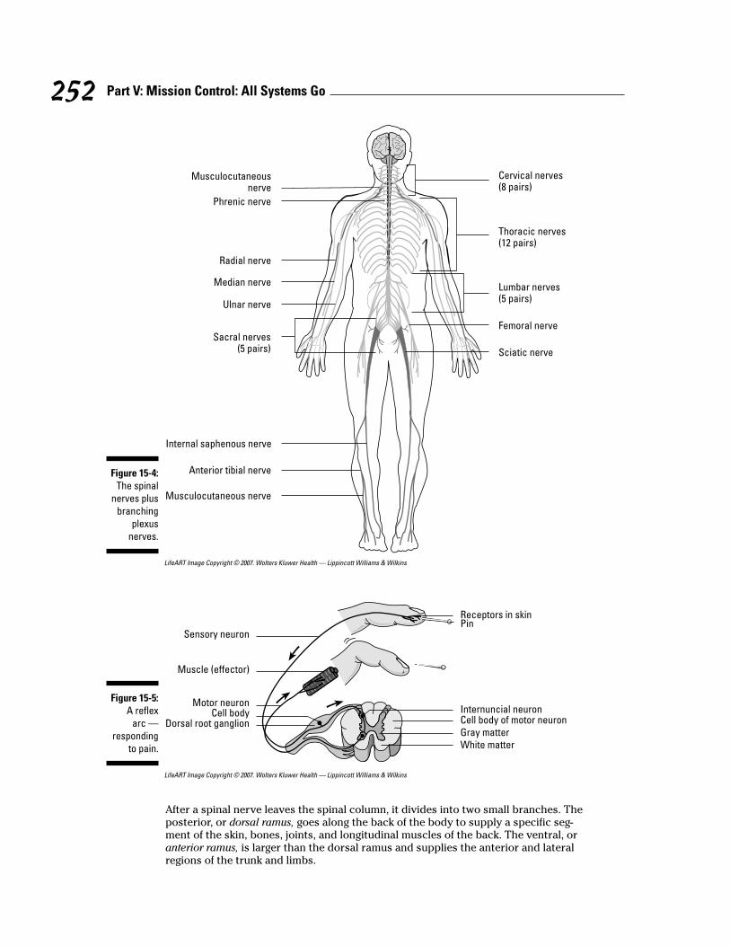
The peripheral nervous system is the network that carries information to and from thespinal cord. Among its key structures are 31 pairs of spinal nerves (see Figure 15-4),each originating in a segment of the spinal cord called a neuromere. Eight of the spinalnerve pairs are cervical (having to do with the neck), 12 are thoracic (relating to thechest, or thorax), five are lumbar (between the lowest ribs and the pelvis), five aresacral (the posterior section of the pelvis), and one is coccygeal (relating to the tail-bone). Spinal nerves connect with the spinal cord by two bundles of nerve fibers, orroots. The dorsal root contains afferent fibers that carry sensory information fromreceptors to the central nervous system. The cell bodies of these sensory neurons lieoutside the spinal cord in a bulging area called the dorsal root ganglion (refer to thecross-section of the spinal cord in Figure 15-2). A second bundle, the ventral root, con-tains efferent motor fibers with cell bodies that lie inside the spinal cord. In eachspinal nerve, the two roots join outside the spinal cord to form what’s called amixed spinal nerve.
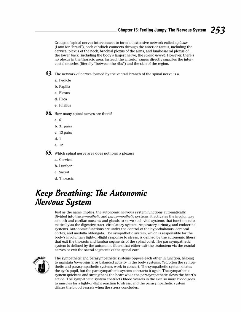
Spinal reflexes, or reflex arcs, occur when a sensory neuron transmits a “danger”signal — like a sensation of burning heat — through the dorsal root ganglion. An inter-nuncial neuron (or association neuron) in the spinal cord passes along the signal to amotor neuron (or efferent fiber) that stimulates a muscle, which immediately pulls theburning body part away from heat (see Figure 15-5).
To spinal cord
51 _____
52 _____
53 _____
54 _____55 _____56 _____
57 _____
58 _____59 _____
60 _____
61 _____
62 _____
Figure 15-3:Sagittal
view of thebrain.
251Chapter 15: Feeling Jumpy: The Nervous System
LifeART Image Copyright © 2007. Wolters Kluwer Health — Lippincott Williams & Wilkins
LifeART Image Copyright © 2007. Wolters Kluwer Health — Lippincott Williams & Wilkins
After a spinal nerve leaves the spinal column, it divides into two small branches. Theposterior, or dorsal ramus, goes along the back of the body to supply a specific seg-ment of the skin, bones, joints, and longitudinal muscles of the back. The ventral, oranterior ramus, is larger than the dorsal ramus and supplies the anterior and lateralregions of the trunk and limbs.
Receptors in skin
Sensory neuron
Muscle (effector)
Motor neuron
Pin
Dorsal root ganglion
Internuncial neuronCell bodyCell body of motor neuronGray matterWhite matter
Figure 15-5:A reflex
arc —responding
to pain.
Cervical nerves(8 pairs)
Thoracic nerves(12 pairs)
Lumbar nerves(5 pairs)
Femoral nerve
Sciatic nerve
Musculocutaneousnerve
Phrenic nerve
Radial nerve
Sacral nerves(5 pairs)
Internal saphenous nerve
Anterior tibial nerve
Musculocutaneous nerve
Median nerve
Ulnar nerve
Figure 15-4:The spinal
nerves plusbranching
plexusnerves.
252 Part V: Mission Control: All Systems Go
Groups of spinal nerves interconnect to form an extensive network called a plexus(Latin for “braid”), each of which connects through the anterior ramus, including thecervical plexus of the neck, brachial plexus of the arms, and lumbosacral plexus ofthe lower back (including the body’s largest nerve, the sciatic nerve). However, there’sno plexus in the thoracic area. Instead, the anterior ramus directly supplies the inter-costal muscles (literally “between the ribs”) and the skin of the region.
63. The network of nerves formed by the ventral branch of the spinal nerve is a
a. Pedicle
b. Papilla
c. Plexus
d. Plica
e. Phallus
64. How many spinal nerves are there?
a. 61
b. 31 pairs
c. 13 pairs
d. 1
e. 12
65. Which spinal nerve area does not form a plexus?
a. Cervical
b. Lumbar
c. Sacral
d. Thoracic
Keep Breathing: The AutonomicNervous System
Just as the name implies, the autonomic nervous system functions automatically.Divided into the sympathetic and parasympathetic systems, it activates the involuntarysmooth and cardiac muscles and glands to serve such vital systems that function auto-matically as the digestive tract, circulatory system, respiratory, urinary, and endocrinesystems. Autonomic functions are under the control of the hypothalamus, cerebralcortex, and medulla oblongata. The sympathetic system, which is responsible for thebody’s involuntary fight-or-flight response to stress, is defined by the autonomic fibersthat exit the thoracic and lumbar segments of the spinal cord. The parasympatheticsystem is defined by the autonomic fibers that either exit the brainstem via the cranialnerves or exit the sacral segments of the spinal cord.
The sympathetic and parasympathetic systems oppose each other in function, helpingto maintain homeostasis, or balanced activity in the body systems. Yet, often the sympa-thetic and parasympathetic systems work in concert. The sympathetic system dilatesthe eye’s pupil, but the parasympathetic system contracts it again. The sympatheticsystem quickens and strengthens the heart while the parasympathetic slows the heart’saction. The sympathetic system contracts blood vessels in the skin so more blood goesto muscles for a fight-or-flight reaction to stress, and the parasympathetic systemdilates the blood vessels when the stress concludes.
253Chapter 15: Feeling Jumpy: The Nervous System
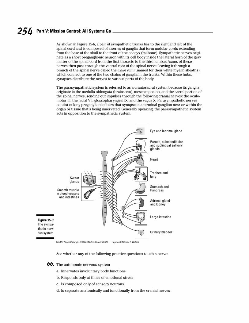
As shown in Figure 15-6, a pair of sympathetic trunks lies to the right and left of thespinal cord and is composed of a series of ganglia that form nodular cords extendingfrom the base of the skull to the front of the coccyx (tailbone). Sympathetic nerves origi-nate as a short preganglionic neuron with its cell body inside the lateral horn of the graymatter of the spinal cord from the first thoracic to the third lumbar. Axons of thesenerves then pass through the ventral root of the spinal nerve, leaving it through abranch of the spinal nerve called the white rami (named for their white myelin sheaths),which connect to one of the two chains of ganglia in the trunks. Within these hubs,synapses distribute the nerves to various parts of the body.
The parasympathetic system is referred to as a craniosacral system because its gangliaoriginate in the medulla oblongata (brainstem), mesencephalon, and the sacral portion ofthe spinal nerves, sending out impulses through the following cranial nerves: the oculo-motor III, the facial VII, glossopharyngeal IX, and the vagus X. Parasympathetic nervesconsist of long preganglionic fibers that synapse in a terminal ganglion near or within theorgan or tissue that’s being innervated. Generally speaking, the parasympathetic systemacts in opposition to the sympathetic system.
LifeART Image Copyright © 2007. Wolters Kluwer Health — Lippincott Williams & Wilkins
See whether any of the following practice questions touch a nerve:
66. The autonomic nervous system
a. Innervates involuntary body functions
b. Responds only at times of emotional stress
c. Is composed only of sensory neurons
d. Is separate anatomically and functionally from the cranial nerves
Eye and lacrimal gland
Sweatglands
Smooth musclein blood vessels
and intestines
Heart
Trachea andlung
Stomach andPancreas
Adrenal glandand kidney
Large intestine
Urinary bladder
Parotid, submandibularand sublingual salivaryglands
Figure 15-6:The sympa-thetic nerv-ous system.
254 Part V: Mission Control: All Systems Go

67. Which of the following statements is true about the autonomic nervous system?
a. It has two parts: the parasympathetic that controls all normal functions, and the sympatheticthat carries out the same functions.
b. It’s the nervous system that controls all reflexes.
c. It doesn’t function when the body’s under stress.
d. It has two divisions that are antagonistic to each other, meaning that one counteracts theeffects of the other one.
e. It controls the contractions of the skeletal, smooth, and cardiac muscle tissue.
68. The divisions of the autonomic nervous system are
a. Sympathetic and peripheral
b. Somatic and peripheral
c. Parasympathetic and peripheral
d. Sympathetic and parasympathetic
69. Which part of the autonomic nervous system can be called a craniosacral system?
a. Ganglia
b. Sympathetic trunks
c. Parasympathetic system
d. Medulla oblongata
Coming To Your SensesThe nervous system must have some way to perceive its environment in order to gen-erate appropriate responses. That’s where the senses come in. Sense receptors arethose numerous organs that respond to stimuli — like increased temperature, bittertastes, and sharp points — by generating a nerve impulse. While there are millions ofgeneral sense receptors found throughout the body that can convey touch, pain, andphysical contact, there are far fewer of the special sense receptors — those located inthe head — that really bring meaning to your world.
Sense receptors are classified by the stimuli they receive, as follows:
! Exteroceptors: Receive stimuli from the external environment. These are sen-sory nerve terminals, such as those in the skin and mucous membranes, that arestimulated by the immediate external environment.
! Interoceptors: Receive stimuli from the internal environment. These can be anyof the sensory nerve terminals located in and transmitting impulses from theviscera.
! Proprioceptors: Part of the “true” internal environment. They’re sensory nerveterminals chiefly found in muscles, joints, and tendons that give informationconcerning movements and position of the body.
! Teleceptors: Sensory nerve terminals stimulated by emanations from distantobjects. They exist in the eyes, ears, and nose.
255Chapter 15: Feeling Jumpy: The Nervous System
EyesAlthough there are many romantic notions about eyes, the truth is that an eyeball issimply a hollow sphere bounded by a trilayer wall and filled with a gelatinous fluidcalled, oddly enough, vitreous humor (see Figure 15-7). The outer fibrous coat is madeup of the sclera in back and the cornea in front. The sclera provides mechanicalsupport, protection, and a base for attachment of eye muscles, and it assists in thefocusing process. The cornea covers the anterior with a clear window.
An intermediate, or vascular, coat called the uvea provides blood and lymphatic fluids tothe eye, regulates light, and also secretes and reabsorbs aqueous humor, a thin wateryliquid that fills the anterior chamber of the eyeball in front of the iris. A pigmented coathas three layers: the iris, containing blood vessels, pigment cells, and smooth musclefibers to control the pupil’s diameter; the ciliary body, which is attached to the peripheryof the iris; and the choroid, a thin, dark brown, vascular layer lining most of the sclera onthe back and sides of the eye. The choroid contains arteries, veins, and capillaries thatsupply the retina with nutrients, and it also contains pigment cells to absorb light andprevent reflection and blurring. An optic nerve enters at the back (posterior) of each eye.
The retina is part of an internal nervous layer that connects with the optic nerve. Thenervous tissue layers along the inner back of the eye contain rods and cones (types ofneurons that analyze visual input). The rods are dim light receptors whereas the conesdetect bright light and construct form, structure, and color. The retina has an opticdisc, which is essentially a blind spot incapable of producing an image.
The crystalline lens consists of concentric layers of protein. It’s biconcave in shape,bulging outward. Located behind the pupil and iris, the lens is held in place by liga-ments attached to the ciliary muscles. When the ciliary muscles contract, the shape ofthe lens changes, altering the visual focus. This process of accommodation allows theeye to see objects both at a distance and close up.
The palpebrae (eyelids) extend from the edges of the eye orbit, into which roughlyfive-sixths of the eyeball is recessed. Eyelids come together at medial and lateralangles of the eye that are called the canthi. In the medial angle of the eye is a pinkregion called the caruncula, or caruncle. The caruncula contains sebaceous glands andsudoriferous (sweat) glands. A mucous membrane called the conjunctiva covers theinner surface of each eyelid and the anterior surface of the eye. Up top and to the sideof the orbital cavities are lacrimal glands that secrete tears that are carried through aseries of lacrimal ducts to the conjunctiva of the upper eyelid. Ultimately, secretionsdrain from the eyes through the nasolacrimal ducts.
EarsHuman ears — otherwise called vestibulocochlear organs — are more than just organsof hearing. They also serve as organs of equilibrium, or balance. Here are the threedivisions of the ear:
! The external ear includes the auricle, or pinna, which is the folded, roundedappendage made of cartilage and skin. Extending into the skull is the ear canal,or external auditory meatus, a short passage through the temporal bone endingat the tympanic membrane, or eardrum. Sebaceous glands near the external open-ing and ceruminous glands in the upper wall produce the brownish substanceknown as earwax, or cerumen.
256 Part V: Mission Control: All Systems Go
! The middle ear is a small, usually air-filled cavity in the skull that’s lined withmucous membrane. It communicates through the Eustachian tube with the phar-ynx. The Eustachian tube keeps air pressure equal on both sides of the eardrum(tympanic membrane), equalizing pressure in the middle ear with atmosphericpressure from outside. Three small bones called auditory ossicles occupy themiddle ear, deriving their names from their shapes: the malleus (hammer), theincus (anvil), and the stapes (stirrup).
! The internal ear is the most complex structure of the entire organ because it’swhere vibrations are translated. It’s composed of a group of interconnectedcanals or channels called the cochlea. Within the cochlea are three canals sepa-rated from each other by thin membranes; two of the canals — the vestibular andthe tympanic — are bony chambers filled with a perilymph fluid, and the thirdcanal — the cochlear canal — is a membranous chamber filled with endolymph.The cochlear canal lies between the vestibular and tympanic canals and containsthe organ of Corti, a spiral-shaped organ made up of cells with projecting hairsthat transmit auditory impulses.
The process of hearing a sound follows these basic steps:
1. Sound waves travel through the auditory canal, striking the eardrum andmaking it vibrate and setting the three ossicle bones into motion.
2. The stapes at the end of the chain strikes against the oval window of thevestibular canal, translating the motion into the perilymph fluid in the vestibu-lar and tympanic canals of the cochlea.
3. The vibrating fluid begins moving the basilar membrane that separates thetwo canals, stimulating the endolymph fluid in the membranous area ofthe cochlea.
4. The stimulated endolymph fluid in turn stimulates the hair cells of the organof Corti, which transmit the impulses to the brain over the auditory nerve.
That’s the hearing part of your ears. Equilibrium requires that some additional partscome into play. Three semicircular canals, each with an ampulla (or small, dilated por-tion) at each end, lie at right angles to each other. The ampullae connect to a fluid-filledsac called a utricle, which in turn connects to another fluid-filled sac called a saccule.Both sacs contain regions called maculae that are lined with sensitive hairs and containconcretions (solid masses) of calcium carbonate called otoliths (or otoconia). Whenlinear acceleration pulls at them, the otoliths press on the hair cells and initiate animpulse to the brain through basal sensory nerve fibers. When the head changes posi-tion, it causes a change in the direction of force on the hairs. Movement of the hairsstimulates dendrites of the vestibulocochlear nerve (the eighth cranial nerve) to carryimpulses to the brain.
257Chapter 15: Feeling Jumpy: The Nervous System
Q. The most sensitive region of theretina producing the greatest visualacuity is the
a. Blind spot
b. Cornea
c. Fovea centralis
d. Macula lutea
e. Lens
A. The correct answer is foveacentralis. It’s loaded with light-sensitive cones.
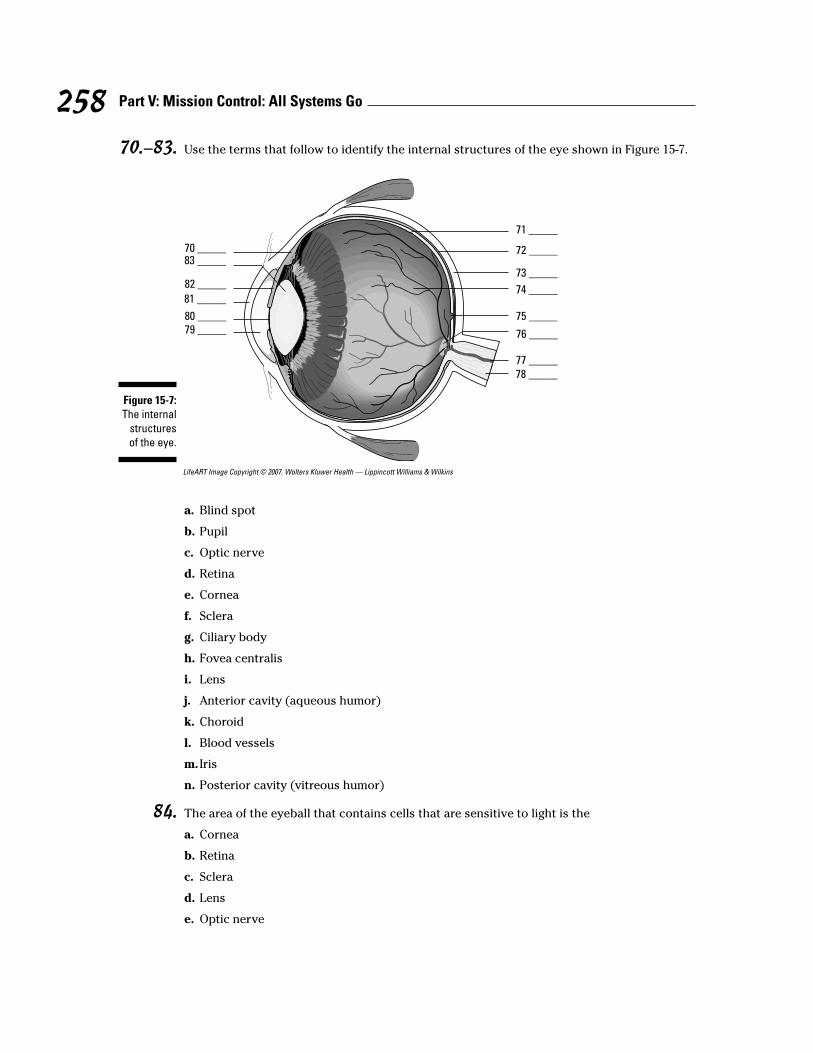
70.–83. Use the terms that follow to identify the internal structures of the eye shown in Figure 15-7.
LifeART Image Copyright © 2007. Wolters Kluwer Health — Lippincott Williams & Wilkins
a. Blind spot
b. Pupil
c. Optic nerve
d. Retina
e. Cornea
f. Sclera
g. Ciliary body
h. Fovea centralis
i. Lens
j. Anterior cavity (aqueous humor)
k. Choroid
l. Blood vessels
m. Iris
n. Posterior cavity (vitreous humor)
84. The area of the eyeball that contains cells that are sensitive to light is the
a. Cornea
b. Retina
c. Sclera
d. Lens
e. Optic nerve
70 _____83 _____
82 _____
81 _____
80 _____79 _____
78 _____77 _____
76 _____
75 _____
74 _____
73 _____
72 _____
71 _____
Figure 15-7:The internal
structuresof the eye.
258 Part V: Mission Control: All Systems Go
85. Which of the following structures is not part of the eyeball?
a. Optic nerve
b. Iris
c. Cornea
d. Pupil
e. Ciliary body
86. The accommodation (focusing) of the eye is accomplished by the
a. Sphincter of the pupil
b. Contraction of the iris
c. Action of the ciliary muscles
d. Dilator of the pupil
e. Contraction of the pupil
87. The structure in the eye that responds to the ciliary muscles during focusing is the
a. Pupil
b. Lens
c. Retina
d. Iris
e. Choroid
88. The middle ear is separated from the external ear by the
a. Tympanic membrane
b. Round window
c. The organ of Corti
d. Oval window
e. Cochlea
89. The structure that contains the receptor cells for the perception of sound is the
a. Tympanic membrane
b. Semicircular canals
c. Mastoid air cells
d. Organ of Corti
e. Middle ear cavity
90. The fluid in the membranous canal of the cochlea is called
a. Aqueous humor
b. Plasma
c. Endolymph
d. Perilymph
e. Vitreous humor
259Chapter 15: Feeling Jumpy: The Nervous System

91. Equilibrium is maintained by receptors in the
a. Cochlea
b. Utricle and saccule
c. Tympanic membrane
d. Organ of Corti
e. Middle ear cavity
92. The small bone in the ear that strikes against the oval window of the vestibular canal, settinginto motion the perilymph fluid in the vestibular and tympanic canals of the cochlea, is the
a. Incus
b. Hammer
c. Malleus
d. Anvil
e. Stapes
93.–105. Use the terms that follow to identify the structures of the ear shown in Figure 15-8.
LifeART Image Copyright © 2007. Wolters Kluwer Health — Lippincott Williams & Wilkins
93 _____
94 _____
95 _____
96 _____
97 _____
98 _____
99 _____
100 _____
101 _____
102 _____103 _____104 _____105 _____
Figure 15-8:The anatomy
of the ear.
260 Part V: Mission Control: All Systems Go
a. Oval window
b. Semicircular canals
c. Cochlea
d. Pinna
e. Malleus
f. Cochlear nerve
g. Stapes
h. Incus
i. Round window
j. Auditory canal
k. Tympanic membrane
l. Vestibular nerve
m. Auditory tube
Answers to Questions on the Nervous SystemThe following are answer to the practice questions presented in this chapter.
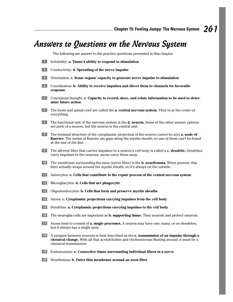
a Irritability: a. Tissue’s ability to respond to stimulation
b Conductivity: d. Spreading of the nerve impulse
c Orientation: c. Sense organs’ capacity to generate nerve impulse to stimulation
d Coordination: b. Ability to receive impulses and direct them to channels for favorableresponse
e Conceptual thought: e. Capacity to record, store, and relate information to be used to deter-mine future action
f The brain and spinal cord are called the a. central nervous system. They’re at the center ofeverything.
g The functional unit of the nervous system is the d. neuron. Some of the other answer optionsare parts of a neuron, but the neuron is the central unit.
h The terminal structure of the cytoplasmic projection of the neuron cannot be a(n) a. node ofRanvier. The nodes of Ranvier are gaps along the myelin sheath, so one of them can’t be foundat the end of the line.
i The afferent fiber that carries impulses to a neuron’s cell body is called a c. dendrite. Dendritescarry impulses to the neurons; axons carry them away.
j The membrane surrounding the axon (nerve fiber) is the b. neurilemma. When present, thisfiber actually wraps around the myelin sheath, so it’s always on the outside.
k Astrocytes: e. Cells that contribute to the repair process of the central nervous system
l Microgliacytes: d. Cells that are phagocytic
m Oligodendrocytes: b. Cells that form and preserve myelin sheaths
n Axons: c. Cytoplasmic projections carrying impulses from the cell body
o Dendrites: a. Cytoplasmic projections carrying impulses to the cell body
p The neuroglia cells are important as b. supporting tissue. They nourish and protect neurons.
q Axons tend to consist of a. single processes. A neuron may have one, many, or no dendrites,but it always has a single axon.
r A synapse between neurons is best described as the e. transmission of an impulse through achemical change. With all that acetylcholine and cholinesterase floating around, it must be achemical transmission.
s Endoneurium: e. Connective tissue surrounding individual fibers in a nerve
t Neurilemma: b. Outer thin membrane around an axon fiber
261Chapter 15: Feeling Jumpy: The Nervous System

u Schwann cell: c. Cell in the sheath of an axon
v Node of Ranvier: d. Depression in the sheath around a fiber
w Myelin sheath: a. Fatty layer around an axon fiber
x All-or-none response: c. Threshold of excitation determines ability to respond
y Cation: d. Positively charged ion on the outer surface of the cell membrane
A Anion: b. Negatively charged ion on the inner surface of the cell membrane
B Polarization: a. Impermeability of cell membrane
C Depolarization: e. Reshuffling of cell membrane ions; permeability of cell membrane
D Cholinesterase: b. Enzyme for breakdown of excitatory chemical
E Choline acetylase: c. Enzyme for reformation of excitatory chemical
F Terminal bulb: e. Contains storage vesicles for excitatory chemical
G Acetylcholine: a. Excitatory chemical necessary for continual nerve pathway
H Synapse: d. Space between neurons
I The cerebrum consists of two major halves called d. cerebral hemispheres. Cerebrum = cere-bral, and two halves = hemispheres.
J The cerebrum is divided into two major halves by the c. longitudinal fissure. Longitudinal isthe most likely position for an equal division.
K In the cerebrum, the e. a, b, and c are correct (right side tends to control the left side of thebody and vice versa, upper area controls lower-body activity, and lower area controlsupper-body activity). Right = left, and up = down. Clear as mud?
L The functions of the occipital lobe of the cerebrum pertain principally to a. visual activity. Toremember, use the word “occipital” to bring to mind the word “optic,” which of course is relatedto visual activity.
M The cerebellum functions primarily as a center of e. motor control.
N White matter: b. Myelinated fibers
O Reticular formation: a. Has the capacity to arouse the brain to wakefulness
P Funiculus: c. Bundles of nerve fibers arranged in tracts
Q Dorsal root ganglion: d. Collection of cell bodies outside of the central nervous system
R Gray matter: e. Unmyelinated fibers, cell bodies, and neuroglia
S Pons: a. Bridge connecting the medulla oblongata and cerebellum
T Cerebellum: d. Controls motor coordination and refinement of muscular movement
262 Part V: Mission Control: All Systems Go
U Medulla oblongata: b. Contains the centers that control cardiac, respiratory, and vasomotorfunctions
V Cerebrum: e. Controls sensory and motor activity of the body
W Mesencephalon: c. Contains the corpora quadrigemina and nuclei for the oculomotor andtrochlear nerves
X The largest quantity of cerebrospinal fluid originates from the c. lateral ventricle. This onerequires rote memorization — sorry!
Y The part of the brain that contains the thalamus, pituitary gland, and the optic chiasm is thea. diencephalon. Think of it as the home of the thalamus, and you can’t go wrong.
z–0 Following is how Figure 15-3, the brain, should be labeled.
51. g. Cerebrum; 52. d. Corpus callosum; 53. b. Thalamus; 54. f. Hypothalamus; 55. j.Pituitary gland; 56. h. Cerebral aqueduct; 57. c. Cerebellum; 58. l. Fourth ventricle; 59. k.Medulla oblongata; 60. a. Pons; 61. i. Midbrain; 62. e. Third ventricle
! The network of nerves formed by the ventral branch of the spinal nerve is a c. plexus. Theword stems from the Latin for “braid,” which makes sense for a network.
@ How many spinal nerves are there? b. 31 pairs. Count them: 8 cervical, 12 thoracic, 5 lumbar,5 sacral — plus 1 tailbone (coccygeal).
# Which spinal nerve area does not form a plexus? d. Thoracic
$ The autonomic nervous system a. innervates involuntary body functions. It’s the only answeroption with a sense of automation.
% Which of the following statements is true about the autonomic nervous system? d. It has twodivisions that are antagonistic to each other, meaning that one counteracts the effects of theother one. As a result, the body achieves homeostasis.
^ The divisions of the autonomic nervous system are d. sympathetic and parasympathetic. Theywork against each other in order to help the body maintain balance.
& Which part of the autonomic nervous system can be called a craniosacral system?c. Parasympathetic system. It originates in both the brainstem and the sacral region.
*–; Following is how Figure 15-7, the internal structures of the eye, should be labeled.
70. g. Ciliary body; 71. d. Retina; 72. k. Choroid; 73. f. Sclera; 74. n. Posterior cavity (vitre-ous humor); 75. h. Fovea centralis; 76. a. Blind spot; 77. l. Blood vessels; 78. c. Optic nerve;79. j. Anterior cavity (aqueous humor); 80. b. Pupil; 81. e. Cornea; 82. m. Iris; 83. i. Lens
: The area of the eyeball that contains cells that are sensitive to light is the b. retina. It’s at theback of the eyeball.
, Which of the following structures is not part of the eyeball? a. Optic nerve. This nerve carriesthe visual signals to the brain.
< The accommodation (focusing) of the eye is accomplished by the c. action of the ciliary mus-cles. They reshape the lens by contracting and relaxing as needed to bring things into focus.
263Chapter 15: Feeling Jumpy: The Nervous System

. The structure in the eye that responds to the ciliary muscles during focusing is the b. lens.Refer to the explanation for the preceding question.
> The middle ear is separated from the external ear by the a. tympanic membrane. Otherwiseknown as the eardrum, this membrane sometimes bursts or tears as a result of infection ortrauma.
/ The structure that contains the receptor cells for the perception of sound is the d. organ ofCorti. Hairs in this structure are what ultimately send the signal down the auditory nerve.
? The fluid in the membranous canal of the cochlea is called c. endolymph. Don’t forget that theprefix endo– means “within.”
` Equilibrium is maintained by receptors in the b. utricle and saccule. These little endolymph-filled sacs have hairs and chunks of calcium carbonate that detect changes in gravitationalforces.
~ The small bone in the ear that strikes against the oval window of the vestibular canal, settinginto motion the perilymph fluid in the vestibular and tympanic canals of the cochlea, is thee. stapes. That’s the only bone that actually touches the window. The other two carry thesignal down the chain to the stapes.
ú–œ Following is how Figure 15-8, the structures of the ear, should be labeled.
93. k. Tympanic membrane; 94. e. Malleus; 95. h. Incus; 96. b. Semicircular canals; 97. l.Vestibular nerve; 98. f. Cochlear nerve; 99. c. Cochlea; 100. i. Round window; 101. m.Auditory tube; 102. a. Oval window; 103. g. Stapes; 104. j. Auditory canal; 105. d. Pinna
264 Part V: Mission Control: All Systems Go