chapter 1 + 2 of environmental physics i the atmosphere - iup
TRANSCRIPT

R EV I EW AR T I C L E
The rhizosphere microbiome: significance of plant beneficial,plant pathogenic, and human pathogenic microorganisms
Rodrigo Mendes1, Paolina Garbeva2 & Jos M. Raaijmakers3
1Laboratory of Environmental Microbiology, Embrapa Environment, Jaguariuna, Brazil; 2Netherlands Institute of Ecology, Royal Dutch Academy of
Arts & Sciences (NIOO-KNAW), Wageningen, The Netherlands; and 3Laboratory of Phytopathology, Bacterial Ecology & Genomics, Wageningen
University, Wageningen, The Netherlands
Correspondence: Jos M. Raaijmakers,
Laboratory of Phytopathology, Bacterial
Ecology & Genomics, Wageningen University,
Droevendaalsesteeg 1, 6708 PB Wageningen,
The Netherlands. Tel.: +31 317 483427; fax:
+31 317 483412;
e-mail: [email protected]
Received 27 January 2013; revised 22 May
2013; accepted 27 May 2013. Final version
published online 22 July 2013.
DOI: 10.1111/1574-6976.12028
Editor: Dieter Haas
Keywords
plant–microbe interactions; plant growth
promotion; food safety; disease-suppressive
soils; core and minimal microbiome;
metagenomics.
Abstract
Microbial communities play a pivotal role in the functioning of plants by influ-
encing their physiology and development. While many members of the rhizo-
sphere microbiome are beneficial to plant growth, also plant pathogenic
microorganisms colonize the rhizosphere striving to break through the protec-
tive microbial shield and to overcome the innate plant defense mechanisms in
order to cause disease. A third group of microorganisms that can be found in
the rhizosphere are the true and opportunistic human pathogenic bacteria,
which can be carried on or in plant tissue and may cause disease when intro-
duced into debilitated humans. Although the importance of the rhizosphere
microbiome for plant growth has been widely recognized, for the vast majority
of rhizosphere microorganisms no knowledge exists. To enhance plant growth
and health, it is essential to know which microorganism is present in the rhizo-
sphere microbiome and what they are doing. Here, we review the main func-
tions of rhizosphere microorganisms and how they impact on health and
disease. We discuss the mechanisms involved in the multitrophic interactions
and chemical dialogues that occur in the rhizosphere. Finally, we highlight sev-
eral strategies to redirect or reshape the rhizosphere microbiome in favor of
microorganisms that are beneficial to plant growth and health.
Introduction
Plants are colonized by an astounding number of (micro)
organisms that can reach cell densities much greater than
the number of plant cells (Fig. 1). Also, the number of
microbial genes in the rhizosphere outnumbers by far the
number of plant genes (Fig. 1). An overwhelming number
of studies have revealed that many plant-associated
microorganisms can have profound effects on seed germi-
nation, seedling vigor, plant growth and development,
nutrition, diseases, and productivity (Fig. 2). Consistent
with the terminology used for microorganisms colonizing
the human body (Qin et al., 2010; Zhao, 2010; Gevers
et al., 2012), the collective communities of plant-associated
microorganisms are referred to as the plant microbiome
or as the plants’ other genome. In this context, plants can
be viewed as superorganisms that rely in part on their
microbiome for specific functions and traits. In return,
plants deposit their photosynthetically fixed carbon into
their direct surroundings, that is, spermosphere, phyllo-
sphere, rhizosphere, and mycorrhizosphere (Nelson, 2004;
Frey-Klett et al., 2007; Raaijmakers et al., 2009; Berendsen
et al., 2012; Vorholt, 2012), thereby feeding the microbial
community and influencing their composition and activi-
ties. To date, the interplay between plants and microor-
ganisms has been studied in depth for various leaf
pathogens, symbiotic rhizobia, and mycorrhizal fungi.
However, for the vast majority of plant-associated micro-
organisms, there is limited knowledge of their impact on
plant growth, health, and disease. Hence, deciphering the
plant microbiome is critical to identify microorganisms
that can be exploited for improving plant growth and
health.
The rhizosphere, that is, the narrow zone surrounding
and influenced by plant roots, is a hot spot for numerous
organisms and is considered as one of the most complex
ª 2013 Federation of European Microbiological Societies FEMS Microbiol Rev 37 (2013) 634–663Published by John Wiley & Sons Ltd. All rights reserved
MIC
ROBI
OLO
GY
REV
IEW
S
Downloaded from https://academic.oup.com/femsre/article-abstract/37/5/634/540803by gueston 26 July 2018

ecosystems on Earth (Hinsinger & Marschner, 2006; Pier-
ret et al., 2007; Jones & Hinsinger, 2008; Hinsinger et al.,
2009; Raaijmakers et al., 2009). Organisms found in the
rhizosphere include bacteria, fungi, oomycetes, nema-
todes, protozoa, algae, viruses, archaea, and arthropods
(Fig. 1; Lynch, 1990; Meeting, 1992; Bonkowski et al.,
2009; Bu�ee et al., 2009; Raaijmakers et al., 2009). Most
members of the rhizosphere microbiome are part of a
complex food web that utilizes the large amount of nutri-
ents released by the plant. Given that these rhizodeposits
(e.g. exudates, border cells, mucilage) are a major driving
force in the regulation of microbial diversity and activity
on plant roots, Cook et al. (1995) postulated that plants
may modulate the rhizosphere microbiome to their bene-
fit by selectively stimulating microorganisms with traits
that are beneficial to plant growth and health. Others
have argued that exudates are passively ‘released’ as over-
flow/waste products of the plant (Hartmann et al., 2009;
Jones et al., 2009; Dennis et al., 2010). So, whether plants
are using exudates to ‘cry for help’ or are ‘just crying’
remains to be addressed.
Rhizosphere organisms that have been well studied for
their beneficial effects on plant growth and health are the
nitrogen-fixing bacteria, mycorrhizal fungi, plant growth-
promoting rhizobacteria (PGPR), biocontrol microorgan-
isms, mycoparasitic fungi, and protozoa. Rhizosphere
organisms that are deleterious to plant growth and health
include the pathogenic fungi, oomycetes, bacteria, and
nematodes. A third group of microorganisms that can be
found in the rhizosphere are the human pathogens. Over
the past decade, there is an increasing number of reports
describing the proliferation of human pathogenic bacteria
Fig. 1. Overview of (micro)organisms present in the rhizosphere zoo. The circle’s size, except for VIRUSES, is a measure of the average number
of genes in the genomes of representative species of each group of organisms; the size (or size range) of their respective genomes is indicated
between parentheses. For each of these (micro)organisms, the approximate numbers for their abundance are indicated between square brackets
(Alexander, 1977; Brady, 1974; Lynch, 1988; Meeting, 1992; Bu�ee et al., 2009). The endophytic microorganisms, including endosymbionts, are
not included. The species selected to illustrate the composition of the rhizosphere microbiome and used to calculate the number of genes and
genome sizes are: PLANT: Glycine max, Populus trichocarpa, Zea mays, Oryza sativa, Arabidopsis thaliana, and Vitis vinifera; PROTOZOA:
Dictyostelium discoideum; VIRUSES: Pseudomonas phage 73, Fusarium graminearum dsRNA mycovirus-4, Agrobacterium phage 7-7-1,
Rhizoctonia solani virus 717; ALGAE: Chlorella variabilis and Chlamydomonas reinhardtii; BACTERIA: Pseudomonas fluorescens, Bradyrhizobium
japonicum, Rhizobium leguminosarum, Bacillus cereus, Bacillus amyloliquefaciens, Burkholderia cenocepacia, and Streptomyces filamentosus;
ARTHROPODES: Metaseiulus occidentalis, Acromyrmex echinatior, and Solenopsis invicta; NEMATODES: Caenorhabditis elegans and Meloidogyne
hapla; FUNGI/OOMYCETES: Laccaria bicolor, Nectria haematococca, Piriformospora indica, Verticillium dahliae, Metarhizium anisopliae, Fusarium
oxysporum, Sporisorium reilianum, Phytophthora sojae, Phytophthora parasitica, Aphanomyces euteiches, Phytophthora cinnamomi, and Pythium
ultimum; ARCHAEA: Candidatus Nitrosoarchaeum koreensis.
FEMS Microbiol Rev 37 (2013) 634–663 ª 2013 Federation of European Microbiological SocietiesPublished by John Wiley & Sons Ltd. All rights reserved
Rhizosphere microbiome – impact on health and disease 635
Downloaded from https://academic.oup.com/femsre/article-abstract/37/5/634/540803by gueston 26 July 2018

in and on plant tissues (van Baarlen et al., 2007; Tyler &
Triplett, 2008; Holden et al., 2009; Teplitski et al., 2011;
Kaestli et al., 2012). Understanding the processes that
shape and drive the composition and dynamics of the
rhizosphere microbiome is therefore an essential step not
only to safeguard plant productivity but also to safeguard
human health. In this review, we will focus on the fre-
quency, diversity, and activities of plant beneficial (‘the
good’), plant pathogenic (‘the bad’), and human patho-
genic (‘the ugly’) microorganisms in the rhizosphere and
how they impact on health and disease. It should be
emphasized that the use of the terms ‘the good’, ‘the
bad’, and ‘the ugly’ is arbitrary as a specific microbial
species may be beneficial or deleterious depending on its
abundance (Maurhofer et al., 1992). Hence, this anthro-
pogenic terminology is merely used to facilitate the dis-
cussion of the complex rhizosphere microbiome
environment. Specific attention is given to mechanisms
involved in multitrophic interactions and chemical dia-
logues that occur in the rhizosphere. Finally, we will dis-
cuss strategies to redirect or reshape the rhizosphere
microbiome in favor of those microorganisms that are
beneficial to plant growth and health. Given the enor-
mous number of publications in this multidisciplinary
field of research, it was not possible to cover the entire
literature. Instead, we decided to highlight several themes
and, when necessary, refer to more comprehensive
reviews on specific aspects of rhizosphere research.
The rhizosphere microbiome – who isthere?
Culture-independent approaches have shown that micro-
bial diversity of soil and rhizosphere microbiomes is
highly underestimated. Next-generation sequencing tech-
nologies have demonstrated that only a minority (c. up to
5%) of bacteria have been cultured by current methodol-
ogies and that a significant proportion of the bacterial
phyla detected by these technologies has no cultured rep-
resentative yet. For example, the rarefaction curve built
from 16S rRNA gene sequencing data obtained in a soil
metagenome study failed to reach saturation and revealed
that, of 150 000 sequencing reads obtained for a soil
clone library, < 1% exhibited overlap with the sequencing
reads of other independent soil clone libraries (Tringe
et al., 2005). In one of the first studies in this research
field, Torsvik et al. (2002) estimated that the number of
bacterial species in a gram of boreal forest soil was
c. 10 000. Following the same strategy with substantial
computational improvements, Gans et al. (2005) pre-
dicted that 1 g of soil can contain more than 1 million
distinct bacterial genomes, exceeding previous estimates
by several orders of magnitude. Two years later, Roesch
et al. (2007) obtained 139 819 bacterial and 9340 crenar-
chaeotal rRNA gene sequences from four distinct soils
and counted, based on diversity estimators, a maximum of
52 000 operational taxonomic units (OTUs). Bacteroidetes,
Betaproteobacteria, and Alphaproteobacteria were the most
abundant bacterial groups in the four soils investigated
(Roesch et al., 2007).
For the rhizosphere, most studies to date have focused
on the number and diversity of bacterial taxa rather than
on other rhizosphere inhabitants. Depending on the tech-
niques used, numbers reported in rhizosphere studies
range from < 100 to more than 55 000 OTUs (Table 1).
For example, a meta-analysis of 19 clone libraries
obtained from the rhizosphere of 14 plant species
revealed more than 1200 distinguishable bacterial taxa
from 35 different taxonomic orders, with the Proteobacte-
ria as the most dominant phylum (Hawkes et al., 2007).
Based on 454 pyrosequencing, Uroz et al. (2010) detected
5619 OTUs in the rhizosphere of oak and showed that
the bacterial community was dominated by the Acidobac-
teria and Proteobacteria. Their study also showed that the
bacterial diversity was higher in the bulk soil than in the
oak rhizosphere (Uroz et al., 2010). Using the same approach,
Uroz et al. (2012) demonstrated that also the ectomy-
corrhizospheres of Xerocomus pruinatus and Scleroderma
Fig. 2. Schematic overview of the functions and impact of plant
beneficial (‘the good’), plant pathogenic (‘the bad’), and human
pathogenic microorganisms (‘the ugly’) on the host plant. The terms
‘the good’, ‘the bad’, and ‘the ugly’ are arbitrary as microbial species
may be beneficial or deleterious depending on its abundance
(Maurhofer et al., 1992). For example, also plant pathogenic and
human pathogenic microorganisms may influence several of the
functions depicted for the plant beneficial microorganisms. This
anthropogenic terminology is merely used to facilitate the description
of the complex rhizosphere microbiome environment.
ª 2013 Federation of European Microbiological Societies FEMS Microbiol Rev 37 (2013) 634–663Published by John Wiley & Sons Ltd. All rights reserved
636 R. Mendes et al.
Downloaded from https://academic.oup.com/femsre/article-abstract/37/5/634/540803by gueston 26 July 2018
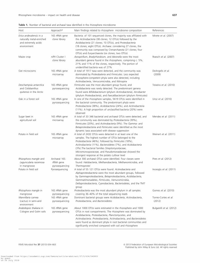
Table 1. Number of bacterial and archaeal taxa identified in the rhizosphere microbiome
Host Approach* Main findings related to rhizosphere microbiome composition References
Erica andevalensis in a
naturally metal-enriched
and extremely acidic
environment
16S rRNA gene
clone library
Bacteria: of 101 sequenced clones, the majority was affiliated with
the Actinobacteria (38 clones; 12 OTUs) followed by the
Acidobacteria (21 clones; 10 OTUs), and Proteobacteria
(18 clones; eight OTUs). Archaea: considering 27 clones, the
community was composed by Crenarchaeota (21 clones; four
OTUs) and Euryarchaeota (six clones; two OTUs)
Mirete et al. (2007)
Maize crop nifH Cluster I
clone library
Azospirillum, Bradyrhizobium, and Ideonella were the most
abundant genera found in the rhizosphere, comprising c. 5%,
21% and 11% of the clones, respectively. The portion of
unidentified bacteria was of 27%
Roesch et al. (2007)
Oat microcosms 16S rRNA gene
microarray
A total of 1917 taxa were detected, and the community was
dominated by Proteobateria and Firmicutes. Less expected
rhizosphere-competent phyla were also detected, including
Actinobacteria, Verrucomicrobia, and Nitrospira
DeAngelis et al.
(2009)
Deschampsia antarctica
and Colobanthus
quitensis in the Arctic
16S rRNA gene
pyrosequencing
Firmicutes was the most abundant group found, and
Acidobacteria was rarely detected. The predominant genera
found were Bifidobacterium (phylum Actinobacteria), Arcobacter
(phylum Proteobacteria), and Faecalibacterium (phylum Firmicutes)
Teixeira et al. (2010)
Oak in a forest soil 16S rRNA gene
pyrosequencing
In one of the rhizosphere samples, 5619 OTUs were identified in
the bacterial community. The predominant phyla were
Proteobacteria (38%), Acidobacteria (24%), and Actinobacteria
(11%). A high proportion of unclassified bacteria (20%) were
observed
Uroz et al. (2010)
Sugar beet in
agricultural soil
16S rRNA gene
microarray
A total of 33 346 bacterial and archaeal OTUs were detected, and
the community was dominated by Proteobacteria (39%),
Firmicutes (20%), and Actinobacteria (9%). The Gamma- and
Betaproteobacteria and Firmicutes were identified as the most
dynamic taxa associated with disease suppression
Mendes et al. (2011)
Potato in field soil 16S rRNA gene
microarray
A total of 2432 OTUs were detected in at least one of the
samples. The highest number of OTUs belonged to the
Proteobacteria (46%), followed by Firmicutes (18%),
Actinobacteria (11%), Bacteroidetes (7%), and Acidobacteria
(3%).The bacterial families Streptomycetaceae,
Micromonosporaceae, and Pseudomonadaceae showed the
strongest response at the potato cultivar level
Weinert et al. (2011)
Rhizophora mangle and
Laguncularia racemosa
in mangroove
Archaeal 16S
rRNA gene
pyrosequencing
About 300 archaeal OTUs were identified. Four classes were
found: Halobacteria, Methanobacteria, Methanomicrobia, and
Thermoprotei
Pires et al. (2012)
Potato in field soil Pyrosequencing A total of 55 121 OTUs were found. Actinobacteria and
Alphaproteobacteria were the most abundant groups, followed
by Gammaproteobacteria, Betaproteobacteria, Acidobacteria,
Gemmatimonadetes, Firmicutes, Verrucomicrobia,
Deltaproteobacteria, Cyanobacteria, Bacteriodetes, and the TM7
group
Inceoglu et al. (2011)
Rhizophora mangle in
mangroove
16S rRNA gene
pyrosequencing
Proteobacteria was the most abundant phylum in all samples
covering 36–40% of the total sequencing reads
Gomes et al. (2010)
Mannillaria carnea
(cactus) in semi-arid
environment
16S rRNA gene
pyrosequencing
Dominant bacterial groups were Acidobacteria, Actinobacteria,
Proteobacteria, and Bacteroidetes
Torres-Cortes et al.
(2012)
Arabidopsis thaliana in
Cologne and Golm soils
16S rRNA gene
pyrosequencing
About 1000 OTUs were estimated in the rhizosphere and 1000
OTUs in root compartments. The rhizosphere was dominated by
Acidobacteria, Proteobacteria, Planctomycetes, and
Actinobacteria. Proteobacteria, Actinobacteria, and Bacteroidetes
were found as dominant phyla in root bacterial communities and
significantly enriched compared with soil and rhizosphere
Bulgarelli et al. (2012)
FEMS Microbiol Rev 37 (2013) 634–663 ª 2013 Federation of European Microbiological SocietiesPublished by John Wiley & Sons Ltd. All rights reserved
Rhizosphere microbiome – impact on health and disease 637
Downloaded from https://academic.oup.com/femsre/article-abstract/37/5/634/540803by gueston 26 July 2018

citrinum hosted significantly more Alpha-, Beta-, and
Gammaproteobacteria than bulk soil. Despite the harsh
abiotic conditions in Antarctic soils, up to 732 OTUs
were detected in the rhizosphere of two vascular plants
(Teixeira et al., 2010). For most of the rhizosphere
samples from the Antarctic, Firmicutes were the most
abundant phylum, whereas in many other rhizosphere
studies, the Proteobacteria are commonly more abundant.
Using a high-density 16S rRNA gene oligonucleotide
microarray, referred to as the PhyloChip (Fig. 3),
DeAngelis et al. (2009) detected 2595 OTUs in the oat
rhizosphere with 1917 OTUs consistently present in all
three replicate samples. The dynamic subset (147 OTUs)
responsive to root growth was dominated by the Alpha-
proteobacteria, Firmicutes, and Actinobacteria (DeAngelis
et al., 2009). PhyloChip analysis has also been used to
access bacterial communities in the rhizosphere of other
plant species, including potato and sugar beet. The work
Table 1. Continued
Host Approach* Main findings related to rhizosphere microbiome composition References
Arabidopsis thaliana
in Mason farm and
Clayton soils
16S rRNA gene
pyrosequencing
18 783 bacterial OTUs were firstly detected, and 778 measurable
OTUs were used for analysis. The rhizosphere microbiome was
dominated by Proteobacteria, Bacteroidetes, Actinobacteria, and
Acidobacteria. The endophytic compartment was dominated by
Actinobacteria, Proteobacteria, and Firmicutes and was depleted
of Acidobacteria, Gemmatimonadetes, and Verrucomicrobia
Lundberg et al. (2012)
OTUs, operational taxonomic units.
*Here, we focused on a select number of microarray and pyrosequencing studies; for more studies that used the clone library approach to access
the dominant bacterial groups in the rhizosphere, we refer to Bu�ee et al. (2009).
Fig. 3. Overall workflow of the PhyloChip
technology to assess the diversity and
abundance of the bacterial communities in the
rhizosphere (adapted from Mendes et al.,
2011).
ª 2013 Federation of European Microbiological Societies FEMS Microbiol Rev 37 (2013) 634–663Published by John Wiley & Sons Ltd. All rights reserved
638 R. Mendes et al.
Downloaded from https://academic.oup.com/femsre/article-abstract/37/5/634/540803by gueston 26 July 2018

by Weinert et al. (2011) showed that the number of
OTUs found in the rhizosphere of three potato cultivars
grown at two distant field sites ranged from 1444 to
2015. The dominant phylum detected was the Proteobac-
teria (46%), followed by Firmicutes (18%), Actinobacteria
(11%), Bacteroidetes (7%), and Acidobacteria (3%). Inter-
estingly, the relative abundance of the top 10 dominant
phyla was similar for all three potato cultivars at both
sites (Weinert et al., 2011). With the increased capacity of
the latest PhyloChip generation (G3), which includes
c. 60 000 OTUs representing 147 phyla and 1123 classes
of Bacteria and Archaea domains (Hazen et al., 2010),
over 33 000 OTUs were detected in the rhizosphere of
sugar beet seedlings grown in soils from agricultural fields
in the Netherlands (Mendes et al., 2011). Similar to the
results obtained by Roesch et al. (2007), the Proteobacte-
ria was the most dominant phylum followed by the
Firmicutes, Actinobacteria, and Bacteroidetes. The unclassi-
fied bacteria represented a relatively large group (16%)
among the OTUs detected in the sugar beet rhizosphere
(Mendes et al., 2011). Recently, the taxonomic classifica-
tion of the Greengenes database has been updated
(McDonald et al., 2012). By reevaluating the proportion
of unclassified taxa found in the sugar beet rhizosphere
using the updated database, the number of unclassified
taxa in the sugar beet rhizosphere decreased to 7.5%
(R. Mendes and J.M. Raaijmakers, unpublished data).
Hence, expanding our basic taxonomic knowledge of
microorganisms is essential to further improve the tech-
nological resolution and capabilities of next-generation
sequencing to study the diversity and functional potential
of plant and other microbiomes.
Consistent with the approaches and concepts in human
microbiome studies, Lundberg et al. (2012) and Bulgarelli
et al. (2012) recently investigated the spatial distribution
of bacterial communities in the rhizosphere of different
Arabidopsis accessions to determine the composition of
the core microbiome. By pyrosequencing 16S rRNA gene
segments of bacteria from bulk soil, rhizosphere, and
endophytic root compartments of more than 600 Arabid-
opsis plants, Lundberg et al. (2012) showed a strong
influence of the soil type on the bacterial communities in
each of the compartments. Their results also showed that
the endophytic root compartment was enriched with
Actinobacteria and Proteobacteria and that the plant’s
developmental stage and genotype can drive differential
recruitment and/or differential exclusion of bacterial com-
munities (Lundberg et al., 2012). Using different PCR
primers, different computational pipelines and physico-
chemically different soils, similar findings were reported
by Bulgarelli et al. (2012).
The discovery that Archaea represent an important
group of ammonia oxidizers in soils (Leininger et al.,
2006) has led to an increasing number of studies on this
microbial group. In a global survey of 146 soils, Bates
et al. (2011) used a set of universal primers for nearly all
bacterial and archaeal taxa and showed that the Archaea
domain comprised an average of 2% of the total 16S
rRNA gene sequences recovered from these soils and that
their relative abundance was higher in soils with lower
C : N ratios. For the rhizosphere, an earlier study by
Chelius & Triplett (2001) identified six unique archaeal
sequences associated with maize roots. In mangroves,
about 300 archaeal OTUs distributed over four classes
(Halobacteria, Methanobacteria, Methanomicrobia, and
Thermoprotei) were recently identified in association with
Rhizophora mangle and Laguncularia racemosa (Pires
et al., 2012). In the rhizosphere of sugar beet seedlings
grown in agricultural soils, we detected 70 archaeal OTUs
representing 0.21% of the total archaeal and bacterial
community accessed by PhyloChip analysis (R. Mendes
and J.M. Raaijmakers, unpublished data). Interestingly,
we found a correlation between the composition of the
Archaeal communities and the level of suppressiveness of
soils to Rhizoctonia damping-off disease. Whether Archaea
play a role in protection of plants against soilborne
pathogens is not yet known.
The rhizosphere microbiome – what arethey doing?
In addition to a comprehensive phylogenetic analysis of
the rhizosphere microbiome, there is a strong need to go
beyond cataloguing microbial communities (‘collecting
stamps’) and to determine which microorganisms are
active during the different developmental stages of plant/
root growth and which functions and pathways are dis-
played in time and space. In the review by Barret et al.
(2011), a variety of molecular approaches to study gene
expression in the rhizosphere was discussed. From their
detailed overview, it is apparent that most of our current
knowledge of genes and functions expressed in the rhizo-
sphere is still based on studies with reporter genes.
Despite their limitations, reporter genes do enable the
evaluation of how specific members of the rhizosphere
microbiome perceive their habitat in terms of chemical,
physical, and biological stimuli. An elegant promoter-
trapping strategy, referred to as in vivo expression tech-
nology (IVET), was adopted to identify Pseudomonas fluo-
rescens genes with elevated levels of expression in the
rhizosphere (Rainey, 1999). Genes induced in P. fluores-
cens during rhizosphere colonization were genes involved
in nutrient acquisition, stress response, and secretion
(Rainey, 1999). In another study, the IVET technology
revealed that proteins involved in environmental sensing,
control of gene expression, metabolic reactions, and
FEMS Microbiol Rev 37 (2013) 634–663 ª 2013 Federation of European Microbiological SocietiesPublished by John Wiley & Sons Ltd. All rights reserved
Rhizosphere microbiome – impact on health and disease 639
Downloaded from https://academic.oup.com/femsre/article-abstract/37/5/634/540803by gueston 26 July 2018

membrane transport were specifically expressed in the
pea-nodulating bacterium Rhizobium leguminosarum A34
during rhizosphere colonization (Barr et al., 2008). Next
to IVET, a number of studies have used a diverse panel
of reporter genes to study specific processes in the rhizo-
sphere, including responses of bacteria to carbon, nitro-
gen, phosphorus availability (Kragelund et al., 1997;
Jensen & Nybroe, 1999; Ramos et al., 2000; Koch et al.,
2001; DeAngelis et al., 2005), temperature, and water
potential (Ullrich et al., 2000; Axtell & Beatie, 2002;
Herron et al., 2010). Bioreporters were also successfully
adopted to study bacterial communication in the rhizo-
sphere (Andersen et al., 2001; Steidle et al., 2001; Withers
et al., 2001; Loh et al., 2002; Steindler & Venturi, 2007;
Ferluga & Venturi, 2009) as well as the in situ production
of antimicrobial compounds (Hay et al., 2000; Kulakova
et al., 2009; Rochat et al., 2010). For more detailed infor-
mation on the results of these and other reporter gene
studies, we refer to Gage et al. (2008), Sorensen et al.
(2009), and van der Meer & Belkin (2010).
To go beyond the ‘one-gene-at-a-time’ approach,
Mark et al. (2005) used whole genome transcriptome
profiling to evaluate the effects of root exudates from
two sugar beet cultivars on gene expression in Pseudomo-
nas aeruginosa. In addition to genes previously identified
in plant–microbe interactions (i.e. metabolism, chemo-
taxis, type III secretion), Mark et al. (2005) showed that
104 genes were significantly altered in response to both
root exudates and that the majority of these genes were
regulated in response to only one of the two exudates.
Recently, a whole genome microarray was also used to
study endophytic colonization of rice by Azoarcus sp.
BH72 (Shidore et al., 2012). Among 3992 protein-coding
genes analyzed, 2.4% was up-regulated and 2.0% was
found down-regulated when exposed to root exudates.
Subsequent mutational analysis indicated that genes
encoding pilin PilX or signal transduction proteins with
GGDEF domains and a serine–threonine kinase were
important in colonization. The authors further suggested
that strain BH72 is primed by root exudates for a
lifestyle as endophyte, that is, microorganisms that live
inside plant tissues without causing any immediate, overt
negative effects (Shidore et al., 2012). Microarrays
have also been used to identify functional activities of
multiple members within a complex microbial commu-
nity. The functional gene array, termed GeoChip, con-
tains more than 10 000 genes covering more than 150
functions involved in nitrogen, carbon, sulfur and phos-
phorus cycling, metal reduction and resistance, and
organic contaminant degradation (He et al., 2007). For
example, the GeoChip 3.0 revealed that infection of
citrus trees by the pathogen Candidatus Liberibacter
asiaticus caused shifts in the composition and functional
potential of rhizosphere microbial communities (Trivedi
et al., 2011).
‘Omics’ approaches that enable the identification of
gene transcripts, proteins, or metabolites have been devel-
oped to provide a more detailed insight into the genes
and functions expressed in the plant microbiome. A
metaproteogenomic approach was first reported for bacte-
rial communities in the phyllosphere of Arabidopsis, soy-
bean, and clover plants (Delmotte et al., 2009). For the
rhizosphere, a recent metaproteomics study revealed com-
plex interactions between plants and rhizosphere microor-
ganisms in different cropping systems (Wang et al.,
2011). MALDI-TOF/TOF-MS resulted in the identifica-
tion of 189 protein spots from rice rhizosphere samples
and approximately one-third of the protein spots could
not be identified (Wang et al., 2011). The origin of each
protein was determined, being from plants (107 proteins),
fauna (10), fungi (29), or bacteria (43). Bacterial proteins
were mostly linked to the Proteobacteria and Actinobacte-
ria. They also found that 50% of the bacterial groups
classified by proteomic analysis were not found in the
genomic-based T-RFLP analysis and vice versa, highlight-
ing the importance of combining different approaches to
access the microbial community (Wang et al., 2011). A
similar approach was used to study the rhizosphere
microbiome of the medicinal plant Rehmannia glutinosa
(Wu et al., 2011) and the phyllosphere plus rhizosphere
microbiomes of rice (Knief et al., 2011). The latter study
resulted in the identification of about 4600 proteins and
revealed the presence of one-carbon conversion processes,
predominantly methanogenesis, in both rhizosphere and
phyllosphere (Knief et al., 2011).
Also, the development of stable isotope probing (SIP)
to track plant-derived carbon into microbial nucleic acids
has provided exciting new insights into the metabolically
active rhizobacterial populations (Rangel-Castro et al.,
2005; Prosser et al., 2006). Applying DNA-SIP to 13CO2-
exposed plants greatly helped to identify bacterial
communities that actively assimilate root exudates in the
rhizosphere of four plant species: wheat, maize, rape, and
clover (Haichar et al., 2008). Bacteria related to Sphingo-
bacteriales and Myxococcus assimilated root exudates of all
four plants, while Sphingomonadales were specific to
monocotyledons (Haichar et al., 2008). Analysis of fungal
and bacterial biomarkers (phospholipid fatty acids)
extracted from the rhizosphere of 13CO2-exposed plants
indicated that also fungi metabolized a significant amount
of root exudates (Bu�ee et al., 2009). Hence, bacteria do
not monopolize the rhizosphere and fungi can respond
rapidly to the provision of easily degradable root exudates
(Broeckling et al., 2008; De Graaff et al., 2010). Studying
the community dynamics of saprotrophic fungi in the
rhizosphere of six potato cultivars, Hannula et al. (2010)
ª 2013 Federation of European Microbiological Societies FEMS Microbiol Rev 37 (2013) 634–663Published by John Wiley & Sons Ltd. All rights reserved
640 R. Mendes et al.
Downloaded from https://academic.oup.com/femsre/article-abstract/37/5/634/540803by gueston 26 July 2018

indeed found that fungi make up a significant part of the
rhizosphere microbial biomass especially during flowering
and senescence. Based on DNA-SIP data, Drigo et al.
(2010) provided a conceptual model in which plant-
assimilated carbon is rapidly transferred to arbuscular
mycorrhizal fungi (AMF), followed by a slower release
from AMF to the active bacterial and fungal rhizosphere
communities. Collectively, these studies exemplify that a
combination of functional approaches provide powerful
tools to infer physiological traits of microbial communities
in situ.
Impact of the rhizosphere microbiomeon plant growth, health, and disease
Rhizosphere microorganisms directly and indirectly influ-
ence the composition and productivity (i.e. biomass) of
natural plant communities (van der Heijden et al., 1998,
2006, 2008; Schnitzer et al., 2011). Hence, microbial spe-
cies richness belowground has been proposed as a predic-
tor of aboveground plant diversity and productivity (De
Deyn et al., 2004; Hooper et al., 2005; van der Heijden
et al., 2008; Lau & Lennon, 2011; Wagg et al., 2011).
Wagg et al. (2011) further suggested that belowground
diversity may act as insurance for maintaining plant pro-
ductivity under different environmental conditions. Due
to their sensitivity to small changes in abiotic conditions,
including environmental stress and perturbation, soil
and rhizosphere microorganisms are considered as bio-
indicators of soil quality. Here, we will discuss which
rhizosphere microorganisms impact on plant growth and
health. We will focus on the plant beneficial (‘the good’),
plant pathogenic (‘the bad’), and human pathogenic (‘the
ugly’) microorganisms.
The good
Rhizosphere microorganisms promote plant growth and
protect plants from pathogen attack by a range of mecha-
nisms (Lugtenberg & Kamilova, 2009; Raaijmakers et al.,
2009). These involve biofertilization, stimulation of root
growth, rhizoremediation, control of abiotic stress, and
disease control. These mechanisms are well documented
for rhizobacteria belonging to the Proteobacteria and Fir-
micutes, that is, Pseudomonas and Bacillus, as well as for
fungi from the Deuteromycetes, that is, Trichoderma and
Gliocladium, and from the Sebacinales order, that is, Piri-
formospora (Kogel et al., 2006; Qiang et al., 2012). Fortu-
nately, more information is being obtained in the past
years on the functions of other soil and rhizosphere
inhabitants, including ‘unusual’ or ‘rare’ microbial genera
such as the Planctomycetes (Hol et al., 2010; Jogler et al.,
2012).
Effects of rhizosphere microorganisms on nutrientacquisition by plants
Members of the rhizosphere microbiome can significantly
influence the nutrient status of plants (Fig. 2). Well-known
examples are the nitrogen-fixing rhizobia and the mycor-
rhizal fungi that facilitate phosphorus uptake (Hawkins
et al., 2000; Richardson et al., 2009; Miransari, 2011). The
importance of symbionts such as mycorrhizal fungi for
translocation of nutrients and minerals from soil to the
plant (Gianinazzi et al., 2010; Adeleke et al., 2012; Johnson
& Graham, 2013), for soil physical structuring and generat-
ing stable soil aggregates (Degens et al., 1996; Miller &
Jastrow, 2000), and for suppression of soilborne plant
pathogens (Whipps, 2001; Pozo & Azcon-Aguilar, 2007) is
well recognized and documented (Smith & Read, 1997;
Varma & Hock, 1998; Kapulnik & Douds, 2000; Brundrett,
2002; van der Heijden & Sanders, 2002; Johnson et al.,
2012; Salvioli & Bonfante, 2013). Next to Rhizobium and
Bradyrhizobium, various other nitrogen-fixing bacterial
genera living in the rhizosphere have been identified (Zehr
et al., 2003; Gaby & Buckley, 2011). For example, analysis
of the cowpea rhizosphere revealed a high genetic diversity
of symbiotic rhizobial species in the western Amazon
(Guimar~aes et al., 2012). Based on glasshouse experiments
and 16S rRNA gene sequencing, they indicated that Brady-
rhizobium, Rhizobium, Burkholderia, and Achromobacter
species were able to nodulate cowpea and were efficient in
biological nitrogen fixation (Guimar~aes et al., 2012).
Despite extensive research on nitrogen fixation by rhizobia,
the transfer of the legume-specific symbiosis to other agri-
culturally important plant species has not been achieved
yet. In their recent review, Geurts et al. (2012) indicated
that understanding the fundamental differences between
the seemingly similar cellular responses induced by
Rhizobium and mycorrhizal fungi will be necessary to
achieve this ‘old dream’.
Rhizosphere microorganisms can also facilitate the
uptake of specific trace elements such as iron. Iron is abun-
dant in soil but, under neutral to alkaline conditions, it
exists primarily in the insoluble ferric oxide form, which is
not available for microbial growth. Due to the scarcity of
available iron in many microbial habitats as well as the tox-
icity of free iron at elevated concentrations, bacteria
employ a variety of mechanisms to regulate intracellular
iron concentrations by secretion of siderophores (Lindsay
& Schwab, 1982; Andrews et al., 2003; Buckling et al.,
2007; Hider & Kong, 2010). On the host side, plants
respond to iron limitation by increasing the solubility of
inorganic iron in the rhizosphere (strategy I) or by releas-
ing phytosiderophores that are subsequently transported
back into the root tissue by a specific uptake system (strat-
egy II) (Walker & Connolly, 2008). In rice, iron can be
FEMS Microbiol Rev 37 (2013) 634–663 ª 2013 Federation of European Microbiological SocietiesPublished by John Wiley & Sons Ltd. All rights reserved
Rhizosphere microbiome – impact on health and disease 641
Downloaded from https://academic.oup.com/femsre/article-abstract/37/5/634/540803by gueston 26 July 2018

acquired by both strategies (Walker & Connolly, 2008).
Various studies have proposed an additional strategy of
iron acquisition by plants involving the use of iron
chelated to microbial siderophores (Marschner &
R€omheld, 1994; Vansuyt et al., 2007; Lemanceau et al.,
2009a, b). This was exemplified in studies with fluorescent
pseudomonads, which promoted iron nutrition via sidero-
phores not only for Graminaceous plants but also for
dicotyledonous plant species (Vansuyt et al., 2007; Shirley
et al., 2011). Also, rhizoferrin, a fungal siderophore pro-
duced by Rhizopus arrhizus, was found to be an efficient
carrier of iron to plants with an efficiency that was compa-
rable to that of synthetic chelates (Yehuda et al., 2000).
Rhizobacteria are also able to activate the plant’s own iron
acquisition machinery as was shown for Bacillus subtilis
GB03 (Zhang et al., 2009). In Arabidopsis, strain GB03
up-regulated transcription of the Fe-deficiency-induced
transcription factor 1 (FIT1), thereby inducing the ferric
reductase FRO2 and the iron transporter IRT1 (Zhang
et al., 2009). For more detailed overviews of the mecha-
nisms by which rhizosphere microorganisms influence iron
uptake by plants, we refer to Lemanceau et al. (2009a, b),
and Marschner et al. (2011).
Most rhizobacterial species are organotrophs, that is,
they obtain the energy from the assimilation of organic
compounds. The availability and accessibility of degrad-
able organic compounds are limited in most soils, and
carbon availability is the most common limiting factor
for soil bacteria growth (Alden et al., 2001; Demoling
et al., 2007; Rousk & Baath, 2007). Bacterial communities
play an essential role in releasing the nutritive cations
from soil minerals required not only for their own nutri-
tion but also for plant nutrition. Mineral weathering
bacteria have been isolated from various environments,
and particularly from rhizosphere and ectomycorrhizo-
sphere (Puente et al., 2004; Calvaruso et al., 2007;
Collignon et al., 2011) and can contribute to plant
growth in nutrient-poor soils (Leveau et al., 2010; Mapelli
et al., 2012).
Supporting plant growth under biotic stress
The rhizosphere provides the frontline defense for plant
roots against attack by soilborne pathogens (Cook et al.,
1995). Various members of the rhizosphere microbiome
can antagonize soilborne pathogens before and during
primary infection, and during secondary spread on and
in root tissue (Fig. 2). The main mechanisms by which
rhizosphere microorganisms ward off plant pathogens are
antibiosis (Haas & D�efago, 2005; Lugtenberg & Kamilova,
2009; Raaijmakers & Mazzola, 2012), competition for
trace elements, nutrients and microsites (Duffy, 2001),
parasitism (Druzhinina et al., 2011; Mela et al., 2011),
interference with quorum sensing affecting virulence (Lin
et al., 2003; Uroz et al., 2009; Chan et al., 2011), and
induced systemic resistance (Conrath, 2006; van Loon,
2007; Yang et al., 2009; Pieterse, 2012; Schenk et al.,
2012).
Most, if not all, rhizobacteria produce metabolites that
inhibit the growth or activity of competing microorgan-
isms. Also, rhizosphere fungi are prolific producers of
antibiotic metabolites (Hoffmeister & Keller, 2007; Brakh-
age & Schroeckh, 2011). Especially, Trichoderma species
have received considerable attention for the production of
antimicrobial compounds (Vyas & Mathus, 2002; Harman
et al., 2004; Mathivanan et al., 2005; Elad et al., 2008;
Druzhinina et al., 2011). Most fungal and bacterial bio-
control strains produce more than one antibiotic com-
pound with overlapping or different degrees of
antimicrobial activity. For example, bacteriocins such as
agrocin 84 produced by Agrobacterium radiobacter
(Reader et al., 2005; Kim et al., 2006) exhibit antibiotic
activities against closely related genera, whereas many
polyketide and nonribosomal peptide antibiotics exhibit
broad-spectrum activities (Gross & Loper, 2009; Raaij-
makers et al., 2010). Interestingly, many antibiotic com-
pounds have different effects on other microorganisms at
subinhibitory concentrations, an observation which led to
an exciting new direction in research on the natural func-
tions of antibiotics. Recent studies have indeed shown
that antibiotics function in a concentration-dependent
manner, acting as growth inhibitors at high concentra-
tions and as mediators of intercellular signaling at low
concentrations (Davies et al., 2006; Fajardo & Martinez,
2008; Romero et al., 2011). Other natural functions
attributed to antibiotics include a role in defense against
predatory protozoa, motility, biofilm formation, and
nutrition (Raaijmakers & Mazzola, 2012).
Among the metabolites produced by rhizosphere
microorganisms, volatile organic compounds (VOCs) are
receiving more attention over the past years. Some of
them were shown to modulate plant growth and to medi-
ate the intricate dialogues between microorganisms and
plants (Bailly & Weisskopf, 2012; Effmert et al., 2012).
Although VOCs appear to represent a small proportion
of the total number of metabolites produced by fungi and
bacteria, their unique properties have been proposed to
play essential functions in long-distance communication
in the rhizosphere and in soil ecosystems. VOCs are small
molecules (< 300 Da) with high vapor pressures able to
diffuse through the water- and gas-filled pores in soil
(Wheatley, 2002; Insam & Seewald, 2010). Various bacte-
rial species including Stenotrophomonas maltophilia, Serra-
tia plymuthica, Pseudomonas trivialis, P. fluorescens,
B. subtilis, and Burkholderia cepacia produce VOCs that
inhibit mycelial growth of fungal plant pathogens (Kai
ª 2013 Federation of European Microbiological Societies FEMS Microbiol Rev 37 (2013) 634–663Published by John Wiley & Sons Ltd. All rights reserved
642 R. Mendes et al.
Downloaded from https://academic.oup.com/femsre/article-abstract/37/5/634/540803by gueston 26 July 2018

et al., 2007, 2009; Vespermann et al., 2007; Zou et al.,
2007; Jamalizadeh et al., 2010). Most work on VOCs to
date, however, is conducted in vitro on nutrient-rich
media and may not be representative of the conditions
that prevail in the rhizosphere. Effects of specific abiotic
conditions on VOC production were shown by Weise
et al. (2012), who reported a discrepancy in the number
and spectrum of volatiles produced by a Xanthomonas
species grown in broth culture and on solid agar media.
Recent work showed that the spectrum of volatiles
released by rhizobacteria can be influenced by the avail-
able pool of root exudates (P. Garbeva, unpublished
data). For example, volatiles produced in soil amended
with artificial root exudates without amino acids had
strong antibacterial effects but mild antifungal effects,
whereas volatiles produced from root exudates supple-
mented with amino acids had strong antifungal effects
(P. Garbeva, unpublished data). Conversely, bacterial
volatiles may promote growth of ectomycorrhizal fungi
(Schrey et al., 2005) and play important regulatory roles
in mycorrhizal network establishment (Bonfante & Anca,
2009). They may also play a role in the tripartite interac-
tions between bacteria, fungi, and nematodes. In this con-
text, Son et al. (2009) showed that Paenibacillus polymyxa
and Paenibacillus lentimorbus exhibited strong antifungal
activities, thereby interfering with the interactions
between Meloidogyne incognita and Fusarium oxysporum
and concomitant nematode infestation of tomato plants.
Recently, Chernin et al. (2011) reported that bacterial
volatiles can also interfere with quorum sensing of phylo-
genetically different bacteria by suppressing the transcrip-
tion of the N-acyl-homoserine lactone synthase genes.
Dimethylsulfide was identified as one of the compounds
that interfered with quorum sensing (Chernin et al.,
2011). Finally, VOCs can also induce systemic resistance
in plants (Ryu et al., 2003, 2004; Han et al., 2006) and
promote plant growth (Ryu et al., 2003; Cho et al., 2008;
Blom et al., 2011a, b; Bailly & Weisskopf, 2012).
Members of the rhizosphere microbiome can also mod-
ulate the plant immune system (De Vleesschauwer &
Hofte, 2009; Pineda et al., 2010; Berendsen et al., 2012;
Zamioudis & Pieterse, 2012). The systemic resistance
response induced in plants by beneficial rhizobacteria is
in many cases regulated by the phytohormones jasmonic
acid (JA) and ethylene (ET) (Zamioudis & Pieterse,
2012). However, some bacterial strains do not induce sys-
temic resistance via the JA/ET pathway but via the sali-
cylic acid (SA)-pathway (Maurhofer et al., 1994;
De Meyer & Hofte, 1997; Maurhofer et al., 1998; De
Meyer et al., 1999; Audenaert et al., 2002; Barriuso et al.,
2008; van de Mortel et al., 2012). Other rhizobacteria
such as Bacillus cereus AR156 induce systemic resistance
by activating both signaling pathways (Niu et al., 2011).
Furthermore, quorum-sensing molecules from rhizobacte-
ria can provoke a range of plant responses, including the
activation of various defense-related genes such as MPK3,
MPK6, WRKY22, WRKY29, and Pdf1.2 (reviewed in
Hartmann & Schikora, 2012). Over the past years, signifi-
cant progress has been made in unraveling the transcrip-
tional and metabolic changes induced in plants by
rhizobacteria. For those bacterial strains that induce resis-
tance via the JA/ET pathways, relatively few transcrip-
tional changes were observed in Arabidopsis (Verhagen
et al., 2004; Cartieaux et al., 2008; ). However, for rhizo-
bacterial strains that induce resistance in Arabidopsis via
the SA pathway, substantial, transcriptional, and meta-
bolic changes were observed (van de Mortel et al., 2012).
By integrating metabolic pathways and transcript profiles,
Weston et al. (2012a, b) further showed that two distinct
strains of P. fluorescens reduced the host plant’s carbon
gain, but provided a fitness benefit when the plants were
challenged with the pathogen Pseudomonas syringae.
These studies indicated that rhizobacteria can have
diverse and profound effects on the immune response
and physiology/metabolism of the host plant (Fig. 2),
enhancing the production of known secondary metabo-
lites but also inducing the biosynthesis of structurally
unknown metabolites (van de Mortel et al., 2012). Analy-
sis of the identity and activities of ‘cryptic’ plant com-
pounds induced by rhizobacteria should be pursued to
resolve their putative functions in induced systemic resis-
tance and other physiological processes.
Supporting plant growth under abiotic stress
It has been postulated that the rhizosphere microbiome
contributes to the ability of some plant species to survive
under extreme conditions (Jorquera et al., 2012). For
example, Achromobacter piechaudii ARV8, a soil isolate
obtained from an arid and saline environment, signifi-
cantly increased the biomass of tomato and pepper seed-
lings exposed to transient drought stress (Mayak et al.,
2004a, b). Also, under conditions of flooding, rhizobacte-
ria were shown to support plant growth (Grichko &
Glick, 2001). In diverse production systems, plant pro-
ductivity can be strongly affected by soil salinity due to
osmotic and drought stress. Halotolerant bacteria thrive
under salt-stress conditions and in association with the
host plant are able to express traits that promote plant
growth. From the rhizosphere of wheat plants grown in a
saline zone, Upadhyay et al. (2009) showed that of 130
rhizobacterial isolates, 24 were tolerant to relatively high
levels (8%) of NaCl. All of the 24 salt-tolerant isolates
produced indole-3-acetic acid, 10 isolates solubilized
phosphorus, eight produced siderophores, six produced
gibberellin, and two isolates contained the nifH gene,
FEMS Microbiol Rev 37 (2013) 634–663 ª 2013 Federation of European Microbiological SocietiesPublished by John Wiley & Sons Ltd. All rights reserved
Rhizosphere microbiome – impact on health and disease 643
Downloaded from https://academic.oup.com/femsre/article-abstract/37/5/634/540803by gueston 26 July 2018

indicating their potential for nitrogen fixation. The domi-
nant bacterial genus isolated under these conditions was
Bacillus (Upadhyay et al., 2009). Halotolerant bacterial
strains were also isolated from halophytic plant species
found in coastal soils in Korea. Several of the obtained
isolates enhanced plant growth under saline stress, and
the reduction in ET production via ACC deaminase activ-
ity was proposed as the underlying mechanism of plant
growth promotion (Siddikee et al., 2010). New halotoler-
ant diazotrophic bacteria harboring indole acetic acid
production, phosphate solubilization, and 1-aminocyclo-
propane-1-carboxylic acid (ACC) deaminase activity were
isolated from roots of the extreme halophyte Salicornia
brachiate (Jha et al., 2012). The isolates were identified as
Brachybacterium saurashtrense sp. nov., Zhihengliuella sp.,
Brevibacterium casei, Haererehalobacter sp., Halomonas
sp., Vibrio sp., Cronobacter sakazakii, Pseudomonas spp.,
Rhizobium radiobacter, and Mesorhizobium sp. (Jha et al.,
2012). For more comprehensive reviews on the beneficial
effects of soil biota on plant responses to saline stress, we
refer to reviews by Dodd & P�erez-Alfocea (2012) and
Berg et al. (2013). In these reviews, several mechanisms
are described by which microorganisms may alter plant
physiological response under saline stress, including their
effects on: (1) water homeostasis by osmolyte accumula-
tion, (2) plant energetics by modulating the source-sink
relationships, (3) root uptake of toxic ions and nutrients
by altering host physiology, modifying physical barriers
around the roots, or by directly reducing foliar accumula-
tion of toxic ions, and (4) crop salt tolerance by altering
hormonal root–shoot signaling.Environments with low temperatures harbor microor-
ganisms adapted to live under such conditions. It is inter-
esting to note that despite the impact of low
temperatures on nodule formation and nitrogen fixation,
native legumes in the high arctic can nodulate and fix
nitrogen at rates comparable to those reported for
legumes in temperate climates (Bordeleau & Pr�evost,
1994). There is great interest in agriculture and horticul-
ture for microbial inoculants that enhance growth of
plants under cold conditions. For example, Burkholderia
phytofirmans PsJN increased grapevine root growth and
physiological activity at temperatures down to 4°C (Barka
et al., 2006). When coinoculated with Bradyrhizobium
japonicum, Serratia proteamaculans stimulated soybean
growth at 15°C, the temperature at which soybean nodule
infection and nitrogen fixation are normally inhibited
(Zhang et al., 1995, 1996). To identify mechanisms
involved in plant growth promotion at low temperatures,
Katiyar & Goel (2003) selected cold-tolerant mutants of
different P. fluorescens strains for their ability to solubilize
phosphorus and to promote plant growth. They identified
two cold-tolerant mutants that were more efficient in
phosphorus solubilization at 10 °C than their respective
wild types (Katiyar & Goel, 2003). Also, Trivedi & Sa
(2008) found two mutants (of 115) that were more effi-
cient than the wild-type strain Pseudomonas corrugata in
phosphorus solubilization across a temperature range
from 4 to 28 °C. In both studies, the identity of the genes
associated with cold tolerance and phosphorus solubiliza-
tion was not mentioned.
Other abiotic factors that may adversely affect plant
growth are pH and high concentrations of toxic com-
pounds. Soils with low pH or contaminated soils are
major challenges in many production systems worldwide.
In the case of pH stress, it was demonstrated that foliar
lesions induced on corn growing in a low-pH soil were
significantly reduced on plants treated with a 2,4-diac-
etylphloroglucinol (DAPG)-producing P. fluorescens
strain. This was the first evidence that DAPG producers,
in addition to their role in pathogen control, can also act
to ameliorate abiotic stress factors (Raudales et al., 2009).
The presence of pollutants in soil has promoted the
search for efficient bioremediation methods as an alterna-
tive for excavation and incineration. Rhizoremediation, a
combination of phytoremediation and bioaugmentation
(Kuiper et al., 2004), is a promising strategy to clean pol-
luted sites. During rhizoremediation, exudates of plants
stimulate the survival and activity of rhizobacteria that
degrade pollutants. A recent study, using a split-root
model and a combination of T-RFLP, DGGE, and 16S
rRNA gene pyrosequencing, showed that Trifolium and
other legumes respond to polycyclic aromatic hydrocar-
bons contamination in a systemic manner (Kawasaki
et al., 2012). Verrucomicrobia and Actinobacteria were
more abundant in the contaminated rhizospheres, and
the betaproteobacterium Denitratisoma was substantially
increased in the presence of the contaminant, suggesting
that this genus may be important in the rhizoremediation
process (Kawasaki et al., 2012). Also, fungi are important
players in rhizoremediation of hydrocarbons as was
shown by inoculation of the endophytic fungus Lewia sp.
in the rhizosphere of Festuca arundinacea (Cruz-Hern�andez
et al., 2012).
In conclusion, members of the rhizosphere microbiome
can alleviate biotic and abiotic stresses on plants (Fig. 2),
providing an environmentally sound alternative for
genetic engineering and plant breeding. However, success-
ful implementation of microbial inoculants is still in its
infancy due to multiple constraints, including variable
efficacy across environments and different plants species,
limited shelf-life, and different registration procedures in
different countries. To resolve several of these constraints,
more fundamental knowledge is required on how benefi-
cial rhizosphere microorganisms communicate with the
host plant, which molecular and metabolic changes are
ª 2013 Federation of European Microbiological Societies FEMS Microbiol Rev 37 (2013) 634–663Published by John Wiley & Sons Ltd. All rights reserved
644 R. Mendes et al.
Downloaded from https://academic.oup.com/femsre/article-abstract/37/5/634/540803by gueston 26 July 2018

induced in plants, and how beneficial microorganisms
affect the population dynamics and virulence of plant
pathogenic microorganisms.
The bad
Soilborne plant pathogens cause major yield reductions in
the production of food, feed, fiber, and fuel crops
(Fig. 2). Two main groups of soilborne plant pathogens
are the nematodes and the fungi, including the true fungi
and the fungal-like oomycetes. In temperate climates,
plant pathogenic fungi, oomycetes, and nematodes are
agronomically more important than plant pathogenic
bacteria, although some bacterial genera (i.e. Pectobacteri-
um, Ralstonia) can cause substantial economic damage in
some crops. For bacterial pathogens that infect plants via
roots, Agrobacterium tumefaciens, Ralstonia solanacearum,
Dickeya dadanthi and Dickeya solani, and Pectobacterium
carotovorum and Pectobacterium atrosepticum are among
the top 10 most notorious (Mansfield et al., 2012). Also,
viruses can infect plants via the roots but require vectors
such as nematodes or zoosporic fungi to enter the root
tissue (Campbell, 1996; Macfarlane, 2003). Compared
with our understanding of the role of rhizodeposits in the
communication between symbionts and plants, informa-
tion on root exudates that activate and attract soilborne
plant pathogens is more scarce and fragmented. Weston
et al. (2012a, b) indicated that the limited knowledge of
the communication between plants and root pathogens is
largely due to a poor understanding of the complex phys-
ical–chemical conditions in soil and rhizosphere environ-
ments. Hence, expanding our analytical skills to elucidate
the chemistry of rhizodeposits and their spatiotemporal
production and distribution patterns, collectively termed
‘ecometabolomics’ (Sardans et al., 2011; Weston et al.,
2012a, b), will be important to resolve the dialogues
between pathogens and plant roots. Here, we will discuss
which processes and chemical cues are important for soil-
borne pathogens to colonize the rhizosphere and to infect
the root tissue. We will focus on few examples of fungi,
oomycetes, and nematodes.
Fungi and oomycetes
For germination, growth and establishment in the rhizo-
sphere, fungal, and oomycete pathogens depend on sev-
eral different cues from the host plant. Dormancy of
fungal spores can be triggered by a range of factors,
including changes in abiotic conditions (i.e. pH) and root
exudates. Wu et al. (2008a, b) found that phenolic com-
pounds like p-hydroxybenzoic, gallic, coumaric, cinnamic,
ferulic, salicylic, and sinamic acids in root exudates stim-
ulated, at low concentrations, conidial germination of
pathogenic fungi; when concentrations increased, an
inhibitory effect was observed. Also, Zhang et al. (2012)
found similar effects of four phenolic acids from cotton
root exudates on germination of Verticillium dahliae
spores. Also, alkaloids from roots of Veratrum taliense
(Liliaceae) were shown to inhibit growth of Phytophthora
capsici and Rhizoctonia cerealis (Zhou et al., 2003). Inter-
estingly, work by Joosten et al. (2009) showed that soil
type and soil microorganisms greatly affected the compo-
sition of alkaloids in roots and shoots of Jacobaea vulga-
ris, in particular retrorsine and retrorsine N-oxide. Both
alkaloids inhibit mycelium growth of several plant-associ-
ated fungi, including F. oxysporum, Fusarium sambuci-
num, and Trichoderma sp. (Hol & Van Veen, 2002).
Based on these results, Joosten & van Veen (2011) postu-
lated that the effect of microorganisms on the alkaloid
composition of plants could have other ecological conse-
quences as these changes may attract specialist herbivores
aboveground while deterring generalists.
Saponins are probably the best examples of chemical
constituents of roots that adversely affect plant pathogenic
fungi. Saponins represent a structurally diverse group of
glycosides with triterpene or steroid backbones. They form
complexes with sterols causing pore formation and loss of
membrane integrity in fungal pathogens (Gonzalez-Lamo-
the et al., 2009; Osbourn et al., 2011). Compelling evi-
dence for a role of saponins in protection of plants against
root-infecting fungi was provided by studies on avenacin
(Bednarek & Osbourn, 2009; Gonzalez-Lamothe et al.,
2009; Osbourn et al., 2011). Avenacin A-1 exhibits anti-
fungal activity and is localized in the epidermal cells of
root tips and emerging lateral roots of oats. The fungal
root pathogen Gaeumannomyces graminis var avenae (Gga)
can detoxify avenacin A-1 and infect oat roots, whereas
Gga-mutants that lack the detoxifying hydrolase, desig-
nated avenacinase, were more sensitive to avenacin A-1
and were no longer able to infect. On the plant side, avena-
cin-deficient mutants showed compromised resistance to
several pathogens. Recent studies further suggested that
avenacin or avenacin intermediates may also elicit other
processes in the plant such as callose deposition (Bednarek
& Osbourn, 2009) which in turn strengthens the defense
response. Other studies indicated that plant metabolites,
like glucosinolates in cruciferous plants, are mobilized to
pathogen infection sites where they are enzymatically con-
verted into biologically active compounds only when they
are released by disruption of the plant tissue (Bednarek &
Osbourn, 2009). Pathways and mechanisms involved in
the safe storage and exudation of secondary metabolites in
plants were highlighted in recent reviews by Sardans et al.
(2011) and Weston et al. (2012a, b).
In contrast to root-infecting fungi, oomycete pathogens
produce motile zoospores that swim toward the plant
FEMS Microbiol Rev 37 (2013) 634–663 ª 2013 Federation of European Microbiological SocietiesPublished by John Wiley & Sons Ltd. All rights reserved
Rhizosphere microbiome – impact on health and disease 645
Downloaded from https://academic.oup.com/femsre/article-abstract/37/5/634/540803by gueston 26 July 2018

root to initiate infection. Next to the effects of specific
compounds in seed and root exudates on zoospore
behavior (Morris & Ward, 1992; Nelson, 2004; Hua et al.,
2008), the work by van West et al. (2002) pointed to
electrotaxis as a key root-targeting mechanism for zoosp-
ores. Plant roots generate external electrical currents due
to the flow of protons and other ions into and out of
growing and wounded regions (van West et al., 2002). In
a series of experiments, van West et al. (2002) showed
that the profile of endogenous electrical fields generated
by plant roots coincided with the sites where electrotactic
species of zoospores accumulated. They also showed that
induced or imposed electrical fields were capable of over-
riding local chemical cues in the rhizosphere that either
mediate attraction or repulsion. They further postulated
that electrotaxis is an important cue for zoosporic patho-
gens to selectively colonize living rather than dead roots,
thereby maximizing their survival rate. Whether electro-
taxis also plays a role in the chemotactic responses of
soilborne pathogenic fungi and bacteria is, to our knowl-
edge, not known yet.
Nematodes
Most nematodes in soil are free living, but some feed on
the root exterior (migratory ectoparasitic), some penetrate
and move in the root interior (migratory endoparasitic),
while others develop a feeding site in the root where they
reproduce (sedentary endoparasites). For plant parasitic
nematodes other than cyst or polyphagous root knot
nematodes, it is critical to exploit chemical gradients to
find their host plant (Rasmann et al., 2012). Their sen-
sory apparatus enables them to orientate, move, and
locate nutrient sources. In the physico-chemically com-
plex soil matrix, volatile as well as water-soluble com-
pounds are important cues for nematode foraging.
Volatile compounds have been suggested to play a major
role in long-range chemotaxis, whereas water-soluble
compounds were proposed to be more suitable for short-
range chemotaxis (Rasmann et al., 2012). Most studies to
date have reported on plant-derived compounds that
attract nematodes, but also nematode repellent com-
pounds have been identified like a-terthienyl, inositol,
and cucurbitacin A (Johnson & Nielsen, 2012; Rasmann
et al., 2012; Turlings et al., 2012). Among the volatiles
emitted by plant roots, CO2 is the main so-called long-
distance kairomone for root location by plant parasitic
nematodes, with a theoretical action-radius of up to 1 m
for a single root and more than 2 m for a complete root
system (Johnson & Nielsen, 2012). Turlings et al. (2012)
postulated that CO2 most likely serves as a ‘response acti-
vator’ that alerts the entomopathogenic nematodes to the
general presence of other organisms and may enhance
their responsiveness to more specific cues. Besides CO2,
many other compounds from different chemical classes
induce chemotaxis in nematodes such as 2,4-dihydroxy-7-
methoxy-1,4-benzoxazin-3-one (DIMBOA), glutamic and
ascorbic acid (Rasmann et al., 2012). Knowledge of the
chemical cues that attracts nematodes and other patho-
gens can and has been exploited to attract these patho-
gens to nonhost crops (Franco et al., 1999). Classic
examples of these trap crops are Asparagus officinalis and
Tagetes species that attract a wide range of nematodes
which, after being lured in, are killed by defensive com-
pounds such as glycosides (Bilgrami, 1997; Rasmann
et al., 2012).
The ugly
In the last decade, disease outbreaks linked to human
pathogen contamination of fresh plant produce are a
growing concern worldwide (Berg et al., 2005; van Baar-
len et al., 2007; Tyler & Triplett, 2008; Whipps et al.
2008; Holden et al., 2009; Teplitski et al., 2009; Critzer
& Doyle, 2010). Salmonellosis is increasingly linked to
contaminated horticultural products, including fruits,
lettuce, cabbage, and other raw salad vegetables. Simi-
larly, Escherichia coli O157:H7, the causal agent of the
life-threatening hemorrhagic colitis and hemolytic ure-
mic syndrome, has been isolated with increased fre-
quency from fresh food products. A series of studies
have clearly shown that human pathogenic bacteria can
enter the food production chain not only after harvest
and during handling of fresh food products, but also
during the preharvest stages of crop production. Prehar-
vest entry can occur via contaminated manure and irri-
gation water, animals, or seeds. Their ability to survive
in soils and to colonize the plant exemplifies that
human pathogenic bacteria are not solely adapted to
propagate and survive in the animal gastrointestinal
tracts. Instead, there appears to be a continuum of
available microsites on different hosts that allows for
cross-kingdom jumps by human pathogenic bacteria
(van Baarlen et al., 2007; Holden et al., 2009; Kaestli
et al., 2012). In this context, Tyler & Triplett (2008)
suggested that human pathogens may use plants ‘as an
alternative host to survive in the environment and as a
vehicle to re-colonize animal hosts once ingested’.
Opportunistic human pathogens in therhizosphere
Next to the ‘true’ human pathogens such as Salmonella
enterica serovar Typhimurium and E. coli O157:H7, the
plant environment is a niche for pathogens that cause dis-
eases only in debilitated or immunocompromised humans.
ª 2013 Federation of European Microbiological Societies FEMS Microbiol Rev 37 (2013) 634–663Published by John Wiley & Sons Ltd. All rights reserved
646 R. Mendes et al.
Downloaded from https://academic.oup.com/femsre/article-abstract/37/5/634/540803by gueston 26 July 2018

These so-called opportunistic or facultative human patho-
gens have been associated with significant case fatality
ratios in patients in Europe and Northern America, and
their impact on human health has increased substantially
over the past two decades (Berg et al., 2005; Teplitski
et al., 2011). Various wild and cultivated plant species have
been reported to host opportunistic human pathogens in
the rhizosphere (Fig. 2), in particular B. (ceno)cepacia,
P. aeruginosa, and S. maltophilia (Berg et al., 2005). How-
ever, also other bacterial species that cause skin, wound,
and urinary tract infections (e.g. B. cereus, Proteus vulgaris)
can be found in rhizosphere environments (Berg et al.,
2005). Although many studies have highlighted the pres-
ence of opportunistic human pathogens in the rhizosphere,
relatively little is known about their virulence relative to
their clinical counterparts. In a recent study on P. aeruginosa
PaBP35, a strain isolated from the aerial shoots of black
pepper plants grown in a remote rain forest in southern
India, we used a panel of discriminatory genotyping meth-
ods such as recN sequencing, multilocus sequence typing,
and comparative genome hybridization to assess the
strain’s identity and to determine its genetic relatedness to
P. aeruginosa strains that originated from clinical habitats
(Kumar et al., 2013). These polyphasic approaches showed
that strain PaBP35 was a singleton among a large collection
of P. aeruginosa strains, clustering distantly from the typi-
cal clinical isolates (Kumar et al., 2013). However, subse-
quent analyses revealed that strain PaBP35 was resistant to
multiple antibiotics, grew at temperatures up to 41°C,produced rhamnolipids, hydrogen cyanide, and phenazine
antibiotics, displayed cytotoxicity on mammalian cells, and
caused infection in an acute murine airway infection
model (Kumar et al., 2013). In contrast, Wu et al. (2011)
found that plant-associated P. aeruginosa M18 was more
susceptible to several antimicrobial agents and easier to be
erased in a mouse acute lung infection model than clinical
strain LESB58. These and other studies highlighted the ver-
satile functional and adaptive behavior of P. aeruginosa
and exemplified that P. aeruginosa strains originating from
different environments can differ in accessory genome
regions, genome expression profiles, virulence activities,
and antibiotic resistance spectrum.
The occurrence of human pathogenic bacteria in the
rhizosphere has been ascribed to several factors, including
the high nutritional content, protection from UV radia-
tion, and the availability of water films for dispersal and
for preventing desiccation (Berg et al., 2005; Tyler &
Triplett, 2008). Others have argued that the abundant
and highly diverse indigenous rhizosphere microbial com-
munities provide a strong barrier against the invasion of
human pathogens. This was exemplified in a study by
Colley et al. (2003) where growth of S. enterica and
E. coli O157:H7 on roots of Arabidopsis thaliana was
strongly inhibited by a plant-associated strain of Entero-
bacter asburiae. Nevertheless, many of the human patho-
genic bacteria can be highly competitive for nutrients and
produce various antimicrobial metabolites allowing them
to colonize and proliferate on plant surfaces in the pres-
ence of the indigenous microbial communities. For exam-
ple, P. aeruginosa was shown to be an excellent colonizer
of the wheat rhizosphere (Troxler et al., 1997). Also,
clinical and plant-associated Stenotrophomonas strains
efficiently colonized the strawberry rhizosphere and even
stimulated root growth and root hair development
(Suckstorff & Berg, 2003). Interestingly, the mechanisms
involved in rhizosphere colonization and antimicrobial
activity of human pathogenic bacteria appear to be
similar to the mechanisms involved in virulence and
colonization of human tissues (Berg et al., 2005; van
Baarlen et al., 2007; Holden et al., 2009). For example,
several of the B. cepacia strains that caused disease in a
lung infection model were also virulent on alfalfa (Bernier
et al., 2003). For S. enterica, several pathogenicity genes
as well as genes related to carbon utilization were differ-
entially regulated in the presence of lettuce root exudates
(Klerks et al., 2007). Combined with the results of che-
motaxis assays, Klerks et al. (2007) postulated that the
root exudates trigger chemotaxis in S. enterica and switch
on genes that play a role in adherence. Several other
genes and traits have been identified to play a role in the
attachment of human pathogens to plant surface, includ-
ing fimbriae, adhesins, and capsule production. For more
comprehensive reviews on this topic, we refer to van
Baarlen et al. (2007), Tyler & Triplett (2008), Holden
et al. (2009), and Teplitski et al. (2011).
Plant colonization by human pathogens
Following attachment, human pathogenic bacteria and in
particular the Enterobacteriaceae can invade the root tis-
sue. For a more extensive list of examples on internaliza-
tion of human pathogens in plants, we refer to Warriner
& Namvar (2010). In contrast to the invasion of animal
hosts, enteric bacteria appear to reside mostly in the apo-
plastic spaces of plant hosts (Holden et al., 2009). Vari-
ous studies have indicated that human pathogenic
bacteria enter the root tissue at sites of lateral root emer-
gence. This was shown for Salmonella and E. coli O157:
H7 on roots of Arabidopsis and lettuce, and for Klebsiella
pneumoniae on multiple plant species (Tyler & Triplett,
2008). Endophytic and systemic colonization of barley by
the foodborne pathogen S. enterica serovar Typhimurium
was also demonstrated by Kutter et al. (2006). Also for
opportunistic human pathogens, damaged roots provide
an easy access point resulting in invasion and endophytic
colonization. For example, P. aeruginosa PaBP35 was able
FEMS Microbiol Rev 37 (2013) 634–663 ª 2013 Federation of European Microbiological SocietiesPublished by John Wiley & Sons Ltd. All rights reserved
Rhizosphere microbiome – impact on health and disease 647
Downloaded from https://academic.oup.com/femsre/article-abstract/37/5/634/540803by gueston 26 July 2018

to colonize the shoots of black pepper stem cuttings at
relatively high densities (> 105 CFU per gram of shoot
segment) already after 8 min of root treatment (Kumar
et al., 2013). The invasive and endophytic behavior of
strain PaBP35 was not limited to black pepper but was
also demonstrated for tomato seedlings (Kumar et al.,
2013). Interestingly, strain PaBP35 established signifi-
cantly higher population densities in the root and shoot
of tomato seedlings than P. aeruginosa PA01 (Kumar
et al., 2013), suggesting some level of specificity in endo-
phytic colonization.
Not only do the sites of lateral root emergence provide
entry points, but damage caused by plant pathogens may
also contribute to plant colonization and invasion by
human pathogenic bacteria. To date, most studies have
focused on the role of pathogen infection of edible plant
produce on human pathogen colonization. For example,
fruits and vegetables infected with soft rot pathogens led
to significant increases in populations of Salmonella and
E. coli O157:H7 (Teplitski et al., 2011). Next to a loss of
cell tissue integrity and a concomitant release of nutrients
from infected plant tissue, degradation of plant tissue by
macerating plant pathogens may also lead to an increase
in pH that could be beneficial to enteric pathogens
(Holden et al., 2009; Teplitski et al., 2011). Furthermore,
plant pathogens may also suppress plant defenses that
otherwise would have limited the invasion and endo-
phytic colonization by human pathogens (Iniguez et al.,
2005). Teplitski et al. (2011) further postulated that con-
trolling plant pathogens would reduce the predisposition
of plant produce to colonization by human pathogens.
Soil health status and occurrence of potentialhuman pathogens
In a first attempt to determine the effect of the soil and
plant health status on the abundance and diversity of
opportunistic human pathogenic bacteria in the rhizo-
sphere, we analyzed the PhyloChip data obtained in the
rhizosphere microbiome study conducted for sugar beet
seedlings grown in soils that are suppressive or conducive
to Rhizoctonia damping-off disease (Mendes et al., 2011).
In these disease-suppressive soils, the fungal pathogen
Rhizoctonia solani did not cause disease or only very little
as compared with the disease-conducive (nonsuppressive)
soil. Disease suppressiveness was microbial in nature as it
was eliminated by selective heat treatments or by c-irradi-ation (Mendes et al., 2011). For both the Rhizoctonia-sup-
pressive and Rhizoctonia-conducive soils, we screened the
PhyloChip data for the presence of 44 potentially human
pathogenic bacteria reported previously by Berg et al.
(2005) for the rhizosphere of other plant species. Based
on these analyses, a total of 249 OTUs distributed over
29 bacterial species were detected in disease-conducive
and disease-suppressive soils. However, most of these
potentially human pathogenic bacterial OTUs were signif-
icantly more abundant in the conducive soil (158; 63.5%)
than in the suppressive soil (91; 36.5%) (Fig. 4a). All Fir-
micutes (25 OTUs), with B. cereus as the dominant group
(88%), were more abundant in the rhizosphere of sugar
beet seedlings grown in the conducive soil. Considering
the total abundance of each species, that is, the sum of
the relative abundance of all OTUs within each species,
nine potentially human pathogens were statistically
more abundant in the nonsuppressive soil, including
Achromobacter xylosoxidans, Alcaligenes faecalis, Alcaligenes
xylosoxidans, Janthinobacterium lividum, Enterobacter
amnigenus, Serratia marcescens, B. cereus, and Staphylococ-
cus aureus (Fig. 4b). Conversely, S. maltophilia was signifi-
cantly more abundant in suppressive than in conducive
rhizosphere soil (Fig. 4b). These results suggest that the
rhizosphere of sugar beet seedlings grown in a disease-
conducive soil harbors more potentially human pathogenic
bacteria than seedlings grown in a disease-suppressive soil.
It should be noted that these preliminary data should be
interpreted with caution as the PhyloChip-based 16S
rRNA gene analysis does not allow accurate classification
at the species level (Philippot et al., 2010). Additional
molecular markers, including virulence genes, should be
used to further enhance the taxonomic resolution of these
analyses and to support the hypothesis that root-infecting
plant pathogens can lead to a substantial increase in the
population densities of opportunistic human pathogens on
and in root tissue.
Shaping the rhizosphere microbiome
From the previous sections, it is obvious that the rhizo-
sphere microbiome is a dynamic blend of beneficial and
pathogenic (plant, human) microorganisms. The compo-
sition, relative abundance, and spatiotemporal dynamics
of these members of the rhizosphere microbiome will not
only impact on plant growth but may also affect human
health. Hence, there is a major interest to develop strate-
gies that reshape the rhizosphere microbiome in favor of
microorganisms that improve plant productivity and that
prevent the proliferation of plant and human pathogens.
Numerous studies conducted over the past three decades
have clearly shown that the plant genotype and the soil
type are two main drivers that shape the rhizosphere mi-
crobiome (Berg & Smalla, 2009; Bakker et al., 2012).
Plants are able to recruit specific members of the soil
microbiome as was elegantly shown for malic-acid-medi-
ated stimulation of beneficial B. subtilis (Rudrappa et al.,
2008). Recently, evidence was provided that beneficial
Pseudomonas putida is not only tolerant but also attracted
ª 2013 Federation of European Microbiological Societies FEMS Microbiol Rev 37 (2013) 634–663Published by John Wiley & Sons Ltd. All rights reserved
648 R. Mendes et al.
Downloaded from https://academic.oup.com/femsre/article-abstract/37/5/634/540803by gueston 26 July 2018

to DIMBOA, the allelochemical that is exuded in rela-
tively high quantities from roots of young maize seedlings
(Neal et al., 2012). However, several metabolites that act
as chemoattractant for beneficial microorganisms may
also trigger the germination and directional growth of
plant pathogens. For example, isoflavones from soybean
roots attract the symbiont B. japonicum but also the
oomycete pathogen Phytophthora sojae (Morris et al.,
1998). Plant flavonoids also stimulate mycorrhizal spore
germination and hyphal branching and can affect quorum
sensing (Faure et al., 2009; Guo et al., 2011; Hassan &
Mathesius, 2012). Similarly, volatiles released from plant
roots attract both plant parasitic and entomopathogenic
nematodes, and may also exhibit direct antimicrobial
activities (Garbeva et al., 2011a; Rasmann et al., 2012;
Turlings et al., 2012).
The effects of soil type on the rhizosphere microbiome
have been demonstrated for a variety of plant species
worldwide (Berg & Smalla, 2009). Soils represent highly
complex and heterogeneous environments, which in turn
affect plant physiology, root exudation patterns, and con-
comitantly the rhizosphere microbiome. A key soil factor
in this is pH. Fierer & Jackson (2006) collected 98 soil
samples from across North and South America and
showed that bacterial diversity was unrelated to site tem-
perature, latitude, and other variables that typically pre-
dict plant and animal diversity. The diversity and richness
of soil bacterial communities differed by ecosystem type
and were largely explained by soil pH. They observed that
bacterial diversity was highest in neutral soils and lower
in acidic soils, with soils from the Peruvian Amazon as
the most acidic and least diverse (Fierer & Jackson,
2006). Also, Lauber et al. (2009) found that the overall
bacterial community composition in 88 different soils was
significantly correlated with soil pH. This influence of pH
on community composition was evident at a coarse level
of taxonomic resolution with the relative abundance of
certain bacterial phyla (e.g. Actinobacteria, Bacterioidetes,
and Acidobacteria) changing in a constant manner across
the soil pH gradient (Lauber et al., 2009). When studying
the abundance and diversity of the amoA gene in 47 well-
characterized soils in the UK with pH ranges from 3.5 to
8.7, Gubry-Rangin et al. (2011) found evidence that also
individual archaeal lineages were adapted to specific pH
(a) (b)
Fig. 4. Occurrence of putative opportunistic human pathogenic bacteria in the rhizosphere of sugar beet seedlings grown in soils suppressive or
conducive to the fungal pathogen Rhizoctonia solani. Of more than 33 000 bacterial OTUs detected in the rhizosphere of sugar beet by
PhyloChip analysis (Mendes et al., 2011), 249 OTUs, distributed over 29 bacterial species, were detected in both soils and classified as potential
human pathogens. (a) Number of OTUs: the bars indicate the number of opportunistic human pathogenic OTUs that are more abundant in
suppressive or conducive soil. For example, all of the 23 OTUs classified as Bacillus cereus were more abundant in the conducive soil. (b) Relative
abundance: the bars indicate the average (N = 4) of the total relative abundance of the putative human pathogenic OTUs. Asterisks indicate
statistically significant differences (P < 0.05, Student’s t-test).
FEMS Microbiol Rev 37 (2013) 634–663 ª 2013 Federation of European Microbiological SocietiesPublished by John Wiley & Sons Ltd. All rights reserved
Rhizosphere microbiome – impact on health and disease 649
Downloaded from https://academic.oup.com/femsre/article-abstract/37/5/634/540803by gueston 26 July 2018

ranges. The apparent influence of soil pH on microbiome
structure has also been documented in several other stud-
ies (Baath & Anderson, 2003; Cookson et al., 2007) and
may be explained, in part, by the relatively narrow pH
growth tolerance of bacterial taxa (Rousk et al., 2010).
Redirecting the rhizosphere microbiome by
plant breeding and genetic engineering
A number of strategies have been proposed to reshape
the microbial composition of the rhizosphere and to redi-
rect microbial activity. Considering the role of root exu-
dates in the attraction of plant pathogens and the
subsequent activation of virulence factors, changing the
quantity and/or quality of root exudates via plant breed-
ing or genetic modification is an obvious approach to
redirect the rhizosphere microbiome. This strategy, also
referred to as ‘rhizosphere engineering’ (Oger et al., 2004;
Ryan et al., 2009; Bakker et al., 2012), requires in-depth
understanding of the chemical diversity of rhizodeposits
and of the molecular communication in the rhizosphere.
Although our understanding of communications and
interactions is progressing, there are still only a few exam-
ples of breeding programs that have considered rhizo-
sphere-related traits and root exudation characteristics
(Smith & Goodman, 1999; Smith et al., 1999; Rengel,
2002; Wissuwa et al., 2009). In this context, Bakker et al.
(2012) stated that currently there is no known breeding
program that evaluates plant lines for their broad interac-
tion with the soil microbiome. Smith et al. (1999) pro-
vided one of the initial studies to investigate the genetic
basis in plants for interactions with rhizobacteria that are
antagonistic to plant pathogens. Using several inbred lines
of tomato, they identified three quantitative trait loci
(QTL) associated with disease suppression by a strain of
B. cereus. They detected significant phenotypic variation
among recombinant inbred lines of tomato and made an
attempt to identify loci associated with resistance to
Pythium torulosum, disease suppression by B. cereus, and
with growth of B. cereus on the seed. Genetic analyses
revealed that three QTL associated with disease suppres-
sion explained 38% of the phenotypic variation among
the recombinant inbred lines. Their results indicated that
genetic variation in host plant species can be exploited to
enhance the beneficial associations between plants and
rhizosphere microorganisms. Their work also suggested
that modern plant breeding may have selected against
plant traits that are essential for hosting and supporting
beneficial microorganisms.
Genetic engineering is a promising alternative to tradi-
tional plant breeding but is time-consuming. Some of the
genetic engineering has been performed to modify the
rhizosphere pH (Gevaudant et al., 2007; Yang et al.,
2007), to modulate organic anion efflux (Tesfaye et al.,
2001; Anoop et al., 2003; Delhaize et al., 2004; Sasaki
et al., 2004; Li et al., 2005), or to facilitate citrate efflux
(Furukawa et al., 2007; Wang et al., 2007). Results from
experiments with transgenic plants manipulated to secrete
specific signal molecules have also shown that plants
communicate with microorganisms in the rhizosphere
(Oger et al., 1997, 2000). For example, plants release
chemical compounds that interfere with bacterial quorum
sensing (Gao et al., 2003; Bauer & Mathesius, 2004). GM
potato plants able to interfere with bacterial quorum
sensing via expression of a lactonase gene, showed a
higher level of resistance to the bacterial plant pathogen
P. carotovorum (Dong et al., 2001). Transgenic potato
plants with increased production of 5-O-glucosyltransfer-
ase (Lorenc-Kukula et al., 2005) and pectate lyase
(Wegener, 2001) also showed increased resistance to
P. carotovorum. To date, however, our knowledge of root
exudation in situ is limited, and the composition of root
exudates is mostly determined in hydroponic culture con-
ditions. Hence, there are few specific targets available yet
for plant breeding. Another bottleneck is the potentially
high variability of root exudation across environments,
soil types, and plant physiological conditions such as
growth stage (Sandnes et al., 2005; Dessureault-Rompre
et al., 2006, 2007; Phillips et al., 2008; Shi et al., 2011).
Redirecting the rhizosphere microbiome by
introducing or stimulating microorganisms
Other strategies to redirect the rhizosphere microbiome
are (1) to introduce beneficial microorganisms in soil,
onto seeds, and planting materials, or (2) to stimulate
resident beneficial rhizosphere microorganisms by soil or
crop management. Many beneficial rhizobacterial and
fungal strains with different traits have been introduced
into soil or onto seeds or planting materials to boost
plant performance (Bhattacharyya & Jha, 2012). Reshap-
ing the rhizosphere microbiome by introducing beneficial
microorganisms that protect the host plant against patho-
gen infections is in many ways comparable with the use
of probiotics in humans. Probiotic bacteria, mainly
Lactobacillus and Bifidobacterium species, have been exten-
sively studied for prevention and treatment of diarrhea
(Guandalini, 2011; Hempel et al., 2012) and inflamma-
tory bowel disease (Calafiore et al., 2012). Related to
plant pathogens, Fgaier & Eberl (2011) recently adopted a
theoretical approach to investigate whether a probiotic
strategy could eradicate a well-established pathogen by
introducing a siderophore-producing bacterium. Their
simulations indicated that a one-time or short-time expo-
sure of the agricultural system to a control agent is not
sufficient to eradicate the pathogen even if temporarily
ª 2013 Federation of European Microbiological Societies FEMS Microbiol Rev 37 (2013) 634–663Published by John Wiley & Sons Ltd. All rights reserved
650 R. Mendes et al.
Downloaded from https://academic.oup.com/femsre/article-abstract/37/5/634/540803by gueston 26 July 2018

very low cell counts of the pathogen can be achieved. If
the treatment was stopped too early, the pathogen popu-
lation can recover and reestablish thereby out-competing
the biocontrol agent. Hence, effective beneficial microor-
ganisms should proliferate and survive in the rhizosphere
microbiome and attain cell densities above a specific
threshold density at a time and place that is critical for
pathogen infection. When introduced into new environ-
ments, however, many microbial strains do not survive or
cannot establish densities in the rhizosphere that are nec-
essary to control soilborne pathogens (Raaijmakers et al.,
2009). One approach to meet these criteria is to develop
so-called designer probiotics, which are genetically engi-
neered microbial strains (Paton et al., 2006; Picard &
Bosco, 2008; Picard et al., 2008; Berlec, 2012). However,
this approach has not been widely adopted due to public
concerns about the use of recombinant microorganisms.
Instead of the ‘one-microorganism’ approach, the use
of assemblages of different microorganisms with comple-
mentary or synergistic traits will probably provide a more
effective and consistent effect. As indicated by Bakker
et al. (2012), such consortia may serve to reduce the per-
iod of time required for the rhizosphere microbiome to
achieve niche saturation and to competitively exclude
pathogens. This concept of so-called concerted rhizo-
sphere communities is gaining more and more momen-
tum. However, to find or select the right players of a
consortium is difficult due to problems with compatibil-
ity between the consortium members. In most of the bio-
control studies published to date, combinations of
microorganisms usually consisted of strains (bacterial,
fungal) that were effective on their own and/or controlled
pathogens by different mechanisms (Raupach & Kloepper,
1998; Jetiyanon & Kloepper, 2002). Studies by Garbeva &
de Boer (2009), however, showed that microorganisms
that are not antagonistic on their own, can exhibit sub-
stantial antimicrobial activities when they are part of a
microbial consortium. Subsequent transcriptome analyses
further revealed that expression of specific genes involved
in signal transduction and secondary metabolite produc-
tion of one of the bacterial consortium members was
strongly affected by the identity of other members (Garb-
eva et al., 2011b). These results illustrated that rhizobac-
teria are able to distinguish among their neighbors and
fine-tune the biosynthesis of antimicrobial metabolites.
These studies also highlight the complexity of designing a
microbial consortium for controlling soilborne plant
pathogens.
Natural disease-suppressive soils probably provide the
best framework to unravel the optimal composition of a
microbial consortium that efficiently protects plants from
soilborne pathogens. Using PhyloChip-based metagenom-
ics, Mendes et al. (2011) detected more than 33 000
bacterial and archaeal species in the rhizosphere of sugar
beet plants grown in a soil suppressive to Rhizoctonia
damping-off disease. Based on the relative abundance of
these bacterial taxa in the rhizosphere of sugar beet plants
grown in soils with different levels of disease suppressive-
ness, they were able to pinpoint bacterial phyla that were
consistently associated with disease suppression, including
the Proteobacteria, Firmicutes, and Actinobacteria. For spe-
cific members of the Gammaproteobacteria, they demon-
strated that the disease-suppressive activity was governed
by the production of a chlorinated lipopeptide, designated
thanamycin (Mendes et al., 2011; Watrous et al., 2012).
For the other consortium members, it is not yet clear
whether and how they contribute to disease suppressive-
ness. Also, the microarray-based analyses by Kyselkov�a
et al. (2009) of soils suppressive to black root rot of
tobacco showed that multiple bacterial genera, including
Azospirillum, Gluconacetobacter, Burkholderia, Comamon-
as, Sphingomonadaceae, and the fluorescent Pseudomonas,
were more prevalent on roots of tobacco plants grown in
disease suppressive than in conducive (nonsuppressive)
soils. Recent studies by Rosenzweig et al. (2012) on a
potato common scab-suppressive soil from Michigan
(USA) revealed a higher number of Lysobacter and of Aci-
dobacteria (groups 4 and 6) in the suppressive soil as
compared with the conducive soil. Although the potential
role of the identified bacterial communities in disease
suppressiveness was not addressed in these two latter
studies, their initial characterizations will help to target
specific groups for functional analyses. Collectively, these
studies indicated that suppressiveness of soils is not a
matter of the exclusive presence of specific groups of
antagonistic microorganisms but is largely determined by
their relative abundance. Ramette et al. (2006) further
hypothesized that suppressiveness may also be governed
by differential effects of environmental factors on the
expression of key biocontrol genes in rhizobacteria rather
than by differences in their population structures. Collec-
tively, these and other studies highlight the need for a
community systems (CoSy) biology approach to resolve
the interplay between individual community members,
the host plant, and the soil environment. In this context,
Zengler & Palsson (2012) indicated that top-down
approaches such as metagenomics and bottom-up
approaches targeting individual species or strains should
be integrated and combined with modeling approaches to
provide a comprehensive coverage and understanding of
the microbial community as a whole. Zilber-Rosenberg &
Rosenberg (2008) proposed the hologenome theory of
evolution, where the hologenome is defined as the sum of
the genetic information of the host and its microbiome.
This proposition fits within the framework of the ‘super-
organism’ (Wilson & Sober, 1989). In the case of animals,
FEMS Microbiol Rev 37 (2013) 634–663 ª 2013 Federation of European Microbiological SocietiesPublished by John Wiley & Sons Ltd. All rights reserved
Rhizosphere microbiome – impact on health and disease 651
Downloaded from https://academic.oup.com/femsre/article-abstract/37/5/634/540803by gueston 26 July 2018

Eberl (2010) described the immunity as the homeostasis
of the superorganism, where the immune system is not a
killer, but rather a force that shapes homeostasis within the
superorganism. Extending this view to plants, we can
assume that the intimate interaction between the host
and its microbiome is a driving force for host develop-
ment and functioning. Considering the adaptive behavior
of pathogen populations to microbial antagonism (Duffy
et al., 2003), Kinkel et al. (2011) proposed a coevolution-
ary framework for inducing or managing natural disease
suppressiveness of soils. They proposed, among others, to
identify the nutrient conditions under which the micro-
bial communities follow an antagonistic coevolutionary
trajectory vs. a coevolutionary displacement trajectory.
They also emphasized that ‘effective antagonistic popula-
tions or phenotypes are likely to vary for different plant
pathogens suggesting that distinct evolutionary and coevolu-
tionary trajectories may be significant to disease suppression
in different cropping systems’. Top-down and bottom-up
analyses of a number of different disease-suppressive soils
will be required to determine whether this latter state-
ment is actually true or whether a specific subset of
microbial taxa contributes to suppressiveness of soils
against multiple plant pathogens.
Conclusions and outlook
Although the importance of the rhizosphere microbiome
in the functioning of plant ecosystems has been widely
recognized, traditional approaches to unravel these
functions are limited in their capacity and for the vast
majority of rhizosphere organisms, no knowledge exists.
Coupling traditional approaches with advanced next-
generation sequencing techniques to assess organismal or
community ecology and physiology will bring new
insights to understand microbial life in the rhizosphere.
Identification of the exudates, signals, and key players in
the rhizosphere microbiome will provide chemical and
microbial markers to elucidate whether and how plants
recruit and stimulate beneficial (micro)organisms. Unrav-
eling the rhizosphere microbiome also holds potential to
improve crop protection and to uncover numerous yet
unknown soil microorganisms, functions, and genes for
diverse applications. Another challenge we face is how to
prevent human pathogen proliferation in plant environ-
ments to critical doses causing human disease. Therefore,
a better understanding of the factors and cues that enable
human pathogens to find a suitable niche on plant
surfaces is essential to safeguard human health.
To keep plant and human pathogens in check, different
and complementary strategies should be developed that
redirect the rhizosphere microbiome in favor of microor-
ganisms that prevent pathogens to germinate, grow,
attach, and invade the root tissue. One potential
approach is to initiate plant breeding programs that are
directed toward unraveling the molecular basis of interac-
tions between plant lines and beneficial members of the
rhizosphere microbiome. The initial studies by Smith
et al. (1999) on QTL mapping of tomato lines for traits
that support beneficial rhizobacteria provide an excellent
framework for this. Combined with in-depth analysis of
the rhizosphere microbiomes of wild relatives of econom-
ically important food crops, it should be feasible to
resolve whether modern plant breeding can select for
plant traits that are essential for hosting beneficial micro-
organisms. This approach of going ‘back to the roots’ will
most likely also lead to the identification of new rhizo-
sphere microorganisms, genes, and traits that can be
exploited for other applications. To reduce the impact of
plant diseases, we propose to design a ‘core microbiome’
that is effective against soilborne pathogens in different
agro-ecosystems. Analogous to the concept of the core
microbiome in human microbiology (Turnbaugh et al.,
2009; Huse et al., 2012; Ursell et al., 2012), we define the
core rhizosphere microbiome in the context of plant
health as the set of microorganisms that are needed to
effectively protect plants from soilborne pathogens. How
many microorganisms should be part of the core microb-
iome is not known, and also, the number of traits needed
to effectively protect plants from pathogens remains elu-
sive. Given that several antagonistic traits are shared by
different rhizosphere microorganisms, there may be a cer-
tain level of functional redundancy among members of
the core microbiome. In this respect, one could also
design a ‘minimal microbiome’. Analogous to the concept
of the minimal genome (Gill et al., 2006; Moya et al.,
2009; Juhas et al., 2011), the minimal microbiome would
comprise the minimal set of microbial traits needed to
fulfill a specific ecosystem service, in this case protection
of plants against soilborne diseases. One may argue that
control of different pathogens on different crops requires
a different subset of antagonistic microorganisms (Kinkel
et al., 2011). This is probably true for the different groups
of soilborne plant pathogens, that is, bacteria, fungi,
oomycetes, and nematodes. However, designing a core
microbiome for each of these pathogen groups separately
may be feasible. This assumption is based on several
observations: studies on natural disease-suppressive soils
have pointed to common players and identical mecha-
nisms and genes in the suppressiveness of soils to differ-
ent fungal pathogens; also, the onset of natural disease
suppressiveness of soils follows a similar pattern for vari-
ous fungal pathogens (Weller et al., 2002), suggesting that
similar processes, mechanisms, and microorganisms may
be required for the transition of a soil from a condu-
cive to a suppressive state. Knowledge of the shifts in
ª 2013 Federation of European Microbiological Societies FEMS Microbiol Rev 37 (2013) 634–663Published by John Wiley & Sons Ltd. All rights reserved
652 R. Mendes et al.
Downloaded from https://academic.oup.com/femsre/article-abstract/37/5/634/540803by gueston 26 July 2018

microbial community composition and activities during
this transition phase will provide the basis to select
potential candidate members of the core microbiome and
to set their initial densities to jump start disease suppres-
siveness. A core microbiome also can be designed for the
other functions listed in Fig. 2 that support and sustain
plant growth and health. Following the work of Burke
et al. (2011), we propose to assemble the core microbio-
mes more from a functional perspective than based on
taxonomic classification only.
References
Adeleke RA, Cloete TE, Bertrand A & Khasa DP (2012) Iron
ore weathering potentials of ectomycorrhizal plants.
Mycorrhiza 22: 535–544.Alden L, Demoling F & Baath E (2001) Rapid method of
determining factors limiting bacterial growth in soil. Appl
Environ Microbiol 67: 1830–1838.Alexander M (1977) Introduction to Soil Microbiology, 2nd edn.
John Wiley & Sons, New York.
Andersen JB, Heydorn A, Hentzer M et al. (2001) Gfp-based
N-acyl homoserine-lactone sensor systems for detection of
bacterial communication. Appl Environ Microbiol 67:
575–585.Andrews SC, Robinson AK & Rodr�ıguez-Qui~nones F (2003)
Bacterial iron homeostasis. FEMS Microbiol Rev 27: 215–237.Anoop VM, Basu U, McCammon MT, McAlister-Henn L &
Taylor GJ (2003) Modulation of citrate metabolism alters
aluminum tolerance in yeast and transgenic canola
overexpressing a mitochondrial citrate synthase. Plant
Physiol 132: 2205–2217.Audenaert K, Pattery T, Cornelis P & Hofte M (2002)
Induction of systemic resistance to Botrytis cinerea in
tomato by Pseudomonas aeruginosa 7NSK2: role of salicylic
acid, pyochelin, and pyocyanin. Mol Plant Microbe Interact
15: 1147–1156.Axtell CA & Beatie GA (2002) Construction and
characterization of a proU-gfp transcriptional fusion that
measures water availability in a microbial habitat. Appl
Environ Microbiol 68: 4604–4612.Baath E & Anderson TH (2003) Comparison of soil fungal/
bacterial ratios in a pH gradient using physiological and
PLFA-based techniques. Soil Biol Biochem 35: 955–963.Bailly A & Weisskopf L (2012) The modulating effect of
bacterial volatiles on plant growth. Plant Signal Behav 7:
1–7.Bakker MG, Manter DK, Sheflin AM, Weir TL & Vivanco JM
(2012) Harnessing the rhizosphere microbiome through
plant breeding and agricultural management. Plant Soil 360:
1–13.Barka EA, Nowak J & Cl�ement C (2006) Enhancement of chilling
resistance of inoculated grapevine plantlets with a plant
growth-promoting rhizobacterium, Burkholderia phytofirmans
strain PsJN. Appl Environ Microbiol 72: 7246–7252.
Barr M, East AK, Leonard M, Mauchline TH & Poole PS (2008)
In vivo expression technology (IVET) selection of genes of
Rhizobium leguminosarum biovar viciae A34 expressed in the
rhizosphere. FEMS Microbiol Lett 282: 219–227.Barret M, Morrissey JP & O’Gara F (2011) Functional
genomics analysis of plant growth-promoting rhizobacterial
traits involved in rhizosphere competence. Biol Fertil Soils
47: 729–743.Barriuso J, Ramos Solano B & Gutierrez Manero FJ (2008)
Protection against pathogen and salt stress by four plant
growth-promoting rhizobacteria isolated from Pinus sp on
Arabidopsis thaliana. Phytopathology 98: 666–672.Bates ST, Berg-Lyons D, Caporaso JG, Walters WA, Knight R
& Fierer N (2011) Examining the global distribution of
dominant archaeal populations in soil. ISME J 5:
908–917.Bauer WD & Mathesius U (2004) Plant responses to bacterial
quorum sensing signals. Curr Opin Plant Biol 7: 429–433.Bednarek P & Osbourn A (2009) Plant–microbe interactions:
chemical diversity in plant defense. Science 324: 746–748.Berendsen RL, Pieterse CMJ & Bakker P (2012) The
rhizosphere microbiome and plant health. Trends Plant Sci
17: 478–486.Berg G & Smalla K (2009) Plant species and soil type
cooperatively shape the structure and function of microbial
communities in the rhizosphere. FEMS Microbiol Ecol 68:
1–13.Berg G, Eberl L & Hartmann A (2005) The rhizosphere as a
reservoir for opportunistic human pathogenic bacteria.
Environ Microbiol 7: 1673–1685.Berg G, Alavi M, Schmidt C, Zachow C, Egamberdieva D,
Kamilova B & Lugtenberg BJJ (2013) Biocontrol and
osmoprotection for plants under salinated conditions.
Molecular Microbial Ecology of the Rhizosphere (de Bruijn FJ,
ed), pp. 587–592. John Wiley & Sons, Inc., Hoboken, NJ.
Berlec A (2012) Novel techniques and findings in the study of
plant microbiota: search for plant probiotics. Plant Sci 193:
96–102.Bernier SP, Silo-Suh L, Woods DE, Ohman DE & Sokol PA
(2003) Comparative analysis of plant and animal models for
characterization of Burkholderia cepacia virulence. Infect
Immun 71: 5306–5313.Bhattacharyya PN & Jha DK (2012) Plant growth-promoting
rhizobacteria (PGPR): emergence in agriculture. World J
Microbiol Biotechnol 28: 1327–1350.Bilgrami AL (1997) Evaluation of the predation abilities of the
mite Hypoaspis calcuttaensis, predaceous on plant and soil
nematodes. Fundam Appl Nematol 20: 96–98.Blom D, Fabbri C, Eberl L & Weisskopf L (2011a)
Volatile-mediated killing of Arabidopsis thaliana by bacteria
is mainly due to hydrogen cyanide. Appl Environ Microbiol
77: 1000–1008.Blom D, Fabbri C, Connor EC et al. (2011b) Production of
plant growth modulating volatiles is widespread among
rhizosphere bacteria and strongly depends on culture
conditions. Environ Microbiol 13: 3047–3058.
FEMS Microbiol Rev 37 (2013) 634–663 ª 2013 Federation of European Microbiological SocietiesPublished by John Wiley & Sons Ltd. All rights reserved
Rhizosphere microbiome – impact on health and disease 653
Downloaded from https://academic.oup.com/femsre/article-abstract/37/5/634/540803by gueston 26 July 2018

Bonfante P & Anca I-A (2009) Plants, mycorrhizal fungi, and
bacteria: a network of interactions. Annu Rev Microbiol 63:
363–383.Bonkowski M, Villenave C & Griffiths B (2009) Rhizosphere
fauna: the functional and structural diversity of intimate
interactions of soil fauna with plant roots. Plant Soil 321:
213–233.Bordeleau L & Pr�evost D (1994) Nodulation and nitrogen
fixation in extreme environments. Plant Soil 161: 115–125.Brady NC (1974) The Nature and Properties of Soils, 9th edn.
Macmillan, New York.
Brakhage AA & Schroeckh V (2011) Fungal secondary
metabolites – strategies to activate silent gene clusters.
Fungal Genet Biol 48: 15–22.Broeckling CD, Broz AK, Bergelson J, Manter DK & Vivanco
JM (2008) Root exudates regulate soil fungal community
composition and diversity. Appl Environ Microbiol 74: 738.
Brundrett MC (2002) Coevolution of roots and mycorrhizas of
land plants. New Phytol 154: 275–304.Buckling A, Harrison F, Vos M, Brockhurst MA, Gardner A,
West SA & Griffin A (2007) Siderophore-mediated
cooperation and virulence in Pseudomonas aeruginosa. FEMS
Microbiol Ecol 62: 135–141.Bu�ee M, De Boer W, Martin F, van Overbeek L & Jurkevitch E
(2009) The rhizosphere zoo: an overview of plant-associated
communities of microorganisms, including phages, bacteria,
archaea, and fungi, and of some of their structuring factors.
Plant Soil 321: 189–212.Bulgarelli D, Rott M, Schlaeppi K et al. (2012) Revealing
structure and assembly cues for Arabidopsis root-inhabiting
bacterial microbiota. Nature 488: 91–95.Burke C, Steinberg P, Rusch D, Kjelleberg S & Thomas T (2011)
Bacterial community assembly based on functional genes
rather than species. P Natl Acad Sci USA 108: 14288–14293.Calafiore A, Gionchetti P, Calabrese C, Tambasco R,
Spuri-Fornarini G, Liguori G, Riso D, Campieri M &
Rizzello F (2012) Probiotics, prebiotics and antibiotics in
the treatment of inflammatory bowel disease. J Gastroenterol
Hepatol 1: 97–106.Calvaruso C, Turpault MP, Leclerc E & Frey-Klett P (2007)
Impact of ectomycorrhizosphere on the functional diversity
of soil bacterial and fungal communities from a forest stand
in relation to nutrient mobilization processes. Microbial Ecol
54: 567–577.Campbell RN (1996) Fungal transmission of plant viruses.
Annu Rev Phytopathol 34: 87–108.Cartieaux F, Contesto C, Gallou A et al. (2008) Simultaneous
interaction of Arabidopsis thaliana with Bradyrhizobium sp.
strain ORS278 and Pseudomonas syringae pv. tomato
DC3000 leads to complex transcriptome changes. Mol Plant
Microbe Interact 21: 244–259.Chan KG, Atkinson S, Mathee K et al. (2011) Characterization
of N-acylhomoserine lactone-degrading bacteria associated
with the Zingiber officinale (ginger) rhizosphere: co-existence
of quorum quenching and quorum sensing in Acinetobacter
and Burkholderia. BMC Microbiol 11: 51.
Chelius MK & Triplett EW (2001) The diversity of archaea
and bacteria in association with the roots of Zea mays L.
Microb Ecol 41: 252–263.Chernin L, Toklikishvili N, Ovadis M, Kim S, Ben-Ari J,
Khmel I & Vainstein A (2011) Quorum-sensing quenching
by rhizobacterial volatiles. Environ Microbiol Rep 3: 698–704.Cho SM, Kang BR, Han SH et al. (2008) 2R,3R-butanediol, a
bacterial volatile produced by Pseudomonas chlororaphis O6,
is involved in induction of systemic tolerance to drought in
Arabidopsis thaliana. Mol Plant Microbe Interact 21: 1067–1075.
Collignon C, Uroz S, Turpault MP & Frey-Klett P (2011)
Seasons differently impact the structure of mineral
weathering bacterial communities in beech and spruce
stands. Soil Biol Biochem 43: 2012–2022.Conrath U (2006) Systemic acquired resistance. Plant Signal
Behav 4: 179–184.Cook RJ, Thomashow LS, Weller DM, Fujimoto D, Mazzola
M, Bangera G & Kim DS (1995) Molecular mechanisms of
defense by rhizobacteria against root disease. P Natl Acad
Sci USA 92: 4197.
Cookson WR, Osman M, Marschner P et al. (2007) Controls
on soil nitrogen cycling and microbial community
composition across land use and incubation temperature.
Soil Biol Biochem 39: 744–756.Cooley MB, Miller WG & Mandrell RE (2003) Colonization of
Arabidopsis thaliana with Salmonella enterica and
enterohemorrhagic Escherichia coli O157:H7 and
competition by Enterobacter asburiae. Appl Environ Microbiol
69: 4915–4926.Critzer FJ & Doyle MP (2010) Microbial ecology of foodborne
pathogens associated with produce. Curr Opin Biotechnol 21:
125–130.Cruz-Hern�andez A, Tomasini-Campocosio A, P�erez-Flores L,
Fern�andez-Perrino F & Guti�errez-Rojas M (2012)
Inoculation of seed-borne fungus in the rhizosphere of
Festuca arundinacea promotes hydrocarbon removal and
pyrene accumulation in roots. Plant Soil 363: 261–270.Davies J, Spiegelman GB & Yim G (2006) The world of
subinhibitory antibiotic concentrations. Curr Opin Microbiol
9: 445–453.De Deyn GB, Raaijmakers CE, van Ruijven J, Berendse F &
van der Putten WH (2004) Plant species identity and
diversity effects on different trophic levels of nematodes in
the soil food web. Oikos 106: 576–586.De Graaff MA, Classen AT, Castro HF & Schadt CW (2010)
Labile soil carbon inputs mediate the soil microbial
community composition and plant residue decomposition
rates. New Phytol 188: 1055–1064.De Vleesschauwer D & Hofte M (2009) Rhizobacteria-induced
systemic resistance. Adv Bot Res 51: 223–281.DeAngelis KM, Ji PS, Firestone MK & Lindow SE (2005) Two
novel bacterial biosensors for detection of nitrate availability
in the rhizosphere. Appl Environ Microbiol 71: 8537–8547.DeAngelis KM, Brodie EL, DeSantis TZ, Andersen GL, Lindow
SE & Firestone MK (2009) Selective progressive response of
ª 2013 Federation of European Microbiological Societies FEMS Microbiol Rev 37 (2013) 634–663Published by John Wiley & Sons Ltd. All rights reserved
654 R. Mendes et al.
Downloaded from https://academic.oup.com/femsre/article-abstract/37/5/634/540803by gueston 26 July 2018

soil microbial community to wild oat roots. ISME J 3:
168–178.Degens BP, Sparling GP & Abbott LK (1996) Increasing the
length of hyphae in a sandy soil increases the amount of
water-stable aggregates. Appl Soil Ecol 3: 149–159.Delhaize E, Ryan PR, Hebb DM, Yamamoto Y, Sasaki T &
Matsumoto H (2004) Engineering high-level aluminum
tolerance in barley with the ALMT1 gene. P Natl Acad Sci
USA 101: 15249–15254.Delmotte N, Knief C, Chaffron S et al. (2009) Community
proteogenomics reveals insights into the physiology of
phyllosphere bacteria. P Natl Acad Sci USA 106: 16428–16433.De Meyer G & Hofte M (1997) Salicylic acid produced by the
rhizobacterium Pseudomonas aeruginosa 7NSK2 induces
resistance to leaf infection by Botrytis cinerea on bean.
Phytopathology 87: 588–593.De Meyer G, Capieau K, Audenaert K, Buchala A, Metraux JP
& Hofte M (1999) Nanogram amounts of salicylic acid
produced by the rhizobacterium Pseudomonas aeruginosa
7NSK2 activate the systemic acquired resistance pathway in
bean. Mol Plant Microbe Interact 12: 450–458.Demoling F, Figueroa D & Baath E (2007) Comparison of
factors limiting bacterial growth in different soils. Soil Biol
Biochem 39: 2485–2495.Dennis PG, Miller AJ & Hirsch PR (2010) Are root exudates
more important than other sources of rhizodeposits in
structuring rhizosphere bacterial communities? FEMS
Microbiol Ecol 72: 313–327.Dessureault-Rompre J, Nowack B, Schulin R & Luster J (2006)
Modified micro suction cup/rhizobox approach for the
in-situ detection of organic acids in rhizosphere soil
solution. Plant Soil 286: 99–107.Dessureault-Rompre J, Nowack B, Schulin R & Luster J (2007)
Spatial and temporal variation in organic acid anion
exudation and nutrient anion uptake in the rhizosphere of
Lupinus albus L. Plant Soil 301: 123–134.Dodd IC & P�erez-Alfocea F (2012) Microbial amelioration of
crop salinity stress. J Exp Bot 63: 3415–3428.Dong YH, Wang LH, Xu JL et al. (2001) Quenching
quorum-sensing-dependent bacterial infection by an N-acyl
homoserine lactonase. Nature 411: 813–817.Drigo B, Pijl AS, Duyts H et al. (2010) Shifting carbon flow
from roots into associated microbial communities in
response to elevated atmospheric CO2. P Natl Acad Sci USA
107: 10938–10942.Druzhinina IS, Seidl-Seiboth V, Herrera-Estrella A et al. (2011)
Trichoderma: the genomics of opportunistic success. Nat Rev
Microbiol 9: 896.
Duffy BK (2001) Competition. Encyclopedia of Plant Pathology
(Maloy OC & Murray TD, eds), pp. 243–244. John Wiley &
Sons, Inc., New York, NY.
Duffy BK, Schouten A & Raaijmakers JM (2003) Pathogen
self-defense: mechanisms to counteract microbial
antagonism. Annu Rev Phytopathol 41: 501–538.Eberl G (2010) A new vision of immunity: homeostasis of the
superorganism. Mucosal Immunol 3: 450–460.
Effmert U, Kalderas J, Warnke R & Piechulla B (2012) Volatile
mediated interactions between bacteria and fungi in the soil.
J Chem Ecol 38: 665–703.Elad Y, Barak R, Chet I & Henis Y (2008) Ultrastructural
studies of the interaction between Trichoderma spp. and
plant pathogenic fungi. J Phytopathol 107: 168–175.Fajardo A & Martinez JL (2008) Antibiotics as signals that
trigger specific bacterial responses. Curr Opin Microbiol 11:
161–167.Faure D, Vereecke D & Leveau JHJ (2009) Molecular
communication in the rhizosphere. Plant Soil 321: 279–303.Ferluga S & Venturi V (2009) OryR is a LuxR-family protein
involved in interkingdom signaling between pathogenic
Xanthomonas oryzae pv. oryzae and rice. J Bacteriol 191:
890–897.Fgaier H & Eberl HJ (2011) Antagonistic control of microbial
pathogens under iron limitations by siderophore producing
bacteria in a chemostat setup. J Theor Biol 273: 103–114.Fierer N & Jackson RB (2006) The diversity and biogeography
of soil bacterial communities. P Natl Acad Sci USA 103:
626–631.Franco J, Main G & Oros R (1999) Trap crops as a component
for the integrated management of Globodera spp. (potato cyst
nematodes) in Bolivia. Nematropica 29: 51–60.Frey-Klett P, Garbaye J & Tarkka M (2007) The mycorrhiza
helper bacteria revisited. New Phytol 176: 22–36.Furukawa J, Yamaji N, Wang H et al. (2007) An
aluminum-activated citrate transporter in barley. Plant Cell
Physiol 48: 1081–1091.Gaby JC & Buckley DH (2011) A global census of nitrogenase
diversity. Environ Microbiol 13: 1790–1799.Gage DJ, Herron PM, Arango Pinedo C & Cardon ZG (2008)
Live reports from the soil grain – the promise and challenge
of microbiosensors. Funct Ecol 22: 983–989.Gans J, Wolinsky M & Dunbar J (2005) Computational
improvements reveal great bacterial diversity and high metal
toxicity in soil. Science 309: 1387–1390.Gao MS, Teplitski M, Robinson JB & Bauer WD (2003)
Production of substances by Medicago truncatula that affect
bacterial quorum sensing. Mol Plant Microbe Interact 16:
827–834.Garbeva P & de Boer W (2009) Inter-specific interactions
between carbon-limited soil bacteria affect behavior and
gene expression. Microb Ecol 58: 36–46.Garbeva P, Hol WHG, Termorshuizen AJ, Kowalchuk GA &
de Boer W (2011a) Fungistasis and general soil biostasis – a
new synthesis. Soil Biol Biochem 43: 469–477.Garbeva P, Silby MW, Raaijmakers JM, Levy SB & de Boer W
(2011b) Transcriptional and antagonistic responses of
Pseudomonas fluorescens Pf0-1 to phylogenetically different
bacterial competitors. ISME J 5: 973–985.Geurts R, Lillo A & Bisseling T (2012) Exploiting an ancient
signalling machinery to enjoy a nitrogen fixing symbiosis.
Curr Opin Plant Biol 15: 438–443.Gevaudant F, Duby G, von Stedingk E, Zhao R, Morsomme P
& Boutry M (2007) Expression of a constitutively activated
FEMS Microbiol Rev 37 (2013) 634–663 ª 2013 Federation of European Microbiological SocietiesPublished by John Wiley & Sons Ltd. All rights reserved
Rhizosphere microbiome – impact on health and disease 655
Downloaded from https://academic.oup.com/femsre/article-abstract/37/5/634/540803by gueston 26 July 2018

plasma membrane H + -ATPase alters plant development
and increases salt tolerance. Plant Physiol 144:
1763–1776.Gevers D, Knight R, Petrosino JF et al. (2012) The human
microbiome project: a community resource for the healthy
human microbiome. PLoS Biol 10: e1001377.
Gianinazzi S, Gollotte A, Binet MN, van Tuinen D, Redecker
D & Wipf D (2010) Agroecology: the key role of arbuscular
mycorrhizas in ecosystem services. Mycorrhiza 20: 519–530.Gill SR, Pop M, DeBoy RT et al. (2006) Metagenomic analysis
of the human distal gut microbiome. Science 312: 1355.
Gomes NCM, Cleary DFR, Pinto FN et al. (2010) Taking root:
enduring effect of rhizosphere bacterial colonization in
mangroves. PLoS ONE 5: e14065. doi: 10.1371/journal.pone.
0014065.
Gonzalez-Lamothe R, Mitchell G, Gattuso M, Diarra MS,
Malouin F & Bouarab K (2009) Plant antimicrobial agents
and their effects on plant and human pathogens. Int J Mol
Sci 10: 3400–3419.Grichko VP & Glick BR (2001) Amelioration of flooding stress
by ACC deaminase-containingplant growth-promoting
bacteria. Plant Physiol Biochem 39: 11–17.Gross H & Loper JE (2009) Genomics of secondary metabolite
production by Pseudomonas spp. Nat Prod Rep 26: 1408–1446.
Guandalini S (2011) Probiotics for prevention and treatment
of diarrhea. J Clin Gastroenterol 45: S149.
Gubry-Rangin C, Hai B, Quince C et al. (2011) Niche
specialization of terrestrial archaeal ammonia oxidizers. P
Natl Acad Sci USA 108: 21206–21211.Guimar~aes AA, Jaramillo PMD, N�obrega RSA, Florentino LA,
Silva KB & de Souza Moreira FM (2012) Genetic and
symbiotic diversity of nitrogen-fixing bacteria isolated from
agricultural soils in the western Amazon by using cowpea as
the trap plant. Appl Environ Microbiol 78: 6726–6733.Guo ZY, Kong CH, Wang JG & Wang YF (2011) Rhizosphere
isoflavones (daidzein and genistein) levels and their relation
to the microbial community structure of mono-cropped
soybean soil in field and controlled conditions. Soil Biol
Biochem 43: 2257–2264.Haas D & D�efago G (2005) Biological control of soil-borne
pathogens by fluorescent pseudomonads. Nat Rev Microbiol
3: 307–319.Haichar FE, Marol C, Berge O et al. (2008) Plant host habitat
and root exudates shape soil bacterial community structure.
ISME J 2: 1221–1230.Han SH, Lee SJ, Moon JH et al. (2006) GacS-dependent
production of 2,3-butanediol by Pseudomonas chlororaphis
O6 is a major determinant for eliciting systemic resistance
against Erwinia carotovora but not against Pseudomonas
syringae pv. tabaci in tobacco. Mol Plant Microbe Interact
19: 924–930.Hannula SE, de Boer W & van Veen JA (2010) In situ
dynamics of soil fungal communities under different
genotypes of potato, including a genetically modified
cultivar. Soil Biol Biochem 42: 2211–2223.
Harman GE, Howell CR, Viterbo A, Chet I & Lorito M (2004)
Trichoderma species – opportunistic, avirulent plant
symbionts. Nat Rev Microbiol 2: 43–56.Hartmann A & Schikora A (2012) Quorum sensing of bacteria
and trans-kingdom interactions of N-acyl homoserine
lactones with eukaryotes. J Chem Ecol 38: 704–713.Hartmann A, Schmid M, van Tuinen D & Berg G (2009)
Plant-driven selection of microbes. Plant Soil 321:
235–257.Hassan S & Mathesius U (2012) The role of flavonoids in
root-rhizosphere signalling: opportunities and challenges for
improving plant–microbe interactions. J Exp Bot 63:
3429–3444.Hawkes CV, DeAngelis KM & Firestone MK (2007) Root
interactions with soil microbial communities and processes.
The Rhizosphere (Cardon Z & Whitbeck J eds), pp. 1–3.Elsevier, New York.
Hawkins HJ, Johansen A & George E (2000) Uptake and
transport of organic and inorganic nitrogen by arbuscular
mycorrhizal fungi. Plant Soil 226: 275–285.Hay AG, Rice JF, Applegate BM, Bright NG & Sayler GS
(2000) A bioluminescent whole-cell reporter for detection of
2,4-dichlorophenoxyacetic acid and 2,4-dichlorophenol in
soil. Appl Environ Microbiol 66: 4589–4594.Hazen TC, Dubinsky EA, DeSantis TZ et al. (2010) Deep-sea
oil plume enriches indigenous oil-degrading bacteria. Science
330: 204–208.He Z, Gentry TJ, Schadt CW et al. (2007) GeoChip: a
comprehensive microarray for investigating
biogeochemical, ecological and environmental processes.
ISME J 1: 67–77.Hempel S, Newberry SJ, Maher AR et al. (2012) Probiotics for
the prevention and treatment of antibiotic-associated
diarrhea: a systematic review and meta-analysis. J Am Med
Assoc 307: 1959–1969.Herron PM, Gage DJ & Cardon ZG (2010) Micro-scale
water potential gradients visualized in soil around plant
root tips using microbiosensors. Plant, Cell Environ 33:
199–210.Hider RC & Kong X (2010) Chemistry and biology of
siderophores. Nat Prod Rep 27: 637–657.Hinsinger P & Marschner P (2006) Rhizosphere – perspectives
and challenges – a tribute to Lorenz Hiltner. Plant Soil 283:
vii–viii.Hinsinger P, Bengough AG, Vetterlein D & Young IM (2009)
Rhizosphere: biophysics, biogeochemistry and ecological
relevance. Plant Soil 321: 117–152.Hoffmeister D & Keller NP (2007) Natural products of
filamentous fungi: enzymes, genes, and their regulation. Nat
Prod Rep 24: 393–416.Hol WHG & Van Veen JA (2002) Pyrrolizidine alkaloids from
Senecio jacobaea affect fungal growth. J Chem Ecol 28: 1763–1772.
Hol WHG, de Boer W, Termorshuizen AJ et al. (2010)
Reduction of rare soil microbes modifies plant-herbivore
interactions. Ecol Lett 13: 292–301.
ª 2013 Federation of European Microbiological Societies FEMS Microbiol Rev 37 (2013) 634–663Published by John Wiley & Sons Ltd. All rights reserved
656 R. Mendes et al.
Downloaded from https://academic.oup.com/femsre/article-abstract/37/5/634/540803by gueston 26 July 2018

Holden N, Pritchard L & Toth I (2009) Colonization outwith
the colon: plants as an alternative environmental reservoir
for human pathogenic enterobacteria. FEMS Microbiol Rev
33: 689–703.Hooper DU, Chapin FS, Ewel JJ et al. (2005) Effects of
biodiversity on ecosystem functioning: a consensus of
current knowledge. Ecol Monogr 75: 3–35.Hua CL, Wang YL, Zheng XB, Dou DL, Zhang ZG, Govers F
& Wang YC (2008) A Phytophthora sojae G-Protein alpha
subunit is involved in chemotaxis to soybean isoflavones.
Eukaryot Cell 7: 2133–2140.Huse SM, Ye Y, Zhou Y & Fodor AA (2012) A core human
microbiome as viewed through 16S rRNA sequence clusters.
PLoS ONE 7: e34242.
Inceoglu O, Abu Al-Soud W, Salles JF, Semenov AV & van
Elsas JD (2011) Comparative analysis of bacterial
communities in a potato field as determined by
pyrosequencing. PLoS ONE 6. doi:10.1371/journal.pone.
0023321.
Iniguez AL, Dong YM, Carter HD, Ahmer BMM, Stone JM &
Triplett EW (2005) Regulation of enteric endophytic
bacterial colonization by plant defenses. Mol Plant Microbe
Interact 18: 169–178.Insam H & Seewald MSA (2010) Volatile organic compounds
(VOCs) in soils. Biol Fertil Soils 46: 199–213.Jamalizadeh M, Etebarian HR, Aminian H et al. (2010)
Biological control of Botrytis mali on apple fruit by use of
Bacillus bacteria, isolated from the rhizosphere of wheat.
Arch Phytopathol Plant Protect 43: 1836–1845.Jensen LE & Nybroe O (1999) Nitrogen availability to
Pseudomonas fluorescens DF57 is limited during
decomposition of barley straw in bulk soil and in the barley
rhizosphere. Appl Environ Microbiol 65: 4320–4328.Jetiyanon K & Kloepper JW (2002) Mixtures of plant
growth-promoting rhizobacteria for induction of systemic
resistance against multiple plant diseases. Biol Control 24:
285–291.Jha B, Gontia I & Hartmann A (2012) The roots of the
halophyte Salicornia brachiata are a source of new
halotolerant diazotrophic bacteria with plant
growth-promoting potential. Plant Soil 356: 265–277.Jogler C, Waldmann J, Huang XL, Jogler M, Glockner FO,
Mascher T & Kolter R (2012) Identification of proteins
likely to be involved in morphogenesis, cell division, and
signal transduction in Planctomycetes by comparative
genomics. J Bacteriol 194: 6419–6430.Johnson NC & Graham JH (2013) The continuum concept
remains a useful framework for studying mycorrhizal
functioning. Plant Soil 363: 411–419.Johnson SN & Nielsen UN (2012) Foraging in the dark –
chemically mediated host plant location by belowground
insect herbivores. J Chem Ecol 38: 604–614.Johnson D, Martin F, Cairney JWG & Anderson IC (2012)
The importance of individuals: intraspecific diversity of
mycorrhizal plants and fungi in ecosystems. New Phytol 194:
614–628.
Jones D & Hinsinger P (2008) The rhizosphere: complex by
design. Plant Soil 312: 1–6.Jones DL, Nguyen C & Finlay RD (2009) Carbon flow in the
rhizosphere: carbon trading at the soil-root interface. Plant
Soil 321: 5–33.Joosten L & van Veen JA (2011) Defensive properties of
pyrrolizidine alkaloids against microorganisms. Phytochem
Rev 10: 127–136.Joosten L, Mulder PPJ, Klinkhamer PGL & van Veen JA
(2009) Soil-borne microorganisms and soil-type affect
pyrrolizidine alkaloids in Jacobaea vulgaris. Plant Soil 325:
133–143.Jorquera MA, Shaharoona B, Nadeem SM, de la Luz Mora M
& Crowley DE (2012) Plant growth-promoting rhizobacteria
associated with ancient clones of creosote bush (Larrea
tridentata). Microb Ecol 64: 1008–1017.Juhas M, Eberl L & Glass JI (2011) Essence of life:
essential genes of minimal genomes. Trends Cell Biol 21:
562–568.Kaestli M, Schmid M, Mayo M et al. (2012) Out of the
ground: aerial and exotic habitats of the melioidosis
bacterium Burkholderia pseudomallei in grasses in Australia.
Environ Microbiol 14: 2058–2070.Kai M, Effmert U, Berg G & Piechulla B (2007) Volatiles of
bacterial antagonists inhibit mycelial growth of the plant
pathogen Rhizoctonia solani. Arch Microbiol 187: 351–360.Kai M, Haustein M, Molina F, Petri A, Scholz B & Piechulla B
(2009) Bacterial volatiles and their action potential. Appl
Microbiol Biotechnol 81: 1001–1012.Kapulnik Y & Douds DD Jr (eds) (2000) Arbuscular
Mycorrhizas: Physiology and Function. Kluwer Academic
Publishers, London.
Katiyar V & Goel R (2003) Solubilization of inorganic
phosphate and plant growth promotion by cold tolerant
mutants of Pseudomonas fluorescens. Microbiol Res 158:
163–168.Kawasaki A, Watson ER & Kertesz MA (2012) Indirect effects
of polycyclic aromatic hydrocarbon contamination on
microbial communities in legume and grass rhizospheres.
Plant Soil 358: 169–182.Kim JG, Park BK, Kim SU et al. (2006) Bases of biocontrol:
sequence predicts synthesis and mode of action of agrocin
84, the Trojan Horse antibiotic that controls crown gall. P
Natl Acad Sci USA 103: 8846–8851.Kinkel LL, Bakker MG & Schlatter DC (2011) A
coevolutionary framework for managing disease-suppressive
soils. Annu Rev Phytopathol 49: 47–67.Klerks MM, Franz E, van Gent-Pelzer M, Zijlstra C & van
Bruggen AHC (2007) Differential interaction of Salmonella
enterica serovars with lettuce cultivars and plant–microbe
factors influencing the colonization efficiency. ISME J 1:
620–631.Knief C, Delmotte N, Chaffron S et al. (2011)
Metaproteogenomic analysis of microbial communities in
the phyllosphere and rhizosphere of rice. ISME J 6: 1378–1390.
FEMS Microbiol Rev 37 (2013) 634–663 ª 2013 Federation of European Microbiological SocietiesPublished by John Wiley & Sons Ltd. All rights reserved
Rhizosphere microbiome – impact on health and disease 657
Downloaded from https://academic.oup.com/femsre/article-abstract/37/5/634/540803by gueston 26 July 2018

Koch B, Worm J, Jensen LE, Hojberg O & Nybroe O (2001)
Carbon limitation induces sigma(S)-dependent gene
expression in Pseudomonas fluorescens in soil. Appl Environ
Microbiol 67: 3363–3370.Kogel KH, Franken P & H€uckelhoven R (2006) Endophyte
or parasite – what decides? Curr Opin Plant Biol 9:
358–363.Kragelund L, Hosbond C & Nybroe O (1997) Distribution of
metabolic activity and phosphate starvation response of
lux-tagged Pseudomonas fluorescens reporter bacteria in the
barley rhizosphere. Appl Environ Microbiol 63: 4920–4928.Kuiper I, Lagendijk EL, Bloemberg GV & Lugtenberg BJJ
(2004) Rhizoremediation: a beneficial plant–microbe
interaction. Mol Plant Microbe Interact 17: 6–15.Kulakova AN, Kulakov LA, McGrath JW & Quinn JP (2009)
The construction of a whole-cell biosensor for
phosphonoacetate, based on the LysR-like transcriptional
regulator PhnR from Pseudomonas fluorescens 23F. Microb
Biotech 2: 234–240.Kumar A, Munder A, Aravind R, Eapen SJ, T€ummler B &
Raaijmakers JM (2013) Friend or foe: genetic and functional
characterization of plant endophytic Pseudomonas
aeruginosa. Environ Microbiol 15: 764–779.Kutter S, Hartmann A & Schmid M (2006) Colonization of
barley (Hordeum vulgare) with Salmonella enterica and
Listeria spp. FEMS Microbiol Ecol 56: 262–271.Kyselkov�a M, Kopeck�y J, Frapolli M, D�efago G,
S�agov�a-Mare�ckov�a M, Grundmann GL & Mo€enne-Loccoz Y
(2009) Comparison of rhizobacterial community
composition in soil suppressive or conducive to tobacco
black root rot disease. ISME J 3: 1127–1138.Lau JA & Lennon JT (2011) Evolutionary ecology of plant–
microbe interactions: soil microbial structure alters selection
on plant traits. New Phytol 192: 215–224.Lauber CL, Hamady M, Knight R & Fierer N (2009)
Pyrosequencing-based assessment of soil pH as a predictor
of soil bacterial community structure at the continental
scale. Appl Environ Microbiol 75: 5111–5120.Leininger S, Urich T, Schloter M et al. (2006) Archaea
predominate among ammonia-oxidizing prokaryotes in
soils. Nature 442: 806–809.Lemanceau P, Bauer P, Kraemer S & Briat JF (2009a) Iron
dynamics in the rhizosphere as a case study for analyzing
interactions between soils, plants and microbes. Plant Soil
321: 513–535.Lemanceau P, Expert D, Gaymard F, Bakker P & Briat JF
(2009b) Role of iron in plant–microbe interactions. Adv Bot
Res 51: 491–549.Leveau JHJ, Uroz S & de Boer W (2010) The bacterial genus
Collimonas: mycophagy, weathering and other adaptive
solutions to life in oligotrophic soil environments. Environ
Microbiol 12: 281–292.Li JY, Xu RK, Xiao SC & Ji GL (2005) Effect of
low-molecular-weight organic anions on exchangeable
aluminum capacity of variable charge soils. J Colloid
Interface Sci 284: 393–399.
Lin YH, Xu JL, Hu J, Wang LH, Ong SL, Leadbetter JR &
Zhang LH (2003) Acyl-homoserine lactone acylase from
Ralstonia strain XJ12B represents a novel and potent class of
quorum-quenching enzymes. Mol Microbiol 47: 849–860.Lindsay WL & Schwab AP (1982) The chemistry of iron in
soils and its availability to plants. J Plant Nutr 5: 821–840.
Loh J, Pierson EA, Pierson LS, Stacey G & Chatterjee A (2002)
Quorum sensing in plant-associated bacteria. Curr Opin
Plant Biol 5: 285–290.Lorenc-Kukula K, Jafra S, Oszmianski J & Szopa J (2005)
Ectopic expression of anthocyanin 5-O-glucosyltransferase in
potato tuber causes increased resistance to bacteria. J Agric
Food Chem 53: 272–281.Lugtenberg B & Kamilova F (2009) Plant-growth-promoting
rhizobacteria. Annu Rev Microbiol 63: 541–556.Lundberg DS, Lebeis SL, Paredes SH et al. (2012) Defining the
core Arabidopsis thaliana root microbiome. Nature 488: 86.
Lynch JM (1988) The terrestrial environment. Microorganisms
in Action: Concepts and Applications in Microbial Ecology
(Lynch JM & Hobbie JE, eds), pp. 75–100. BlackwellScientific, Oxford.
Lynch JM (1990) The Rhizosphere. John Wiley & Sons, New
York.
Macfarlane SA (2003) Molecular determinants of the
transmission of plant viruses by nematodes. Mol Plant
Pathol 4: 211–215.Mansfield J, Genin S, Magori S et al. (2012) Top 10 plant
pathogenic bacteria in molecular plant pathology. Mol Plant
Pathol 13: 614–629.Mapelli F, Marasco R, Balloi A, Rolli E, Cappitelli F,
Daffonchio D & Borin S (2012) Mineral-microbe
interactions: biotechnological potential of bioweathering. J
Biotechnol 157: 473–481.Mark GL, Dow JM, Kiely PD et al. (2005) Transcriptome
profiling of bacterial responses to root exudates identifies
genes involved in microbe–plant interactions. P Natl Acad
Sci USA 102: 17454–17459.Marschner H & R€omheld V (1994) Strategies of plants for
acquisition of iron. Plant Soil 165: 261–274.Marschner P, Crowley D & Rengel Z (2011) Rhizosphere
interactions between microorganisms and plants govern iron
and phosphorus acquisition along the root axis – model and
research methods. Soil Biol Biochem 43: 883–894.Mathivanan N, Prabavathy VR & Vijayanandraj VR (2005)
Application of talc formulations of Pseudomonas fluorescens
Migula and Trichoderma viride pers. Ex SF Gray decrease
the sheath blight disease and enhance the plant growth and
yield in rice. Phytopathology 153: 697–701.Maurhofer M, Keel C, Schnider U, Voisard C, Haas D &
Defago G (1992) Influence of enhanced antibiotic
production in Pseudomonas fluorescens strain CHA0 on its
disease suppressive capacity. Phytopathology 82: 190–195.Maurhofer M, Hase C, Meuwly P, Metraux JP & Defago G
(1994) Induction of systemic resistans of tabacco to tabacco
necrosis virus by the root-colonizing Pseudomonas
ª 2013 Federation of European Microbiological Societies FEMS Microbiol Rev 37 (2013) 634–663Published by John Wiley & Sons Ltd. All rights reserved
658 R. Mendes et al.
Downloaded from https://academic.oup.com/femsre/article-abstract/37/5/634/540803by gueston 26 July 2018

fluorescens strain CHA0 – influence of the GacA gene and of
pyoverdine production. Phytopathology 84: 139–146.Maurhofer M, Reimmann C, Schmidli-Sacherer P, Heeb S,
Haas D & Defago G (1998) Salicylic acid biosynthetic genes
expressed in Pseudomonas fluorescens strain P3 improve the
induction of systemic resistance in tobacco against tobacco
necrosis virus. Phytopathology 88: 678–684.Mayak S, Tirosh T & Glick BR (2004a) Plant
growth-promoting bacteria that confer resistance to water
stress in tomatoes and peppers. Plant Sci 166: 525–530.Mayak S, Tirosh T & Glick BR (2004b) Plant
growth-promoting bacteria confer resistance in tomato
plants to salt stress. Plant Physiol Biochem 42: 565–572.McDonald D, Price MN, Goodrich J et al. (2012) An
improved Greengenes taxonomy with explicit ranks for
ecological and evolutionary analyses of bacteria and archaea.
ISME J 6: 610–618.Meeting FB (1992) Soil Microbial Ecology: Applications in
Agricultural and Environmental Management. Marcel Dekker,
New York.
Mela F, Fritsche K, de Boer W, van Veen JA, de Graaff LH,
van den Berg M & Leveau JHJ (2011) Dual transcriptional
profiling of a bacterial/fungal confrontation: Collimonas
fungivorans versus Aspergillus niger. ISME J 5: 1494–1504.Mendes R, Kruijt M, de Bruijn I et al. (2011) Deciphering the
rhizosphere microbiome for disease-suppressive bacteria.
Science 332: 1097–1100.Miller RM & Jastrow JD (2000) Mycorrhizal fungi influence
soil structure. Arbuskular Mycorrhizas: Physiology and
Function (Kapulnik Y & Douds DDJr, eds), pp. 3–18.Kluwer Academic Publishers, London.
Miransari M (2011) Arbuscular mycorrhizal fungi and
nitrogen uptake. Arch Microbiol 193: 77–81.Mirete S, de Figueras CG & Gonzalez-Pastor JE (2007) Novel
nickel resistance genes from the rhizosphere metagenome of
plants adapted to acid mine drainage. Appl Environ
Microbiol 73: 6001–6011.Morris PF & Ward EWB (1992) Chemoattraction of zoospores
of the soybean pathogen, Phytophthora sojae by isoflavones.
Physiol Mol Plant Pathol 40: 17–22.Morris PF, Bone E & Tyler BM (1998) Chemotropic and
contact responses of Phytophthora sojae hyphae to soybean
isoflavonoids and artificial substrates. Plant Physiol 117:
1171–1178.Moya A, Gil R, Latorre A, Pereto J, Pilar Garcillan-Barcia M &
de la Cruz F (2009) Toward minimal bacterial cells:
evolution vs. design. FEMS Microbiol Rev 33: 225–235.Neal AL, Ahmad S, Gordon-Weeks R & Ton J (2012)
Benzoxazinoids in root exudates of maize attract
Pseudomonas putida to the rhizosphere. PLoS ONE 7: e35498.
Nelson EB (2004) Microbial dynamics and interactions in the
spermosphere. Annu Rev Phytopathol 42: 271–309.Niu DD, Liu HX, Jiang CH, Wang YP, Wang QY, Jin HL &
Guo JH (2011) The plant growth-promoting rhizobacterium
Bacillus cereus AR156 induces systemic resistance in
Arabidopsis thaliana by simultaneously activating
salicylate-and jasmonate/ethylene-dependent signaling
pathways. Mol Plant Microbe Interact 24: 533–542.Oger P, Petit A & Dessaux Y (1997) Genetically engineered
plants producing opines alter their biological environment.
Nat Biotechnol 15: 369–372.Oger P, Mansouri H & Dessaux Y (2000) Effect of crop
rotation and soil cover on alteration of the soil microflora
generated by the culture of transgenic plants producing
opines. Mol Ecol 9: 881–890.Oger PM, Mansouri H, Nesme X & Dessaux Y (2004)
Engineering root exudation of lotus toward the production
of two novel carbon compounds leads to the selection of
distinct microbial populations in the rhizosphere. Microb
Ecol 47: 96–103.Osbourn A, Goss RJM & Field RA (2011) The saponins –
polar isoprenoids with important and diverse biological
activities. Nat Prod Rep 28: 1261–1268.Paton AW, Morona R & Paton JC (2006) Designer probiotics
for prevention of enteric infections. Nat Rev Microbiol 4:
193–200.Philippot L, Andersson SGE, Battin TJ et al. (2010) The
ecological coherence of high bacterial taxonomic ranks. Nat
Rev Microbiol 8: 523–529.Phillips RP, Erlitz Y, Bier R & Bernhardt ES (2008) New
approach for capturing soluble root exudates in forest soils.
Funct Ecol 22: 990–999.Picard C & Bosco M (2008) Genotypic and phenotypic
diversity in populations of plant-probiotic Pseudomonas spp.
colonizing roots. Naturwissenschaften 95: 1–16.Picard C, Baruffa E & Bosco M (2008) Enrichment and
diversity of plant-probiotic microorganisms in the
rhizosphere of hybrid maize during four growth cycles. Soil
Biol Biochem 40: 106–115.Pierret A, Doussan C, Capowiez Y, Bastardie F & Pag�es L
(2007) Root functional architecture: a framework for
modeling the interplay between roots and soil. Vadose Zone
J 6: 269–281.Pieterse CMJ (2012) Prime time for transgenerational defense.
Plant Physiol 158: 545.
Pineda A, Zheng S-J, van Loon JJA, Pieterse CMJ & Dicke
M (2010) Helping plants to deal with insects: the role of
beneficial soil-borne microbes. Trends Plant Sci 15:
507–514.Pires ACC, Cleary DFR, Almeida A et al. (2012) Denaturing
gradient gel electrophoresis and barcoded pyrosequencing
reveal unprecedented archaeal diversity in mangrove
sediment and rhizosphere samples. Appl Environ Microbiol
78: 5520–5528.Pozo MJ & Azcon-Aguilar C (2007) Unraveling
mycorrhiza-induced resistance. Curr Opin Plant Biol 10:
393–398.Prosser JI, Rangel-Castro JI & Killham K (2006) Studying
plant–microbe interactions using stable isotope technologies.
Curr Opin Biotechnol 17: 98–102.Puente ME, Bashan Y, Li CY & Lebsky VK (2004) Microbial
populations and activities in the rhizoplane of
FEMS Microbiol Rev 37 (2013) 634–663 ª 2013 Federation of European Microbiological SocietiesPublished by John Wiley & Sons Ltd. All rights reserved
Rhizosphere microbiome – impact on health and disease 659
Downloaded from https://academic.oup.com/femsre/article-abstract/37/5/634/540803by gueston 26 July 2018

rock-weathering desert plants. I. Root colonization and
weathering of igneous rocks. Plant Biol 6: 629–642.Qiang X, Weiss M, Kogel KH & Schafer P (2012)
Piriformospora indica a mutualistic basidiomycete with an
exceptionally large plant host range. Mol Plant Pathol 13:
508–518.Qin J, Li R, Raes J et al. (2010) A human gut microbial gene
catalogue established by metagenomic sequencing. Nature
464: 59–65.Raaijmakers J & Mazzola M (2012) Diversity and natural
functions of antibiotics produced by beneficial and
pathogenic soil bacteria. Annu Rev Phytopathol 50:
403–424.Raaijmakers JM, Paulitz TC, Steinberg C, Alabouvette C &
Mo€enne-Loccoz Y (2009) The rhizosphere: a playground
and battlefield for soilborne pathogens and beneficial
microorganisms. Plant Soil 321: 341–361.Raaijmakers JM, de Bruijn I, Nybroe O & Ongena M (2010)
Natural functions of lipopeptides from Bacillus and
Pseudomonas: more than surfactants and antibiotics. FEMS
Microbiol Rev 34: 1037–1062.Rainey PB (1999) Adaptation of Pseudomonas fluorescens to the
plant rhizosphere. Environ Microbiol 1: 243–257.Ramette A, Mo€enne-Loccoz Y & D�efago G (2006) Genetic
diversity and biocontrol potential of fluorescent
pseudomonads producing phloroglucinols and HCN from
Swiss soils naturally suppressive or conducive to
Thielaviopsis basicola-mediated black root rot of tobacco.
FEMS Microbiol Ecol 55: 369–381.Ramos C, Molbak L & Molin S (2000) Bacterial activity in the
rhizosphere analyzed at the single-cell level by monitoring
ribosome contents and synthesis rates. Appl Environ
Microbiol 66: 801–809.Rangel-Castro JI, Killham K, Ostle N et al. (2005) Stable
isotope probing analysis of the influence of liming on root
exudate utilization by soil microorganisms. Environ
Microbiol 7: 828–838.Rasmann S, Ali JG, Helder J & van der Putten WH (2012)
Ecology and evolution of soil nematode chemotaxis. J Chem
Ecol 38: 615–628.Raudales RE, Stone E & McSpadden Gardener BB (2009) Seed
treatment with 2,4-diacetylphloroglucinol-producing
pseudomonads improves crop health in low-pH soils by
altering patterns of nutrient uptake. Phytopathology 99: 506–511.
Raupach GS & Kloepper JW (1998) Mixtures of plant
growth-promoting rhizobacteria enhance biological control
of multiple cucumber pathogens. Phytopathology 88: 1158–1164.
Reader JS, Ordoukhanian PT, Kim JG, de Cr�ecy-Lagard V,
Hwang I, Farrand S & Schimmel P (2005) Major biocontrol
of plant tumors targets tRNA synthetase. Science 309: 1533.
Rengel Z (2002) Breeding for better symbiosis. Plant Soil 245:
147–162.Richardson AE, Barea JM, McNeill AM & Prigent-Combaret C
(2009) Acquisition of phosphorus and nitrogen in the
rhizosphere and plant growth promotion by
microorganisms. Plant Soil 321: 305–339.Rochat L, Pechy-Tarr M, Baehler E, Maurhofer M & Keel C
(2010) Combination of fluorescent reporters for
simultaneous monitoring of root colonization and
antifungal gene expression by a biocontrol Pseudomonad on
cereals with flow cytometry. Mol Plant Microbe Interact 23:
949–961.Roesch LFW, Fulthorpe RR, Riva A et al. (2007)
Pyrosequencing enumerates and contrasts soil microbial
diversity. ISME J 1: 283–290.Romero D, Traxler MF, Lopez D & Kolter R (2011)
Antibiotics as signal molecules. Chem Rev 111: 5492–5505.
Rosenzweig N, Tiedje JM, Quensen JF III, Meng Q & Hao JJ
(2012) Microbial communities associated with potato
common scab-suppressive soil determined by
pyrosequencing analyses. Plant Dis 96: 718–725.Rousk J & Baath E (2007) Fungal and bacterial growth in soil
with plant materials of different C/N ratios. FEMS Microbiol
Ecol 62: 258–267.Rousk J, Baath E, Brookes PC et al. (2010) Soil bacterial and
fungal communities across a pH gradient in an arable soil.
ISME J 4: 1340–1351.Rudrappa T, Czymmek KJ, Pare PW & Bais HP (2008)
Root-secreted malic acid recruits beneficial soil bacteria.
Plant Physiol 148: 1547–1556.Ryan PR, Dessaux Y, Thomashow LS & Weller DM (2009)
Rhizosphere engineering and management for sustainable
agriculture. Plant Soil 321: 363–383.Ryu CM, Farag MA, Hu CH, Reddy MS, Wei HX, Pare PW &
Kloepper JW (2003) Bacterial volatiles promote growth in
Arabidopsis. P Natl Acad Sci USA 100: 4927–4932.Ryu CM, Farag MA, Hu CH, Reddy MS, Kloepper JW & Par�e
PW (2004) Bacterial volatiles induce systemic resistance in
Arabidopsis. Plant Physiol 134: 1017–1026.Salvioli A & Bonfante P (2013) Systems biology and “omics”
tools: a cooperation for next-generation mycorrhizal studies.
Plant Sci 203: 107–114.Sandnes A, Eldhuset TD & Wollebaek G (2005) Organic acids
in root exudates and soil solution of Norway spruce and
silver birch. Soil Biol Biochem 37: 259–269.Sardans J, Penuelas J & Rivas-Ubach A (2011) Ecological
metabolomics: overview of current developments and future
challenges. Chemoecology 21: 191–225.Sasaki T, Yamamoto Y, Ezaki B et al. (2004) A wheat gene
encoding an aluminum-activated malate transporter. Plant J
37: 645–653.Schenk ST, Stein E, Kogel KH & Schikora A (2012)
Arabidopsis growth and defense are modulated by bacterial
quorum sensing molecules. Plant Signal Behav 7: 178–181.Schnitzer SA, Klironomos JN, HilleRisLambers J et al. (2011)
Soil microbes drive the classic plant diversity-productivity
pattern. Ecology 92: 296–303.Schrey SD, Schellhammer M, Ecke M, Hampp R & Tarkka
MT (2005) Mycorrhiza helper bacterium Streptomyces AcH
505 induces differential gene expression in the
ª 2013 Federation of European Microbiological Societies FEMS Microbiol Rev 37 (2013) 634–663Published by John Wiley & Sons Ltd. All rights reserved
660 R. Mendes et al.
Downloaded from https://academic.oup.com/femsre/article-abstract/37/5/634/540803by gueston 26 July 2018

ectomycorrhizal fungus Amanita muscaria. New Phytol
168: 205–216.Shi S, Condron L, Larsen S et al. (2011) In situ sampling of
low molecular weight organic anions from rhizosphere of
radiata pine (Pinus radiata) grown in a rhizotron system.
Environ Exp Bot 70: 131–142.Shidore T, Dinse T, Ohrlein J, Becker A & Reinhold-Hurek B
(2012) Transcriptomic analysis of responses to exudates
reveal genes required for rhizosphere competence of the
endophyte Azoarcus sp strain BH72. Environ Microbiol 14:
2775–2787.Shirley M, Avoscan L, Bernaud E, Vansuyt G & Lemanceau P
(2011) Comparison of iron acquisition from Fe-pyoverdine
by strategy I and strategy II plants. Botany 89: 731–735.Siddikee M, Chauhan P, Anandham R, Han GH & Sa T
(2010) Isolation, characterization, and use for plant growth
promotion under salt stress, of ACC deaminase-producing
halotolerant bacteria derived from coastal soil. J Microbiol
Biotechnol 20: 1577–1584.Smith KP & Goodman RM (1999) Host variation for
interactions with beneficial plant-associated microbes. Annu
Rev Phytopathol 37: 473–491.Smith SE & Read DJ (1997) Mycorrhizal Symbiosis. Academic
Press, New York.
Smith KP, Handelsman J & Goodman RM (1999) Genetic
basis in plants for interactions with disease-suppressive
bacteria. P Natl Acad Sci USA 96: 4786–4790.Son SH, Khan Z, Kim SG & Kim YH (2009) Plant
growth-promoting rhizobacteria, Paenibacillus polymyxa and
Paenibacillus lentimorbus suppress disease complex caused
by root-knot nematode and fusarium wilt fungus. J Appl
Microbiol 107: 524–532.Sorensen J, Nicolaisen MH, Ron E & Simonet P (2009)
Molecular tools in rhizosphere microbiology-from single-cell
to whole-community analysis. Plant Soil 321: 483–512.Steidle A, Sigl K, Schuhegger R et al. (2001) Visualization of
N-acylhomoserine lactone-mediated cell-cell communication
between bacteria colonizing the tomato rhizosphere. Appl
Environ Microbiol 67: 5761–5770.Steindler L & Venturi V (2007) Detection of quorum-sensing
N-acyl homoserine lactone signal molecules by bacterial
biosensors. FEMS Microbiol Lett 266: 1–9.Suckstorff I & Berg G (2003) Evidence for dose-dependent
effects on plant growth by Stenotrophomonas strains from
different origins. J Appl Microbiol 95: 656–663.Teixeira LCRS, Peixoto RS, Cury JC, Sul WJ, Pellizari VH,
Tiedje J & Rosado AS (2010) Bacterial diversity in
rhizosphere soil from Antarctic vascular plants of Admiralty
Bay, maritime Antarctica. ISME J 4: 989–1001.Teplitski M, Barak JD & Schneider KR (2009) Human enteric
pathogens in produce: un-answered ecological questions
with direct implications for food safety. Curr Opin
Biotechnol 20: 166–171.Teplitski M, Warriner K, Bartz J & Schneider KR (2011)
Untangling metabolic and communication networks:
interactions of enterics with phytobacteria and their
implications in produce safety. Trends Microbiol 19: 121–127.
Tesfaye M, Temple SJ, Allan DL, Vance CP & Samac DA (2001)
Overexpression of malate dehydrogenase in transgenic alfalfa
enhances organic acid synthesis and confers tolerance to
aluminum. Plant Physiol 127: 1836–1844.Torres-Cortes G, Millan V, Fernandez-Gonzalez AJ et al.
(2012) Bacterial community in the rhizosphere of the cactus
species Mammillaria carnea during dry and rainy seasons
assessed by deep sequencing. Plant Soil 357: 275–288.Torsvik V, Ovreas L & Thingstad TF (2002) Prokaryotic
diversity – magnitude, dynamics, and controlling factors.
Science 296: 1064–1066.Tringe SGC, von Mering A, Kobayashi AA et al. (2005)
Comparative metagenomics of microbial communities.
Science 308: 554–557.Trivedi P & Sa T (2008) Pseudomonas corrugata (NRRL
B-30409) mutants increased phosphate solubilization,
organic acid production, and plant growth at lower
temperatures. Curr Microbiol 56: 140–144.Trivedi P, He Z, Van Nostrand JD, Albrigo G, Zhou J & Wang
N (2011) Huanglongbing alters the structure and functional
diversity of microbial communities associated with citrus
rhizosphere. ISME J 6: 363–383.Troxler J, Azelvandre P, Zala M, Defago G & Haas D (1997)
Conjugative transfer of chromosomal genes between
fluorescent pseudomonads in the rhizosphere of wheat. Appl
Environ Microbiol 63: 213–219.Turlings TCJ, Hiltpold I & Rasmann S (2012) The importance
of root-produced volatiles as foraging cues for
entomopathogenic nematodes. Plant Soil 358: 47–56.Turnbaugh PJ, Hamady M, Yatsunenko T et al. (2009) A core
gut microbiome in obese and lean twins. Nature 457: 480–484.
Tyler HL & Triplett EW (2008) Plants as a habitat for
beneficial and/or human pathogenic bacteria. Annu Rev
Phytopathol 46: 53–73.Ullrich MS, Schergaut M, Boch J & Ullrich B (2000)
Temperature-responsive genetic loci in the plant pathogen
Pseudomanas syringae pv. glycinea. Microbiology 146: 2457–2468.
Upadhyay SK, Singh DP & Saikia R (2009) Genetic diversity of
plant growth promoting rhizobacteria isolated from
rhizospheric soil of wheat under saline condition. Curr
Microbiol 59: 489–496.Uroz S, Dessaux Y & Oger P (2009) Quorum sensing and
quorum quenching: the yin and yang of bacterial
communication. Chembiochem 10: 205–216.Uroz S, Bu�ee M, Murat C, Frey-Klett P & Martin F (2010)
Pyrosequencing reveals a contrasted bacterial diversity
between oak rhizosphere and surrounding soil. Environ
Microbiol Rep 2: 281–288.Uroz S, Oger P, Morin E & Frey-Klett P (2012) Distinct
ectomycorrhizospheres share similar bacterial communities
as revealed by pyrosequencing-based analysis of 16S rRNA
genes. Appl Environ Microbiol 78: 3020–3024.
FEMS Microbiol Rev 37 (2013) 634–663 ª 2013 Federation of European Microbiological SocietiesPublished by John Wiley & Sons Ltd. All rights reserved
Rhizosphere microbiome – impact on health and disease 661
Downloaded from https://academic.oup.com/femsre/article-abstract/37/5/634/540803by gueston 26 July 2018

Ursell LK, Metcalf JL, Parfrey LW & Knight R (2012) Defining
the human microbiome. Nutr Rev 70: S38–S44.van Baarlen P, van Belkum A, Summerbell RC, Crous PW &
Thomma B (2007) Molecular mechanisms of pathogenicity:
how do pathogenic microorganisms develop cross-kingdom
host jumps? FEMS Microbiol Rev 31: 239–277.van de Mortel JE, de Vos RCH, Dekkers E et al. (2012)
Metabolic and transcriptomic changes induced in
Arabidopsis by the rhizobacterium Pseudomonas fluorescens
SS101. Plant Physiol 160: 2173–2188.van der Heijden MGA, Boller T, Wiemken A & Sanders IR
(1998) Different arbuscular mycorrhizal fungal species are
potential determinants of plant community structure.
Ecology 79: 2082–2091.van der Heijden MGA & Sanders IR (2002) Mycorrhizal
Ecology. Springer, New York.
van der Heijden MGA, Bakker R, Verwaal J, Scheublin TR,
Rutten M, van Logtestijn R & Staehelin C (2006) Symbiotic
bacteria as a determinant of plant community structure and
plant productivity in dune grassland. FEMS Microbiol Ecol
56: 178–187.van der Heijden MGA, Bardgett RD & van Straalen NM
(2008) The unseen majority: soil microbes as drivers of
plant diversity and productivity in terrestrial ecosystems.
Ecol Lett 11: 296–310.van der Meer JR & Belkin S (2010) Where microbiology meets
microengineering: design and applications of reporter
bacteria. Nat Rev Microbiol 8: 511–522.van Loon L (2007) Plant responses to plant growth-promoting
rhizobacteria. Eur J Plant Pathol 119: 243–254.van West P, Morris BM, Reid B et al. (2002) Oomycete plant
pathogens use electric fields to target roots. Mol Plant
Microbe Interact 15: 790–798.Vansuyt G, Robin A, Briat JF, Curie C & Lemanceau P (2007)
Iron acquisition from Fe-pyoverdine by Arabidopsis
thaliana. Mol Plant Microbe Interact 20: 441–447.Varma A & Hock B (eds) (1998) Mycorrhiza: Structure, Function,
Molecular Biology and Biotechnology. Springer, New York.
Verhagen BWM, Glazebrook J, Zhu T, Chang HS, van Loon
LC & Pieterse CMJ (2004) The transcriptome of
rhizobacteria-induced systemic resistance in Arabidopsis. Mol
Plant Microbe Interact 17: 895–908.Vespermann A, Kai M & Piechulla B (2007) Rhizobacterial
volatiles affect the growth of fungi and Arabidopsis thaliana.
Appl Environ Microbiol 73: 5639–5641.Vorholt JA (2012) Microbial life in the phyllosphere. Nat Rev
Microbiol 10: 828–840.Vyas RK & Mathus K (2002) Trichoderma spp. in cumin
rhizosphere and their potential in suppression of wilt.
Indian Phytopathol 55: 455–457.Wagg C, Jansa J, Schmid B & van der Heijden MGA (2011)
Belowground biodiversity effects of plant symbionts support
aboveground productivity. Ecol Lett 14: 1001–1009.Walker EL & Connolly EL (2008) Time to pump iron:
iron-deficiency-signaling mechanisms of higher plants. Curr
Opin Plant Biol 11: 530–535.
Wang J, Raman H, Zhou M et al. (2007) High-resolution
mapping of the Alp locus and identification of a candidate
gene HvMATE controlling aluminium tolerance in barley
(Hordeum vulgare L.). Theor Appl Genet 115: 265–276.Wang HB, Zhang ZX, Li H et al. (2011) Characterization of
metaproteomics in crop rhizospheric soil. J Proteome Res 10:
932–940.Warriner K & Namvar A (2010) The tricks learnt by human
enteric pathogens from phytopathogens to persist within the
plant environment. Curr Opin Biotechnol 21: 131–136.Watrous J, Roach P, Alexandrov T et al. (2012) Mass spectral
molecular networking of living microbial colonies. P Natl
Acad Sci USA 109: E1743–E1752.Wegener CB (2001) Transgenic potatoes expressing an Erwinia
pectate lyase gene – results of a 4-year field experiment.
Potato Res 44: 401–410.Weinert N, Piceno Y, Ding GC et al. (2011) PhyloChip
hybridization uncovered an enormous bacterial diversity in
the rhizosphere of different potato cultivars: many common
and few cultivar-dependent taxa. FEMS Microbiol Ecol 75:
497–506.Weise T, Kai M, Gummesson A et al. (2012) Volatile organic
compounds produced by the phytopathogenic bacterium
Xanthomonas campestris pv. vesicatoria 85-10. J Org Chem 8:
579–596.Weller DM, Raaijmakers JM, Gardener BBMS & Thomashow
LS (2002) Microbial populations responsible for specific soil
suppressiveness to plant pathogens. Annu Rev Phytopathol
40: 309–348.Weston DJ, Pelletier DA, Morrell-Falvey JL et al. (2012a)
Pseudomonas fluorescens induces strain-dependent and
strain-independent host plant responses in defense
networks, primary metabolism, photosynthesis, and fitness.
Mol Plant Microbe Interact 25: 765–778.Weston LA, Ryan PR & Watt M (2012b) Mechanisms for
cellular transport and release of allelochemicals from plant
roots into the rhizosphere. J Exp Bot 63: 3445–3454.Wheatley RE (2002) The consequences of volatile organic
compound mediated bacterial and fungal interactions.
Antonie Van Leeuwenhoek 81: 357–364.Whipps JM (2001) Ecological and biotechnological
considerations in enhancing disease biocontrol. Enhancing
Biocontrol Agents and Handling Risks, Vol. 339 (M. Vurro, J.
Gressel, T. Butt, GE Harman, A. Pilgeram, RJ St. Leger &
DL Nuss, eds), pp. 43–51. IOP Press, Amsterdam.
Whipps JM, Hand P, Pink D & Bending GD (2008)
Phyllosphere microbiology with special reference to diversity
ans plant genotype. J Appl Microbiol 105: 1744–1755.Wilson DS & Sober E (1989) Reviving the superorganism. J
Theor Biol 136: 337–356.Wissuwa M, Mazzola M & Picard C (2009) Novel approaches
in plant breeding for rhizosphere-related traits. Plant Soil
321: 409–430.Withers H, Swift S & Williams P (2001) Quorum sensing as
an integral component of gene regulatory networks in
Gram-negative bacteria. Curr Opin Microbiol 4: 186–193.
ª 2013 Federation of European Microbiological Societies FEMS Microbiol Rev 37 (2013) 634–663Published by John Wiley & Sons Ltd. All rights reserved
662 R. Mendes et al.
Downloaded from https://academic.oup.com/femsre/article-abstract/37/5/634/540803by gueston 26 July 2018

Wu HS, Liu DY, Ling N et al. (2008a) Effects of vanillic acid
on the growth and development of Fusarium oxysporum f.
sp niveum. Allelopathy J 22: 111–121.Wu HS, Raza W, Fan JQ et al. (2008b) Antibiotic effect of
exogenously applied salicylic acid on in vitro soilborne patho-
gen, Fusarium oxysporum f.sp.niveum. Chemosphere 74: 45–50.Wu L, Wang H, Zhang Z, Lin R & Lin W (2011) Comparative
metaproteomic analysis on consecutively Rehmannia
glutinosa-monocultured rhizosphere soil. PLoS ONE 6: e20611.
Yang H, Knapp J, Koirala P et al. (2007) Enhanced
phosphorus nutrition in monocots and dicots
over-expressing a phosphorus-responsive type
IH+-pyrophosphatase. Plant Biotechnol J 5: 735–745.Yang J, Kloepper JW & Ryu CM (2009) Rhizosphere bacteria
help plants tolerate abiotic stress. Trends Plant Sci 14: 1–4.Yehuda Z, Shenker M, Hadar Y & Chen YN (2000) Remedy of
chlorosis induced by iron deficiency in plants with the
fungal siderophore rhizoferrin. J Plant Nutr 23: 1991–2006.Zamioudis C & Pieterse CMJ (2012) Modulation of host
immunity by beneficial microbes. Mol Plant Microbe Interact
25: 139–150.Zehr JP, Jenkins BD, Short SM & Steward GF (2003) Nitroge-
nase gene diversity and microbial community structure: a
cross-system comparison. Environ Microbiol 5: 539–554.Zengler K & Palsson BO (2012) A road map for the
development of community systems (CoSy) biology. Nat
Rev Microbiol 10: 366–372.
Zhang F, Lynch DH & Smith DL (1995) Impact of low root
temperatures in soybean [Glycine max (L.) Merr.] on
nodulation and nitrogen fixation. Environ Exp Bot 35: 279–285.
Zhang F, Dashti N, Hynes R & Smith DL (1996) Plant growth
promoting rhizobacteria and soybean [Glycine max (L.)
Merr.] nodulation and nitrogen fixation at suboptimal root
zone temperatures. Ann Bot 77: 453–460.Zhang H, Sun Y, Xie X, Kim MS, Dowd SE & Par�e PW (2009)
A soil bacterium regulates plant acquisition of iron via
deficiency-inducible mechanisms. Plant J 58: 568–577.Zhang Y, Wang L, Yao Y, Yan J & He ZH (2012) Phenolic
acid profiles of Chinese wheat cultivars. J Cereal Sci 56:
629–635.Zhao L (2010) The tale of our other genome. Nature 465:
879–880.Zhou CX, Liu JY, Ye WC, Liu CH & Tan RX (2003)
Neoverataline A and B, two antifungal alkaloids with a
novel carbon skeleton from Veratrum taliense. Tetrahedron
59: 5743–5747.Zilber-Rosenberg I & Rosenberg E (2008) Role of
microorganisms in the evolution of animals and plants: the
hologenome theory of evolution. FEMS Microbiol Rev 32:
723–735.Zou C-S, Mo M-H, Gu Y-Q, Zhou J-P & Zhang K-Q (2007)
Possible contributions of volatile-producing bacteria to soil
fungistasis. Soil Biol Biochem 39: 2371–2379.
FEMS Microbiol Rev 37 (2013) 634–663 ª 2013 Federation of European Microbiological SocietiesPublished by John Wiley & Sons Ltd. All rights reserved
Rhizosphere microbiome – impact on health and disease 663
Downloaded from https://academic.oup.com/femsre/article-abstract/37/5/634/540803by gueston 26 July 2018