ch 3 cervical insufficiency - med.wayne.edu · origin of the concept of cervical incompetence ......
TRANSCRIPT

1
Cervical Insufficiency
Sonia S. Hassan, MD1,4
, Roberto Romero, MD1,2,3
, Francesca Gotsch, MD5, Lorraine
Nikita, RN1, and Tinnakorn Chaiworapongsa, MD
1,4
1Perinatology Research Branch, Eunice Kennedy Shriver National Institute of Child Health and
Human Development/National Institutes of Health/Department of Health and Human Services,
Bethesda, MD and Detroit, MI, USA; 2Center for Molecular Medicine and Genetics, Wayne State
University, Detroit, Michigan, USA; 3Department of Epidemiology, Michigan State University,
East Lansing, Michigan, USA., 4Department of Obstetrics and Gynecology, Wayne State
University, Detroit, Michigan, USA, 5Department of Obstetrics and Gynecology
Azienda Ospedaliera Universitaria Integrata Verona, Italy
2
Introduction
The uterine cervix has a central role in the maintenance of pregnancy and in normal
parturition. Preterm cervical ripening may lead to cervical insufficiency or preterm
delivery. Moreover, delayed cervical ripening has been implicated in a prolonged latent
phase of labor at term. This chapter will review the anatomy and physiology of the
uterine cervix during pregnancy and focus on the diagnostic and therapeutic challenges of
cervical insufficiency and the role of cerclage in obstetrics.
Anatomy
The uterus is composed of three parts: corpus, isthmus and cervix. The corpus is the
upper segment of the organ and predominantly contains smooth muscle (myometrium).
The isthmus lies between the anatomical internal os of the cervix and the histological
internal os, and during labor, gives rise to the lower uterine segment. The anatomical
internal os refers to the junction between the uterine cavity and the cervical canal, while
the histologic internal os is the region where the epithelium changes from endometrial to
endocervical.1 The term “fibromuscular junction” was introduced by Danforth, who
identified the boundary between the connective tissue of the cervix and the myometrium.
The fibromuscular junction is in close proximity to the histological internal os.2 See
Figures 1a-1d.3

3

4

5
Function of the uterine cervix
The main function of the uterine cervix is to serve as a barrier to the expulsion of the
conceptus. The endocervical glands generate mucous which forms the mucous plug, an
anatomical and biochemical barrier to microorganisms. “Cervical ripening” is a term used
to describe the changes in cervical dilatation, effacement and consistency which generally
precede the onset of spontaneous labor. This process is associated with complex changes
in the extracellular matrix aimed at increasing cervical compliance. The conventional
view has been that uterine contractions lead to cervical changes, a concept based on the
relationship between increased uterine contractility and cervical dilatation during
spontaneous labor at term. However, the process of cervical ripening begins weeks before
the onset of labor. Similarly, preterm cervical ripening can occur without a demonstrable
increase in uterine contractility. Experimental evidence indicates that cervical changes
can occur even if the cervix is transected from the myometrium; therefore, these two
components of the uterus (fundus and cervix) can undergo changes in preparation for
labor which are independent from each other.
A brief summary of the biology of cervical ripening and remodeling
The uterine cervix is essentially a connective tissue organ. Smooth muscle accounts
for less than 8% of the distal part of the cervix.4 Cervical competency, defined as the
ability of the cervix to retain the conceptus during pregnancy, is unlikely to depend upon
a traditional muscular sphincteric mechanism. Experiments in which strips of human
cervix have been incubated with vasopressin (a hormone that induces smooth muscle
contractility) indicate that the contractile response of the cervix is substantially lower
than that of tissue obtained from the isthmus of the uterine fundus.5 It is now well-

6
established that the normal function of the cervix during pregnancy depends upon
extracellular matrix.
The connective tissue remodeling of the uterine cervix during pregnancy has been
proposed to occur in four stages: 1) softening; 2) ripening; 3) dilatation; and 4) repair6.
These phases are overlapping and cannot be sharply separated during gestation (Figure
2). The interested reader is referred to the reviews and original work by numerous
authors2, 6-31
for a detailed discussion of the biochemical and cellular events underlying
cervical remodeling during pregnancy and labor.
Word et al. have proposed that early in pregnancy, tensile strength of the softened
cervix is maintained by increasing collagen synthesis and cervical growth6. Collagens
type I and III confer tensile strength. During cervical ripening, the cervix becomes thin
and pliable, and the collagen concentrations are decreased. This decrease is due to a
relative increase in hydrophilic glycosaminoglycans and non-collagenous proteins. The

7
increased expression of aquaporin water channels leads to tissue hydration; this, in turn,
disperses collagen fibers and increases collagen solubility and its susceptibility to
endogenous proteases. The primary glycosaminoglycan involved is decorin, which
protects collagen fibers; however, later in gestation, decorin decreases and hyaluronan
increases. The latter can weaken the interaction between collagen and fibronectin,
contributing to collagen dispersal.6 Hyaluronan has been found in human endocervical
mucous. Mahendroo’s laboratory has demonstrated that, in mice, the hyaluronan content
of the cervix is increased along with expression of the enzyme hyaluronan synthase 2.
Low molecular weight hyaluronan can bind CD44, activate macrophages to produce
chemokines that attract inflammatory cells.32, 33
Thus, the current understanding is that
once collagen is solubilized, an inflammatory cascade is initiated. Studies in humans are
necessary to determine the biochemistry of these processes. Strong evidence suggests that
a suspension of progesterone action can lead to cervical ripening.34, 35
Origin of the concept of Cervical Incompetence/Insufficiency
One of the first descriptions of “cervical incompetence” has been attributed to Cole,
Culpepper and Rowland in 1658,36
who wrote in a chapter on the state of being barren:
“the second fault in women which hindered conception is when the seed is not retained or
the orifice of the womb is so slack that it cannot rightly contract itself to keep in the seed;
which is chiefly caused by abortion or hard labor and childbirth, whereby the fibers of the
womb are broken in pieces one from another and the inner orifice of the womb overmuch
slackened.”37
The term “cervical incompetence” was mentioned by Gream in an article
published in the Lancet in 1865.38
Interestingly, the earliest observations were made
nearly 300 years before surgical treatment was developed by Shirodkar,39
and
subsequently, McDonald.40
Although the term “cervical incompetence” has been used for

8
many years,41
this condition is now referred to as “cervical insufficiency” to avoid the
negative connotation that the term “incompetence” may imply to patients.42
Defining cervical insufficiency
Definitions of cervical insufficiency have been proposed by many authors and vary
slightly. Such definitions need to be examined critically, particularly in light of recent
observations with ultrasound and results of studies that have reframed the concept of
cervical insufficiency.
The clinical diagnosis of cervical insufficiency has been traditionally applied to
patients with a history of recurrent mid-trimester spontaneous abortions and/or early
preterm deliveries in which “the basic process is thought to be the failure of the cervix to
remain closed during pregnancy.”41
The assumption is that cervical dilatation and
effacement have occurred in the absence of increased uterine contractility.41
The
presenting symptom is sometimes considered to be a feeling of vaginal pressure, possibly
caused by protruding membranes in the mid-trimester of pregnancy. Sometimes the
membranes rupture. Typically, there is no vaginal bleeding, the fetuses are often born
alive, and labor is short.40, 41, 43
We find difficulty in establishing a causal relationship between the clinical
presentation outlined above and its attribution to a primary cervical disease (i.e.,
“insufficiency”).42
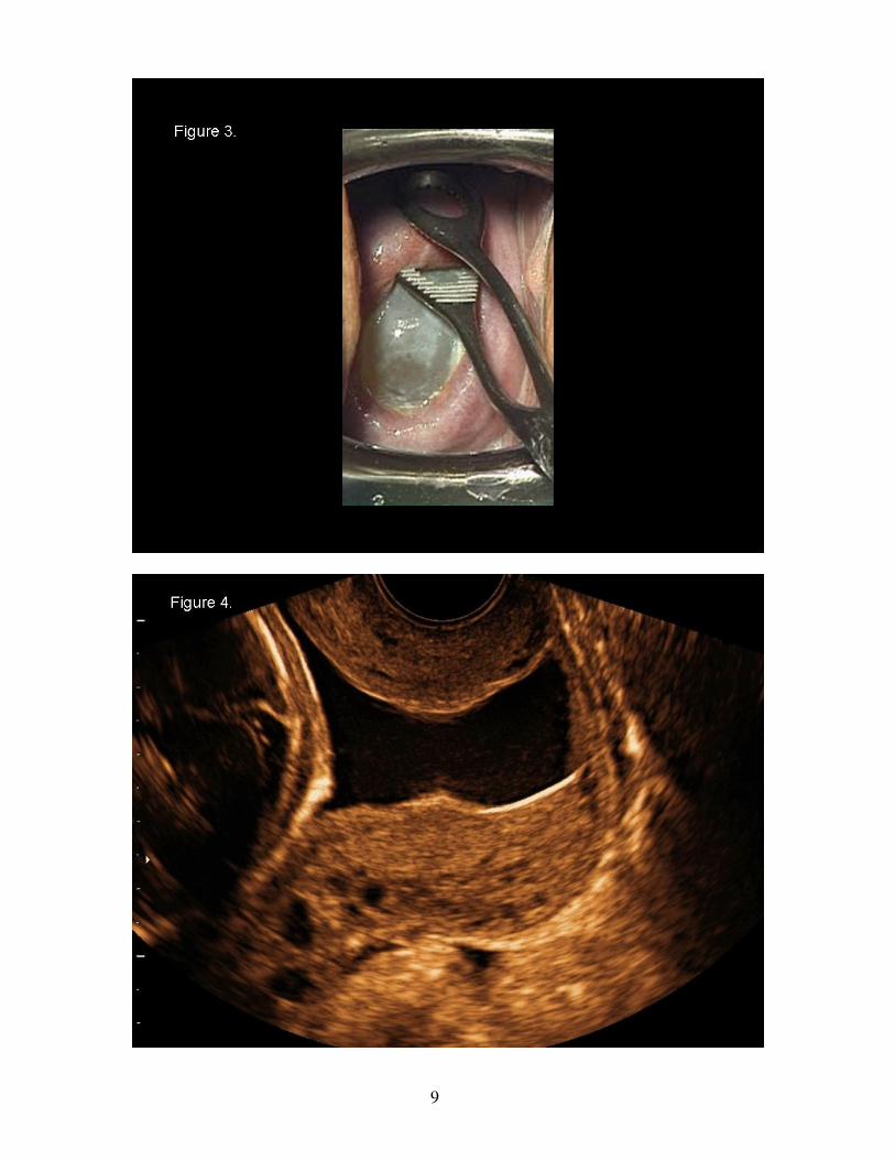
Clearly, a condition exists in which patients present before 24 weeks
of gestation with a dilated and effaced cervix in which the membranes protrude into the
vagina (Figure 3). This condition can often be visualized on ultrasound as illustrated in
Figure 4.
9

10
Harger, in a review of the literature, defined “cervical insufficiency” as “the inability
of the uterine cervix to retain a pregnancy in the absence of contractions or labor.”44
However, the use of this definition in clinical practice is problematic. How can an
obstetrician identify “the inability of the cervix to retain the pregnancy”? There is no
clinical test that would allow this determination. Some have proposed that the response to
treatment offers criteria for the diagnosis. Namely, a patient with a history recurrent mid-
trimester pregnancy loss who is subsequently treated with a cerclage and delivers at term
could be considered to have cervical insufficiency, and hence, the claim that such patients
need a cerclage in every subsequent pregnancy. Yet, observational studies do not support
this conclusion.
One approach by some authors to examine the accuracy of the diagnosis of cervical
insufficiency is to determine pregnancy outcome in patients with this diagnosis who did
not have treatment in subsequent pregnancies. In 1961, Dunn and Dans45
reported the
outcome of 30 patients who had two or more consecutive midtrimester abortions without
an intervening first trimester abortion. Interestingly, 13 of the 30 patients had three
consecutive midtrimester abortions. The authors reasoned that if cervical insufficiency
was the operative mechanism for pregnancy failure, it would be expected that all
subsequent untreated pregnancies would result in pregnancy loss; however, this was not
the case. Indeed, in 50% of cases, patients delivered neonates who exceeded 28 weeks of
gestation and weighed >1000 g. Thus, the authors proposed that the spontaneous “cure
rate” of this entity would be 50%. The Editor of Obstetrical and Gynecological Survey
published a commentary46
in which he argued that a more appropriate endpoint for
spontaneous cure would be delivery at term in the subsequent pregnancy. Although he
recalculated the spontaneous “cure rate” to be 20%, a clear message of the study is that,

11
even in patients with two and sometimes three midtrimester abortions, term pregnancy is
possible without a cerclage.
In 1984, Socol et al.47
reported the outcome of a group of patients who had previously
delivered a live born infant between 20-32 weeks of gestation. In the subsequent
pregnancy, some patients were treated with a cervical cerclage and others were not (at the
discretion of the practitioner). The rate of preterm delivery was 36% (5/14) in patients
who had a cerclage, and 38% (10/26) in those managed expectantly. One interpretation of
these data is that delivery at term was frequently possible in patients with a previous mid-
trimester pregnancy loss or early preterm delivery without a cervical cerclage.
Yet, obstetricians often face the clinical dilemma of how to manage a patient who had
a previous successful pregnancy with a cervical cerclage. Was the first cerclage truly
necessary? Would the patient have delivered at term regardless of the placement of the
cerclage? These questions are pervasive because of the subjective nature of the diagnosis.
For example, some obstetricians place a cerclage after a mid-trimester pregnancy loss
with rupture of membranes, while others consider that this is not enough evidence of
cervical insufficiency to justify the operation.
In a 1994 article entitled “Once a cerclage, not always a cerclage”, Fejgin et al48
reported the pregnancy outcome of patients who had a history of at least one pregnancy
in which a McDonald cerclage had been placed, and the physician caring for the
subsequent pregnancy was uncertain as to whether the procedure was necessary. A
committee of three obstetricians reviewed the history, a hysterosalpingogram when
available, and the results of a pelvic examination. There were 35 patients who had 58
pregnancies with a cerclage, and 52 subsequent pregnancies without the procedure. The
outcome of pregnancies without a cerclage was better than those with a cerclage.

12
Collectively, these retrospective studies question the accuracy of the diagnosis of
cervical insufficiency and/or the effectiveness of cerclage. The results of these studies
(and others) are at the root of the controversy about the nature of this clinical entity, its
diagnosis and the precise role of cerclage in clinical medicine.
There is no objective diagnostic test for cervical insufficiency. The diagnosis is
often made in a patient in the midtrimester who presents with dilatation of the cervix and
different degrees of membrane prolapse. The evaluation of the non-pregnant patient
represents an unsolved challenge. Several methods have been proposed for the
identification of the patient at risk for cervical insufficiency, including: 1) the passage of
Hegar dilators (6 to 8 mm) or Pratt dilators through the internal cervical os;49-51
2) the use
of a balloon test;52
or 3) the ability of the cervix to hold an inflated Foley catheter during
hysterosalpingography.53
However, there is a paucity of scientific evidence to support the
value of these tests in predicting subsequent pregnancy outcome.44
Sonographic cervical length
Digital examination of the cervix was the method used to determine cervical status
(effacement, dilatation, position, and consistency) before the introduction of ultrasound.
Bishop developed his cervical scoring system primarily to predict when spontaneous
labor at term would occur, and found that the higher the score, the sooner the labor would
start.54
Wood et al. were the first to report that a short cervix was a risk factor for preterm
labor and delivery.55
A large study reported by Papiernik consisted of serial digital
examinations in 8,303 women, where dilatation of the internal os was the strongest risk
factor for preterm delivery. A short cervix (≤1cm) also increased the risk.56
These
findings have been confirmed by others.57-62
However, digital examination is subjective
and has limitations. For example, the coefficient of variation for effacement has been
reported to be 26%.63
Moreover, evaluation of effacement requires placing the examining

13
finger in close proximity to the fetal membranes. Sonographic imaging of the cervix is
less invasive and more objective in assessing cervical length as well as changes in the
anatomy of the internal os.64
Transvaginal sonography is superior to transabdominal for examination of the
cervix.65
Numerous studies have proven that the shorter the sonographic cervical length
in the mid-trimester, the higher the risk of spontaneous preterm labor/delivery.66-70
However, there is no agreement concerning what constitutes a sonographic short cervix.
For example, Iams et al.67
proposed that a cervix of 26 mm or shorter at 24 weeks of
gestation increases the risk for spontaneous preterm delivery (Relative Risk: 6.19, 95%
CI: 3.84-9.97). The prevalence of spontaneous preterm delivery (defined as less than 35
weeks) in this study was 4.3%, and the positive predictive value was 17.8% for a cervical
length ≤ 25 mm at 24 weeks of gestation.67
Thus, most women with a short cervix
(defined as 25mm or less) will not deliver a preterm neonate. Other investigators have
proposed a cut-off of 15 mm, because a cervical length of 15 mm or less is associated
with nearly a 50% risk of spontaneous preterm delivery at 32 weeks of gestation or less
when neonatal morbidity is substantial.68, 70
It is important to stress that sonographic cervical length is not a screening test for
spontaneous preterm delivery, because only a fraction of all patients who will have a
spontaneous preterm birth have a short cervix in the mid-trimester. Sonographic cervical
length is only a method for risk assessment for spontaneous preterm delivery and not a
screening test. Cervical length can modify the a priori risk for preterm delivery. For
example, a woman with a history of preterm delivery or one with a twin or triplet
gestation will have a higher risk for preterm delivery than a patient with the same cervical
length, but without such history.71-80
It is now possible to provide women with an
14
individualized estimation of risk for preterm delivery based upon cervical length and
whether they have a history of preterm birth.81
Cervical sufficiency/insufficiency as a continuum
The hypothesis that cervical competence or sufficiency represents a spectrum was
studied by Parikh and Mehta, who used digital examination of the cervix and concluded
that degrees of cervical competence did not exist.82
Iams et al., using sonographic
examination of the cervix, concluded that cervical competence was a continuum.83
The
authors reported a strong relationship between cervical length in pregnancy and previous
obstetrical history. This relationship is nearly linear. However, patients with a typical
history of an incompetent cervix appear to represent a different group than those who
delivered preterm.83
Similar results have been reported by Guzman et al.84
Collectively,
these studies suggest that there is a relationship between a history of preterm delivery and
the cervical length in a subsequent pregnancy. Inasmuch as patients with a short cervix are
at increased risk for a mid-trimester pregnancy loss or spontaneous preterm delivery with
intact or rupture of membranes, a short cervix could be considered as the expression of a
spectrum of cervical diseases or functions. However, it is noteworthy that some women
with a short cervix have an adverse pregnancy outcome, while others have an
uncomplicated term delivery.65-70, 83-97
Indeed, approximately 50% of women with a cervix
of 15 mm or less deliver after 32 weeks.70
This indicates that cervical length is only one of
the factors determining the degree of cervical competence and that a short cervix should
not be equated with “cervical insufficiency.”
Cervical insufficiency is a syndrome
In a manner similar to preterm labor, pre-eclampsia, small for gestational age (SGA),
fetal death, and preterm premature rupture of membranes (PROM), the clinical conditions

15
that describe cervical insufficiency can be considered “an obstetrical syndrome.”98
Cervical ripening in the mid-trimester may be the result of: 1) the loss of connective
tissue after a cervical operation such as conization99-101
or Loop electrosurgical excision
procedure (LEEP) procedure;101
2) a congenital disorder such as cervical hypoplasia after
diethylstilbestrol (DES) exposure;102-105
3) intrauterine infection;106-108
4) a suspension of
progesterone action.34, 35
There is experimental evidence that progesterone can reverse
cervical compliance induced by the administration of dexamethasone to pregnant
sheep.114
Moreover, recent studies have indicated that progesterone administration to
women with a short cervix can reduce the rate of preterm birth;109, 110
and 5) a cervical
disorder that manifests itself with the clinical presentation of cervical insufficiency. Each
of these different causes of the syndrome could be affected by genetic or environmental
factors (Figure 5).42
Moreover, more than one mechanism of disease may be operative in
a specific patient. The possibility of novel and yet to be discovered mechanisms of
disease playing a role must also be considered.

16
Previous trauma as a cause for cervical insufficiency: Mechanical dilatation of the
cervix before gynecologic procedures, laser ablation, LEEP and cold-knife conization
may increase the risk for a preterm birth.100, 111-118
In a recent retrospective study by Shin
et al, the rate of preterm delivery was significantly higher in women with a history of a
conization when compared to those without one (32.1% [18/56] vs. 15.2%
[3,355/22,070], p<0.001). Yet, the use of a McDonald cerclage (n=25) was not associated
with a reduction in the rate of preterm delivery when compared to those without cerclage
(n=31) (expectantly managed group vs. cerclage group; <28 week, 6.5% vs. 8.0%,
p=1.000; <34 week, 19.4% vs. 20.0%, p=1.000; <37 week, 29.0% vs. 36.0%,
p=0.579).119
17
Congenital cervical insufficiency: Exposure to diethylstilbestrol (DES) in utero has
been reported to increase the risk of second trimester pregnancy loss five-fold (6.3% in
DES exposed versus 1.6% in the control group). 120
There is evidence of familial aggregation in cervical insufficiency. In one study,
approximately 25% of patients with cervical insufficiency had a first degree relative with
a similar diagnosis.121
This has been attributed to genetic factors involved in the
regulation of extracellular matrix; specifically, polymorphisms in the genes encoding for
collagen 1 alpha 1 (COLIAI) and transforming growth factor beta (TGFβ),121
and
recently, IL-10.122
A genetic predisposition to cervical insufficiency has been reported in
women with Ehlers-Danlos syndrome123, 124
and Marfan syndrome.125-128
“Cervical insufficiency” as a clinical manifestation of intrauterine infection: A
proportion of patients presenting with asymptomatic cervical dilatation in the mid-
trimester have microbial invasion of the amniotic cavity (MIAC)106, 107
that can be as high
as 51.5%.106
Microbial invasion of the amniotic cavity may be due to premature cervical
dilatation with the exposure of the chorioamniotic membranes to the microbial flora of
the lower genital tract. Microorganisms may gain access to the amniotic cavity by
crossing intact membranes.106
Under these circumstances, infection would be a secondary
phenomenon to primary cervical disease. An alternative is that intrauterine infection or
one caused by activation of microorganisms present within the uterine cavity129
in the
second trimester of pregnancy produces myometrial contractility and cervical ripening.
Since uterine contractions are usually clinically silent in the mid-trimester of pregnancy,
the clinical picture of an infection-induced spontaneous abortion may be
indistinguishable from that of an incompetent cervix.106, 130
The most frequently isolated
organisms from the amniotic fluid in women with suspected cervical insufficiency
18
include Ureaplasma urealyticum and Ureaplasma parvum106, 131, 132
. We have established
that 9% (5/57) of women with a short endocervix (less than 25 mm) have
microbiologically-proven intra-amniotic infection,108
suggesting that these infections are
sub-clinical and may precede the development of the clinical picture of acute “cervical
insufficiency” (dilated and effaced cervix with bulging membranes).
Cerclage: Cervical cerclage was introduced in 1955 by V.N. Shirodkar, Professor of
Midwifery and Gynecology at the Grant Medical College in Bombay, India.39
The
procedure was developed in response to his observation that “some women abort
repeatedly between the fourth and seventh months and no amount of rest and treatment
with hormones seemed to help them in retaining the product of conception.39
Shirodkar
referred to a group of 30 women who had had at least four abortions (some between 9
and 11 weeks). He stated that in his opinion, “95% of cases were due to a weak cervical
sphincter and the other few to an underdeveloped or malformed uterus, etc.”39
Shirodkar
emphasized that his work was confined to women in whom he could prove the existence
of weakness of the internal os by “repeated internal examinations.”39
Ian McDonald, from
the Royal Melbourne Hospital, reported in 1957 his experience with 70 patients who had
a suture of the cervix for inevitable miscarriage.40
The history of this procedure is
relevant since 50 years after its introduction, cerclage is being used for indications
different from those originally intended, and there is conflicting evidence about its
efficacy for the new indications (e.g., prevention of preterm birth in women with a
sonographic short cervix).39, 40, 90-92, 95-97, 133-154
There are several approaches to cervical cerclage: 1) the Shirodkar method;39
2) the
McDonald method;40
3) the Wurm procedure;155
and 4) transabdominal156
. The latter has

19
been performed using a laparotomy approach and later described, laparoscopically157, 158
.
The most widely used procedure is the McDonald cerclage.
Complications of cerclage:
The risks associated with the use of cerclage include those that can occur intra-
operatively (anesthetic complications, blood loss, rupture of membranes), in the post-
operative period (subclinical or clinical chorioamnionitis, preterm PROM, cervical
laceration, preterm contractions), and at the time of labor and delivery (cervical
laceration, cervical dystocia, sepsis, uterine rupture).159-170
The rate of each complication
varies as a function of the indication for the cerclage, type of cerclage and the state of the
cervix and amniotic membranes at the time of surgery.161, 164, 167
Complications
commonly reported include: preterm premature rupture of membranes (elective 2.8-
36.3%, emergency 2.8-52.2%),162, 167-170
preterm contractions (36-39%),168
cervical
lacerations (elective 0-14%, emergency 0-23.8%),166-169, 171
uterine rupture (<0.1%),172
chorioamnionitis (elective 5.2-14.9%, emergency 4-39.1%),162, 167, 169, 170, 172
sepsis
(0.34%),166
and bleeding (14.9%).168
Harger169
reported the outcome of 251 patients; 202 who underwent a cerclage placed
based upon history and 49 cerclages were placed due to cervical dilation. The mean blood
loss in patients was 30 ml in patients who underwent a McDonald cerclage and 44 ml in
those who had a Shirodkar procedure. Two patients who had undergone an emergency
cerclage lost 150 ml of blood. The risk of pregnancy loss ‘apparently caused by elective
procedures’ was 2% (4/202). Acute chorioamnionitis occurred in 1.2% of patients.
Cervical lacerations occurred more frequently at the time of labor in women who had a
Shirodkar or McDonald cerclage than in those without a cerclage (n=55,688) (Shirodkar:
11% and McDonald: 14% vs. control group: 2.18%, p< 0.001). Furthermore, the authors

20
argued that some cesarean sections in women who underwent cerclage (McDonald or
Shirodkar) may be attributed to cervical scarring as a result of the surgery.
Charles and Edwards noted an increased risk of complications in patients undergoing
cerclage after 19 weeks of gestation. The rate of rupture of the membranes within 5 days
of the procedure was 19.5%, preterm premature rupture of the membranes (26-34 weeks
of gestation) 52.2%, and chorioamnionitis 39.1% in these patients.162
In a retrospective review of 482 singleton pregnancies who underwent cerclage
placement (McDonald cerclage [n=377], Shirodkar [n=104]) over a 6-year period,
Treadwell et al167
reported that premature rupture of the membranes occurred in 38% of
patients and was the most frequent complication. The prognostic factors determining
gestational age at delivery were: gestational age at the time of cerclage, cervical dilation,
and number of prior pregnancy losses before 24 weeks of gestation. The rate of cervical
laceration was 6.7% and was similar for both emergency and elective cerclages. Of
interest, the primary cesarean delivery rate of patients who had undergone a Shirodkar
cerclage was higher than those who had undergone a McDonald cerclage (31% vs 17%,
p<.005).
Cerclage in patients with acute cervical insufficiency: Only one randomized clinical
trial has tested the effect of cerclage in patients with acute cervical insufficiency. Patients
were identified if they presented with a dilated cervix and membranes at or below a
dilated external os before 27 weeks of gestation. Patients were treated either with
emergency cerclage and indomethacin (n=13), or bedrest only (n=10). Indomethacin was
only given to one group. Preterm delivery before 34 weeks of gestation was significantly
less frequent in patients allocated to have an emergency cerclage and indomethacin than
in those managed expectantly [54% (7/13) versus 0% (0/10); p=0.02]. Antibiotics were

21
administered to both groups of patients.173
Acute cervical insufficiency, however, can
present with different degrees of severity. A patient with a dilated cervix in which the
membranes are visible but within the uterine cavity is not the same as the patient who has
prolapsed “hourglass” membranes in the vagina with a fetal presenting part. The latter
group represents a greater surgical challenge, and is often associated with subclinical
intrauterine infection. One recommendation is to place the patient in the Trendelenburg
position and to allow reduction of the membranes to the amniotic cavity. Since patients
with intra-amniotic inflammation/infection have a poor prognosis if a cerclage is placed,
an amniocentesis is performed to look for infection/inflammation. The work-up includes
an amniotic fluid Gram stain, white blood cell count, glucose and culture for aerobic and
anaerobic bacteria as well as genital mycoplasmas. After the placement of a cerclage
patients are often followed by transvaginal ultrasound. The length of the cervix after a
cerclage has some prognostic value about the likelihood of success.174-177
Cerclage to prevent preterm delivery in women with a short cervix without a history
of preterm delivery: The largest randomized clinical trial in which cerclage was used in
patients with a sonographic cervical length ≤15 mm was conducted by the Fetal Medicine
Foundation of the United Kingdom.150
Cervical length was determined in low-risk
patients at a median gestational age of 23 weeks, and those with a cervix ≤15 mm were
randomized to either expectant management (n = 126) or cerclage group (n = 127). The
rate of preterm delivery at less than 33 weeks of gestation was not significantly different
(expectant management group 26% [33/126] vs cerclage group 22% [28/127]). The
conclusion of this study is that cerclage placement in patients with a short cervix without
risk factors for preterm delivery does not reduce the rate of spontaneous preterm birth or
the rate of perinatal death.150

22
Prophylactic cerclage and cerclage in patients with a history of preterm delivery
and a short cervix: The role of prophylactic cerclage in high-risk patients without a
sonographic short cervix for the prevention of preterm delivery/midtrimester abortion (by
history) is unclear.144, 152, 165, 178, 179
While the largest trial conducted prior to the
introduction of ultrasound evaluation of the cervix suggested a modest beneficial
effect,179
other trials165, 178
and systematic reviews41
prior to the use of ultrasound have
indicated that the evidence of effectiveness for prophylactic cerclage is either weak or
non-existent.
In the Cervical Incompetence Prevention Randomized Cerclage Trial (CIPRACT)
study, Althuisius et al135, 180
randomized 73 pregnant women at less than 15 weeks of
gestation with risk factors for “cervical incompetence” to have a prophylactic cerclage (n
= 23) or be observed (n = 44). Four patients had a spontaneous abortion during the first
trimester and 2 were lost for follow-up. The risk factors for “cervical incompetence”
included the history of preterm delivery before 34 weeks’ gestation, previous preterm
PROM before 32 weeks’ gestation, history of cold knife conization, DES exposure, and a
Müllerian duct abnormality. Prophylactic cerclages (i.e. McDonald) were generally
placed between 10 to 12 weeks (later if enrolled at a later gestational age) using a braided
polyester thread. In both groups, the cervical length was evaluated every 2 weeks after
randomization. The rate of preterm delivery (<34 weeks) was similar in both groups
(prophylactic cerclage 13% [3 of 23] vs observation 14% [6 of 44]; p > 0.05) as well as
the neonatal survival (prophylactic cerclage 91% [21 of 23] vs observation 93% [41 of
44]; p > 0.05). Patients allocated to the observation group were followed with serial
sonography. If the cervical length shortened (<25 mm) before the 27th week of gestation,
they were randomized to both therapeutic cerclage and indomethacin with bed rest (n =

23
20) or bed rest alone (n = 16). Patients who received a cerclage and indomethacin had a
lower rate of preterm delivery at less than 34 weeks and composite neonatal morbidity
(neonatal intensive care unit [NICU] admission or neonatal death) (0% vs 44% [7 of 16],
P = .002; and 5% [1 of 19] vs 50% [8 of 16], P = .005).
In contrast, Rust et al91, 181
randomized 113 patients presenting with a short cervix
(<25 mm) or funneling (≥25%) between 16 and 24 weeks into the therapeutic cerclage
group (n = 55) and the no cerclage group (n = 58). The population included patients with
and without risk factors for preterm birth. All patients underwent amniocentesis to
exclude intra-amniotic infection and received 48 hours of therapy with indomethacin and
antibiotics. There were no significant differences between the 2 groups with respect to the
rate of preterm delivery at less than 34 weeks’ gestation (35% vs 36.2%), readmission for
preterm labor (52% vs 53%), placental abruption (11% vs 14%), chorioamnionitis (20%
vs 10%), and the perinatal death rate (13% vs 12%).
The CIPRACT study135, 152
enrolled only patients at risk for preterm delivery, while in
the trial of Rust et al,91, 181
13% of patients were at low risk. The positive predictive value
for a short cervix to predict preterm delivery is higher in patients with a history of
preterm birth. The fact that the rate of preterm delivery in the control group of the
CIPRACT study was higher than that of the study of Rust et al (43.8% vs 36.2%) may
explain, at least in part, the different results between these 2 studies.
Berghella et al146
conducted a randomized clinical trial of the use of McDonald
cerclage in women with one or more risk factors for preterm birth (one or more prior
deliveries at less than 35 weeks, 2 or more curettages, history of DES exposure, cone
biopsy, Müllerian anomaly, or twin gestation). Sixty-one patients with a cervical length
less than 25 mm or funneling greater than 25% were randomized to cerclage or bed rest.
The authors reported that 47 pregnancies (77%) were high-risk singleton gestations.

24
There was no significant difference in the rate of preterm birth prior to 35 weeks
in women who underwent cerclage (14 of 31; 45%) compared to those in the bed rest
group (14 of 30; 47%) (RR 0.94; 95% CI = 0.34 to 2.58). Similarly, patients with a
singleton gestation and a prior preterm birth at less than 35 weeks of gestation and a
cervical length less than 25 mm (n = 31 women) also had no benefit from cerclage
placement (40% vs 56%; RR 0.52; 95% CI = 0.12 to 2.17).146
Owen et al.153
reported a randomized clinical trial in which women with a
prior spontaneous preterm birth before 34 weeks of gestation and a short cervix (defined
as a cervical length of < 25 mm) were randomly assigned to have a cerclage or to be
managed expectantly. The primary endpoint for the trial was birth at <35 weeks of
gestation. Of the 302 patients randomized, 148 were allocated to the cerclage group, and
153 to the non-cerclage group. There was a non-significant decrease in the primary
endpoint (delivery at less than 35 weeks of gestation) in women allocated to cerclage than
in the non-cerclage group (32% vs. 42%: odds ratio 0.67; 95% CI, 0.42-1.07; p=0.09). A
post hoc analysis demonstrated that the rate of preterm delivery at <35 weeks was
significantly lower in women with a cervical length below 15 mm, and there was no
demonstrable effect in women with a cervical length between 16-24 mm. This study
reported that cerclage reduced the rate of pre-viable birth and perinatal mortality. The
results of the secondary analysis (<15 mm) are considered to be hypothesis-generating.
A meta-analysis (published in March 2011) of five trials in which women had
singleton gestations, previous spontaneous preterm birth and cervical length <25 mm
before 24 weeks of gestation demonstrated that placement of a cerclage was associated
with a lower rate of preterm birth before 35 weeks of gestation (28.4% [71/250] versus
41.3% [105/254]; RR 0.70, 95% CI, 0.55-0.89). Moreover, an index of perinatal mortality
and morbidity (composite index) was significantly reduced in patients who had a cerclage

25
(15.6% in the cerclage group versus 24.8% in patients without cerclage, RR 0.64, 95%
CI, 0.45-0.91). Subgroup analysis demonstrated that in women with a cervical length of
15.9 mm or less, cerclage was associated with significant prevention of preterm birth at
<37, 35, 32 and 28 weeks. The authors also examined the effect of cerclage as a function
of cervical length. In patients with a sonographic cervical length of 16-24.9 mm, cerclage
was associated with a significant reduction in the rate of preterm birth at <37 and <24
weeks of gestation.154
The authors stated that, based on their findings, ‘the effect of this
intervention is important but clearly not the solution to the whole problem of preterm
birth,’ and call for ‘further understanding of the pathophysiology of spontaneous preterm
birth.’ Thus, cerclage may be effective in reducing the rate of preterm birth in a subset of
patients.
Pessary in women with cervical insufficiency and/or a short cervix:
The first report of the use of a pessary for the treatment of cervical ‘insufficiency’
was in 1959.182
Advocates for pessaries to prevent preterm delivery argued that their use
should be considered in women with suspected cervical insufficiency because cerclage
had not been proven to be efficacious in the prevention of preterm birth, and pessaries
would not have the complications known to occur with cerclage. Vaginal pessaries are
inexpensive, can be readily placed and removed and allow outpatient management
(without anesthesia).183
Several studies have evaluated the use of a pessary to prevent preterm delivery in
women with suspected cervical insufficiency based upon history. Most of these reports
consist of small case series.183-191
Arabin et al.187
reported sonographic cervical length measurements on patients
with a prior spontaneous preterm birth before 36 weeks of gestation or early symptoms of

26
preterm labor (pressure or contractions) and twin pregnancies. Patients with a cervical
length < 15 mm between 22 and 28 weeks of gestation were offered the use of a silicone
pessary (Arabin pessary). The outcome of 11 patients who had a pessary was compared
with a gestational age and cervical length-matched group. The control group consisted of
12 singleton pregnancies and 23 twin gestations. Placement of a pessary was associated
with an older gestational age at delivery in both the singleton and twin gestations (for
singletons, the mean gestational age at delivery was 38 weeks [36 6/7 – 41] vs. 33 4/7
weeks [26-38], p=0.02; for twins – 35 6/7 weeks (33-37 4/7) vs. 33 2/7 weeks [24 4/7 –
37 2/7], p=0.02). None of the 12 singleton gestations with a pessary delivered prior to 36
weeks of gestation (pessary 0% [0/12] vs. control 50% [6/12], p < 0.001).187
Based on
these results, several trials are now in progress to determine if a pessary can reduce the
rate of preterm birth in women with a short cervix in both singleton and twin gestations
(Carreras, Nicolaides, Palacio, Moratonas, Nizard). At this time, there is no evidence that
cerclage is effective in twin gestations.
Progesterone: The role of vaginal progesterone in the prevention of preterm birth in
women with a short cervix is discussed in the chapter focusing on preterm labor in this
book.

27
Acknowledgments: This work was supported (in part) by the Perinatology Research
Branch, Division of Intramural Research, Eunice Kennedy Shriver National Institute of
Child Health and Human Development, NIH, DHHS.

28
Legends:
Figures 1a – 1d:
1a: Schematic representing the location of the fibromuscular junction and the cervical
changes seen throughout pregnancy and labor.3
1b – 1d: Ultrasound images displaying the changes seen in the cervix during pregnancy
and labor.
Figure 2: Stages of Cervical Function During Pregnancy and the Puerperium as proposed
by Word et al.6
Figure 3: A patient who presented prior to 24 weeks of gestation with a dilated and
effaced cervix in which the membranes are protruding into the vagina.
Figure 4: An ultrasound image demonstrating a dilated cervix in a patient with suspected
cervical insufficiency.
Figure 5: The proposed causes of the “cervical insufficiency syndrome”.42

29
References
1. CALDER A. The human cervix in pregnancy: a clinical perspective. In: Ellwood D,
Anderson A, eds. The cervix in pregnancy and labor: Churchill Livingstone,
1981.
2. DANFORTH DN, BUCKINGHAM JC, RODDICK JW, JR. Connective tissue changes
incident to cervical effacement. Am J Obstet Gynecol 1960;80:939-945.
3. DANFORTH DN, HENDRICKS CH. Obstetrics and Gynecology: Harper and Row,
1977.
4. SCHWALM H, DUBRAUSZKY V. The structure of the musculature of the human
uterus--muscles and connective tissue. Am J Obstet Gynecol 1966;94:391-404.
5. DANFORTH DN. The distribution and functional activity of the cervical
musculature. Am J Obstet Gynecol 1954;68:1261-71.
6. WORD RA, LI XH, HNAT M, CARRICK K. Dynamics of cervical remodeling during
pregnancy and parturition: mechanisms and current concepts. Semin Reprod Med
2007;25:69-79.
7. DANFORTH DN. The fibrous nature of the human cervix, and its relation to the
isthmic segment in gravid and nongravid uteri. Am J Obstet Gynecol
1947;53:541-60.
8. MAILLOT KV, ZIMMERMANN BK. The solubility of collagen of the uterine cervix
during pregnancy and labour. Arch Gynakol 1976;220:275-280.
9. JUNQUEIRA LC, ZUGAIB M, MONTES GS, TOLEDO OM, KRISZTAN RM,
SHIGIHARA KM. Morphologic and histochemical evidence for the occurrence of
collagenolysis and for the role of neutrophilic polymorphonuclear leukocytes
during cervical dilation. Am J Obstet Gynecol 1980;138:273-281.
10. LIGGINS G, ELWOOD DA, ANDERSON ABM. Cervical ripening as an inflammatory
reaction. The Cervix in Pregnancy and Labour: Clinical and Biochemical
Investigations. Edinburgh: Churchill-Livingstone, 1981.
11. LEPPERT PC, KELLER S, CERRETA J, MANDL I. Conclusive evidence for the
presence of elastin in human and monkey cervix. Am J Obstet Gynecol
1982;142:179-82.
12. EKMAN G, ULDBJERG N, MALMSTROM A, ULMSTEN U. Increased postpartum
collagenolytic activity in cervical connective tissue from women treated with
prostaglandin E2. Gynecol Obstet Invest 1983;16:292-8.
13. LEPPERT PC, KELLER S, CERRETA J, HOSANNAH Y, MANDL I. The content of
elastin in the uterine cervix. Arch Biochem Biophys 1983;222:53-8.
14. ULDBJERG N, CARLSTEDT I, EKMAN G, MALMSTROM A, ULMSTEN U, WINGERUP
L. Dermatan sulphate and mucin glycopeptides from the human uterine cervix.
Gynecol Obstet Invest 1983;16:199-209.
15. ULDBJERG N, EKMAN G, MALMSTROM A, OLSSON K, ULMSTEN U. Ripening of
the human uterine cervix related to changes in collagen, glycosaminoglycans, and
collagenolytic activity. Am J Obstet Gynecol 1983;147:662-666.
16. TIMPL R, FUJIWARA S, DZIADEK M, AUMAILLEY M, WEBER S, ENGEL J. Laminin,
proteoglycan, nidogen and collagen IV: structural models and molecular
interactions. Ciba Found Symp 1984;108:25-43.
17. LEPPERT PC, YU SY. Apoptosis in the cervix of pregnant rats in association with
cervical softening. Gynecol Obstet Invest 1994;37:150-4.
18. HWANG JJ, MACINGA D, RORKE EA. Relaxin modulates human cervical stromal
cell activity. J Clin Endocrinol Metab 1996;81:3379-84.

30
19. MAHENDROO MS, CALA KM, RUSSELL DW. 5 alpha-reduced androgens play a
key role in murine parturition. Mol Endocrinol 1996;10:380-92.
20. RECHBERGER T, ABRAMSON SR, WOESSNER JF, JR. Onapristone and prostaglandin
E2 induction of delivery in the rat in late pregnancy: a model for the analysis of
cervical softening. Am J Obstet Gynecol 1996;175:719-23.
21. SENNSTROM MK, BRAUNER A, LU Y, GRANSTROM LM, MALMSTROM AL, EKMAN
GE. Interleukin-8 is a mediator of the final cervical ripening in humans. Eur J
Obstet Gynecol Reprod Biol 1997;74:89-92.
22. WESTERGREN-THORSSON G, NORMAN M, BJORNSSON S, et al. Differential
expressions of mRNA for proteoglycans, collagens and transforming growth
factor-beta in the human cervix during pregnancy and involution. Biochim
Biophys Acta 1998;1406:203-13.
23. MAHENDROO MS, PORTER A, RUSSELL DW, WORD RA. The parturition defect in
steroid 5alpha-reductase type 1 knockout mice is due to impaired cervical
ripening. Mol Endocrinol 1999;13:981-92.
24. ULDBJERG N, FORMAN A, REECE EA, HOBBINS JC. Biomechanical and
biochemical changes of the uterus and cervix during pregnancy. Medicine of the
fetus and mother. Philadelphia, PA: Lippincott-Raven Publishers, 1999 (vol 2nd).
25. OSMAN I, YOUNG A, LEDINGHAM MA, et al. Leukocyte density and pro-
inflammatory cytokine expression in human fetal membranes, decidua, cervix and
myometrium before and during labour at term. Mol Hum Reprod 2003;9:41-45.
26. SAKAMOTO Y, MORAN P, SEARLE RF, BULMER JN, ROBSON SC. Interleukin-8 is
involved in cervical dilatation but not in prelabour cervical ripening. Clin Exp
Immunol 2004;138:151-157.
27. STJERNHOLM-VLADIC Y, STYGAR D, MANSSON C, et al. Factors involved in the
inflammatory events of cervical ripening in humans. Reprod Biol Endocrinol
2004;2:74.
28. TIMMONS BC, MAHENDROO M. Processes regulating cervical ripening differ from
cervical dilation and postpartum repair: insights from gene expression studies.
Reprod Sci 2007;14:53-62.
29. HASSAN SS, ROMERO R, TARCA AL, et al. The transcriptome of cervical ripening
in human pregnancy before the onset of labor at term: identification of novel
molecular functions involved in this process. J Matern Fetal Neonatal Med
2009;22:1183-93.
30. HASSAN SS, ROMERO R, TARCA AL, et al. The molecular basis for sonographic
cervical shortening at term: identification of differentially expressed genes and the
epithelial-mesenchymal transition as a function of cervical length. Am J Obstet
Gynecol 2010;203:472 e1-472 e14.
31. ELOVITZ MA, GONZALEZ J. Medroxyprogesterone acetate modulates the immune
response in the uterus, cervix and placenta in a mouse model of preterm birth. J
Matern Fetal Neonatal Med 2008;21:223-30.
32. OBARA M, HIRANO H, OGAWA M, et al. Changes in molecular weight of
hyaluronan and hyaluronidase activity in uterine cervical mucus in cervical
ripening. Acta Obstet Gynecol Scand 2001;80:492-6.
33. UCHIYAMA T, MATSUMOTO T, SUZUKI Y, ISHIDA M, OBARA T, KANAYAMA T.
Endogenous hyaluronan: a cytokine-like factor present in rabbit uterine cervix
during pregnancy. Biol Pharm Bull 2004;27:1907-12.
34. CHWALISZ K. The use of progesterone antagonists for cervical ripening and as an
adjunct to labour and delivery. Hum Reprod 1994;9 Suppl 1:131-61.

31
35. CLARK K, JI H, FELTOVICH H, JANOWSKI J, CARROLL C, CHIEN EK. Mifepristone-
induced cervical ripening: structural, biomechanical, and molecular events. Am J
Obstet Gynecol 2006;194:1391-8.
36. ANONYMOUS. In: Culpeper N, Cole A, Rowland W, eds. The practice of physick.
London, UK: George Sawbridge, 1678.
37. ALTHUISIUS S. Cervical incompetence, you better believe it. Department of
Obstetrics and Gynecology, Division of Maternal-Fetal Medicine. Amsterdam,
the Netherlands: VU Medical Center, 2001.
38. GREAM GT. Dilatation or Division of the Cervix Uteri. The Lancet 1865:381
-381.
39. SHIRODKAR VN, ET AL. A new method of operative treatment for habitual
abortions in the second trimester of pregnancy. Antiseptic 1955;52 299-300.
40. MCDONALD IA. Suture of the cervix for inevitable miscarriage. J Obstet Gynaecol
Br Emp. 1957;64:346-350.
41. GRANT A. Cervical cerclage to prolong pregnancy. In: Chalmers I, Enkin M,
Keirse MJNC, eds. Effective care in pregnancy and childbirth. New York, NY:
Oxford University Press, 1989.
42. ROMERO R, ESPINOZA J, EREZ O, HASSAN S. The role of cervical cerclage in
obstetric practice: can the patient who could benefit from this procedure be
identified? Am J Obstet Gynecol 2006;194:1-9.
43. BENGTSSON LP. Cervical insufficiency. Acta Obstet Gynecol Scand.
1968;47:Suppl-35.
44. ACOG Practice Bulletin. Cervical insufficiency. Obstet Gynecol 2003;102:1091-
1099.
45. DUNN LJ, DANS P. Subsequent obstetrical performance of patients meeting the
historical criteria for cervical incompetence. Bull Sloane Hosp Women
1961;7:43-5.
46. DUNN LJ, DANS P. Subsequent obstetrical performance of patients meeting the
historical criteria for cervical incompetence. Bull Sloane Hosp Women 1961;7
43-5; Editorial Comment in Obstetrical and Gynecological Survey, p. 797.
47. SOCOL ML, DOOLEY SL, TAMURA RK, DEPP OR. Perinatal outcome following
prior delivery in the late second or early third trimester. Am J Obstet Gynecol
1984;150:228-231.
48. FEJGIN MD, GABAI B, GOLDBERGER S, BEN NUN I, BEYTH Y. Once a cerclage, not
always a cerclage. J Perinat Med. 1994;39:880-882.
49. PAGE EW. Incompetent internal os of the cervix causing late abortion and
premature labor; technic for surgical repair. Obstet Gynecol 1958;12:509-515.
50. TOAFF R, TOAFF ME, BALLAS S, OPHIR A. Cervical incompetence: diagnostic and
therapeutic aspects. Isr J Med Sci 1977;13:39-49.
51. KIWI R, NEUMAN MR, MERKATZ IR, SELIM MA, LYSIKIEWICZ A. Determination
of the elastic properties of the cervix. Obstet Gynecol 1988;71:568-574.
52. ZLATNIK FJ, BURMEISTER LF, FEDDERSEN DA, BROWN RC. Radiologic
appearance of the upper cervical canal in women with a history of premature
delivery. II. Relationship to clinical presentation and to tests of cervical
compliance. J Reprod Med. 1989;34:525-530.
53. BERGMAN P, SVENNERUD S. Traction test for demonstrating incompetence of the
internal os of the cervix. Int J Fertil 1957;2:163-167.
54. BISHOP EH. Pelvic Scoring for Elective Induction. Obstet Gynecol 1964;24:266-
8.

32
55. WOOD C, BANNERMAN RH, BOOTH RT, PINKERTON JH. The Prediction of
Premature Labor by Observation of the Cervix and External Tocography. Am J
Obstet Gynecol 1965;91:396-402.
56. PAPIERNIK E, BOUYER J, COLLIN D, WINISDOERFFER G, DREYFUS J. Precocious
cervical ripening and preterm labor. Obstet Gynecol 1986;67:238-242.
57. ANDERSON AB, TURNBULL AC. Relationship between length of gestation and
cervical dilatation, uterine contractility, and other factors during pregnancy. Am J
Obstet Gynecol 1969;105:1207-1214.
58. BOUYER J, PAPIERNIK E, DREYFUS J, COLLIN D, WINISDOERFFER B, GUEGUEN S.
Maturation signs of the cervix and prediction of preterm birth. Obstet Gynecol
1986;68:209-214.
59. LEVENO KJ, COX K, ROARK ML. Cervical dilatation and prematurity revisited.
Obstet Gynecol 1986;68:434-435.
60. STUBBS TM, VAN DORSTEN JP, MILLER MC, III. The preterm cervix and preterm
labor: relative risks, predictive values, and change over time. Am J Obstet
Gynecol 1986;155:829-834.
61. HOLBROOK RH, JR., FALCON J, HERRON M, LIRETTE M, LAROS RK, JR., CREASY
RK. Evaluation of the weekly cervical examination in a preterm birth prevention
program. Am J Perinatol 1987;4:240-244.
62. CATALANO PM, ASHIKAGA T, MANN LI. Cervical change and uterine activity as
predictors of preterm delivery. Am J Perinatol 1989;6:185-190.
63. HOLCOMB WL, JR., SMELTZER JS. Cervical effacement: variation in belief among
clinicians. Obstet Gynecol. 1991;78:43-45.
64. GOMEZ R, GALASSO M, ROMERO R, et al. Ultrasonographic examination of the
uterine cervix is better than cervical digital examination as a predictor of the
likelihood of premature delivery in patients with preterm labor and intact
membranes. Am J Obstet Gynecol 1994;171:956-964.
65. ANDERSEN HF. Transvaginal and transabdominal ultrasonography of the uterine
cervix during pregnancy. J Clin Ultrasound 1991;19:77-83.
66. ANDERSEN HF, NUGENT CE, WANTY SD, HAYASHI RH. Prediction of risk for
preterm delivery by ultrasonographic measurement of cervical length. Am J
Obstet Gynecol 1990;163:859-867.
67. IAMS JD, GOLDENBERG RL, MEIS PJ, et al. The length of the cervix and the risk of
spontaneous premature delivery. National Institute of Child Health and Human
Development Maternal Fetal Medicine Unit Network. N Engl J Med
1996;334:567-572.
68. HEATH VC, SOUTHALL TR, SOUKA AP, ELISSEOU A, NICOLAIDES KH. Cervical
length at 23 weeks of gestation: prediction of spontaneous preterm delivery.
Ultrasound Obstet Gynecol. 1998;12:312-317.
69. TAIPALE P, HIILESMAA V. Sonographic measurement of uterine cervix at 18-22
weeks' gestation and the risk of preterm delivery. Obstet Gynecol 1998;92:902-
907.
70. HASSAN SS, ROMERO R, BERRY SM, et al. Patients with an ultrasonographic
cervical length < or =15 mm have nearly a 50% risk of early spontaneous preterm
delivery. Am J Obstet Gynecol 2000;182:1458-1467.
71. GOLDENBERG RL, IAMS JD, MIODOVNIK M, et al. The preterm prediction study:
risk factors in twin gestations. National Institute of Child Health and Human
Development Maternal-Fetal Medicine Units Network. Am J Obstet Gynecol
1996;175:1047-1053.

33
72. SOUKA AP, HEATH V, FLINT S, SEVASTOPOULOU I, NICOLAIDES KH. Cervical
length at 23 weeks in twins in predicting spontaneous preterm delivery. Obstet
Gynecol 1999;94:450-454.
73. GUZMAN ER, WALTERS C, O'REILLY-GREEN C, et al. Use of cervical
ultrasonography in prediction of spontaneous preterm birth in twin gestations. Am
J Obstet Gynecol 2000;183:1103-1107.
74. GUZMAN ER, WALTERS C, O'REILLY-GREEN C, et al. Use of cervical
ultrasonography in prediction of spontaneous preterm birth in triplet gestations.
Am J Obstet Gynecol 2000;183:1108-1113.
75. TO MS, SKENTOU C, CICERO S, LIAO AW, NICOLAIDES KH. Cervical length at 23
weeks in triplets: prediction of spontaneous preterm delivery. Ultrasound Obstet
Gynecol 2000;16:515-518.
76. YANG JH, KUHLMAN K, DALY S, BERGHELLA V. Prediction of preterm birth by
second trimester cervical sonography in twin pregnancies. Ultrasound Obstet
Gynecol 2000;15:288-291.
77. MAYMON R, HERMAN A, JAUNIAUX E, FRENKEL J, ARIELY S, SHERMAN D.
Transvaginal sonographic assessment of cervical length changes during triplet
gestation. Hum Reprod 2001;16:956-960.
78. OWEN J, YOST N, BERGHELLA V, et al. Mid-trimester endovaginal sonography in
women at high risk for spontaneous preterm birth. JAMA 2001;286:1340-1348.
79. SKENTOU C, SOUKA AP, TO MS, LIAO AW, NICOLAIDES KH. Prediction of
preterm delivery in twins by cervical assessment at 23 weeks. Ultrasound Obstet
Gynecol 2001;17:7-10.
80. VAYSSIERE C, FAVRE R, AUDIBERT F, et al. Cervical length and funneling at 22
and 27 weeks to predict spontaneous birth before 32 weeks in twin pregnancies: a
French prospective multicenter study. Am J Obstet Gynecol 2002;187:1596-1604.
81. CELIK E, TO M, GAJEWSKA K, SMITH GC, NICOLAIDES KH. Cervical length and
obstetric history predict spontaneous preterm birth: development and validation of
a model to provide individualized risk assessment. Ultrasound Obstet Gynecol
2008;31:549-554.
82. PARIKH MN, MEHTA AC. Internal cervical os during the second half of
pregnancy. J Obstet Gynaecol Br Emp. 1961;68:818-821.
83. IAMS JD, JOHNSON FF, SONEK J, SACHS L, GEBAUER C, SAMUELS P. Cervical
competence as a continuum: a study of ultrasonographic cervical length and
obstetric performance. Am J Obstet Gynecol 1995;172:1097-1103.
84. GUZMAN ER, MELLON R, VINTZILEOS AM, ANANTH CV, WALTERS C, GIPSON K.
Relationship between endocervical canal length between 15-24 weeks gestation
and obstetric history. J Matern Fetal Med. 1998;7:269-272.
85. KUSHNIR O, VIGIL DA, IZQUIERDO L, SCHIFF M, CURET LB. Vaginal
ultrasonographic assessment of cervical length changes during normal pregnancy.
Am J Obstet Gynecol 1990;162:991-993.
86. OKITSU O, MIMURA T, NAKAYAMA T, AONO T. Early prediction of preterm
delivery by transvaginal ultrasonography. Ultrasound Obstet Gynecol.
1992;2:402-409.
87. GUZMAN ER, PISATOWSKI DM, VINTZILEOS AM, BENITO CW, HANLEY ML,
ANANTH CV. A comparison of ultrasonographically detected cervical changes in
response to transfundal pressure, coughing, and standing in predicting cervical
incompetence. Am J Obstet Gynecol 1997;177:660-665.
34
88. GUZMAN ER, VINTZILEOS AM, MCLEAN DA, MARTINS ME, BENITO CW,
HANLEY ML. The natural history of a positive response to transfundal pressure in
women at risk for cervical incompetence. Am J Obstet Gynecol 1997;176:634-
638.
89. GUZMAN ER, MELLON C, VINTZILEOS AM, ANANTH CV, WALTERS C, GIPSON K.
Longitudinal assessment of endocervical canal length between 15 and 24 weeks'
gestation in women at risk for pregnancy loss or preterm birth. Obstet Gynecol
1998;92:31-37.
90. BERGHELLA V, DALY SF, TOLOSA JE, et al. Prediction of preterm delivery with
transvaginal ultrasonography of the cervix in patients with high-risk pregnancies:
does cerclage prevent prematurity? Am J Obstet Gynecol 1999;181:809-815.
91. RUST OA, ATLAS RO, JONES KJ, BENHAM BN, BALDUCCI J. A randomized trial of
cerclage versus no cerclage among patients with ultrasonographically detected
second-trimester preterm dilatation of the internal os. Am J Obstet Gynecol
2000;183:830-835.
92. HASSAN SS, ROMERO R, MAYMON E, et al. Does cervical cerclage prevent
preterm delivery in patients with a short cervix? Am J Obstet Gynecol
2001;184:1325-1329.
93. MACDONALD R, SMITH P, VYAS S. Cervical incompetence: the use of transvaginal
sonography to provide an objective diagnosis. Ultrasound Obstet Gynecol.
2001;18:211-216.
94. TO MS, SKENTOU C, LIAO AW, CACHO A, NICOLAIDES KH. Cervical length and
funneling at 23 weeks of gestation in the prediction of spontaneous early preterm
delivery. Ultrasound Obstet Gynecol. 2001;18:200-203.
95. BERGHELLA V, HAAS S, CHERVONEVA I, HYSLOP T. Patients with prior second-
trimester loss: prophylactic cerclage or serial transvaginal sonograms? Am J
Obstet Gynecol 2002;187:747-751.
96. WILLIAMS M, IAMS JD. Cervical length measurement and cervical cerclage to
prevent preterm birth. Clin Obstet Gynecol 2004;47:775-783.
97. ALTHUISIUS SM. The short and funneling cervix: when to use cerclage? Curr Opin
Obstet Gynecol 2005;17:574-578.
98. ROMERO R. Prenatal Medicine: the child is the father of the man. Prenatal and
Neonatal Medicine 1996;1:8-11.
99. MOINIAN M, ANDERSCH B. Does cervix conization increase the risk of
complications in subsequent pregnancies? Acta Obstet Gynecol Scand.
1982;61:101-103.
100. KRISTENSEN J, LANGHOFF-ROOS J, WITTRUP M, BOCK JE. Cervical conization and
preterm delivery/low birth weight. A systematic review of the literature. Acta
Obstet Gynecol Scand. 1993;72:640-644.
101. RAIO L, GHEZZI F, DI NARO E, GOMEZ R, LUSCHER KP. Duration of pregnancy
after carbon dioxide laser conization of the cervix: influence of cone height.
Obstet Gynecol 1997;90:978-982.
102. CRAIG CJ. Congenital abnormalities of the uterus and foetal wastage. S Afr Med J
1973;47:2000-2005.
103. MANGAN CE, BOROW L, BURTNETT-RUBIN MM, EGAN V, GIUNTOLI RL, MIKUTA
JJ. Pregnancy outcome in 98 women exposed to diethylstilbestrol in utero, their
mothers, and unexposed siblings. Obstet Gynecol 1982;59:315-319.

35
104. LUDMIR J, LANDON MB, GABBE SG, SAMUELS P, MENNUTI MT. Management of
the diethylstilbestrol-exposed pregnant patient: a prospective study. Am J Obstet
Gynecol 1987;157:665-669.
105. LEVINE RU, BERKOWITZ KM. Conservative management and pregnancy outcome
in diethylstilbestrol-exposed women with and without gross genital tract
abnormalities. Am J Obstet Gynecol 1993;169:1125-1129.
106. ROMERO R, GONZALEZ R, SEPULVEDA W, et al. Infection and labor. VIII.
Microbial invasion of the amniotic cavity in patients with suspected cervical
incompetence: prevalence and clinical significance. Am J Obstet Gynecol
1992;167:1086-1091.
107. MAYS JK, FIGUEROA R, SHAH J, KHAKOO H, KAMINSKY S, TEJANI N.
Amniocentesis for selection before rescue cerclage. Obstet Gynecol 2000;95:652-
655.
108. HASSAN S, ROMERO R, HENDLER I, et al. A sonographic short cervix as the only
clinical manifestation of intra-amniotic infection. J Perinat Med 2006;34:13-19.
109. FONSECA EB, CELIK E, PARRA M, SINGH M, NICOLAIDES KH. Progesterone and
the risk of preterm birth among women with a short cervix. N Engl J Med
2007;357:462-469.
110. HASSAN SS, ROMERO R, VIDYADHARI D, et al. Vaginal progesterone for the
prevention of preterm birth in women with a sonographic short cervix: a
multicentre, randomised, double-blind, placebo-controlled trial; submitted for
publication.
111. SADLER L, SAFTLAS A. Cervical surgery and preterm birth. J Perinat Med
2007;35:5-9.
112. ALBRECHTSEN S, RASMUSSEN S, THORESEN S, IRGENS LM, IVERSEN OE.
Pregnancy outcome in women before and after cervical conisation: population
based cohort study. BMJ 2008;337:a1343.
113. ARBYN M, KYRGIOU M, SIMOENS C, et al. Perinatal mortality and other severe
adverse pregnancy outcomes associated with treatment of cervical intraepithelial
neoplasia: meta-analysis. Bmj 2008;337:a1284.
114. JOLLEY JA, WING DA. Pregnancy management after cervical surgery. Curr Opin
Obstet Gynecol 2008;20:528-33.
115. JAKOBSSON M, GISSLER M, PAAVONEN J, TAPPER AM. Loop electrosurgical
excision procedure and the risk for preterm birth. Obstet Gynecol 2009;114:504-
10.
116. NAM KH, KWON JY, KIM YH, PARK YW. Pregnancy outcome after cervical
conization: risk factors for preterm delivery and the efficacy of prophylactic
cerclage. J Gynecol Oncol 2010;21:225-9.
117. ORTOFT G, HENRIKSEN T, HANSEN E, PETERSEN L. After conisation of the cervix,
the perinatal mortality as a result of preterm delivery increases in subsequent
pregnancy. BJOG 2010;117:258-67.
118. ARMARNIK S, SHEINER E, PIURA B, MEIROVITZ M, ZLOTNIK A, LEVY A. Obstetric
outcome following cervical conization. Arch Gynecol Obstet 2011:1848-3.
119. SHIN MY, SEO ES, CHOI SJ, et al. The role of prophylactic cerclage in preventing
preterm delivery after electrosurgical conization. J Gynecol Oncol;21:230-6.
120. KAUFMAN RH, ADAM E, HATCH EE, et al. Continued follow-up of pregnancy
outcomes in diethylstilbestrol-exposed offspring. Obstet Gynecol 2000;96:483-9.

36
121. WARREN JE, SILVER RM, DALTON J, NELSON LT, BRANCH DW, PORTER TF.
Collagen 1Alpha1 and transforming growth factor-beta polymorphisms in women
with cervical insufficiency. Obstet Gynecol 2007;110:619-24.
122. WARREN JE, NELSON LM, STODDARD GJ, ESPLIN MS, VARNER MW, SILVER RM.
Polymorphisms in the promoter region of the interleukin-10 (IL-10) gene in
women with cervical insufficiency. Am J Obstet Gynecol 2009;201:372 e1-5.
123. LEDUC L, WASSERSTRUM N. Successful treatment with the Smith-Hodge pessary
of cervical incompetence due to defective connective tissue in Ehlers-Danlos
syndrome. Am J Perinatol 1992;9:25-7.
124. DE VOS M, NUYTINCK L, VERELLEN C, DE PAEPE A. Preterm premature rupture of
membranes in a patient with the hypermobility type of the Ehlers-Danlos
syndrome. A case report. Fetal Diagn Ther 1999;14:244-7.
125. PATERNOSTER DM, SANTAROSSA C, VETTORE N, DALLA PRIA S, GRELLA P.
Obstetric complications in Marfan's syndrome pregnancy. Minerva Ginecol
1998;50:441-3.
126. RAHMAN J, RAHMAN FZ, RAHMAN W, AL-SULEIMAN SA, RAHMAN MS. Obstetric
and gynecologic complications in women with Marfan syndrome. J Reprod Med
2003;48:723-8.
127. MEIJBOOM LJ, DRENTHEN W, PIEPER PG, et al. Obstetric complications in Marfan
syndrome. Int J Cardiol 2006;110:53-9.
128. TZIALIDOU I, OEHLER K, SCHARF A, et al. Marfan syndrome in pregnancy:
presentation of four cases and discussion. Z Geburtshilfe Neonatol 2007;211:36-
41.
129. ROMERO R, ESPINOZA J, MAZOR M. Can endometrial infection/inflammation
explain implantation failure, spontaneous abortion, and preterm birth after in vitro
fertilization? Fertil Steril 2004;82:799-804.
130. ROMERO R, MAZOR M, GOMEZ R, GONZALEZ R, GALASSO M, COTTON D. Cervix,
incompetence and premature labor. The Fetus 1993;3 1-10.
131. LEE SE, ROMERO R, PARK CW, JUN JK, YOON BH. The frequency and
significance of intraamniotic inflammation in patients with cervical insufficiency.
Am J Obstet Gynecol 2008;198:633 e1-8.
132. OH KJ, LEE SE, JUNG H, KIM G, ROMERO R, YOON BH. Detection of ureaplasmas
by the polymerase chain reaction in the amniotic fluid of patients with cervical
insufficiency. J Perinat Med 2010;38:261-8.
133. QUINN MJ. Vaginal ultrasound and cervical cerclage: a prospective study.
Ultrasound Obstet Gynecol 1992;2:410-416.
134. HEATH VC, SOUKA AP, ERASMUS I, GIBB DM, NICOLAIDES KH. Cervical length
at 23 weeks of gestation: the value of Shirodkar suture for the short cervix.
Ultrasound Obstet Gynecol. 1998;12:318-322.
135. ALTHUISIUS SM, DEKKER GA, VAN GEIJN HP, BEKEDAM DJ, HUMMEL P. Cervical
incompetence prevention randomized cerclage trial (CIPRACT): study design and
preliminary results. Am J Obstet Gynecol 2000;183:823-829.
136. HIBBARD JU, SNOW J, MOAWAD AH. Short cervical length by ultrasound and
cerclage. J Perinatol. 2000;20:161-165.
137. BLAIR O, FLETCHER H, KULKARNI S. A randomised controlled trial of outpatient
versus inpatient cervical cerclage. J Obstet Gynaecol. 2002;22:493-497.
138. GROOM KM, SHENNAN AH, BENNETT PR. Ultrasound-indicated cervical cerclage:
outcome depends on preoperative cervical length and presence of visible
membranes at time of cerclage. Am J Obstet Gynecol 2002;187:445-449.

37
139. HARGER JH. Cerclage and cervical insufficiency: an evidence-based analysis.
Obstet Gynecol 2002;100:1313-1327.
140. TO MS, PALANIAPPAN V, SKENTOU C, GIBB D, NICOLAIDES KH. Elective cerclage
vs. ultrasound-indicated cerclage in high-risk pregnancies. Ultrasound Obstet
Gynecol. 2002;19:475-477.
141. BELEJ-RAK T, OKUN N, WINDRIM R, ROSS S, HANNAH ME. Effectiveness of
cervical cerclage for a sonographically shortened cervix: a systematic review and
meta-analysis. Am J Obstet Gynecol 2003;189:1679-1687.
142. DRAKELEY AJ, ROBERTS D, ALFIREVIC Z. Cervical cerclage for prevention of
preterm delivery: meta-analysis of randomized trials. Obstet Gynecol
2003;102:621-627.
143. DRAKELEY AJ, ROBERTS D, ALFIREVIC Z. Cervical stitch (cerclage) for preventing
pregnancy loss in women. Cochrane Database Syst Rev 2003:CD003253.
144. ODIBO AO, ELKOUSY M, URAL SH, MACONES GA. Prevention of preterm birth by
cervical cerclage compared with expectant management: a systematic review.
Obstet Gynecol Surv 2003;58:130-136.
145. OWEN J, IAMS JD, HAUTH JC. Vaginal sonography and cervical incompetence.
Am J Obstet Gynecol 2003;188:586-596.
146. BERGHELLA V, ODIBO AO, TOLOSA JE. Cerclage for prevention of preterm birth
in women with a short cervix found on transvaginal ultrasound examination: a
randomized trial. Am J Obstet Gynecol 2004;191:1311-1317.
147. GROOM KM, BENNETT PR, GOLARA M, THALON A, SHENNAN AH. Elective
cervical cerclage versus serial ultrasound surveillance of cervical length in a
population at high risk for preterm delivery. Eur J Obstet Gynecol Reprod Biol
2004;112:158-161.
148. HIGGINS SP, KORNMAN LH, BELL RJ, BRENNECKE SP. Cervical surveillance as an
alternative to elective cervical cerclage for pregnancy management of suspected
cervical incompetence. Aust N Z J Obstet Gynaecol 2004;44:228-232.
149. PRAMOD R, OKUN N, MCKAY D, et al. Cerclage for the short cervix demonstrated
by transvaginal ultrasound: current practice and opinion. J Obstet Gynaecol Can
2004;26:564-570.
150. TO MS, ALFIREVIC Z, HEATH VC, et al. Cervical cerclage for prevention of
preterm delivery in women with short cervix: randomised controlled trial. Lancet
2004;363:1849-1853.
151. BAXTER JK, AIROLDI J, BERGHELLA V. Short cervical length after history-
indicated cerclage: is a reinforcing cerclage beneficial? Am J Obstet Gynecol
2005;193:1204-1207.
152. ALTHUISIUS SM, DEKKER GA, HUMMEL P, BEKEDAM DJ, VAN GEIJN HP. Final
results of the Cervical Incompetence Prevention Randomized Cerclage Trial
(CIPRACT): therapeutic cerclage with bed rest versus bed rest alone. Am J Obstet
Gynecol 2001;185:1106-1112.
153. OWEN J, HANKINS G, IAMS JD, et al. Multicenter randomized trial of cerclage for
preterm birth prevention in high-risk women with shortened midtrimester cervical
length. Am J Obstet Gynecol 2009;201:375 e1-8.
154. BERGHELLA V, RAFAEL T, SZYCHOWSKI J, RUST O, OWEN J. Cerclage for short
cervix on ultrasonography in women with singleton gestations and previous
preterm birth. Obstet Gynecol 2011;117:663-74.

38
155. HEFNER JD, PATOW WE, LUDWIG JM, JR. A new surgical procedure for the
correction of the incompetent cervix during pregnancy. The Wurm procedure.
Obstet Gynecol 1961;18:616-20.
156. BENSON RC, DURFEE RB. TRANSABDOMINAL CERVICO UTERINE
CERCLAGE DURING PREGNANCY FOR THE TREATMENT OF
CERVICAL INCOMPETENCY. Obstet Gynecol 1965;25:145-155.
157. LESSER KB, CHILDERS JM, SURWIT EA. Transabdominal cerclage: a laparoscopic
approach. Obstet Gynecol 1998;91:855-6.
158. SCIBETTA JJ, SANKO SR, PHIPPS WR. Laparoscopic transabdominal
cervicoisthmic cerclage. Fertil Steril 1998;69:161-3.
159. KUHN R, PEPPERELL R. Cervical ligation: a review of 242 pregnancies. Aust NZ J
Obstet Gynecol 1977;17:79-83.
160. AARNOUDSE JG, HUISJES HJ. Complications of cerclage. Acta Obstet Gynecol
Scand. 1979;58:255-257.
161. PETERS WA, III, THIAGARAJAH S, HARBERT GM, JR. Cervical cerclage: twenty
years' experience. South Med J 1979;72:933-937.
162. CHARLES D, EDWARDS WR. Infectious complications of cervical cerclage. Am J
Obstet Gynecol 1981;141:1065-1071.
163. HARGER JH, ARCHER DF, MARCHESE SG, MURACCA-CLEMENS M, GARVER KL.
Etiology of recurrent pregnancy losses and outcome of subsequent pregnancies.
Obstet Gynecol 1983;62:574-581.
164. MARGINE JF, KEMPERS RD, WILLIAMS TJ. Cervical cerclage: 20 years' experience
at the Mayo Clinic. Minn Med 1983:599-602.
165. RUSH RW, ISAACS S, MCPHERSON K, JONES L, CHALMERS I, GRANT A. A
randomized controlled trial of cervical cerclage in women at high risk of
spontaneous preterm delivery. Br J Obstet Gynaecol 1984;91:724-730.
166. CHRYSSIKOPOULOS A, BOTSIS D, VITORATOS N, LOGHIS C. Cervical
incompetence: a 24-year review. Int J Gynaecol Obstet 1988;26:245-253.
167. TREADWELL MC, BRONSTEEN RA, BOTTOMS SF. Prognostic factors and
complication rates for cervical cerclage: a review of 482 cases. Am J Obstet
Gynecol 1991;165:555-558.
168. AUDU BM, CHAMA CM, KYARI OA. Complications of cervical cerclage in women
with cervical incompetence. Int J Gynaecol Obstet 2003;83:299-300.
169. HARGER JH. Comparison of success and morbidity in cervical cerclage
procedures. Obstet Gynecol. 1980;56:543-548.
170. KURUP M, GOLDKRAND JW. Cervical incompetence: elective, emergent, or urgent
cerclage. Am J Obstet Gynecol 1999;181:240-246.
171. SMITH SG, SCRAGG WH, JR. Premature cervical dilatation and the McDonald
cerclage. Obstet Gynecol 1969;33:535-40.
172. LAUERSEN NH, FUCHS F. Experience with Shirodkar's operation and postoperative
alcohol treatment. Acta Obstet Gynecol Scand 1973;52:77-81.
173. ALTHUISIUS SM, DEKKER GA, HUMMEL P, VAN GEIJN HP. Cervical incompetence
prevention randomized cerclage trial: emergency cerclage with bed rest versus
bed rest alone. Am J Obstet Gynecol 2003;189:907-910.
174. RANA J, DAVIS SE, HARRIGAN JT. Improving the outcome of cervical cerclage by
sonographic follow-up. J Ultrasound Med. 1990;9:275-278.
175. GUZMAN ER, HOULIHAN C, VINTZILEOS A, IVAN J, BENITO C, KAPPY K. The
significance of transvaginal ultrasonographic evaluation of the cervix in women
treated with emergency cerclage. Am J Obstet Gynecol 1996;175:471-476.

39
176. KIKUCHI A, KOZUMA S, MARUMO G, MACHIDA Y, YANO T, TAKETANI Y. Local
dynamic changes of the cervix associated with incompetent cervix before and
after Shirodkar's operation. J Clin Ultrasound 1998;26:371-3.
177. DIJKSTRA K, FUNAI EF, O'NEILL L, REBARBER A, PAIDAS MJ, YOUNG BK.
Change in cervical length after cerclage as a predictor of preterm delivery. Obstet
Gynecol 2000;96:346-350.
178. LAZAR P, GUEGUEN S, DREYFUS J, RENAUD R, PONTONNIER G, PAPIERNIK E.
Multicentred controlled trial of cervical cerclage in women at moderate risk of
preterm delivery. Br J Obstet Gynaecol 1984;91:731-735.
179. Final report of the Medical Research Council/Royal College of Obstetricians and
Gynaecologists multicentre randomised trial of cervical cerclage. MRC/RCOG
Working Party on Cervical Cerclage. Br J Obstet Gynaecol 1993;100:516-523.
180. ALTHUISIUS S, DEKKER G, HUMMEL P, BEKEDAM D, KUIK D, VAN GEIJN H.
Cervical Incompetence Prevention Randomized Cerclage Trial (CIPRACT):
effect of therapeutic cerclage with bed rest vs. bed rest only on cervical length.
Ultrasound Obstet Gynecol 2002;20:163-167.
181. RUST OA, ATLAS RO, REED J, VAN GAALEN J, BALDUCCI J. Revisiting the short
cervix detected by transvaginal ultrasound in the second trimester: why cerclage
therapy may not help. Am J Obstet Gynecol 2001;185:1098-1105.
182. CROSS R. Treatment of habitual abortion due to cervical incompetence. The
Lancet 1959;2:127.
183. KIMBER-TROJNAR Z, PATRO-MALYSZA J, LESZCZYNSKA-GORZELAK B,
MARCINIAK B, OLESZCZUK J. Pessary use for the treatment of cervical
incompetence and prevention of preterm labour. J Matern Fetal Neonatal Med
2010;23:1493-9.
184. VITSKY M. The Incompetent Cervical Os and the Pessary. Am J Obstet Gynecol
1963;87:144-7.
185. OSTER S, JAVERT CT. Treatment of the incompetent cervix with the Hodge
pessary. Obstet Gynecol 1966;28:206-8.
186. VITSKY M. Pessary treatment of the incompetent cervical os. Obstet Gynecol
1968;31:732-3.
187. ARABIN B, HALBESMA JR, VORK F, HUBENER M, VAN EYCK J. Is treatment with
vaginal pessaries an option in patients with a sonographically detected short
cervix? J Perinat Med 2003;31:122-33.
188. ACHARYA G, ESCHLER B, GRONBERG M, HENTEMANN M, OTTERSEN T, MALTAU
JM. Noninvasive cerclage for the management of cervical incompetence: a
prospective study. Arch Gynecol Obstet 2006;273:283-7.
189. DHARAN VB, LUDMIR J. Alternative treatment for a short cervix: the cervical
pessary. Semin Perinatol 2009;33:338-42.
190. SIEROSZEWSKI P, JASINSKI A, PERENC M, BANACH R, OSZUKOWSKI P. The Arabin
pessary for the treatment of threatened mid-trimester miscarriage or premature
labour and miscarriage: a case series. J Matern Fetal Neonatal Med 2009;22:469-
72.
191. ABDEL-ALEEM H, SHAABAN OM, ABDEL-ALEEM MA. Cervical pessary for
preventing preterm birth. Cochrane Database Syst Rev 2010:CD007873.