ch. 10 anatomy of the muscular system. the incredible human machine
TRANSCRIPT
Ch. 10Ch. 10
Anatomy of the muscular system
Anatomy of the muscular system

The incredible human machine
The incredible human machine
QuickTime™ and a decompressor
are needed to see this picture.
IntroductionIntroduction
• Myology - Study of muscles• Energy for Muscular contraction -
ATP• Three types of muscle - skeletal,
cardiac, smooth• Number of skeletal muscles - more
than 600• Weight of muscles - 40-50%
• Myology - Study of muscles• Energy for Muscular contraction -
ATP• Three types of muscle - skeletal,
cardiac, smooth• Number of skeletal muscles - more
than 600• Weight of muscles - 40-50%
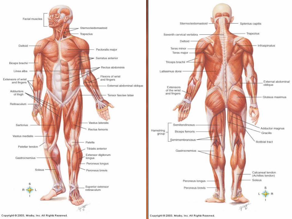
Full muscular system
Full muscular system
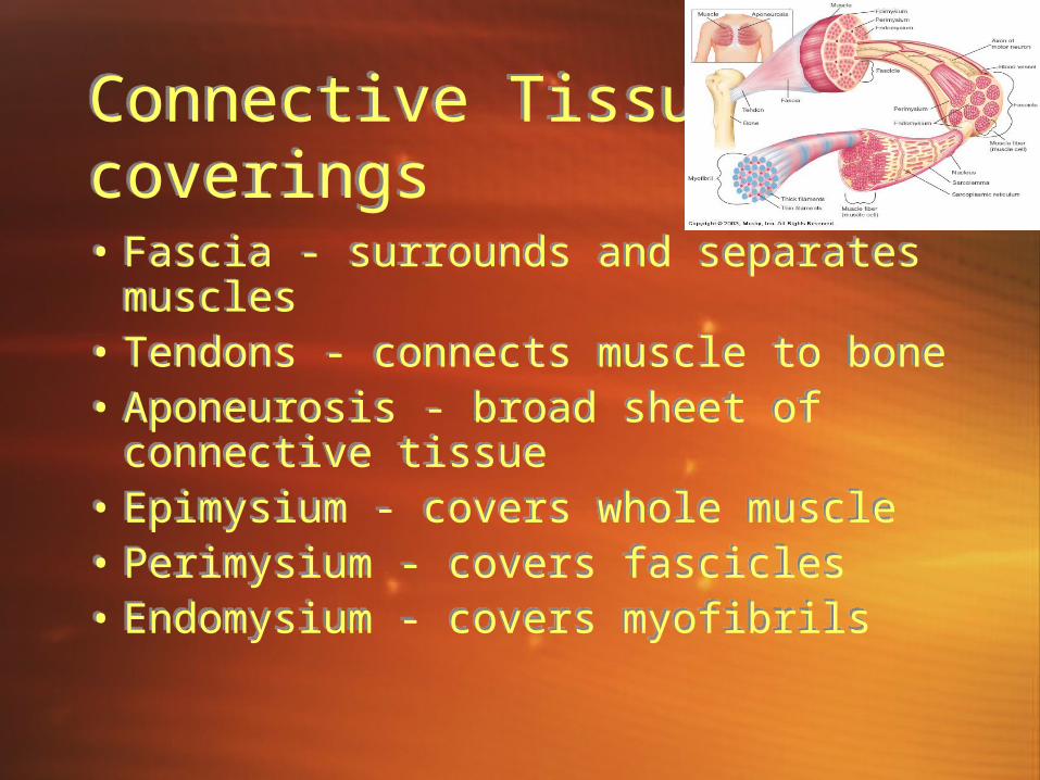
Connective Tissue coveringsConnective Tissue coverings• Fascia - surrounds and separates
muscles• Tendons - connects muscle to bone• Aponeurosis - broad sheet of
connective tissue • Epimysium - covers whole muscle• Perimysium - covers fascicles • Endomysium - covers myofibrils
• Fascia - surrounds and separates muscles
• Tendons - connects muscle to bone• Aponeurosis - broad sheet of
connective tissue • Epimysium - covers whole muscle• Perimysium - covers fascicles • Endomysium - covers myofibrils
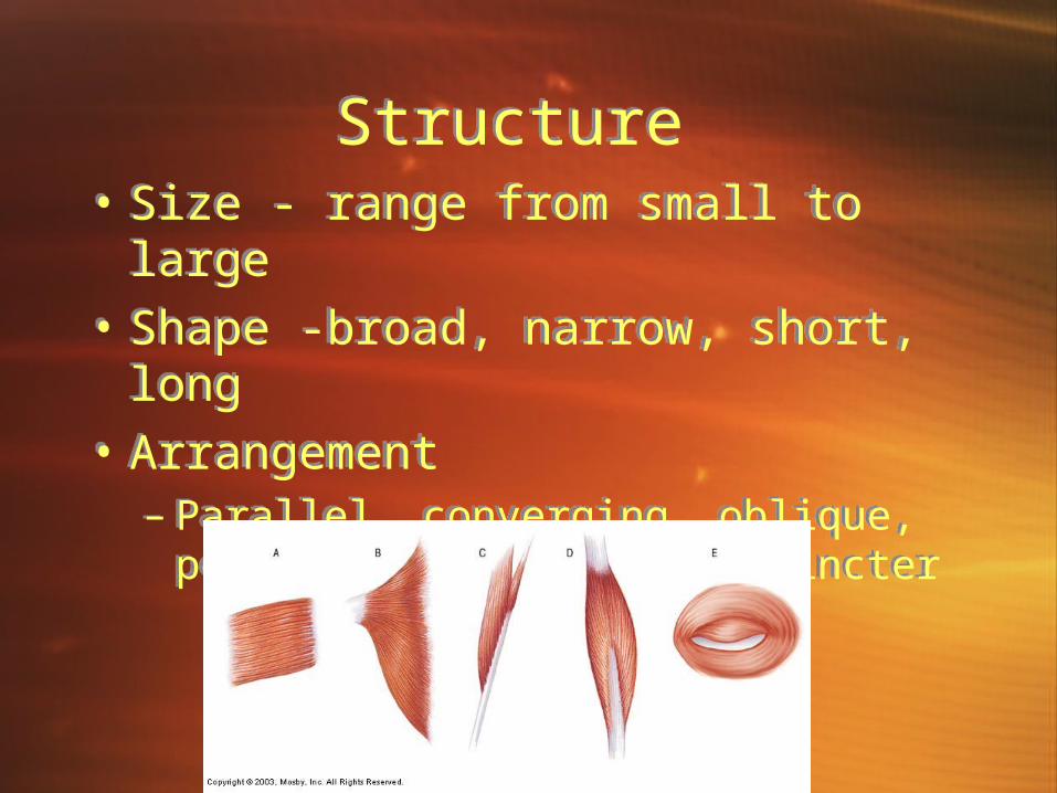
Structure Structure • Size - range from small to large• Shape -broad, narrow, short, long• Arrangement
– Parallel, converging, oblique, pennate, bi- pennate, sphincter
• Size - range from small to large• Shape -broad, narrow, short, long• Arrangement
– Parallel, converging, oblique, pennate, bi- pennate, sphincter
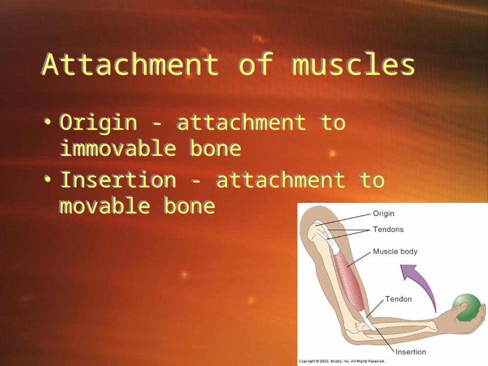
Attachment of musclesAttachment of muscles
• Origin - attachment to immovable bone
• Insertion - attachment to movable bone
• Origin - attachment to immovable bone
• Insertion - attachment to movable bone
Muscle actionsMuscle actions
• Prime movers (agonists) - main action
• Antagonists - opposite action• Synergists - helper muscle
• Prime movers (agonists) - main action
• Antagonists - opposite action• Synergists - helper muscle
The power of muscleThe power of muscle
QuickTime™ and a decompressor
are needed to see this picture.
Lever systemsLever systems
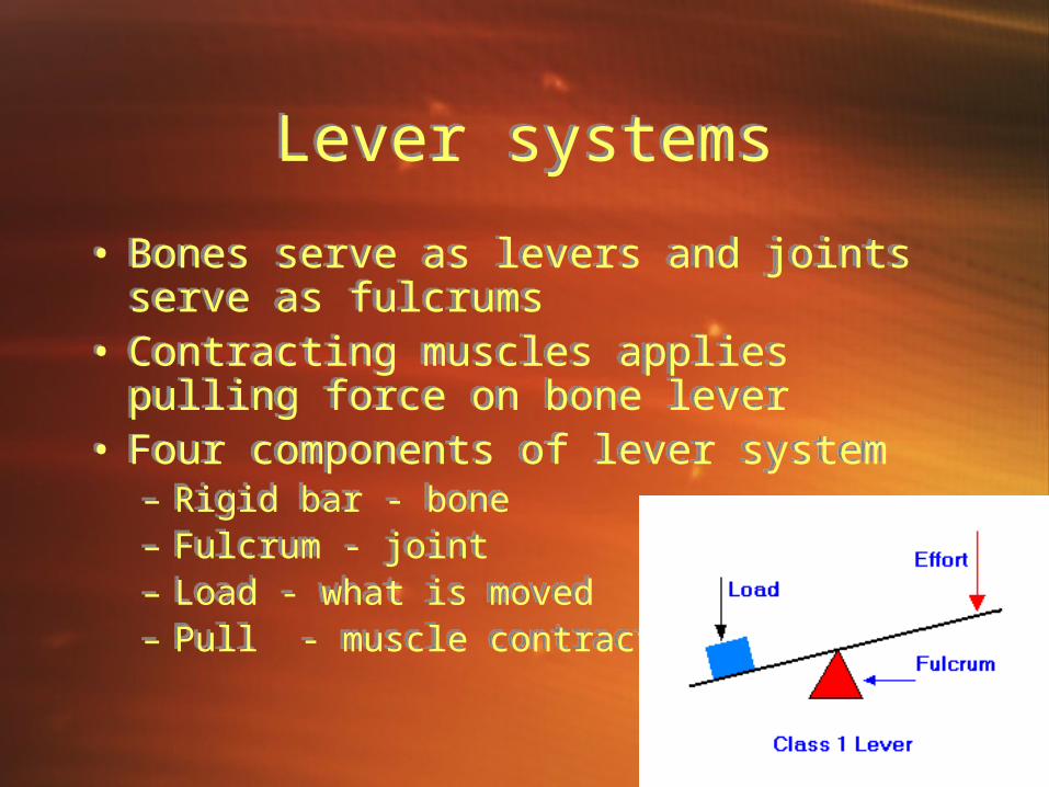
• Bones serve as levers and joints serve as fulcrums
• Contracting muscles applies pulling force on bone lever
• Four components of lever system– Rigid bar - bone– Fulcrum - joint– Load - what is moved– Pull - muscle contraction
• Bones serve as levers and joints serve as fulcrums
• Contracting muscles applies pulling force on bone lever
• Four components of lever system– Rigid bar - bone– Fulcrum - joint– Load - what is moved– Pull - muscle contraction
How muscles are namedHow muscles are named
• Latin based or English• Naming - supply hints as to muscle
action– Location, function, shape– Direction of fibers– Number of heads– Points of attachment– Relative size
• Latin based or English• Naming - supply hints as to muscle
action– Location, function, shape– Direction of fibers– Number of heads– Points of attachment– Relative size

• Epicranius - raises eyebrows• Orbicularis oculi - closes eye• Orbicularis oris - draws lips
together• Buccinator - smiling• Zygomaticus - laughing
• Epicranius - raises eyebrows• Orbicularis oculi - closes eye• Orbicularis oris - draws lips
together• Buccinator - smiling• Zygomaticus - laughing
Muscles of facial
expression
Muscles of facial
expression
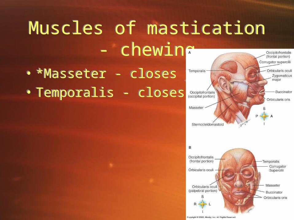
Muscles of mastication - chewing
Muscles of mastication - chewing
• *Masseter - closes jaw• Temporalis - closes jaw
• *Masseter - closes jaw• Temporalis - closes jaw
Muscles that move the head
Muscles that move the head
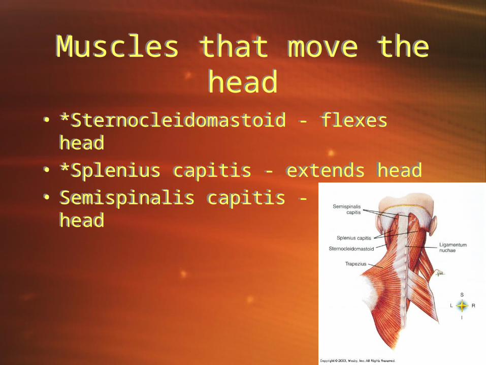
• *Sternocleidomastoid - flexes head• *Splenius capitis - extends head• Semispinalis capitis - extends head
• *Sternocleidomastoid - flexes head• *Splenius capitis - extends head• Semispinalis capitis - extends head
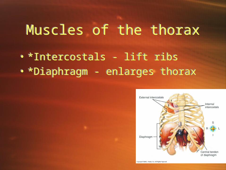
Muscles of the thoraxMuscles of the thorax
• *Intercostals - lift ribs• *Diaphragm - enlarges thorax
• *Intercostals - lift ribs• *Diaphragm - enlarges thorax
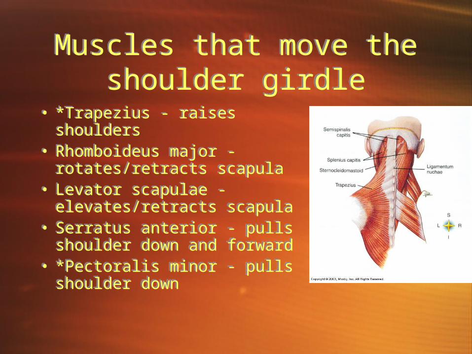
Muscles that move the shoulder girdle
Muscles that move the shoulder girdle
• *Trapezius - raises shoulders
• Rhomboideus major - rotates/retracts scapula
• Levator scapulae - elevates/retracts scapula
• Serratus anterior - pulls shoulder down and forward
• *Pectoralis minor - pulls shoulder down
• *Trapezius - raises shoulders
• Rhomboideus major - rotates/retracts scapula
• Levator scapulae - elevates/retracts scapula
• Serratus anterior - pulls shoulder down and forward
• *Pectoralis minor - pulls shoulder down
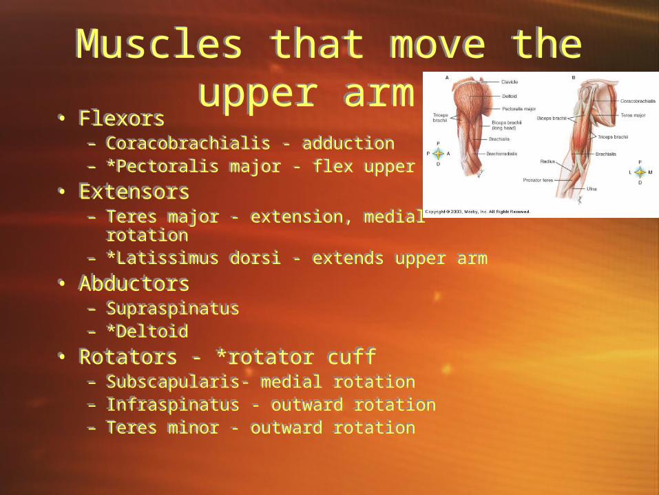
Muscles that move the upper arm
Muscles that move the upper arm
• Flexors– Coracobrachialis - adduction– *Pectoralis major - flex upper arm
• Extensors – Teres major - extension, medial rotation– *Latissimus dorsi - extends upper arm
• Abductors– Supraspinatus– *Deltoid
• Rotators - *rotator cuff– Subscapularis- medial rotation– Infraspinatus - outward rotation– Teres minor - outward rotation
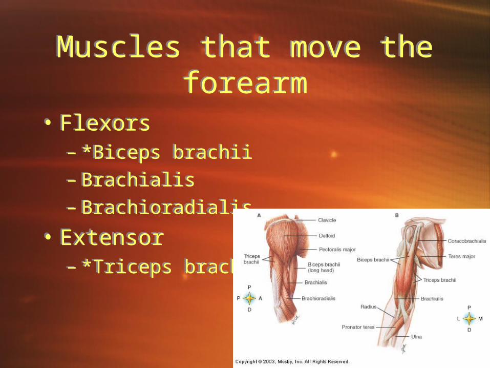
Muscles that move the forearm
Muscles that move the forearm
• Flexors– *Biceps brachii– Brachialis– Brachioradialis
• Extensor– *Triceps brachii
• Flexors– *Biceps brachii– Brachialis– Brachioradialis
• Extensor– *Triceps brachii
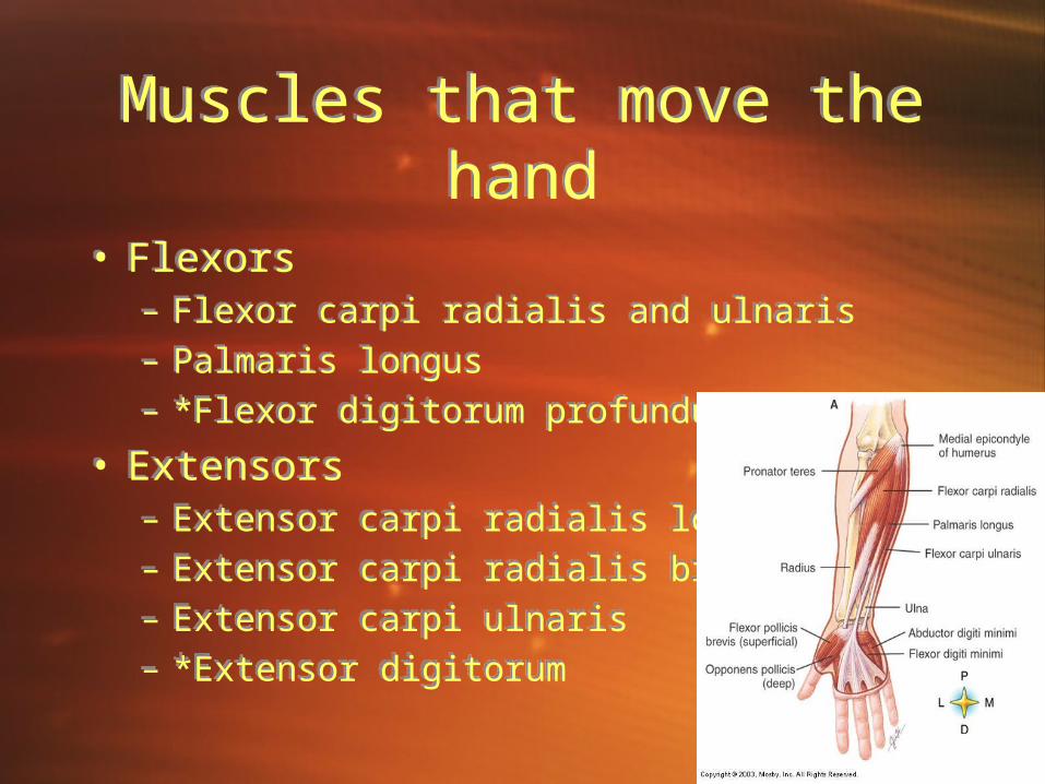
Muscles that move the hand
Muscles that move the hand
• Flexors– Flexor carpi radialis and ulnaris– Palmaris longus– *Flexor digitorum profundus
• Extensors– Extensor carpi radialis longus– Extensor carpi radialis brevis– Extensor carpi ulnaris– *Extensor digitorum
• Flexors– Flexor carpi radialis and ulnaris– Palmaris longus– *Flexor digitorum profundus
• Extensors– Extensor carpi radialis longus– Extensor carpi radialis brevis– Extensor carpi ulnaris– *Extensor digitorum
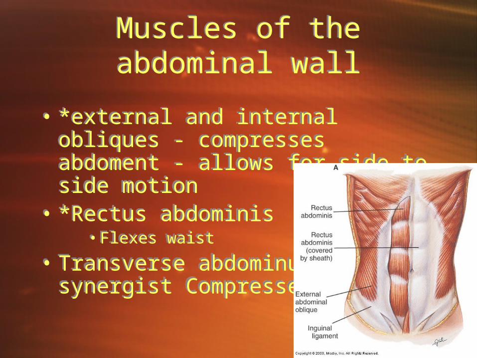
Muscles of the abdominal wall
Muscles of the abdominal wall
• *external and internal obliques - compresses abdoment - allows for side to side motion
• *Rectus abdominis • Flexes waist
• Transverse abdominus synergist Compresses abdomen
• *external and internal obliques - compresses abdoment - allows for side to side motion
• *Rectus abdominis • Flexes waist
• Transverse abdominus synergist Compresses abdomen
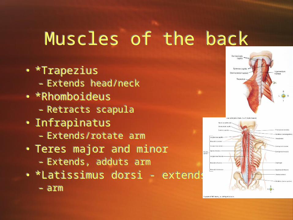
Muscles of the backMuscles of the back
• *Trapezius – Extends head/neck
• *Rhomboideus– Retracts scapula
• Infrapinatus– Extends/rotate arm
• Teres major and minor– Extends, adduts arm
• *Latissimus dorsi - extends– arm
• *Trapezius – Extends head/neck
• *Rhomboideus– Retracts scapula
• Infrapinatus– Extends/rotate arm
• Teres major and minor– Extends, adduts arm
• *Latissimus dorsi - extends– arm
Muscles of the pelvic floorMuscles of the pelvic floor
• Levator ani• Superficial transversus perinei• Bulbospongiosus• Ischiocavernoses
• Levator ani• Superficial transversus perinei• Bulbospongiosus• Ischiocavernoses
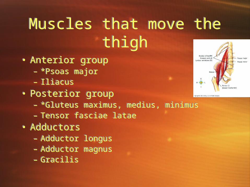
Muscles that move the thigh
Muscles that move the thigh
• Anterior group– *Psoas major– Iliacus
• Posterior group– *Gluteus maximus, medius, minimus– Tensor fasciae latae
• Adductors– Adductor longus– Adductor magnus– Gracilis
• Anterior group– *Psoas major– Iliacus
• Posterior group– *Gluteus maximus, medius, minimus– Tensor fasciae latae
• Adductors– Adductor longus– Adductor magnus– Gracilis
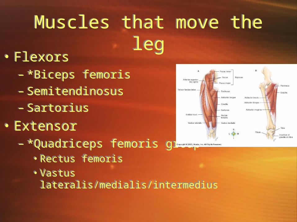
Muscles that move the legMuscles that move the leg• Flexors
– *Biceps femoris – Semitendinosus – Sartorius
• Extensor– *Quadriceps femoris group
• Rectus femoris• Vastus lateralis/medialis/intermedius
• Flexors– *Biceps femoris – Semitendinosus – Sartorius
• Extensor– *Quadriceps femoris group
• Rectus femoris• Vastus lateralis/medialis/intermedius
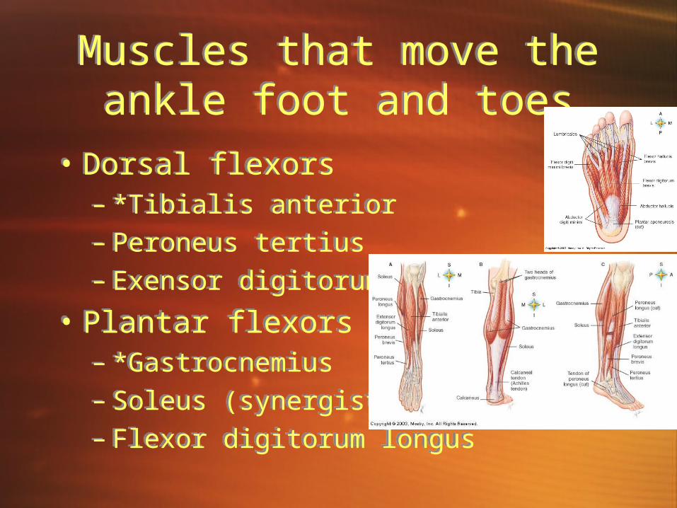
Muscles that move the ankle foot and toes
Muscles that move the ankle foot and toes
• Dorsal flexors– *Tibialis anterior– Peroneus tertius– Exensor digitorum longus
• Plantar flexors– *Gastrocnemius– Soleus (synergist)– Flexor digitorum longus
• Dorsal flexors– *Tibialis anterior– Peroneus tertius– Exensor digitorum longus
• Plantar flexors– *Gastrocnemius– Soleus (synergist)– Flexor digitorum longus
Ch. 11 The physiology of the muscular system
Ch. 11 The physiology of the muscular system

IntroductionIntroduction
• Purpose - move framework of body, produce heat, facilitate posture
• Characteristics – Excitability - ability to be stimulated– Contractibility - ability to shorten
producing movement– Extensibility - ability to stretch and
return to resting length.
• Purpose - move framework of body, produce heat, facilitate posture
• Characteristics – Excitability - ability to be stimulated– Contractibility - ability to shorten
producing movement– Extensibility - ability to stretch and
return to resting length.

Structure of muscle fibersStructure of muscle fibers• Description - each fiber - muscle cell
(spans a joint)• Sarcolemma - cell membrane• Sarcoplasm - cytoplasm of muscle cell
– Contains mitochondria, nuclei, myofibrils
• Myofibrils - filaments– Myosin - thick– Actin - thin
• Sarcomere - unit within myofibril – Extends from z line to z line. – Z lines produce striations
• Sarcoplasmic reticulum (endoplasmic reticulum)– Contains transverse tubules (nerve impulse)
• Description - each fiber - muscle cell (spans a joint)
• Sarcolemma - cell membrane• Sarcoplasm - cytoplasm of muscle cell
– Contains mitochondria, nuclei, myofibrils
• Myofibrils - filaments– Myosin - thick– Actin - thin
• Sarcomere - unit within myofibril – Extends from z line to z line. – Z lines produce striations
• Sarcoplasmic reticulum (endoplasmic reticulum)– Contains transverse tubules (nerve impulse)
Neuromuscular JunctionNeuromuscular Junction• Where neuron and muscle
fiber meet.• Abundant mitochondria
present for ATP production• Neurotransmitters - chemical
communicators located at the end of neuron in the Cytoplasm
• Motor Unit– Mucle fibers contract at once
when triggered by neurotransmitters
– Recruitment - increase in number of motor units activated.
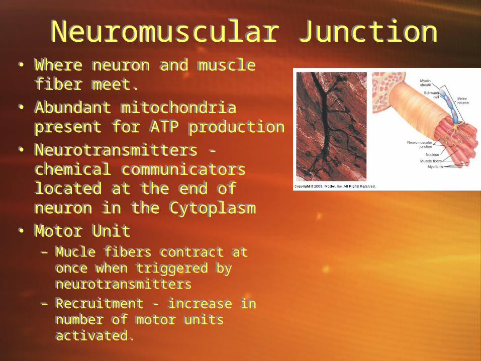
• Where neuron and muscle fiber meet.
• Abundant mitochondria present for ATP production
• Neurotransmitters - chemical communicators located at the end of neuron in the Cytoplasm
• Motor Unit– Mucle fibers contract at once
when triggered by neurotransmitters
– Recruitment - increase in number of motor units activated.

Skeletal Muscle Contraction
Skeletal Muscle Contraction• Shortening of sarcomeres results in
muscle pulling against attachments– Myosin - two twisted strands with
crossbridges– Actin contains myosin binding sites
• Sliding filament theory– Myosin cross-bridge attaches to binding site
on actin filament and bends– Pulls actin filament, releases and attaches
to next binding site, pulling again.– Energy from atp used to prepare the cross-
bridges.
• Shortening of sarcomeres results in muscle pulling against attachments– Myosin - two twisted strands with
crossbridges– Actin contains myosin binding sites
• Sliding filament theory– Myosin cross-bridge attaches to binding site
on actin filament and bends– Pulls actin filament, releases and attaches
to next binding site, pulling again.– Energy from atp used to prepare the cross-
bridges.

Stimulus for contractionStimulus for contraction– Neurotransmitter - Acetylcholine released from synaptic
vesicles at end of axon of neuron.• Note - botulinus toxin prevents acetylcholine release.
– Receptors detect neurotransmitter– Impulse spreads over sarcolemma then travels through
transverse to sarcoplasmic reticulum– Calcium released by sarcoplasmic reticulum– High calcium moves Troponin and tropomyosin aside, exposing
binding site– Myosin Crossbridge attaches to binding site– Crossbridge shortens pulling filaments across each other– Sarcomere shortens– Acetylcholinesterase decomposes acetylcholine– Calcium returns to sarcoplasmic reticulum– Link between actin and myosin is broken.
– Neurotransmitter - Acetylcholine released from synaptic vesicles at end of axon of neuron.
• Note - botulinus toxin prevents acetylcholine release.
– Receptors detect neurotransmitter– Impulse spreads over sarcolemma then travels through
transverse to sarcoplasmic reticulum– Calcium released by sarcoplasmic reticulum– High calcium moves Troponin and tropomyosin aside, exposing
binding site– Myosin Crossbridge attaches to binding site– Crossbridge shortens pulling filaments across each other– Sarcomere shortens– Acetylcholinesterase decomposes acetylcholine– Calcium returns to sarcoplasmic reticulum– Link between actin and myosin is broken.
Muscle contractionMuscle contraction
QuickTime™ and a decompressor
are needed to see this picture.
Oxygen supply and cellular respirationOxygen supply and cellular respiration
• During rest - enough oxygen to support aerobic cellular respiration.
• Oxygen deficiency during exercise– Lactic acid end product of anaerobic respiration. - lactic acid
diffuses out of muscle cells and is carried to liver.
• Oxygen debt - amount of oxygen that liver cells require to convert lactic acid into glucose plus amt. muscle cells need to make atp to original concentration.
• Muscle fatigue-muscle loses ability to contract during strenuous exercise.
• Result of lactic acid accumulation (lower ph)• Muscle cramp - lack of ATP required to return calcium
ions back to sarcoplasmic reticulum so muscle fibers can relax.
• Heat production - energy produced by cellular respiration is lost as heat.

Muscular responsesMuscular responses
• One method of studying muscle function - remove single fiber and connect to device that records response to electrical stimuli.
• Muscle fibers remain unresponsive till they reach Threshold stimulus (stimulus of a certain strength)
• All or none response• Summation - series of stimuli of increasing frequency• Recruitment of motor units-increase in number of activated
motor units• Sustained contractions - muscle tone - achieved by continuous
state of sustained contraction.• Treppe-staircase phenomenon - twitch contractions 1 second
apart• Tetanus-multiple wave summation/no relaxation
Recording a muscular contraction
Recording a muscular contraction
• Myogram - recording of electrically stimulated muscle contraction
• Myograph - machine that records the contraction
• Twitch-single short contraction• Latent period - time delay followed by
period of contraction and relaxation.
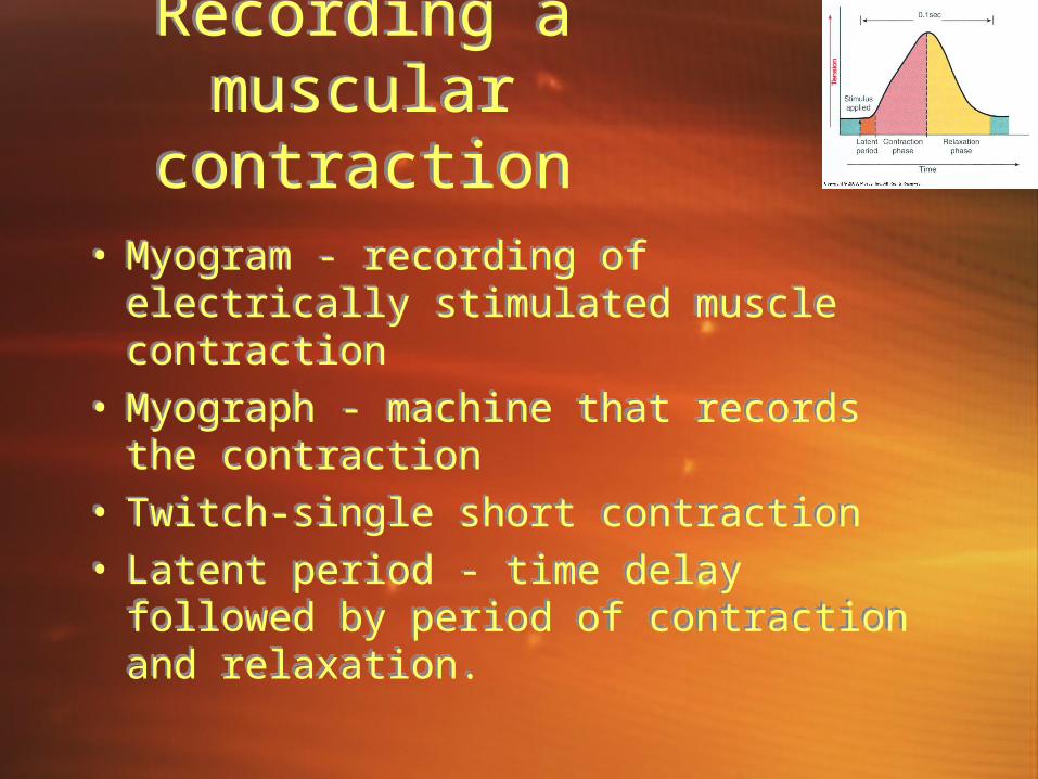
• Myogram - recording of electrically stimulated muscle contraction
• Myograph - machine that records the contraction
• Twitch-single short contraction• Latent period - time delay followed by
period of contraction and relaxation.

Muscle toneMuscle tone
• Tonic contraction-continual partial contraction of a muscle
• Flaccid - less tone than normal• Spastic-more tone than normal• Negative feedback mechanism controls tone• Graded strength - affected by:
– Metabolic condition of fibers– Number of fibers contracting– Number of motor units recruited
• Tonic contraction-continual partial contraction of a muscle
• Flaccid - less tone than normal• Spastic-more tone than normal• Negative feedback mechanism controls tone• Graded strength - affected by:
– Metabolic condition of fibers– Number of fibers contracting– Number of motor units recruited

Physical trainingPhysical training
• Strength training- results in hypertrophy - enlargement of fibers
• Endurance training - increases ability to sustain moderate contractions for longer time. Increase of mitochondria.
• Atrophy - result of disuse– Decreased capillary networks, decreased
mitochondria, decreased filaments.
• Strength training- results in hypertrophy - enlargement of fibers
• Endurance training - increases ability to sustain moderate contractions for longer time. Increase of mitochondria.
• Atrophy - result of disuse– Decreased capillary networks, decreased
mitochondria, decreased filaments.
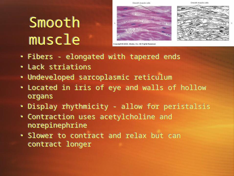
Smooth muscleSmooth muscle
• Fibers - elongated with tapered ends• Lack striations• Undeveloped sarcoplasmic reticulum• Located in iris of eye and walls of hollow
organs• Display rhythmicity - allow for peristalsis• Contraction uses acetylcholine and
norepinephrine• Slower to contract and relax but can contract
longer
• Fibers - elongated with tapered ends• Lack striations• Undeveloped sarcoplasmic reticulum• Located in iris of eye and walls of hollow
organs• Display rhythmicity - allow for peristalsis• Contraction uses acetylcholine and
norepinephrine• Slower to contract and relax but can contract
longer
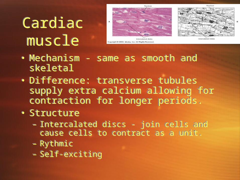
Cardiac muscleCardiac muscle
• Mechanism - same as smooth and skeletal
• Difference: transverse tubules supply extra calcium allowing for contraction for longer periods.
• Structure– Intercalated discs - join cells and cause cells
to contract as a unit.– Rythmic– Self-exciting
• Mechanism - same as smooth and skeletal
• Difference: transverse tubules supply extra calcium allowing for contraction for longer periods.
• Structure– Intercalated discs - join cells and cause cells
to contract as a unit.– Rythmic– Self-exciting