cervical spine trauma
DESCRIPTION
cervical spine traumaTRANSCRIPT

Indian J Orthop. 2007 Oct-Dec; 41(4): 255–267.
doi: 10.4103/0019-5413.36985
PMCID: PMC2989526
Cervical spine trauma
Joel A Torretti and Dilip K Sengupta
University Orthopedics Center 101 Regent Ct State College, PA 16801, USA
Department of Orthopaedic Surgery, Dartmouth-Hitchcock Medical Center, One Medical Center Drive, Lebanon
Correspondence: Dr. Dilip K. Sengupta, Department of Orthopaedic Surgery, Dartmouth-Hitchcock Medical Center, One Medical Center
Drive, Lebanon, E-mail: [email protected]
Copyright © Indian Journal of Orthopaedics
This is an open-access article distributed under the terms of the Creative Commons Attribution License, which permits unrestricted use,
distribution, and reproduction in any medium, provided the original work is properly cited.
Abstract
Cervical spine trauma is a common problem with a wide range of severity from minor ligamentous injuryto frank osteo-ligamentous instability with spinal cord injury. The emergent evaluation of patients at riskrelies on standardized clinical and radiographic protocols to identify injuries; elucidate associatedpathology; classify injuries; and predict instability, treatment and outcomes. The unique anatomy of eachregion of the cervical spine demands a review of each segment individually. This article examines bothupper cervical spine injuries, as well as subaxial spine trauma. The purpose of this article is to provide areview of the broad topic of cervical spine trauma with reference to the classic literature, as well as tosummarize all recently available literature on each topic.
Identification of References for Inclusion:
A Pubmed and Ovid search was performed for each topic in the review to identify recently publishedarticles relevant to the review. In addition prior reviews and classic references were evaluated individuallyfor inclusion of classic papers, classifications and previously unidentified references.
Keywords: Cervical spine injuries, subaxial cervical spine trauma, upper cervical spine trauma
E
It has been reported that the cervical spine is injured in 2.4% of blunt trauma victims. Certain
demographic factors are known to be associated with blunt cervical spine injury: age greater than 65 years,male sex and white ethnicity. To date, only one population-based study of spinal column injuries has been
performed in a complete population. Hu et al. reported on patients in the Manitoba Health Insurance Planfrom 1981–1984. The annual incidence rate was 64/100,000 with two peaks, one in the second and third
decade of the male population and another in elderly females. The most common mechanism of injury wasnoted to be accidental falls, with motor vehicle/transport injuries being the second most common. Inanother study, which is the largest multi-center trial to date, the most common site of injury was theatlantoaxial region, with the most commonly injured levels in the subaxial cervical spine being C6 and C7.
One-third of the injuries identified in this study were considered clinically insignificant. Despite thissurprising number of clinically minor injuries, the cervical spine remains the most common level for spinalcord injury (SCI), representing 55% of all SCIs.
*
*
1
2
3
4
5
Cervical spine trauma http://www.ncbi.nlm.nih.gov/pmc/articles/PMC2989526/?report=printable
1 of 23 6/2/2015 9:36 PM
Clinical evaluation/missed injuries
The reported frequency of missed injuries in the cervical spine varies from 4% to 30%. The most
common reason cited for missed injuries is an inadequate radiographic examination. Characteristic injury
patterns which are commonly missed include odontoid, teardrop, facet and hangman's fractures. Despite
these common patterns, it has been recognized that even in the absence of fractures, clinically significantinstability can exist. Spinal cord injury without radiographic abnormality has been found to occur in 0.08%of adults with blunt cervical spine trauma. When injuries are missed on initial assessment, a delay in
diagnosis occurs that puts the patient at risk for progressive instability and neurologic deterioration. In oneseries by Davis et al., 29% of patients with missed injuries developed permanent neurologic sequelae. It
is clear that a systematic approach to the evaluation of suspected cervical spine injuries is important toavoid these pitfalls.
Current protocols for evaluation of suspected cervical spine injury combine information from the history,clinical examination and radiographic evaluation to predict the presence of instability, identify neurologicaldeficits and guide the need for intervention. During the course of evaluation, patients should be maintainedin a supine position with rigid collar immobilization or other stable neutral immobilization, while standardAdvanced Trauma Life Support protocols are performed. The immediate clinical examination of the spineshould include inspection and palpation of the spine, as well as a complete neurological examination. Inaddition, a cranial nerve examination should always be performed. Cranial nerve (CN) palsies related toCNs VI, VII, IX, X, XI and XII can occur in association with upper cervical spine injuries.
Clinical examination of patients, although critical to all initial evaluation protocols, has severe limitations,with a sensitivity of 77% in blunt trauma patients in one series. To adequately assess a patient, he/ she
must be awake and alert, nonintoxicated and without distracting injury. Patients specifically at risk forcervical spine injury include those with facial fractures/ trauma, closed head injury and blunt/ penetratingneck injury. The severity/ violence of the injury mechanism, as well as its Injury Severity Score (ISS), areimportant to consider and are more predictive of a significant cervical injury than other frequentlydescribed factors. Clinical protocols for determining the need for radiography have been developed, such
as the NEXUS Low Risk Criteria and the Canadian C-spine Rule, which are used to aid in emergency roomtriage. A recent large prospective cohort study demonstrated superiority of the Canadian C-spine Rule overthe NEXUS criteria with regard to sensitivity/ specificity and reducing the incidence of unwarrantedradiography.
Radiographic evaluation
Once the initial trauma evaluation has been performed, it is imperative that an appropriate radiographicevaluation be obtained. The type of radiographic assessment has evolved as imaging techniques haveadvanced, with resultant changes in what is considered the current standard of care in many institutions. Inthe past, plain radiographs which included lateral, AP and odontoid views were considered the standard ofcare for initial trauma evaluation. Although relatively inexpensive and easy to obtain, they provided poorvisualization of the craniocervical and cervicothoracic junction and resulted in missed injury rates of15-30% in some studies. Spiral CT is widely available and has largely supplanted the role of
traditional plain radiography in many institutions. The ability of spiral CT to detect subtle injuries has beendemonstrated to be superior to plain radiography. Spiral CT has been found to have a sensitivity of 99%
and specificity of 100%, with the risk of missed spine injury being 0.04%; while plain film radiography
has a sensitivity of 70%. Spiral CT has also been shown to decrease the time in the radiology department.
The cost-effectiveness of utilizing spiral CT has also been evaluated, and it has been shown to be at leastequivalent, if not superior, to plain film radiography. The advantages of cervical spine spiral CT are
especially apparent in the evaluation of regions poorly visualized by plain films, such as the
6,7
8
9
10
11
12,13
14
15
16
17,18
19
20
21
21–23
Cervical spine trauma http://www.ncbi.nlm.nih.gov/pmc/articles/PMC2989526/?report=printable
2 of 23 6/2/2015 9:36 PM

occipitocervical region, the facets/ lamina and the cervicothoracic junction. For these reasons, it is theauthors' opinion that spiral CT is the preferred method for evaluation of suspected cervical spine injuries inthe acute setting.
When evaluating radiographs of patients with suspected subaxial cervical trauma, key radiographic featuresneed to be assessed: the presence of soft tissue swelling anterior to the vertebral bodies; a loss of thenormal smooth cervical lordosis with special attention to the normal lordotic lines; disc space narrowing;segmental kyphosis; antero/retrolisthesis of one vertebral body relative to another and/or splaying of thespinous processes. The information from these evaluations provides indirect assessments of spinal stability.Stability of the spine has been defined by White and Panjabi as “the ability of the spine under physiologicloads to maintain an association between vertebral segments in such a way that there is neither damage norsubsequent irritation of the spinal cord or nerve roots and, in addition, there is no development ofincapacitating deformity or pain due to structural changes.” Given this framework, they have provided ascoring system that has been widely adopted in predicting the presence of instability on cervicalradiographs with evidence of segmental kyphosis greater than 11 degrees and anterolisthesis greater than3.5 mm of one vertebral body on another as strong indicators of instability. In addition to considerations
of stability, a suspicion of noncontiguous spinal injury must be maintained in all patients with knowncervical injuries, given a reported incidence of 15-20% of noncontiguous spinal injury, with evaluation
of the remaining spine being performed according to individual institutional protocol.
Classification
Classification systems have been developed in an attempt to predict instability, standardize the discussionof injury types and provide a means for applying a consistent approach to these injuries. These systems aredivided between the upper cervical spine and subaxial cervical spine. Due to its unique anatomy the uppercervical spine has its own classifications unique to each injury pattern and level. The subaxial cervicalspine has a variety of systems which each attempt to provide a comprehensive approach. One of the firstand the most commonly employed systems for classifying injuries to the subaxial cervical spine is theAllen and Ferguson classification, which provides a mechanistic classification. It was proposed based
upon a retrospective review of a single author's case series from 1960 to 1974. It included a description of a“major injury vector,” the inferred applied force based upon the interpretation of static radiographs, as wellas a descriptive phylogeny within each vector to clarify the spectrum of injury. This resulted in the creationof six injury types: compression-flexion, compression-extension, vertical-compression, distraction-flexion,distraction-extension and lateral flexion. Each of these has a subclassification of injury types based upontheir characteristic patterns.
A similar mechanistic approach was proposed by the application of the AO thoracolumbar fractureclassification to the subaxial cervical spine. This was also based upon the review of an author's case
series; and despite a few basic differences in some of the fracture patterns and predictions of instability, itwas found to provide a reasonable framework for classification of these diverse injuries. This system
groups the injury patterns into three major groups: type A (14.7%), which are “compression only” lesionsof the vertebral body; type B (43.9%), which are flexion-extension-distraction injuries (representinganterior and posterior element injury with distraction); and type C (41.2%), which are rotational injuries.Despite type-A injury pattern being the predominant pattern in thoracolumbar injuries, it is the leastcommon pattern in the subaxial cervical spine. The most common patterns are types B and C.
More recently there is a novel system proposed by Moore et al., which adopts a morphologic, rather than
a mechanistic, approach to the classification of the injury. They divide the spine into four columns: theanterior column; the lateral columns, consisting of the lateral masses and paired facets; and the posteriorcolumn, the posterior bony arch and supporting ligaments. Each column is then scored based upon the
24
25,26
27
28
29
30
Cervical spine trauma http://www.ncbi.nlm.nih.gov/pmc/articles/PMC2989526/?report=printable
3 of 23 6/2/2015 9:36 PM
severity of injury. Their scoring system was found to be reproducible in their initial case series, althoughthe utility of this approach will need to be evaluated in further studies.
Currently, the Allen and Ferguson classification is the most commonly employed method and the mostpractical in clinical applications for the cervical spine, where it provides a framework for interpretation ofthe injury mechanism and anticipated instability. Most surgeons consider injuries within the context ofthese classification heuristics but do not depend on their specific subclassification to predict the treatment.Rather, they use a combination of the predicted stability of an injury, the presence of neurological deficitand the injury pattern to guide their management decisions.
Functional anatomy
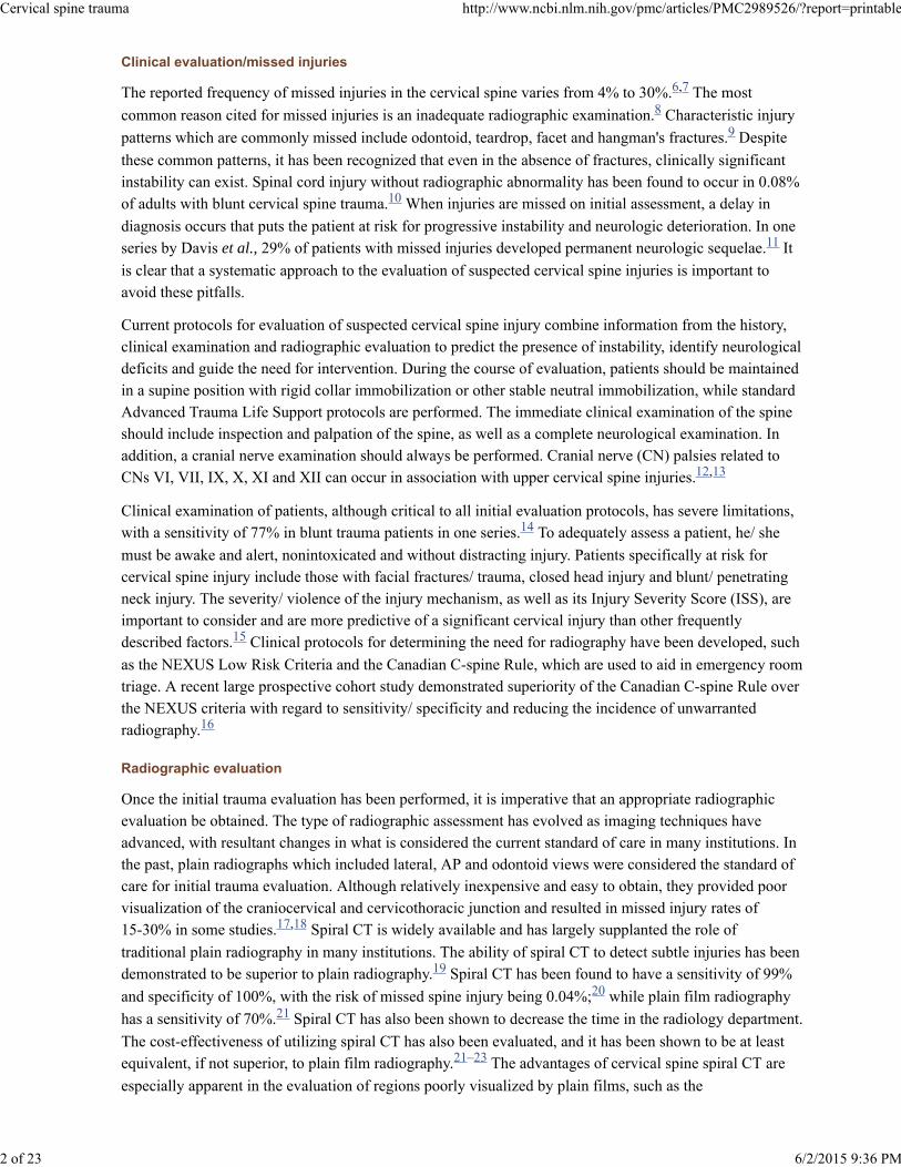
The upper cervical spine has unique anatomic features that distinguish it from the remainder of the cervicalspine [Figure 1]. Its motion segments make up a large amount of total cervical spine motion and, as aresult, predispose it to a unique set of injuries. The occipitoatlantal range of motion is 25 degrees inflexion-extension and 5 degrees in rotation, whereas the atlantoaxial range of motion makes up a largeproportion of the cervical motion in rotation, accounting for approximately 40-50% of cervical rotation.The atlantoaxial articulation also contributes 20 degrees to the flexion-extension range of motion. The
occipital condyles project laterally from the base of the skull and articulate with the facets of the atlas (C1),forming paired synovial joints. The atlas has rather shallow facets, resulting in less constraint than otherfacet complexes; but their capsules are reinforced by the paired alar ligaments, which are unique to thislevel. The alar ligaments extend cephalad from the dens to the medial aspect of the occipital condyles aspaired structures. They limit the axial rotation, as well as the side bending of the occipitoatlantoaxialcomplex. The cephalad extension of the anterior longitudinal ligament (ALL) and the posterior
longitudinal ligament (PLL) also provides ligamentous constraint to the occipitocervical complex with theircontiguous extensions, which are called the anterior atlanto-occipital membrane and tectorial membranerespectively. The atlas is also constrained by the attachments of the ALL and longus colli musclesanteriorly and the ligamentum nuchae posteriorly. The vertebral arteries are paired structures which exit theforamen transversarium cephalad to C1 and travel along the rostral aspect of its posterior arch in a groovebefore traveling medially and cephalad into the foramen magnum. The atlas articulates anteriorly with thedens via a synovial articulation which is reinforced by the transverse ligament. This ligament is the primaryconstraint against anterior translation of C1 on C2. The atlas also articulates with the axis via pairedsynovial facet joints, which have capsules that contribute to their stability. It is the unique anatomy of
these vertebrae that allows for their coupled motion in rotation, flexion-extension and lateral bending,whilst protecting the spinal cord, paired vertebral arteries and cranial nerves as they traverse this region.
The subaxial cervical spine has consistent anatomic features between its levels until the cervicothoracicjunction, where there is a transition from a relatively mobile segment to a rigid one. The anterior columnprovides support and stability, which is a function of the vertebral bodies, intervertebral discs and theattachments of the ALL and PLL. The vertebral body carries two-thirds of the vertebral load. The
posterior bony elements include the lamina, facets and spinous processes. They provide attachments for thecapsuloligamentous structures, which include the supraspinous and interspinous ligaments, ligamentumflavum and facet capsules. These structures contribute to stability by providing resistance to tensile forcesand are commonly described as creating the posterior tension band. The facet joints provide the primaryrestraint against anterior subluxation. The range of motion in the lower cervical spine is greatest at C4-5and C5-6, although the relative contribution of each level is fairly evenly distributed amongst all the levels.The transverse foramen is located in the transverse processes and provides the conduit for the vertebralarteries bilaterally, beginning at C6.
The anatomic features of each cervical motion segment predispose the levels to different patterns of injury
31
32
33
34
Cervical spine trauma http://www.ncbi.nlm.nih.gov/pmc/articles/PMC2989526/?report=printable
4 of 23 6/2/2015 9:36 PM
Occipitocervical dissociation
Occipital condyle fractures
and, as a result, require an evaluation of each individually.
Upper Cervical Spine Injuries (Base of skull-C2)
Occipitocervical dissociation is an uncommon injury which can be difficult toidentify and has a high mortality. The most common mechanism of injury is that of a pedestrian struck by acar, with a high incidence in pediatric patients. Various radiographic parameters have been described fordetermining the presence of occipitoatlantal subluxation/ dislocation, with the most wellknown methodbeing the Power's ratio (BC/ OA). This is calculated as the ratio of the distance from the basion (B) to the
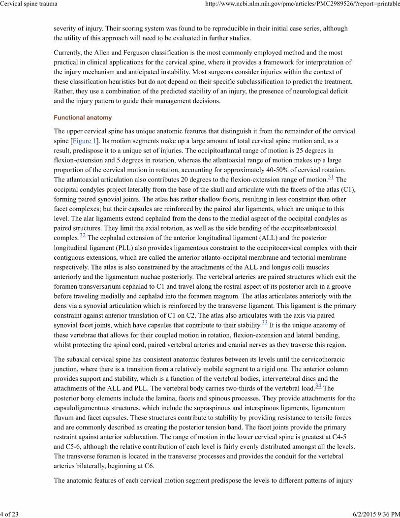
posterior arch (C) of C1 to the distance from the anterior arch of C1 (A) to the opisthion (O). A ratio of BC/OA greater than 1.0 indicates anterior subluxation. A more reliable method for assessing the presence ofcraniovertebral dislocation is the Harris rule of 12's. Harris described a line drawn cephalad from the
posterior body of C2 (posterior axial line). The distance from the basion to the posterior axial line(basion-axis interval) and the distance from the basion to the tip of the dens (basion-dens interval) shouldeach be less than 12 mm [Figure 2]. An increase in this distance indicates instability. The classification ofthese injuries is based upon the displacement of the occiput. Type I injuries are anterior subluxations andare the most common. Type II injuries have vertical distraction greater than 2 mm of the atlanto-occipitaljoint. Type III injuries are posterior dislocations and are rarely reported. Once an injury is identified,
prompt management is of the utmost importance. Traction is contraindicated. Treatment consists ofimmediate halo vest application with reduction of the subluxation and confirmation by CT scanning. Anocciput-to-C2 fusion is required in most cases to provide longterm stability. This can be accomplished byuse of a wiring technique, such as the Bohlman Wire technique, although this requires the use of a halopostoperatively. Another option is to perform rigid fixation with a plate-screw construct or a screw-rodconstruct. Various systems are now available that allow rod attachment to occipital plates, with case reportsof their utility. The advantage of these methods is that immobilization in a rigid cervical collar is all that
is required. A method of fixation from the occiput to the lateral mass of the atlas has been described butawaits further evaluation.
Occipital condyle fractures have previously been viewed as relativelyuncommon injuries; but with the increased utilization of CT scanning with reconstructions in the evaluationof suspected spine trauma patients, an increased incidence has been noted. It has been reported to occur in3-15% of trauma patients. The most commonly employed classification system for these injuries is
that proposed by Anderson and Montesano. They described three types. Type I is an impaction fracture,
which is a result of axial loading and lateral bending. This injury is not considered to be unstable. Type II isa basilar skull fracture that extends into the occipital condyle. This is also a stable injury, given that the alarligaments and the tectorial membrane are intact. A type III occipital condyle fracture is a tension injury,resulting in an avulsion of the occipital condyle. If there is associated disruption of the alar ligaments andtectorial membrane, then the potential for instability exists. For this reason, a type III fracture is consideredpotentially unstable. Type I and II fractures are typically treated conservatively with immobilization in arigid cervical collar for 6–8 weeks. Type III fractures should be treated with halo-vest immobilization ifthere is a suspicion of ligamentous instability, although this can be difficult to determine accurately in someinjuries. If there is evidence of craniovertebral subluxation, some authors advocate immediateocciput-to-C2 fusion.
Atlas fractures
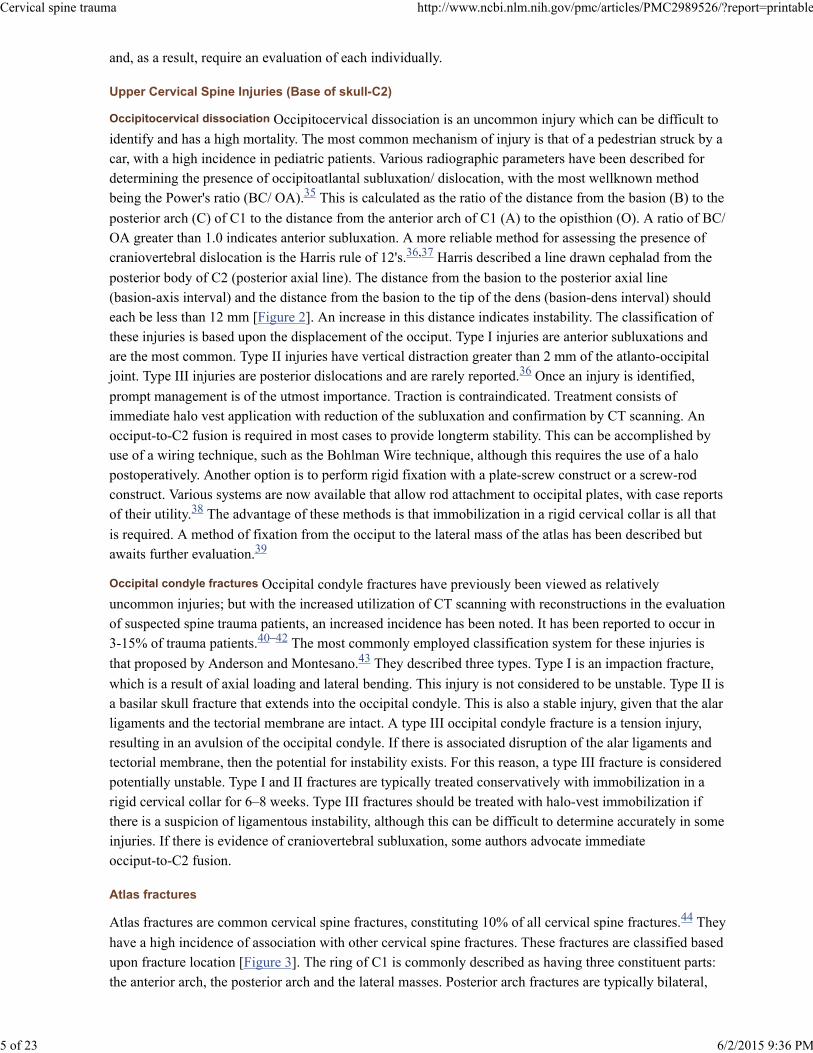
Atlas fractures are common cervical spine fractures, constituting 10% of all cervical spine fractures. They
have a high incidence of association with other cervical spine fractures. These fractures are classified basedupon fracture location [Figure 3]. The ring of C1 is commonly described as having three constituent parts:the anterior arch, the posterior arch and the lateral masses. Posterior arch fractures are typically bilateral,
35
36,37
36
38
39
40–42
43
44
Cervical spine trauma http://www.ncbi.nlm.nih.gov/pmc/articles/PMC2989526/?report=printable
5 of 23 6/2/2015 9:36 PM
Atlantoaxial rotatory instability
Atlantodens instability
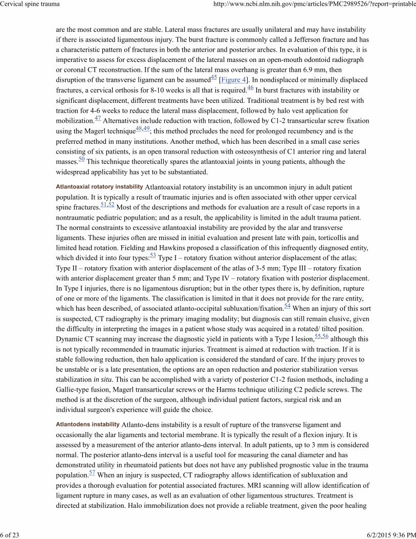
are the most common and are stable. Lateral mass fractures are usually unilateral and may have instabilityif there is associated ligamentous injury. The burst fracture is commonly called a Jefferson fracture and hasa characteristic pattern of fractures in both the anterior and posterior arches. In evaluation of this type, it isimperative to assess for excess displacement of the lateral masses on an open-mouth odontoid radiographor coronal CT reconstruction. If the sum of the lateral mass overhang is greater than 6.9 mm, thendisruption of the transverse ligament can be assumed [Figure 4]. In nondisplaced or minimally displaced
fractures, a cervical orthosis for 8-10 weeks is all that is required. In burst fractures with instability or
significant displacement, different treatments have been utilized. Traditional treatment is by bed rest withtraction for 4-6 weeks to reduce the lateral mass displacement, followed by halo vest application formobilization. Alternatives include reduction with traction, followed by C1-2 transarticular screw fixation
using the Magerl technique ; this method precludes the need for prolonged recumbency and is the
preferred method in many institutions. Another method, which has been described in a small case seriesconsisting of six patients, is an open transoral reduction with osteosynthesis of C1 anterior ring and lateralmasses. This technique theoretically spares the atlantoaxial joints in young patients, although the
widespread applicability has yet to be substantiated.
Atlantoaxial rotatory instability is an uncommon injury in adult patientpopulation. It is typically a result of traumatic injuries and is often associated with other upper cervicalspine fractures. Most of the descriptions and methods for evaluation are a result of case reports in a
nontraumatic pediatric population; and as a result, the applicability is limited in the adult trauma patient.The normal constraints to excessive atlantoaxial instability are provided by the alar and transverseligaments. These injuries often are missed in initial evaluation and present late with pain, torticollis andlimited head rotation. Fielding and Hawkins proposed a classification of this infrequently diagnosed entity,which divided it into four types: Type I – rotatory fixation without anterior displacement of the atlas;
Type II – rotatory fixation with anterior displacement of the atlas of 3-5 mm; Type III – rotatory fixationwith anterior displacement greater than 5 mm; and Type IV – rotatory fixation with posterior displacement.In Type I injuries, there is no ligamentous disruption; but in the other types there is, by definition, ruptureof one or more of the ligaments. The classification is limited in that it does not provide for the rare entity,which has been described, of associated atlanto-occipital subluxation/fixation. When an injury of this sort
is suspected, CT radiography is the primary imaging modality; but diagnosis can still remain elusive, giventhe difficulty in interpreting the images in a patient whose study was acquired in a rotated/ tilted position.Dynamic CT scanning may increase the diagnostic yield in patients with a Type I lesion, although this
is not typically recommended in traumatic injuries. Treatment is aimed at reduction with traction. If it isstable following reduction, then halo application is considered the standard of care. If the injury proves tobe unstable or is a late presentation, the options are an open reduction and posterior stabilization versusstabilization in situ. This can be accomplished with a variety of posterior C1-2 fusion methods, including aGallie-type fusion, Magerl transarticular screws or the Harms technique utilizing C2 pedicle screws. Themethod is at the discretion of the surgeon, although individual patient factors, surgical risk and anindividual surgeon's experience will guide the choice.
Atlanto-dens instability is a result of rupture of the transverse ligament andoccasionally the alar ligaments and tectorial membrane. It is typically the result of a flexion injury. It isassessed by a measurement of the anterior atlanto-dens interval. In adult patients, up to 3 mm is considerednormal. The posterior atlanto-dens interval is a useful tool for measuring the canal diameter and hasdemonstrated utility in rheumatoid patients but does not have any published prognostic value in the traumapopulation. When an injury is suspected, CT radiography allows identification of subluxation and
provides a thorough evaluation for potential associated fractures. MRI scanning will allow identification ofligament rupture in many cases, as well as an evaluation of other ligamentous structures. Treatment isdirected at stabilization. Halo immobilization does not provide a reliable treatment, given the poor healing
45
46
47
48,49
50
51,52
53
54
55,56
57
Cervical spine trauma http://www.ncbi.nlm.nih.gov/pmc/articles/PMC2989526/?report=printable
6 of 23 6/2/2015 9:36 PM
potential of these injuries. For this reason, it is recommended that individuals with significant instability
undergo a C1-2 fusion using one of the aforementioned methods.
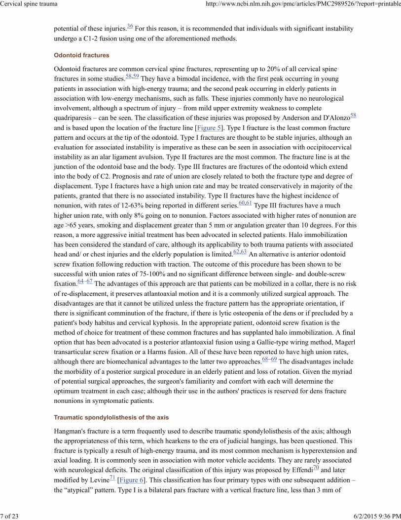
Odontoid fractures
Odontoid fractures are common cervical spine fractures, representing up to 20% of all cervical spinefractures in some studies. They have a bimodal incidence, with the first peak occurring in young
patients in association with high-energy trauma; and the second peak occurring in elderly patients inassociation with low-energy mechanisms, such as falls. These injuries commonly have no neurologicalinvolvement, although a spectrum of injury – from mild upper extremity weakness to completequadriparesis – can be seen. The classification of these injuries was proposed by Anderson and D'Alonzo
and is based upon the location of the fracture line [Figure 5]. Type I fracture is the least common fracturepattern and occurs at the tip of the odontoid. Type I fractures are thought to be stable injuries, although anevaluation for associated instability is imperative as these can be seen in association with occipitocervicalinstability as an alar ligament avulsion. Type II fractures are the most common. The fracture line is at thejunction of the odontoid base and the body. Type III fractures are fractures of the odontoid which extendinto the body of C2. Prognosis and rate of union are closely related to both the fracture type and degree ofdisplacement. Type I fractures have a high union rate and may be treated conservatively in majority of thepatients, granted that there is no associated instability. Type II fractures have the highest incidence ofnonunion, with rates of 12-63% being reported in different series. Type III fractures have a much
higher union rate, with only 8% going on to nonunion. Factors associated with higher rates of nonunion areage >65 years, smoking and displacement greater than 5 mm or angulation greater than 10 degrees. For thisreason, a more aggressive initial treatment has been advocated in selected patients. Halo immobilizationhas been considered the standard of care, although its applicability to both trauma patients with associatedhead and/ or chest injuries and the elderly population is limited. An alternative is anterior odontoid
screw fixation following reduction with traction. The outcome of this procedure has been shown to besuccessful with union rates of 75-100% and no significant difference between single- and double-screwfixation. The advantages of this approach are that patients can be mobilized in a collar, there is no risk
of re-displacement, it preserves atlantoaxial motion and it is a commonly utilized surgical approach. Thedisadvantages are that it cannot be utilized unless the fracture pattern has the appropriate orientation, ifthere is significant comminution of the fracture, if there is lytic osteopenia of the dens or if precluded by apatient's body habitus and cervical kyphosis. In the appropriate patient, odontoid screw fixation is themethod of choice for treatment of these common fractures and has supplanted halo immobilization. A finaloption that has been advocated is a posterior atlantoaxial fusion using a Gallie-type wiring method, Magerltransarticular screw fixation or a Harms fusion. All of these have been reported to have high union rates,although there are biomechanical advantages to the latter two approaches. The disadvantages include
the morbidity of a posterior surgical procedure in an elderly patient and loss of rotation. Given the myriadof potential surgical approaches, the surgeon's familiarity and comfort with each will determine theoptimum treatment in each case; although their use in the authors' practices is reserved for dens fracturenonunions in symptomatic patients.
Traumatic spondylolisthesis of the axis
Hangman's fracture is a term frequently used to describe traumatic spondylolisthesis of the axis; althoughthe appropriateness of this term, which hearkens to the era of judicial hangings, has been questioned. Thisfracture is typically a result of high-energy trauma, and its most common mechanism is hyperextension andaxial loading. It is commonly seen in association with motor vehicle accidents. They are rarely associatedwith neurological deficits. The original classification of this injury was proposed by Effendi and later
modified by Levine [Figure 6]. This classification has four primary types with one subsequent addition –
the “atypical” pattern. Type I is a bilateral pars fracture with a vertical fracture line, less than 3 mm of
36
58,59
58
60,61
62,63
64–67
68–69
70
71
Cervical spine trauma http://www.ncbi.nlm.nih.gov/pmc/articles/PMC2989526/?report=printable
7 of 23 6/2/2015 9:36 PM

Flexion injuries
(a) Flexion-compression injuries
displacement and no angulation. Type II injuries have a vertical fracture line with displacement of greaterthan 3 mm and significant angulation. There is often an associated fracture of the anterior and superiorendplate of C3. Type IIa fractures differ from Type II fractures in that they demonstrate an oblique fracturepattern of the pars, with no displacement but significant angulation – typically greater than 15 degrees. Theimportance of this pattern is the proposed injury vector. It is thought to be due to a flexion-distractionmoment with resultant disc disruption and rupture of the PLL. As a result, traction is contraindicated forreduction. This is the least common pattern, representing 10% of “hangman's” fractures. A Type III fractureis a Type I fracture with bilateral C2-3 facet dislocations. A final type has been described more recently byStarr and Eismont and is considered an “atypical” fracture pattern, in which the fracture propagates throughthe posterior body of C2, rather than the pars. This has been labeled Type Ia. Treatment of Type I and Ia
fractures is typically only collar immobilization. Type II and IIa fractures require reduction beforeimmobilization. In Type II fractures, this is achieved with traction followed by halo application. It hastraditionally been advocated that reduction of displaced Type II fractures be followed by 4-6 weeks of bedrest and traction prior to mobilization. This has recently been evaluated, and it was demonstrated thatpatients with Type II fractures with angulation of less than 12 degrees could be successfully mobilizedacutely in a halo. Another alternative to prolonged recumbency is immediate operative stabilization once
a reduction is achieved. This can be performed with a variety of methods. One method is directosteosynthesis of the fracture with transpedicular lag screws. The disadvantage to this approach is that itdoes not address any potential instability of the disc space. An alternative is to perform an anterior C2-3arthrodesis, but this leaves the posterior fracture unaddressed. A final method is to perform a posterior lagscrew fixation of C2 with C3 lateral mass screws. A biomechanical comparison of these methods wasrecently performed, and both C2-3 rod construct and anterior plating were found to provide significantlygreater stability to the injured segment than pars screws alone. It is the authors' practice to mobilize
patients acutely in a halo and surgically stabilize ones that develop recurrent displacement, with an anteriorapproach being the most commonly employed method. A prospective comparison of clinical outcomes hasyet to be performed, but retrospective case series indicate relative clinical equivalence between the variousstabilization methods for unstable fractures. Type III injuries are felt to be the only absolute surgical
indication in management of traumatic spondylolisthesis of the axis. These require an open reduction andstabilization using one of the aforementioned methods. The union rate of Type I fractures is close to 100%.Type II fractures have the possibility for nonunion, depending on the degree of initial angulation/displacement.
Subaxial cervical spine trauma (C3-T1)
Subaxial cervical spine injuries represent a broad array of injury patterns and degrees of instability. Thecurrent classification systems that are most commonly employed are mechanistic classifications, which,while useful for categorizing the injury patterns, do not reliably predict stability and management. For thisreason, the discussion of specific injuries will review the potential for instability and managementapproaches for each common pattern of injury.
Flexion-compression injuries are one of the major classification groupsproposed by Ferguson and Allen and represent a continuum of injury patterns, with minor degrees oftrauma producing simple vertebral body compression fractures and more severe injuries resulting in atriangular “teardrop” fracture or a quadrangular fracture with posterior ligamentous disruption. The most
severe pattern results in posterior subluxation of the posterior vertebral body into the canal; acute kyphosis;and disruption of the ALL, PLL and posterior ligaments. The rate of spinal cord injury in Allen andFerguson's compressive flexion series was noted to range from none in the mildest injury pattern to 91%
in the most severe. As a result, it is difficult to generalize treatment recommendations for these broadcategories. Treatment is dependent upon the need for decompression, restoration of stability and
72
73
74
75
27
27
Cervical spine trauma http://www.ncbi.nlm.nih.gov/pmc/articles/PMC2989526/?report=printable
8 of 23 6/2/2015 9:36 PM

(b) Flexion-distraction injuries
maintenance of normal alignment. In the mildest forms of injury, simple collar immobilization is adequate.An MRI is useful in more severe injury patterns to assess the intervertebral disc and ligamentous structures.Stabilization may be obtained with halo-vest immobilization or may require operative anterior, posterior orcombined approaches based upon the surgeon's determination of instability and need for decompression. Arecent retrospective cohort study evaluated the mean kyphosis and outcome of treatment in patients treatedwith halo vest versus anterior corpectomy and plating. The operative group had an improved meankyphosis with no major operative complications. Mild injuries are treated by the authors in a collar; while
more severe injuries are treated with an anterior approach, corpectomy, anterior column restoration withallograft or a cage and plating.
Flexion-distraction injuries also represent a spectrum of pathology from mildposterior ligamentous sprains to bilateral facet dislocations. These are the most common injury patterns inAllen and Ferguson's classification. The mildest form of injury in this class is facet subluxation and can
be missed on initial evaluation. As a result, it can occasionally present as late occult instability, due to thepoor healing potential of posterior ligamentous injuries. Unilateral facet dislocations and facet fracture-dislocations represent the next pattern seen in the spectrum of injury. They typically present withtranslation of 25% of one vertebral body on another and have a pathognomonic “sail” or “bow tie” sign onlateral radiographs. C6-7 is the level most commonly affected, and it often has neurological signs of
unilateral nerve root compression; although they can manifest varying degrees of spinal cord injury.Bilateral facet dislocations have a higher incidence of neurologic injury. These injuries require reductionwith traction. Before undertaking closed reduction, it is imperative that the patient be awake, alert andcooperative so that neurological status can be monitored. If the patient is not able to provide anexamination during reduction, some authors recommend a prereduction MRI; although this is controversial.It has been demonstrated that acute reduction can be performed safely without a risk of neurologicaldeterioration. Closed reduction is typically recommended by starting with 10-15 pounds and gradually
increasing the weight with frequent radiographs and neurological checks. It has been demonstrated that upto 140 pounds can safely be applied in obtaining a reduction. At times a closed reduction is not possible.
In these circumstances, an open reduction may need to be performed. This can be accomplished with eitheran anterior or posterior approach. Once a dislocation is reduced, operative stabilization has beendemonstrated to be superior to nonoperative management in maintaining a reduction. It has also been
shown that patients with a nondisplaced facet fracture with less than 1 mm of diastasis can be managedwith an orthosis and close radiographic followup. A ligamentous injury or larger facet fragment withdisplacement may warrant operative stabilization. A recent study by Spector et al. evaluated factors on CTscanning that correlated with failure of nonoperative management. They found that unilateral facet
fractures that involved greater than 40% of the absolute height of the intact lateral mass or fragments thatwere >1 cm were at increased risk of failure of nonoperative treatment. Operative stabilization can beperformed anteriorly with diskectomy and plating or posteriorly with lateral mass screws fixation or facet/spinous process wiring. The advantages of anterior stabilization are that it allows removal of a discherniation and may save a fusion level. Posterior stabilization restores the posterior tension band buttypically requires an additional level of fixation. A biomechanical comparison in a cadaver injury modelfound that lateral mass plating reduced the range of motion in the injury segment fourfold relative toanterior plating, implying a much more stable construct with the posterior fixation. Results of operative
stabilization have been reported to be variable. A recent radiographic evaluation of facet injuries byJohnson et al. demonstrated a loss of fixation in 13% of flexion-distraction injuries, including bothunilateral and bilateral facet injuries, treated with anterior plating. Failure correlated to the presence of
endplate compression fractures and facet fractures. As a result it is difficult to generalize a treatmentalgorithm for all patients with these diverse injuries; rather, specific characteristics of the individual patientand surgeon experience will dictate the most prudent approach.
76
27
36
77,78
79
80
81
82
83
Cervical spine trauma http://www.ncbi.nlm.nih.gov/pmc/articles/PMC2989526/?report=printable
9 of 23 6/2/2015 9:36 PM
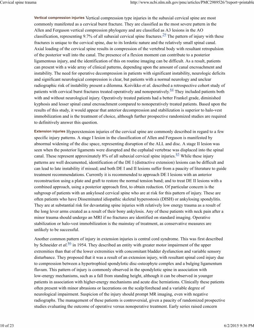
Vertical compression injuries
Extension injuries
Vertical compression type injuries in the subaxial cervical spine are mostcommonly manifested as a cervical burst fracture. They are classified as the most severe pattern in theAllen and Ferguson vertical compression phylogeny and are classified as A3 lesions in the AOclassification, representing 9.7% of all subaxial cervical spine fractures. The pattern of injury with these
fractures is unique to the cervical spine, due to its lordotic nature and the relatively small spinal canal.Axial loading of the cervical spine results in compression of the vertebral body with resultant retropulsionof the posterior wall into the canal. The presence of a flexion moment can contribute to a posteriorligamentous injury, and the identification of this on routine imaging can be difficult. As a result, patientscan present with a wide array of clinical patterns, depending upon the amount of canal encroachment andinstability. The need for operative decompression in patients with significant instability, neurologic deficitsand significant neurological compression is clear, but patients with a normal neurology and unclearradiographic risk of instability present a dilemma. Koivikko et al. described a retrospective cohort study ofpatients with cervical burst fractures treated operatively and nonoperatively. They included patients both
with and without neurological injury. Operatively treated patients had a better Frankel grade, diminishedkyphosis and lesser spinal canal encroachment compared to nonoperatively treated patients. Based upon theresults of this study, it would appear that anterior decompression and stabilization is superior to halo-vestimmobilization and is the treatment of choice, although further prospective randomized studies are requiredto definitively answer this question.
Hyperextension injuries of the cervical spine are commonly described in regard to a fewspecific injury patterns. A stage I lesion in the classification of Allen and Ferguson is manifested byabnormal widening of the disc space, representing disruption of the ALL and disc. A stage II lesion wasseen when the posterior ligaments were disrupted and the cephalad vertebrae was displaced into the spinalcanal. These represent approximately 8% of all subaxial cervical spine injuries. While these injury
patterns are well documented, identification of the DE I (distractive extension) lesions can be difficult andcan lead to late instability if missed; and both DE I and II lesions suffer from a paucity of literature to guidetreatment recommendations. Currently it is recommended to approach DE I lesions with an anteriorreconstruction using a plate and graft to restore the normal tension band; and to treat DE II lesions with acombined approach, using a posterior approach first, to obtain reduction. Of particular concern is thesubgroup of patients with an ankylosed cervical spine who are at risk for this pattern of injury. These areoften patients who have Disseminated idiopathic skeletal hyperostosis (DISH) or ankylosing spondylitis.They are at substantial risk for devastating spine injuries with relatively low energy trauma as a result ofthe long lever arms created as a result of their bony ankylosis. Any of these patients with neck pain after aminor trauma should undergo an MRI if no fractures are identified on standard imaging. Operativestabilization or halo-vest immobilization is the mainstay of treatment, as conservative measures areunlikely to be successful.
Another common pattern of injury in extension injuries is central cord syndrome. This was first describedby Schneider et al. in 1954. They described an entity with greater motor impairment of the upper
extremities than that of the lower extremities with concomitant bladder dysfunction and variable sensorydisturbance. They proposed that it was a result of an extension injury, with resultant spinal cord injury dueto compression between a hypertrophied spondylotic disc-osteophyte complex and a bulging ligamentumflavum. This pattern of injury is commonly observed in the spondylotic spine in association withlow-energy mechanisms, such as a fall from standing height, although it can be observed in youngerpatients in association with higher-energy mechanisms and acute disc herniations. Clinically these patientsoften present with minor abrasions or lacerations on the scalp/forehead and a variable degree ofneurological impairment. Suspicion of the injury should prompt MR imaging, even with negativeradiographs. The management of these patients is controversial, given a paucity of randomized prospectivestudies evaluating the outcome of operative versus nonoperative treatment. Early series raised concern
29
84
85
86
Cervical spine trauma http://www.ncbi.nlm.nih.gov/pmc/articles/PMC2989526/?report=printable
10 of 23 6/2/2015 9:36 PM
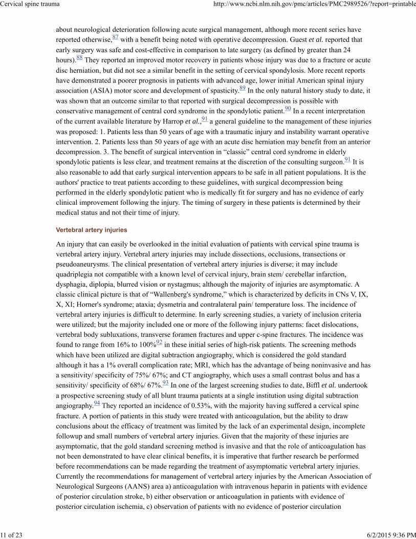
about neurological deterioration following acute surgical management, although more recent series havereported otherwise, with a benefit being noted with operative decompression. Guest et al. reported that
early surgery was safe and cost-effective in comparison to late surgery (as defined by greater than 24hours). They reported an improved motor recovery in patients whose injury was due to a fracture or acute
disc herniation, but did not see a similar benefit in the setting of cervical spondylosis. More recent reportshave demonstrated a poorer prognosis in patients with advanced age, lower initial American spinal injuryassociation (ASIA) motor score and development of spasticity. In the only natural history study to date, it
was shown that an outcome similar to that reported with surgical decompression is possible withconservative management of central cord syndrome in the spondylotic patient. In a recent interpretation
of the current available literature by Harrop et al., a general guideline to the management of these injuries
was proposed: 1. Patients less than 50 years of age with a traumatic injury and instability warrant operativeintervention. 2. Patients less than 50 years of age with an acute disc herniation may benefit from an anteriordecompression. 3. The benefit of surgical intervention in “classic” central cord syndrome in elderlyspondylotic patients is less clear, and treatment remains at the discretion of the consulting surgeon. It is
also reasonable to add that early surgical intervention appears to be safe in all patient populations. It is theauthors' practice to treat patients according to these guidelines, with surgical decompression beingperformed in the elderly spondylotic patient who is medically fit for surgery and has no evidence of earlyclinical improvement following the injury. The timing of surgery in these patients is determined by theirmedical status and not their time of injury.
Vertebral artery injuries
An injury that can easily be overlooked in the initial evaluation of patients with cervical spine trauma isvertebral artery injury. Vertebral artery injuries may include dissections, occlusions, transections orpseudoaneurysms. The clinical presentation of vertebral artery injuries is diverse; it may includequadriplegia not compatible with a known level of cervical injury, brain stem/ cerebellar infarction,dysphagia, diplopia, blurred vision or nystagmus; although the majority of injuries are asymptomatic. Aclassic clinical picture is that of “Wallenberg's syndrome,” which is characterized by deficits in CNs V, IX,X, XI; Horner's syndrome; ataxia; dysmetria and contralateral pain/ temperature loss. The incidence ofvertebral artery injuries is difficult to determine. In early screening studies, a variety of inclusion criteriawere utilized; but the majority included one or more of the following injury patterns: facet dislocations,vertebral body subluxations, transverse foramen fractures and upper c-spine fractures. The incidence wasfound to range from 16% to 100% in these initial series of high-risk patients. The screening methods
which have been utilized are digital subtraction angiography, which is considered the gold standardalthough it has a 1% overall complication rate; MRI, which has the advantage of being noninvasive and hasa sensitivity/ specificity of 75%/ 67%; and CT angiography, which uses a small contrast bolus and has asensitivity/ specificity of 68%/ 67%. In one of the largest screening studies to date, Biffl et al. undertook
a prospective screening study of all blunt trauma patients at a single institution using digital subtractionangiography. They reported an incidence of 0.53%, with the majority having suffered a cervical spine
fracture. A portion of patients in this study were treated with anticoagulation, but the ability to drawconclusions about the efficacy of treatment was limited by the lack of an experimental design, incompletefollowup and small numbers of vertebral artery injuries. Given that the majority of these injuries areasymptomatic, that the gold standard screening method is invasive and that the role of anticoagulation hasnot been demonstrated to have clear clinical benefits, it is imperative that further research be performedbefore recommendations can be made regarding the treatment of asymptomatic vertebral artery injuries.Currently the recommendations for management of vertebral artery injuries by the American Association ofNeurological Surgeons (AANS) area a) anticoagulation with intravenous heparin in patients with evidenceof posterior circulation stroke, b) either observation or anticoagulation in patients with evidence ofposterior circulation ischemia, c) observation of patients with no evidence of posterior circulation
87
88
89
90
91
91
92
93
94
Cervical spine trauma http://www.ncbi.nlm.nih.gov/pmc/articles/PMC2989526/?report=printable
11 of 23 6/2/2015 9:36 PM

ischemia. The authors do not routinely screen asymptomatic patients due to the lack of a widely accepted
screening method that is noninvasive. In symptomatic patients, it is our practice to follow the AANSguidelines in the management of these injuries in conjunction with neurosurgical consultation.
Acknowledgments
Dr. Joel Torretti has no financial relationships with industry to disclose.
Footnotes
Source of Support: Nil
Conflict of Interest: None.
R
1. Goldberg W, Mueller C, Panacek E, Tigges S, Hoffman JR, Mower WR, et al. Distribution and patternsof blunt traumatic cervical spine injury. Ann Emerg Med. 2001;38:17–21. [PubMed: 11423806]
2. Lowery DW, Wald MM, Browne BJ, Tigges S, Hoffman JR, Mower WR, et al. Epidemiology of cervicalspine injury victims. Ann Emerg Med. 2001;38:12–6. [PubMed: 11423805]
3. Hu R, Mustard CA, Burns C. Epidemiology of incident spinal fracture in a complete population. Spine.1996;21:492–9. [PubMed: 8658254]
4. Goldberg W, Mueller C, Panacek E, Tigges S, Hoffman JR, Mower WR, et al. Distribution and patternsof blunt traumatic cervical spine injury. Ann Emerg Med. 2001;38:17–21. [PubMed: 11423806]
5. Sekhon LH, Fehlings MG. Epidemiology, demographics and pathophysiology of acute spinal cord injury.Spine. 2001;26:S2–12. [PubMed: 11805601]
6. Bohlmann HH. Acute fractures and dislocations of the cervical spine: An analysis of three hundredpatients and review of the literature. J Bone Joint Surg Am. 1979;61:1119–42. [PubMed: 511875]
7. Gerrelts BD, Petersen EU, Mabry J, Petersen SR. Delayed diagnosis of cervical spine injuries. J Trauma.1991;31:1622–6. [PubMed: 1749033]
8. Reid DC, Henderson R, Saboe L, Miller JD. Etiology and clinical course of missed spine fractures. JTrauma. 1987;27:980–6. [PubMed: 3656481]
9. Clark CR, Ingram CM, el-Khoury GY, Ehara S. Radiographic evaluation of cervical spine injuries.Spine. 1988;13:742–7. [PubMed: 3194781]
10. Hendey GW, Wolfson AB, Mower WR, Hoffman JR. National Emergency X-radiography UtilizationStudy Group. Spinal cord injury without radiographic abnormality: Results of the National EmergencyX-Radiography Utilization Study in blunt cervical trauma. J Trauma. 2002;53:1–4. [PubMed: 12131380]
11. Davis JW, Phreaner DL, Hoyt DB, Mackersie RC. The Etiology of missed cervical spine injuries. JTrauma. 1993;34:342–6. [PubMed: 8483172]
12. McCleary AJ. A fracture of the odontoid process complicated by tenth and twelfth cranial nerve palsies:A case report. Spine. 1993;18:932–5. [PubMed: 8316898]
13. Arias MJ. Bilateral traumatic abducens nerve palsy without skull fracture and with cervical spinefracture: Case report and review of the literature. Neuorsurgery. 1985;16:232–4.
14. Duane TM, Dechert T, Wolfe LG, Aboutanos MB, Malhotra AK, Ivatury RR. Clinical Examination andits reliability in identifying cervical spine fractures. J Trauma. 2007;62:1405–10. [PubMed: 17563656]
95
Cervical spine trauma http://www.ncbi.nlm.nih.gov/pmc/articles/PMC2989526/?report=printable
12 of 23 6/2/2015 9:36 PM
15. Albrecht RM, Malik S, Kingsley DD, Hart B. Severity of cervical spine ligamentous injury correlateswith mechanism of injury, not with severity of blunt head trauma. Am Surg. 2003;69:261–5.[PubMed: 12678485]
16. Stiell IG, Clement CM, McKnight RD, Brison R, Schull MJ, Rowe BH, et al. The Canadian C-spinerule versus the NEXUS low-risk Criteria in patients with trauma. N Engl J Med. 2003;349:2510–8.[PubMed: 14695411]
17. Reid DC, Henderson R, Saboe L, Miller JD. Etiology and clinical course of missed spine fractures. JTrauma. 1987;27:980–6. [PubMed: 3656481]
18. Davis JW, Phreaner DL, Hoyt DB, Mackersie RC. The Etiology of missed cervical spine injuries. JTrauma. 1993;34:342–6. [PubMed: 8483172]
19. McCulloch PT, France J, Jones DL, Krantz W, Nguyen TP, Chambers C, et al. Helical ComputedTomography alone compared with plain radiographs with adjunct computed tomography to evaluate thecervical spine after high-energy trauma. J Bone Joint Surg Am. 2005;87:2388–94. [PubMed: 16264112]
20. Sanchez B, Waxman K, Jones T, Conner S, Chang R, Becerra S. Cervical spine clearance in blunttrauma: Evaluation of a computed tomography-based protocol. J Trauma. 2005;59:179–83.[PubMed: 16096560]
21. Antevil JL, Sise MJ, Sack DI, Kidder B, Hopper A, Brown CV. Spiral computed tomography for theinitial evaluation of spine trauma: A new standard of care? J Trauma. 2006;61:382–7. [PubMed: 16917454]
22. Brandt MM, Wahl WL, Yeom K, Kazerooni E, Wang SC. Computed tomographic scanning reducescost and time of complete spine evaluation. J Trauma. 2004;56:1022–8. [PubMed: 15179241]
23. Grogan EL, Morris JA, Dittus RS, Moore DE, Poulose BK, Diaz JJ, et al. Cervical spine evaluation inurban trauma centers: Lowering institutional costs and complications through helical CT scan. J Am CollSurg. 2005;200:160–5. [PubMed: 15664088]
24. White AA, 3rd, Johnson RM, Panjabi MM, Southwick WO. Biomechanical analysis of clinical stabilityin the cervical spine. Clin Orthop Relat Res. 1975;109:85–96. [PubMed: 1132209]
25. Vaccaro AR, An HS, Lin S, Sun S, Balderston RA, Cotler JM. Noncontiguous injuries of the spine. JSpinal Disord. 1992;5:320–9. [PubMed: 1520991]
26. Sharma OP, Oswanski MF, Yazdi JS, Jindal S, Taylor M. Assessment for additional spinal trauma inpatients with cervical spine injury. Am Surg. 2007;73:70–4. [PubMed: 17249461]
27. Allen BL, Jr, Ferguson RL, Lehmann TR, O'Brien RP. A mechanistic classification of closed indirectfractures and dislocations of the lower cervical spine. Spine. 1982;7:1–27. [PubMed: 7071658]
28. Magerl F, Aebi M, Gertzbein SD, Harms J, Nazarian S. A comprehensive classification of thoracic andlumbar injuries. Eur Spine J. 1994;3:184–201. [PubMed: 7866834]
29. Aebi M, Arlet V, Webb J. AOSpine Manual Clinical Applications. vol 2. Davos: AO Publishing; 2007.
30. Moore TA, Vaccaro AR, Anderson PA. Classification of lower cervical spine injuries. Spine.2006;31:37–43. [PubMed: 16685235]
31. White AA 3rd, Panjabi MM, editors. Kinematics of the spine, in Clinical Biomechanics of the Spine.2nd ed. Philadelphia: Lippincott; 1990.
32. Dvorak J, Panjabi MM. Functional anatomy of the alar ligaments. Spine. 1987;12:183–9.[PubMed: 3589810]
Cervical spine trauma http://www.ncbi.nlm.nih.gov/pmc/articles/PMC2989526/?report=printable
13 of 23 6/2/2015 9:36 PM

33. Jackson RS, Daxes DM, Rhyne AL, 3rd, Darden BV., 2nd Upper cervical spine injuries. J Am AcadOrthop surg. 2002;10:271–80. [PubMed: 15089076]
34. Clark CR. The cervical spine. New York: Lippincott-Raven; 1998.
35. Powers B, Miller MD, Kramer RS, Martinez S, Gehweiler JA., Jr Traumatic anterior atlanto-occipitaldislocation. J Neurosurg. 1979;4:12–7.
36. Harris JR, Carson GC, Wagner LK. Radiographic diagnosis of traumatic occipitovertebral dissociation:Normal occipitovertebral relationships on lateral radiographs of supine subjects. AJR Am J Roentgenol.1994;162:881–6. [PubMed: 8141012]
37. Harris JH, Jr, Carson GC, Wagner LK, Kerr N. Radiologic diagnosis of traumatic occipitovertebraldissociation: 2: Comparison of three methods of detecting occipitovertebral relationships on lateralradiographs of supine subjects. AJR Am J Roentgenol. 1994;162:887–92. [PubMed: 8141013]
38. Payer M, Sottas CC. Traumatic atlanto-occipital dislocation: Presentation of a new posterioroccipitoatlantoaxial fixation technique in an adult survivor: Technical case report. Neurosurgery.2005;56:E203. [PubMed: 15799814]
39. Anderson AJ, Towns GM, Chiverton N. Traumatic occipitocervical disruption: A new technique forstabilization: Case report and review of the literature. J Bone Joint Surg Br. 2006;88:1464–8.[PubMed: 17075091]
40. Bloom AI, Neeman Z, Slasky BS, Floman Y, Milgrom M, Rivkind A, et al. Fracture of the occipitalcondyles and associated craniocervical ligament injury: incidence, CT imaging and implications. ClinRadiol. 1997;52:198–202. [PubMed: 9091254]
41. Blacksin MF, Lee HJ. Frequency and significance of fractures of the upper cervical spine detected byCT in patients with severe neck trauma. AJR Am J Roentgenol. 1995;165:1201–4. [PubMed: 7572503]
42. Leone A, Cerase A, Colosimo C, Laura L, Puca A, Marano P. Occipital condylar fractures: A review.Radiology. 2000;216:635–44. [PubMed: 10966689]
43. Anderson PS, Montesano PX. Morphology and treatment of occipital condyle fractures. Spine.1988;13:731–6. [PubMed: 3194779]
44. Levine AM, Edwards CC. Treatment of injuries in the C1-C2 complex. Orthop Clin North Am.1986;17:31–44. [PubMed: 3945481]
45. Spence KF, Jr, Decker S, Sell KW. Bursting atlantal fracture associated with rupture of the transverseligament. J Bone Joint Surg Am. 1970;52:543–9. [PubMed: 5425648]
46. Lee TT, Green BA, Petrin DR. Treatment of stable burst fracture of the atlas (Jefferson fracture) withrigid cervical collar. Spine. 1998;23:1963–7. [PubMed: 9779528]
47. Levine AM, Edwards CC. Fractures of the Atlas. J Bone Joint Surg Am. 1991;73:680–91.[PubMed: 2045393]
48. McGuire RA, Jr, Harkley HL. Unstable Jefferson's fracture treated with transarticular screws.Orthopedics. 1995;18:207–9. [PubMed: 7746757]
49. Hein C, Richter HP, Rath SA. Atlantoaxial screw fixation for the treatment of isolated and combinedunstable Jefferson fractures-experiences with 8 patients. Acta Neurochir (Wien) 2002;144:1187–92.[PubMed: 12434175]
50. Ruf M, Melcher R, Harms J. Transoral reduction and osteosynthesis C1 as a function preserving option
Cervical spine trauma http://www.ncbi.nlm.nih.gov/pmc/articles/PMC2989526/?report=printable
14 of 23 6/2/2015 9:36 PM

in the treatment of unstable Jefferson fractures. Spine. 2004;29:823–7. [PubMed: 15087806]
51. Feuntes S, Bouillot P, Palombi O, Ducolombier A, Desgeorges M. Traumatic atlantoaxial rotatorydislocation with odontoid fracture: Case report and review. Spine. 2001;26:830–4. [PubMed: 11295908]
52. Moore KR, Frank EH. Traumatic atlantoaxial rotatory subluxation and dislocation. Spine.1995;20:1928–30. [PubMed: 8560343]
53. Fielding JW, Hawkins RJ. Atlanto-axial rotatory fixation (fixed rotatory subluxation of the atlanto-axialjoint) J Bone Joint Surg Am. 1977;59:37–44. [PubMed: 833172]
54. Bouillot P, Fuentes S, Dufour H, Manera L, Grisoli F. Imaging features in combined atlantoaxial andoccipitoatlantal rotatory subluxation: A rare entity: Case report. J Neurosurg. 1999;90:258–60.[PubMed: 10199260]
55. Dvorak J, Hayek J, Zehnder R. CT functional diagnostics of the rotatory instability of the uppercervical spine: An evaluation on healthy adults and patients with suspected instability. Spine.1987;12:726–31. [PubMed: 3686227]
56. Roche CJ, O'Malley M, Dorgan JC, Carty HM. A pictorial review of atlanto-axial rotatory fixaton: Keypoints for the radiologist. Clin Radiol. 2001;56:947–58. [PubMed: 11795922]
57. Bono CM, Vaccaro AR, Fehlings M, Fisher C, Dvorak M, Ludwig S, et al. Measurement Techniquesfor Upper Cervical Spine Injuries: Consensus statement of the spine trauma study group. Spine.2007;32:593–600. [PubMed: 17334296]
58. Anderson LD, D'Alonzo RT. Fractures of the odontoid process of the axis. J Bone Joint Surg Am.1974;56:1663–74. [PubMed: 4434035]
59. Clark CR, White AA., 3rd Fractures of the dens: A multicenter study. J Bone Joint Surg Am.1985;67:1340–8. [PubMed: 4077905]
60. Seybold EA, Bayley JC. Functional outcome of surgically and conservatively managed dens fractures.Spine. 1998;23:1837–46. [PubMed: 9762740]
61. Julien TD, Frankel B, Traynelis VC, Ryken TC. Evidence-based analysis of odontoid fracturemanagement. Nurosurg Focus. 2000;8:1–6.
62. Kuntz C, Mirza SK, Jarell AD, Chapman JR, Shaffrey CI, Newell DW. Type II odontoid fractures in theelderly: early failure of nonsurgical treatment. Neurosurg Focus. 2000;8:1–6.
63. Tashjian RZ, Majercik S, Biffl WL, Palumbo MA, Cioffi WG. Halo-vest immobilization increases earlymorbidity and mortality in elderly odontoid fractures. J Trauma. 2006;60:199–203. [PubMed: 16456456]
64. Fuji E, Kobayashi K, Hirabayashi K. Treatment of fractures of the odontoid process. Spine.1988;13:604–9. [PubMed: 3175749]
65. Aebi M, Etter C, Cosica M. Fractures of the odontoid process: Treatment with anterior screw fixation.Spine. 1989;14:1065–70. [PubMed: 2588054]
66. Henry AD, Bohly J, Grosse A. Fixation of odontoid fractures by an anterior screw. J Bone Joint SurgBr. 1999;81:472–7. [PubMed: 10872369]
67. Moon MS, Moon JL, Sun DH, Moon YW. Treatment of dens fracture in adults: A report of thirty-twocases. Bull Hosp Jt Dis. 2006;63:108–12. [PubMed: 16878829]
68. Claybrooks R, Kayanja M, Milks R, Benzel E. Atlantoaxial fusion: A biomechanical analysis of two
Cervical spine trauma http://www.ncbi.nlm.nih.gov/pmc/articles/PMC2989526/?report=printable
15 of 23 6/2/2015 9:36 PM

C1-2 fusion techniques. Spine J. 2007.
69. Melcher RP, Puttlitz CM, Kleinstruck F, Lotz JC, Harms J, Bradford DS. Biomechanical testing ofposterior atlantoaxial fixation techniques. Spine. 2002;27:2435–40. [PubMed: 12435971]
70. Effendi B, Roy D, Cornish B, Dussault RG, Laurin CA. Fractures of the ring of the axis: Aclassification based on the analysis of 131 cases. J Bone Joint Surg Br. 1981;63:319–27.[PubMed: 7263741]
71. Levine AM, Edwards CC. The management of traumatic spondylolisthesis of the axis. J Bone JointSurg Am. 1985;67:217–26. [PubMed: 3968113]
72. Starr JK, Eismont FJ. Atypical hangman's fractures. Spine. 1993;18:1954–7. [PubMed: 8272942]
73. Vaccaro AR, Madigan L, Bauerle WB, Blescia A, Cotler JM. Early halo immobilization of displacedtraumatic spondylolisthesis of the axis. Spine. 2002;27:2229–33. [PubMed: 12394899]
74. Duggal N, Chamberlain RH, Perez-Garza LE, Espinoza-Larios A, Sonntag VK, Crawford NR.Hangman's fracture: A biomechanical comparison of stabilization techniques. Spine. 2007;32:182–7.[PubMed: 17224812]
75. Moon MS, Moon JL, Moon YW, Sun DH, Choi WT. Traumatic spondylolisthesis of the axis: 42 cases.Bull Hosp Jt Dis. 2001-2002;60:61–6. [PubMed: 12003355]
76. Fisher CG, Dvorak MF, Leith J, Wing PC. Comparison of Outcomes for unstable lower cervical flexionteardrop fractures managed with halo thoracic vest versus anterior corpectomy and plating. Spine.2002;27:160–6. [PubMed: 11805662]
77. Grant GA, Mirza SK, Chapman JR, Winn HR, Newell DW, Jones DT, et al. Risk of early closedreduction in cervical spine subluxation injuries. J Neurosurg. 1999;90:13–8. [PubMed: 10413120]
78. Star AM, Jones AA, Cotler JM, Balderston RA, Sinha R. Immediate closed reduction of cervical spinedislocations using traction. Spine. 1990;15:1068–72. [PubMed: 2263974]
79. Cotler JM, Herbison GJ, Nasuti JF, Ditunno JF, Jr, An H, Wolff BE. Closed reduction of traumaticcervical spine dislocation using traction weights up to 140 pounds. Spine. 1993;18:386–90.[PubMed: 8475443]
80. Beyer CA, Cabanella ME, Berquist TH. Unilateral facet dislocations and fracture-dislocations of thecervical spine. J Bone Joint Surg Br. 1991;73:977–81. [PubMed: 1955448]
81. Spector LR, Kim DH, Affonso J, Albert TJ, Hilibrand AS, Vaccaro AR. Use of computed tomographyto predict failure of nonoperative treatment of unilateral facet fractures of the cervical spine. Spine.2006;31:2827–35. [PubMed: 17108837]
82. Duggal N, Chamberlain RH, Park SC, Sonntag VK, Dickman CA, Crawford NR. Unilateral cervicalfacet dislocation: Biomechanics of fixation. Spine. 2005;30:164–8.
83. Johnson MG, Fisher CG, Boyd M, Pitzen T, Oxland TR, Dvorak MF. The radiographic failure of singlesegment anterior cervical plate fixation in traumatic cervical flexion distraction injuries. Spine.2004;29:2815–20. [PubMed: 15599284]
84. Koivikko MP, Myllynen P, Karjalainen M, Vornanen M, Santvirta S. Conservative and operativetreatment in cervical burst fractures. Arch Orthop Trauma Surg. 2000;120:448–51. [PubMed: 10968537]
85. Vaccaro AR, Klein GR, Thaller JB, Rushton SA, Cotler JM, Albert TJ. Distraction extension injuries ofthe cervical spine. J Spine Disord. 2001;14:193–200.
Cervical spine trauma http://www.ncbi.nlm.nih.gov/pmc/articles/PMC2989526/?report=printable
16 of 23 6/2/2015 9:36 PM

86. Schneider RC, Cherry G, Pantek H. The syndrome of acute central cervical spinal cord injury, withspecial reference to the mechanisms involved in hyperextension injuries of the cervical spine. J Neurosurg.1954;11:546–77. [PubMed: 13222164]
87. Chen T, Lee S, Lui T, Wong C, Yeh Y, Tzaan W, et al. Efficacy of surgical treatment in traumaticcentral cord syndrome. Surg Nurol. 1997;48:435–41.
88. Guest J, Eleraky MA, Apostolides PJ, Dickman CA, Sonntag VK. Traumatic central cord syndrome:Results of surgical management. J Neurosurg. 2002;97:25–32. [PubMed: 12120648]
89. Dvorak MF, Fisher CG, Hoekema J, Boyd M, Noonan V, Wing PC, et al. Factors predicting motorrecovery and functional outcome after traumatic central cord syndrome: A long term follow-up. Spine.2005;30:2303–11. [PubMed: 16227894]
90. Newey ML, Sen PK, Fraser RD. The long term outcome after central cord syndrome: A study of thenatural history. J Bone Joint Surg Br. 2000;82:851–5. [PubMed: 10990310]
91. Harrop JS, Sharan A, Ratliff J. Central cord injury: Pathophysiology, management and outcomes. SpineJ. 2006;6:198S–206S. [PubMed: 17097539]
92. Inamasu J, Guiot BH. Vertebral artery injury after blunt cervical trauma: An update. Surg Neurol.2006;65:238–46. [PubMed: 16488240]
93. Biffl WL, Ray CE, Moore EE, Mesterk M, Johnson JL, Burch JM. Noninvasive diagnosis of bluntcerebrovascular injuries: A preliminary report. J Trauma. 2002;53:850–6. [PubMed: 12435934]
94. Biffl WL, Moore EE, Elliot JP, Ray C, Offner PJ, Francoise RJ, et al. The devastating potential of bluntvertebral arterial injuries. Ann Surg. 2000;231:672–81. [PMCID: PMC1421054] [PubMed: 10767788]
95. AANS. Management of vertebral artery injuries after non-penetrating cervical trauma. J Neurosurg.2002;50:173–8.
Figures and Tables
Cervical spine trauma http://www.ncbi.nlm.nih.gov/pmc/articles/PMC2989526/?report=printable
17 of 23 6/2/2015 9:36 PM

Figure 1
Upper cervical spine anatomy. A. Sagittal view; B. Posterior view; C. Anterior view (with anterior arch of C1 cut away)34
Cervical spine trauma http://www.ncbi.nlm.nih.gov/pmc/articles/PMC2989526/?report=printable
18 of 23 6/2/2015 9:36 PM

Figure 2
Line diagramme shows Harris Rule of 12's. Illustration for calculating the basion-dens interval (BDI) and the basion-axisinterval (BAI)57
Cervical spine trauma http://www.ncbi.nlm.nih.gov/pmc/articles/PMC2989526/?report=printable
19 of 23 6/2/2015 9:36 PM
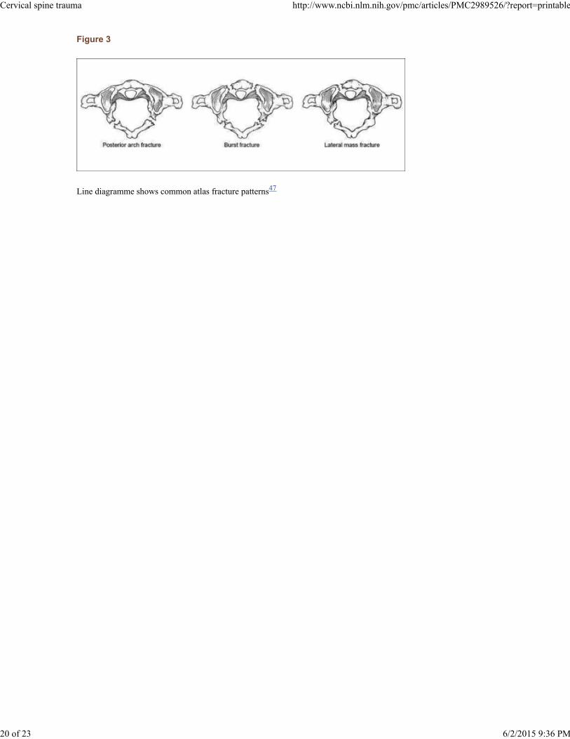
Figure 3
Line diagramme shows common atlas fracture patterns47
Cervical spine trauma http://www.ncbi.nlm.nih.gov/pmc/articles/PMC2989526/?report=printable
20 of 23 6/2/2015 9:36 PM

Figure 4
Line diagramme shows method for calculation of lateral mass overhang57
Cervical spine trauma http://www.ncbi.nlm.nih.gov/pmc/articles/PMC2989526/?report=printable
21 of 23 6/2/2015 9:36 PM

Figure 5
Line diagramme shows odontoid fracture classification58
Cervical spine trauma http://www.ncbi.nlm.nih.gov/pmc/articles/PMC2989526/?report=printable
22 of 23 6/2/2015 9:36 PM

Figure 6
Line diagramme shows classification of traumatic spondylolisthesis of the axis
Articles from Indian Journal of Orthopaedics are provided here courtesy of Medknow Publications
71
Cervical spine trauma http://www.ncbi.nlm.nih.gov/pmc/articles/PMC2989526/?report=printable
23 of 23 6/2/2015 9:36 PM