cervical spine trauma elda baptistelli de carvalho, md, pgy-3 university of toronto
TRANSCRIPT

Cervical Spine Trauma
Elda Baptistelli de Carvalho, MD, PGY-3
University of Toronto

Objectives
Clinical indication for each imaging modality
Identify anatomy of cervical spine Approach to C-spine radiography
interpretation Classification of spine injuries

Who gets radiographs?
Midline cervical tenderness Focal neurologic deficits Altered LOC Evidence of intoxication Painful distracting injury

Who gets CT?
Dangerous mechanisms/high energy mechanisms:
-fall from elevation = or > 3 feet/5 stairs
-axial load to head (diving)
-MVC high speed (>100 km/h), ejection
-motorized recreational vehicles
-bicycle collision

Who gets MRI?
Unexplained neurologic symptoms/signs For visualizing soft tissues, neural elements and
unsuspected disk herniation To differentiate cord edema x hemorrhage x infarction To better characterize epidural hematoma

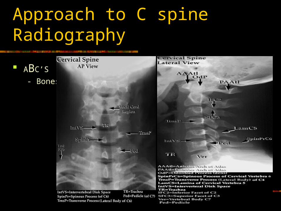
Anatomy

Approach to C spine radiograph
ABC’S
-Adequacy

Approach to C spine radiograph
ABC’S
-Adequacy

Approach to C spine Radiograph
ABC’S
-Alignment
Approach to C spine Radiography
ABC’S
- Bones

Approach to C spine radiograph
ABC’S - Cartilage

Approach to C spine radiograph
ABC’S
-Soft Tissue
Rule 2-6 (C2-6 mm)
6-2 (C6-2 cm)

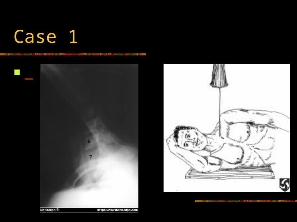
Case 1

Mechanism of Fractures
Hyperflexion Hyperextension Axial Compression

Classification

ClassificationBy Mechanism of injury /Stability
Type of Injury Fractures Stability
Flexion Anterior subluxation
Unilateral facet dislocationBilateral facet dislocationWedge compression fractureFlexion teardrop fractureClay Shoveler's fractureOdontoid
stable or delayed instability
stableunstablestableunstablestableunstable
Extension Hangman's fracture unstable
Compression Jefferson fractureBurst fracture
unstablestable


Case 2

Clay shoveler fracture
Stable fracture Hyperflexion ( shoveling snow) Sudden exertion of muscular attachment Avulsion # of spinous process of C7>C6>T1 Rule out extension to lamina, facet #, unilateral jump
facet


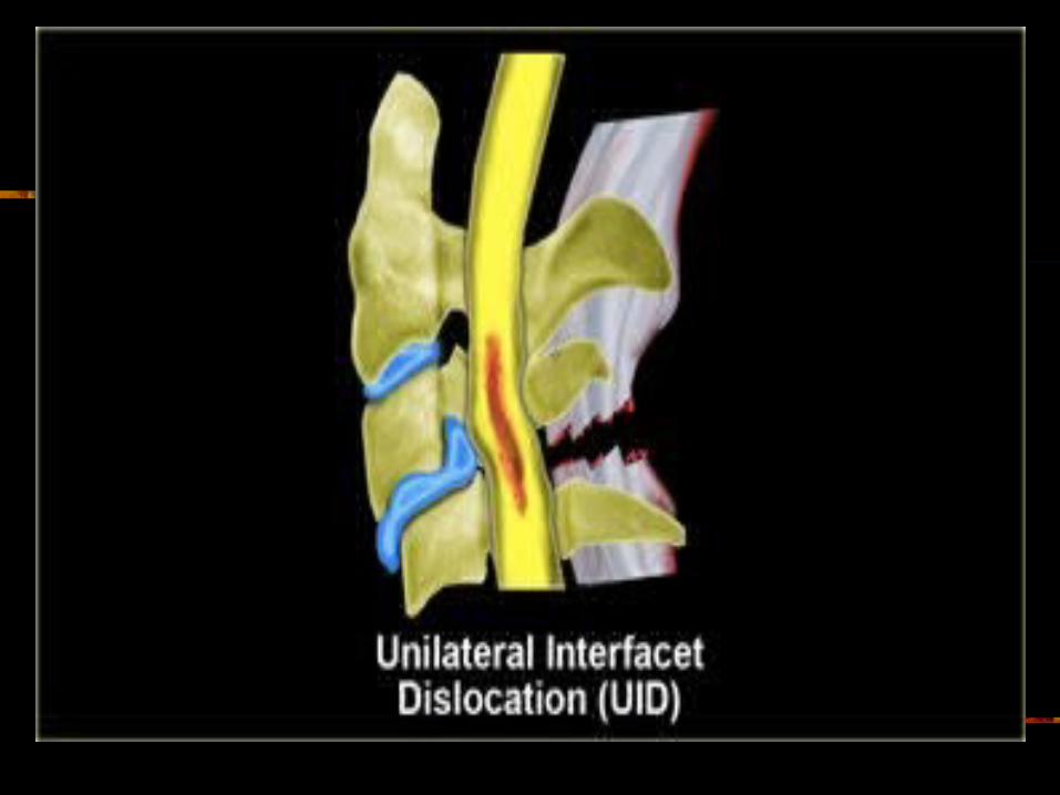
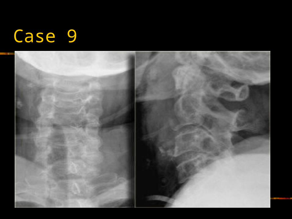
Unilateral Facet Dislocation
Hyperflexion + rotation Superior facet slides over inferior facet and becomes locked Anterior subluxation of superior vertebral body –25% AP diameter Stable injury 30% with associated neurologic deficit MRI: disk extrusion leading to cord compression


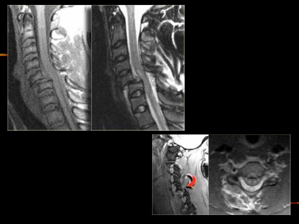


Bilateral Facet Dislocation
Extreme hyperflexion Anterior dislocation of articular masses (disruption of
posterior ligament complex,PLL,disk and ALL. Complete dislocation: dislocated vertebra anteriorly
displaced ½ of AP diameter of vertebral body Unstable ( high incidence of cord damage)


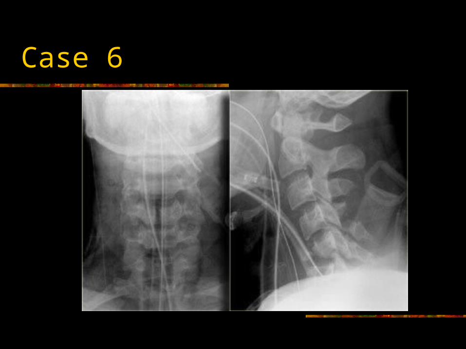
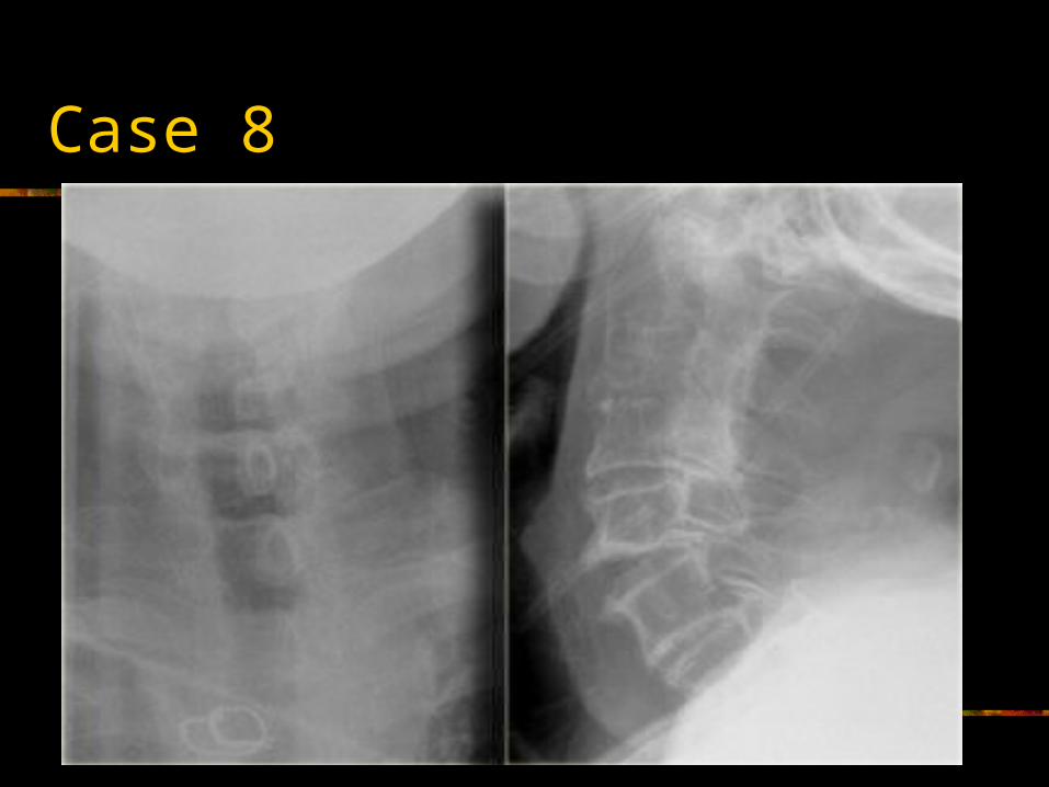
Case 6


Flexion Tear Drop
Flexion+compression (MVA) Teardrop fragment comes from the anteroinferior aspect
of the vertebral body Larger posterior part displaced backward into the spinal
canal Facets joints and interspinous distances usually
widened, disk space may be narrowed 70% of patients with neurologic injuries Unstable fracture (complete disruption of ligaments and
anterior cord syndrome)
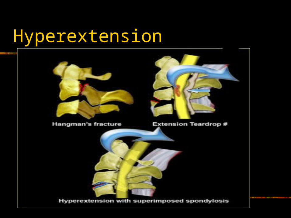

Hangman’s fracture
Most common cervical spine fracture Usually hyperextension Diving Unstable, however seldom associated with cord injury
(AP diameter of spinal canal greatest at C1/C2 level and # pedicles allow decompression)
Hangman’s + uni/bilateral facet dislocation: high rate of neurologic complications


Hyperextension injury
Widening of disk space anteriorly and narrowing posteriorly
“open book” Central cord injury= disproportionated weakness in arms
and normal strength in the legs Injuries can be devastating, however are uncommon
hemorrhagic


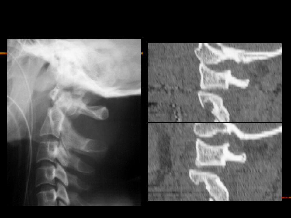
Extension Teardrop Fracture
ALL pulls bony fragment away from inferior aspect of the vertebra because sudden extension
Fragment is true avulsion x fragment from flexion teardrop (compression)
Diving accidents Lower cervical spine Central cord syndrome (buckling of ligamenta flava into
spinal canal) Stable in flexion; highly unstable in extension
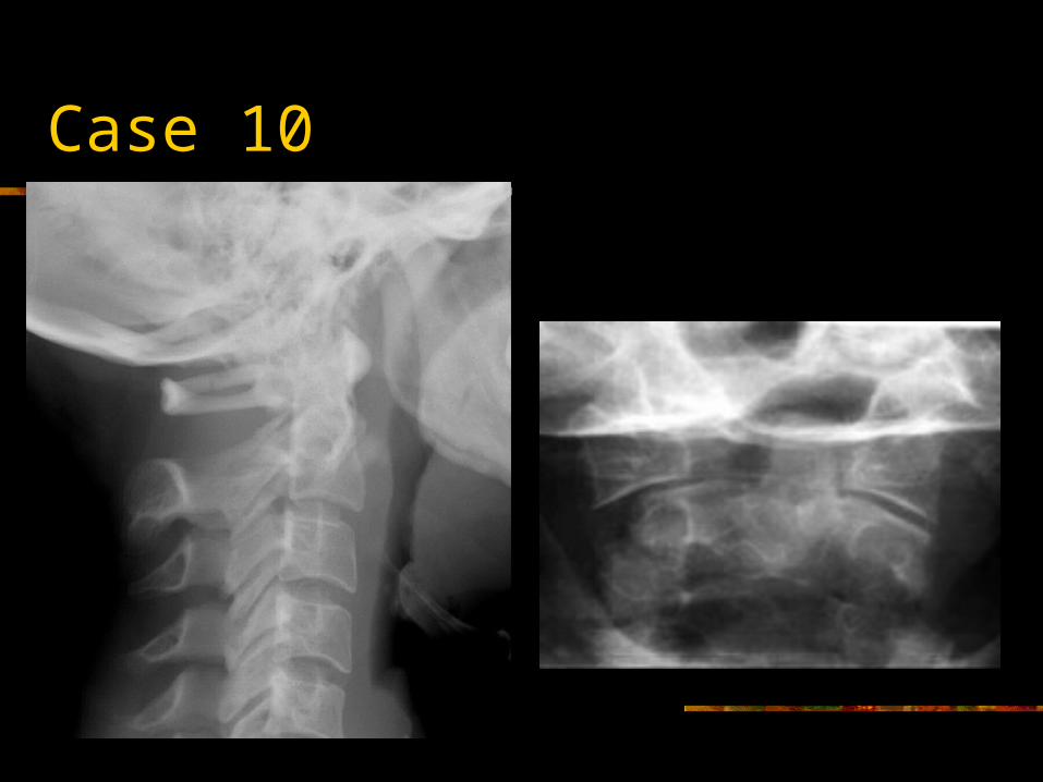


Jefferson Fracture
Burst fracture of ring of C1 Axial loading in the occiput No associated neuro deficts ( C1 ring is wide!) Diving, MVA, fall onto height > 2mm dislocation of lateral masses of C1 or odontoid
view is diagnostic, 1-2 mm is equivocal ( rotation of head?)
Predental space > 3 mm: disruption of transverse ligament
1/3 associated with C2 fracture

Case 11


Atlanto-Occipital Dislocation
Very rare in surviving patients More common in Kids Hyperextension+distraction Disruption of tectorial ligaments CR: rule of 12: tip of dens-basion
Basion-post line< 12mm Atlanto-occipital condyle distance<5mm

Summary
Be systematic (follow ABC’S!!!!) Know anatomy and mechanism of trauma If dangerous mechanism-CT Unexplained neuro symptoms-MRI Don’t clear C spine on call if not sure!!

References
http://www.wheelessonline.com/ortho http://www.radiologyassistant.nl http://www.learningradiolgy.com http://www.radiographics.rsnajnls.org Emergency Radiology-Schwartz Primer to Diagnostic Imaging