so what do you expect with a cervical lesion?10_26_40_am.pdf · cervical traction • gardner-wells...
TRANSCRIPT


So what do you expect with a cervical lesion?
• Quadriplegia or quadriparesis
• Bowel/bladder retention (spastic)
• Various degrees of breathing difficulties
• Neurogenic and/or spinal shock

Case scenario
• 22 y/o female
• Motor vehicle accident (hit a pole at 60mph)
• Short term loss of consciousness (10’)
• Not able to move or feel her legs
• No bladder / bowel control or sensation
• Sensory level at the umbilicus

Thoracic injuries (T2-L1)
• Paraparesis or paraplegia
• UMN (upper motor neuron) signs

Case scenario
• 22 y/o female
• Motor vehicle accident
• Not able to move or feel her legs below the knee
• Could flex thighs against gravity
• No bladder / bowel control or sensation
• Sensory level above the knee on L, below the knee on R

Cauda equina injuries (L2 or below)
• Paraparesis or paraplegia
• LMN (lower motor neuron) signs
• Thigh flexion is almost always preserved to some degree

What is the difference between cauda equina and conus medullaris
syndrome?

Goal of spine trauma care
• Protect further injury during evaluation and management
• Identify spine injury or document absence of spine injury
• Optimize conditions for maximal neurologic recovery
Suspected Spinal Injury
• High speed crash
• Unconscious
• Multiple injuries
• Neurological deficit
• Spinal pain/tenderness
• Up to 15% of spinal injuries have a second (possibly non
adjacent) fracture elsewhere in the spine

Initial Management
• Immobilization
• Rigid collar
• Sandbags and straps
• Spine board
• Log-roll to turn
• Prevent hypotension
• Pressors: Dopamine, not Neosynephrine
• Fluids to replace losses; do not overhydrate
• Maintain oxygenation
• O2 per nasal canula
• If intubation is needed, do NOT move the neckAdvance
Trauma Life Support (ATLS) guidelines

Management in the hospital
• NGT to suction • Prevents aspiration
• Decompresses the abdomen (paralytic ileus is common in the first days)
• Foley • Urinary retention is common
• Methylprednisolone (Solu-Medrol) • Only if started within 8 hours of injury
• Exclusion criteria • Cauda equina syndrome
• GSW
• Pregnancy
• Age <13 years
• Patient on maintenance steroids

Radiolographic evaluation
X-ray Guidelines (cervical)
AABBCDS
• Adequacy, Alignment
• Bone abnormality, Base of skull
• Cartilage
• Disc space
• Soft tissue

Adequacy
• Must visualize entire C-spine
• A film that does not show the upper border of T1 is inadequate
• Caudal traction on the arms may help
• If can not, get swimmer’s view or CT

Alignment
• The anterior vertebral
line, posterior vertebral
line, and spinolaminar
line should have a
smooth curve with no
steps or discontinuities
• Malalignment of the
posterior vertebral bodies
is more significant than
that anteriorly, which may
be due to rotation
• A step-off of >3.5mm is
significant anywhere
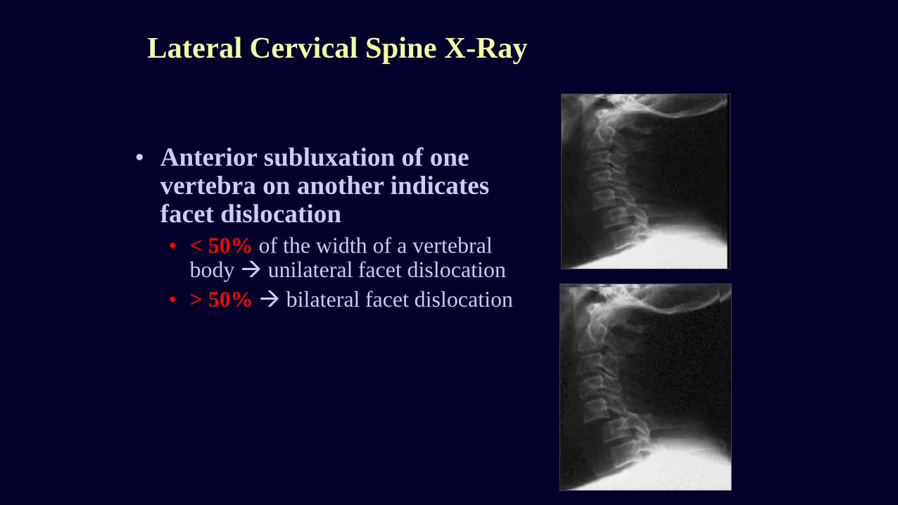
Lateral Cervical Spine X-Ray
• Anterior subluxation of one vertebra on another indicates facet dislocation
• < 50% of the width of a vertebral body unilateral facet dislocation
• > 50% bilateral facet dislocation

Bones

Disc
• Disc Spaces
• Should be uniform
• Assess spaces
between the spinous
processes

Soft tissue
• Nasopharyngeal space (C1) • 10 mm (adult)
• Retropharyngeal space (C2-
C4) • 5-7 mm
• Retrotracheal space (C5-
C7) • 14 mm (children) • 22 mm (adults)

AP C-spine Films
• Spinous processes
should line up
• Disc space should be
uniform
• Vertebral body height
should be uniform.
Check for oblique
fractures.

Open mouth view
• Adequacy: all of the
dens and lateral
borders of C1 & C2
• Alignment: lateral
masses of C1 and
C2
• Bone: Inspect dens
for lucent fracture
lines

CT scan
• Good in acute situations
• Shows bone very well
• Sagittal reconstruction is mandatory
• Soft tissues (discs, spinal cord) are poorly
visualized
• Do NOT give contrast in trauma patients
(contrast is bright, mimicking blood)

MRI
• Almost never an emergency
• Exception: cauda equina syndrome
• Shows tumors and soft tissues (e.g., herniated
discs) much better than CT scan
• May be used to clear c-spine in comatose
patients

Lumbar Puncture
• Sedate the patient and make your life easier
• Measure opening pressure with legs straight
• Always get head CT prior to LP to r/o increased ICP or brain
tumor

Cervical Spine Clearance
• Occiput to T1 need to be cleared
• ER, Neurosurgery or Orthopedics physician
• If the patient • Is awake and oriented
• Has no distracting injuries
• Has no drugs on board
• Has no neck pain
• Is neurologically intact
then the c-spine can be cleared clinically, without any need for XRays
• CT and/or MRI is necessary if the patient is comatose or has neck pain
• Subluxation >3.5mm is usually unstable

Gardner-Wells tongs

Cervical Traction
• Gardner-Wells tongs
• Provides temporary stability of the cervical spine
• Contraindicated in unstable hyperextension injuries
• Weight depends on the level (usually 5lb/level, start with 3lb/level, do not
exceed 10lb/level)
• Cervical collar can be removed while patient is in traction
• Pin care: clean q shift with appropriate solution, then apply povidone-
iodine ointment
• Take XRays at regular intervals and after every move from bed
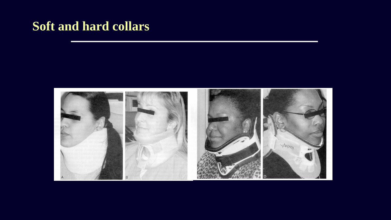
Soft and hard collars

Minerva vest and halo-vest

Jefferson Fracture
• Burst fracture of C1 ring
• Unstable fracture
• Increased lateral ADI on lateral film if ruptured transverse ligament and displacement of C1 lateral masses on open mouth view
• Need CT scan

Burst Fracture
• Fracture of C3-C7 from axial loading
• Spinal cord injury is common from posterior displacement of fragments into the spinal canal
• Unstable

Clay Shoveler’s Fracture
• Flexion fracture of
spinous process
• C7>C6>T1
• Stable fracture

Flexion Teardrop Fracture
• Flexion injury causing a fracture of the anteroinferior portion of the vertebral body
• Unstable because usually associated with posterior ligamentous injury

Bilateral Facet Dislocation
• Flexion injury
• Subluxation of dislocated
vertebra of greater than
½ the AP diameter of the
vertebral body below it
• High incidence of spinal
cord injury
• Extremely unstable

Hangman’s Fracture
• Extension injury
• Bilateral fractures of
C2 pedicles
(white arrow)
• Anterior dislocation of
C2 vertebral body
(red arrow)
• Unstable
Odontoid Fractures
• Complex mechanism of injury
• Generally unstable
• Type 1 fracture through the tip
• Rare
• Type 2 fracture through the base
• Most common
• Type 3 fracture through the base and body
of axis
• Best prognosis
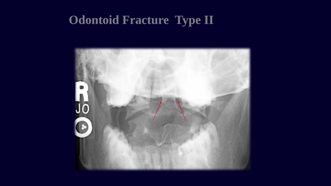
Odontoid Fracture Type II

Odontoid Fracture Type III

Surgical Decompression and/or Fusion
• Indications
• Decompression of the neural elements (spinal cord/nerves)
• Stabilization of the bony elements (spine)
• Timing
• Emergent
• Incomplete lesions with progressive neurologic deficit
• Elective
• Complete lesions (3-7 days post injury)
• Central cord syndrome (2-3 weeks post injury)
Long term care
• Rehab for maximizing motor function
• Bladder/bowel training
• Psychological and social support