central dogma - university of arizonaeebweb.arizona.edu/courses/ecol223/protein synthesis...
TRANSCRIPT

Copyright © 2003 Pearson Education, Inc. publishing as Benjamin Cummings
Cytoplasm
Nucleus
DNA
DNA is the genetic material within the nucleus.
Central Dogma
RNA
Protein
Replication
The process of replication creates new copies of DNA.
TranscriptionThe process of transcription creates an RNA using DNA information.
TranslationThe process of translation creates a protein using RNA information.

Copyright © 2003 Pearson Education, Inc. publishing as Benjamin Cummings
Transcription• DNA is used as a template for creation
of RNA using
• the enzyme RNA polymerase.DNA
5’
3’
5’
3’
G T C A T T C G G
C A G T A A G C C

Copyright © 2003 Pearson Education, Inc. publishing as Benjamin Cummings
Transcription• RNA polymerase reads the nucleotides
on the
• template strand from 3’ to 5’ and creates an RNA
• Molecule in a 5’ to 3’ direction that looks like the coding strand.G T C A T T C G G
C A G T A A G C C

Copyright © 2003 Pearson Education, Inc. publishing as Benjamin Cummings
Transcription• The new RNA molecule is formed by incorporating
• nucleotides that are complementary to the template strand.
DNA coding strand
DNA template strand
DNA
5’
3’
5’
3’
G T C A T T C G G
C A G T A A G C C
G
RNA
5’
GG U C A U U C
3’

Copyright © 2003 Pearson Education, Inc. publishing as Benjamin Cummings
Two types of nucleic acids•RNA
•Usually single-stranded
•Has uracil as a base
•Ribose as the sugar
•Carries protein-encoding information
•Can be catalytic
•DNA
•Usually double-stranded
•Has thymine as a base
•Deoxyribose as the sugar
•Carries RNA-encoding information
•Not catalytic

Copyright © 2003 Pearson Education, Inc. publishing as Benjamin Cummings
Two types of nucleic acids
# of strands
kind of sugar
bases used

Copyright © 2003 Pearson Education, Inc. publishing as Benjamin Cummings
rRNA is part of ribosome, used to translate mRNA into protein

Copyright © 2003 Pearson Education, Inc. publishing as Benjamin Cummings
tRNA is a connection between anticodon and amino acid

Copyright © 2003 Pearson Education, Inc. publishing as Benjamin Cummings

Copyright © 2003 Pearson Education, Inc. publishing as Benjamin Cummings
TATA binding protein
Initiation of transcription
DNA GG TATA CCC
Transcription begins
Promoter Gene sequenceto be transcribed
TATA box
Transcription begins at the 3’ end of the gene in aregion called the promoter.
When a complete transcription complex is formed RNA polymerase binds and transcription begins.
The promoter recruits TATA protein, a DNA binding protein, which in turn recruits other proteins.
Transcription factor

Copyright © 2003 Pearson Education, Inc. publishing as Benjamin Cummings
• Noncodingsegments called introns are spliced out
• A cap and a tail are added to the ends to protect against degradation in the cytoplasm
10.10 Eukaryotic RNA is processed before leaving the nucleus
Figure 10.10
DNA
RNAtranscriptwith capand tail
mRNA
Exon Intron IntronExon Exon
TranscriptionAddition of cap and tail
Introns removed
Exons spliced together
Coding sequenceNUCLEUS
CYTOPLASM
Tail
Cap

Copyright © 2003 Pearson Education, Inc. publishing as Benjamin Cummings

Copyright © 2003 Pearson Education, Inc. publishing as Benjamin Cummings
Fig. 10.20

Copyright © 2003 Pearson Education, Inc. publishing as Benjamin Cummings
• Virtually all organisms share the same genetic code
• All organisms use the same 20 aa
• Each codon specifies a particular aa
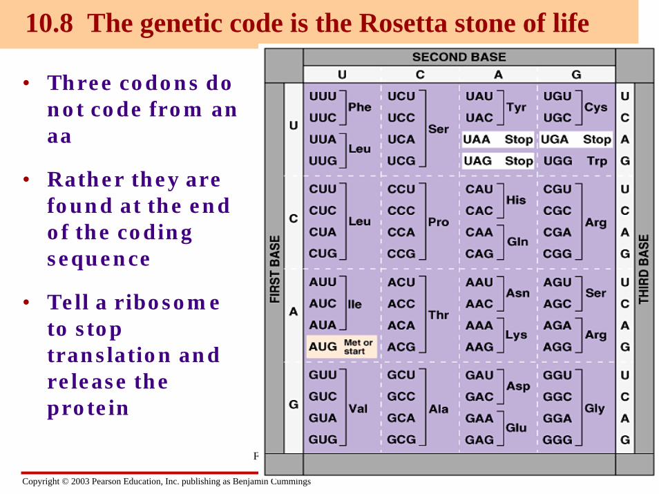
10.8 The genetic code is the Rosetta stone of life
Figure 10.8A
Copyright © 2003 Pearson Education, Inc. publishing as Benjamin Cummings
• Three codons do not code from an aa
• Rather they are found at the end of the coding sequence
• Tell a ribosome to stop translation and release the protein
10.8 The genetic code is the Rosetta stone of life
Figure 10.8A

Copyright © 2003 Pearson Education, Inc. publishing as Benjamin Cummings
• Tryptophan and Methionine have only 1 codon each
• All the rest have more than one
• AUG has a dual function
• 3 stop codons that code for termination of protein synthesis
• Redundancy in the code but no ambiguity
Figure 10.8A

Copyright © 2003 Pearson Education, Inc. publishing as Benjamin Cummings
Translation• The process of reading the RNA sequence of an
mRNA and creating the amino acid sequence of a protein is called translation.
Transcription
Codon Codon Codon
Translation
DNA
T T C A G T C A G
DNAtemplatestrand
mRNA
A A G U C A G U C MessengerRNA
Protein Lysine Serine ValinePolypeptide(amino acidsequence)

Copyright © 2003 Pearson Education, Inc. publishing as Benjamin Cummings
• In the cytoplasm, a ribosome attaches to the mRNA and translates its message into a polypeptide
• The process is aided by transfer RNAs
10.11 Transfer RNA molecules serve as interpreters during translation
Figure 10.11A
Hydrogen bond
Amino acid attachment site
RNA polynucleotide chain
Anticodon

Copyright © 2003 Pearson Education, Inc. publishing as Benjamin Cummings
A codon of three nucleotides determines choice of amino acid

Copyright © 2003 Pearson Education, Inc. publishing as Benjamin Cummings
Translation is composed of three steps
• Initiation translation begins at start codon(AUG=methionine)
Elongation the ribosome uses the tRNAanticodon to match codons to amino acids and adds those amino acids to the growing peptide chain
Termination translation ends at the stop codonUAA, UAG or UGA

Copyright © 2003 Pearson Education, Inc. publishing as Benjamin Cummings
• mRNA, a specific tRNA, and the ribosome subunits assemble during initiation
Figure 10.13B
1
Initiator tRNA
mRNA binding site
Startcodon Small ribosomal
subunit
2
P site
LargeRibosomalsubunit
A site

Copyright © 2003 Pearson Education, Inc. publishing as Benjamin Cummings
Translation initiation
Leadersequence
mRNA5’ 3’
mRNA
A U GU U C G U C G G A C G AU G U A A G A
Small ribosomal subunit
Assembling to begin translation
Met
U A C
Initiator tRNA

Copyright © 2003 Pearson Education, Inc. publishing as Benjamin Cummings
Translation Elongation
CU A
Met
mRNA5’ 3’
Amino acidLarge ribosomal subunit
C C U
tRNA
Ribosome
Gly
U U U CG G G G GGA A A A A
P A

Copyright © 2003 Pearson Education, Inc. publishing as Benjamin Cummings
Translation Elongation
CU A
Met
mRNA5’ 3’
C C U
Gly
U U U CG G G G GGA A A A A
AAC
Cys
P A

Copyright © 2003 Pearson Education, Inc. publishing as Benjamin Cummings
Translation Elongation
mRNA5’ 3’
CC
U
MetGly
CU U
Lys
Lengtheningpolypeptide(amino acid chain)
A AC
Cys
U U U CG G G G GGA A A A A
P A

Copyright © 2003 Pearson Education, Inc. publishing as Benjamin Cummings
Translation Elongation
mRNA5’
U U U CG G G G GGA A A A A U A A
Stop codon
C UG
Arg
CU U
Lys
MetGly
Cys
Releasefactor
A
AC
P
A

Copyright © 2003 Pearson Education, Inc. publishing as Benjamin Cummings
Translation Termination
mRNA5’
CU U
Met Gly CysLys
Stop codonRibosome reaches stop codon
C UG
Arg
U U U CG G G G GGA A A A A U A A
ReleasefactorP
A

Copyright © 2003 Pearson Education, Inc. publishing as Benjamin Cummings
Translation Termination
UU U C
G G G G GGA
A A A A U A A
C UG
Met GlyCys
LysArg
ReleasefactorP
Once stop codon is reached, elements disassemble.
A

Copyright © 2003 Pearson Education, Inc. publishing as Benjamin Cummings

Copyright © 2003 Pearson Education, Inc. publishing as Benjamin Cummings

Copyright © 2003 Pearson Education, Inc. publishing as Benjamin Cummings
Levels of protein structurePrimary structure sequence of amino
acidsSecondary structure shapes formed with
regions of the protein (helices, coil, sheets)
Tertiary structure shape of entire folded protein due to interactions between particular peptides
Quaternary structure structures formed by interaction of several proteins togethere.g. Functional hemoglobin istwo alpha-hemoglobin proteins andtwo beta-hemoglobin proteins

Copyright © 2003 Pearson Education, Inc. publishing as Benjamin Cummings
10_14d.jpg

Copyright © 2003 Pearson Education, Inc. publishing as Benjamin Cummings
Levels of protein structure

Copyright © 2003 Pearson Education, Inc. publishing as Benjamin Cummings
Misfolding of protein impairs function
•Misfolded prion protein disrupts functions of other normally folded prion proteins. •Aberrant conformation can passed on propagating likean “infectious” agent.

Copyright © 2003 Pearson Education, Inc. publishing as Benjamin Cummings
10_18.jpg