cellulitis? - healthcare conferences uk · pdf filecellulitis? an evidence based approach to...
TRANSCRIPT

Cellulitis?
An evidence based approach to diagnosis and treatment of red legs
Linda Nazarko Nurse Consultant
North West Healthcare NHS Trust
5th December 2014 ICO Conference Centre, London

Aims and objectives
To be aware of:
What cellulitis is and what it is not
The importance of diagnosis
Antimicrobial stewardship – why it matters
How to diagnose cellulitis
How to treat
Where to treat
Unresponsive cellulitis
Unusual organisms
Considerations when managing co-
morbidities

Lower leg cellulitis: A growing problem
49,500
74,000
87,749
0
10,000
20,000
30,000
40,000
50,000
60,000
70,000
80,000
90,000
2003 2006 2010
Rising admissions celluliits
77 percent increase in the last seven years 87,749 admissions 400,000 bed days Mean length of stay 7.1 days Cost £172-£254 million a year on inpatient treatment
What is cellulitis?
Cellulitis is a spreading bacterial infection of the dermis and subcutaneous tissues.
(Morris, 2003)
What causes cellulitis
The bacteria that most commonly cause cellulitis are:
Streptococci (esp. Strep. pyogenes)
Staphylococcus aureus
Other bacteria including Gram-negative bacilli, Strep. pneumoniae and anaerobes such as pseudomonas can also cause cellulitis
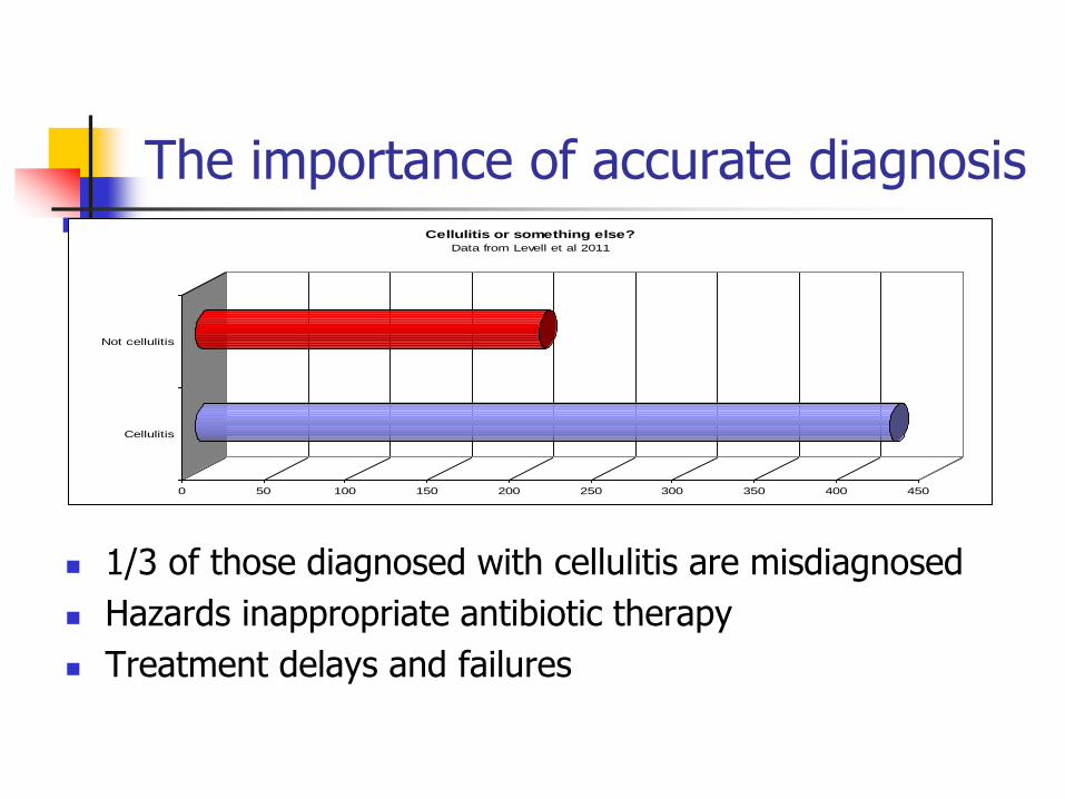
The importance of accurate diagnosis
0 50 100 150 200 250 300 350 400 450
Cellulitis
Not cellulitis
Cellulitis or something else?
Data from Levell et al 2011
1/3 of those diagnosed with cellulitis are misdiagnosed
Hazards inappropriate antibiotic therapy
Treatment delays and failures

Guide to diagnosis & treatment

What else could it be?
Eczema
Lymphoedema
Lipodermatosclerosis
Other
Venous eczema
Lymphoedema
Lipodermatosclerosis
Other
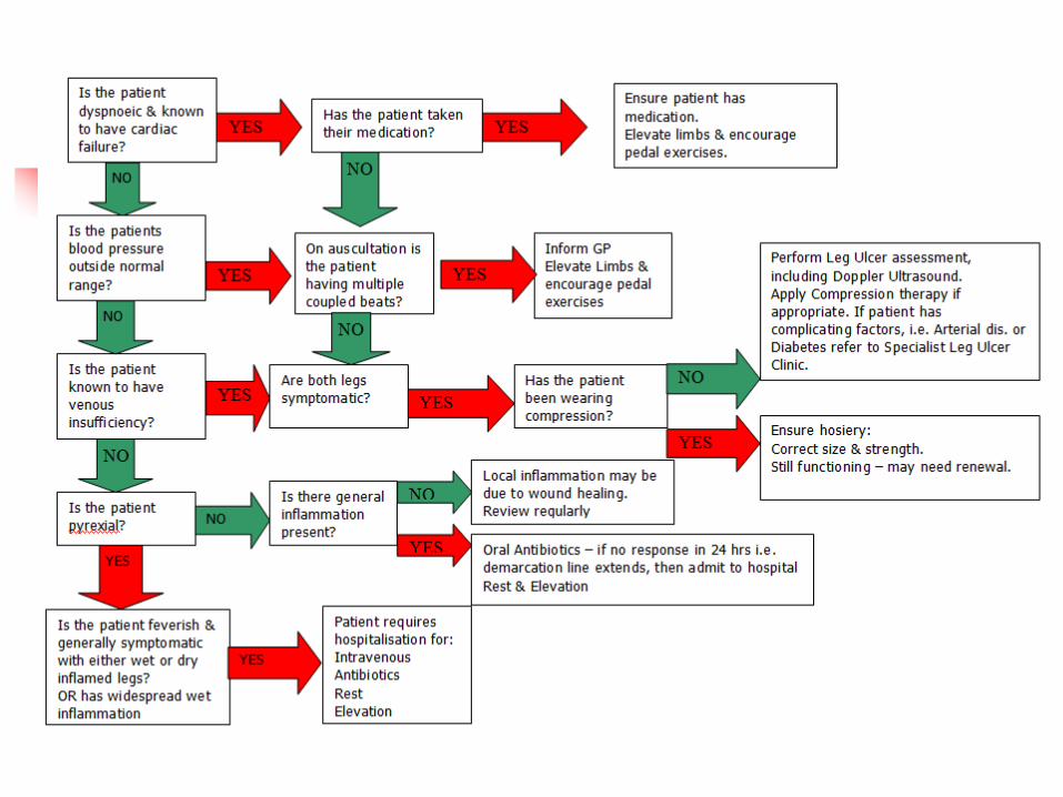
How to diagnose
Cellulitis is diagnosed on the basis of
clinical features
Take your time and check clinical
features
Consider alternative diagnosis

Clinical features cellulitis Feature Comment
Unilateral Bilateral – consider venous eczema
Sudden onset May have flu like symptoms
Red, hot, swollen, tender,
demarcation
Mark leg to check when receding
Scratch, cut, ulcer, fungal infection of
feet
Check for portal of entry
Systemically unwell? Check fever, malaise, rigours vomiting
Enlarged lymph glands, lymphangitis Spreads proximally from area of
cellulitis

Cellulitis quick guide Unilateral – bilateral
consider alternative diagnosis
Sudden onet Raised WCC Warm, red confined to
leg Clear demarcation Tender May be pyrexial Portal entry- ulcer,
scratch, fungal infection
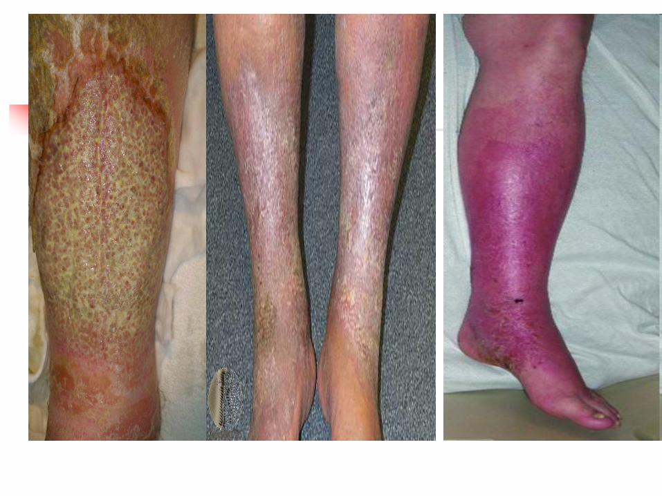

A = Cellulitis B= Acute infection chronic venous eczema C= Acute infection chronic venous eczema D= Contact dermatitis caused by paste bandages
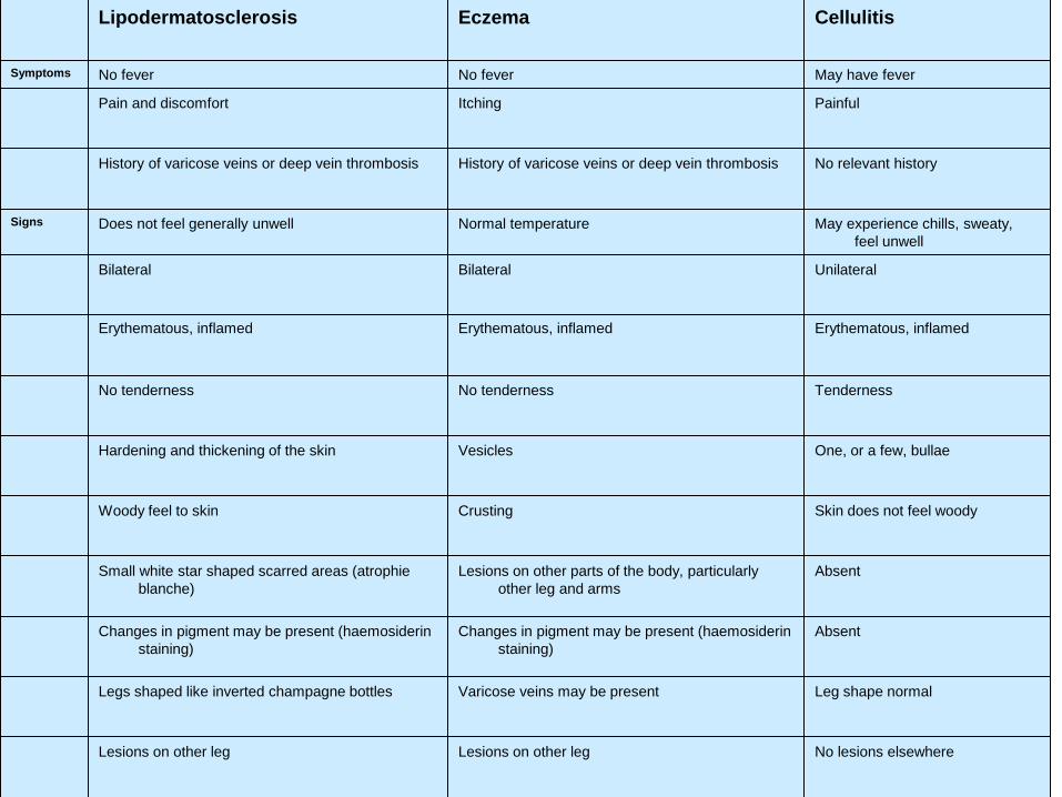
Lipodermatosclerosis Eczema Cellulitis
Symptoms No fever No fever May have fever
Pain and discomfort Itching Painful
History of varicose veins or deep vein thrombosis History of varicose veins or deep vein thrombosis No relevant history
Signs Does not feel generally unwell Normal temperature May experience chills, sweaty,
feel unwell
Bilateral Bilateral Unilateral
Erythematous, inflamed Erythematous, inflamed Erythematous, inflamed
No tenderness No tenderness Tenderness
Hardening and thickening of the skin Vesicles One, or a few, bullae
Woody feel to skin Crusting Skin does not feel woody
Small white star shaped scarred areas (atrophie
blanche)
Lesions on other parts of the body, particularly
other leg and arms
Absent
Changes in pigment may be present (haemosiderin
staining)
Changes in pigment may be present (haemosiderin
staining)
Absent
Legs shaped like inverted champagne bottles Varicose veins may be present Leg shape normal
Lesions on other leg Lesions on other leg No lesions elsewhere
Portal entry Not applicable Not applicable Usually unknown, but break in
skin, ulcers, trauma,
athlete’s foot implicated
Investigatio
ns White cell count normal
CRP normal in chronic lipodermatosclerosis and
slightly elevated in acute lipodermatosclerosis
White cell count normal
CRP normal
Skin swabs—Staphylococcus aureus common
White cell count high
CRP High
Blood culture negative Blood culture negative Blood culture usually negative
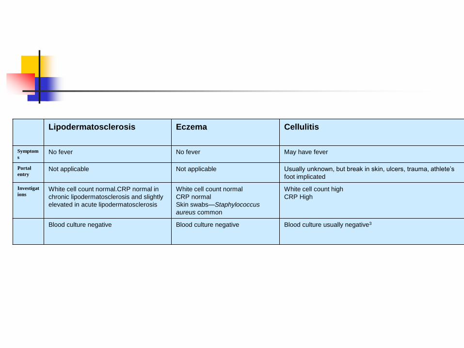
Lipodermatosclerosis Eczema Cellulitis
Symptom
s No fever No fever May have fever
Portal
entry Not applicable Not applicable Usually unknown, but break in skin, ulcers, trauma, athlete’s
foot implicated
Investigat
ions White cell count normal.CRP normal in
chronic lipodermatosclerosis and slightly
elevated in acute lipodermatosclerosis
White cell count normal
CRP normal
Skin swabs—Staphylococcus
aureus common
White cell count high
CRP High
Blood culture negative Blood culture negative Blood culture usually negative3

Infected venous eczema

Lipodermatosclerosis
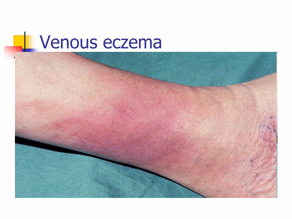
Venous eczema

Eron’s Classification of Cellulitis
Class Description
One Patients have no signs of systemic toxicity, have no uncontrolled long term conditions and can usually take oral antibiotics at home.
Two Patients are either unwell or well but have a condition such as peripheral vascular disease, chronic venous insufficiency or morbid obesity which affect recovery
Three Patients may be unwell and have symptoms such as acute confusion, tachycardia, breathlessness, hypotension or may have unstable conditions that may interfere with a response to therapy or have a limb threatening infection due to vascular compromise.
Four Patients have septicaemia or severe life threatening infection such as necrotizing fasciitis.

Treatment by class Class Description
One Oral flucloxacillin 500mg QDS Or Clarithromycin 500mg 12 hourly if penicillin allergy
Two IV flucloxacillin 1-2gm 6 hourly Or Clindamycin 450mg 8 hourly OPAT Ceftriaxone 1gm daily IV
Three IV flucloxacillin 2gm 6 hourly Or Clindamycin 900mg 8 hourly Or Clarithrmycin 500mg 12 hourly
Four As guided by microbiology

Classification and place treatment


One antibiotic or two?
Penicillin + Flucloxacillin common practice
Rationale treats staphylococcal and streptococcal but flucloxacillin effective against streptococcal infections
Lehman and Mukherjee’s 2005 study found no difference clinical outcomes those treated solely with flucloxacillin

IV to oral switch
No benefit IV after 4 days
Indicators for oral switch – less intense erythema, erythema receding, skin cooler. Pain and swelling settling. Inflammatory markers settling. Pyrexia settling.

Unresponsive cellulitis
Wrong diagnosis
Wrong antibiotic
Immunocompromised
host

Treatment of venous eczema
Compression therapy to correct venous stasis.
Vein surgery, endovenous laser ablation or sclerotherapy
Weight reduction
Emollient therapy

Treatment of acute lipodermatosclerosis
This is an inflammatory condition not an infective condition
Treatments include, stanozolol (2 mg bid for 8 weeks), topical steroids, topical capsaicin, weight loss, compression, improved mobility

Treatment of chronic lipodermatosclerosis
Compression therapy to correct venous stasis.
Vein surgery, endovenous laser ablation or sclerotherapy
Weight reduction
Ultrasound therapy
Fibrinolytic agents such as stanozolol
Pentoxyfylline to increase blood flow
Clobetasol propionate or other high potency steroid
Intralesional triamcinolone injections reduce inflammation
Capsaicin to reduce pain
Horse chestnut extract

Sweet’s syndrome Fever; Leucocytosis; Acute, tender, red plaques; Papillary dermal infiltrate of
neutrophils. Classical, malignancy related
and idiopathic Treated with steroids Potassium iodide and
colchicine are alternative first-line therapies and indomethacin (indometacin), clofazimine, cyclosporine (ciclosporin), and dapsone are second-line treatments
Other bacterial causes cellulitis
B streptococci (e.g., Streptococcus agalactiae) more common in older people.
Pseudomonas aeruginosa more common in those with diabetes and older people.
Animal bites can lead to Pasteurella multocida (cat bite) Capnocytophaga sp (dog bite).
Fresh water bathing, paddling Aeromonas hydrophila;
Warm salt water, by Vibrio vulnificus

Lets try an antibiotic What harm can it do?
0
50
100
150
200
1999 2001 2002 2003 2004 2005 2006 2007
C. Difficile rates
Primary aim healthcare “Primum non nocere”
Patients deserve an accurate diagnosis and appropriate treatment
We need to use resources wisely
0
10
20
30
40
50
60
70
Co-amoxiclav Ampicillin Cefalexin Cefpodoxime Ciprofloxacin Gentamycin Nitrofurantoin Trimethoprim
E. coli UTI antibiotic resistance

Antibiotic stewardship
“Every antibiotic expected by a patient, every unnecessary prescription written by a doctor, every uncompleted course of antibiotics, and every inappropriate or unnecessary use in animals or agriculture is potentially signing a death warrant for a future patient.”
(Donaldson, 2008)
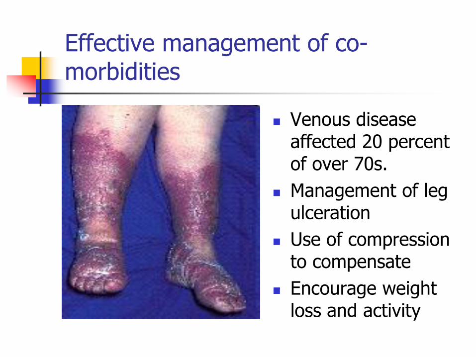
Effective management of co-morbidities
Venous disease affected 20 percent of over 70s.
Management of leg ulceration
Use of compression to compensate
Encourage weight loss and activity
Take home messages
400,000 bed days - £254 million spent on
cellulitis – one third inappropriately
One third of “cellulitis” is misdiagnosed
This exposes patients to the hazards of
inappropriate antibiotic therapy and
treatment delay/failure
We must ensure that we use antibiotics
prudently and are aware of the evidence
base.

Thank you for listening
Any questions?