cell, vol. 97, 503–514, may 14, 1999, copyright 1999 by...
TRANSCRIPT

Cell, Vol. 97, 503–514, May 14, 1999, Copyright 1999 by Cell Press
Mammalian Telomeres End in a Large Duplex Loop
solutions to the end replication problem have been doc-Jack D. Griffith,*‡ Laurey Comeau,*umented, generally involving some form of recombina-Soraya Rosenfield,* Rachel M. Stansel,*tion of tandem terminal repeats (Morin and Cech, 1988;Alessandro Bianchi,†§ Heidi Moss,†Lundblad and Szostak, 1989). The end replication prob-and Titia de Lange†
lem of human telomeres has received particular atten-*Lineberger Comprehensive Cancer Centertion for its possible significance to aging and cancer (deUniversity of North Carolina at Chapel HillLange et al., 1990; Harley et al., 1990; Hastie et al., 1990;and Curriculum in Genetics and Molecular BiologyCounter et al., 1994; reviewed in de Lange and DePinho,Chapel Hill, North Carolina 27599-72951999). Maintenance of the telomeric TTAGGG repeats†The Rockefeller Universityat human chromosome ends, either by telomeraseNew York, New York 10021(Bodnar et al., 1998) or an alternative mechanism (ALT;Bryan et al., 1997), is essential for long-term replicativesurvival of cells in vitro, and a telomerase-deficientSummarymouse strain displays several phenotypes consistentwith impaired tissue homeostasis (Lee et al., 1998; Ru-Mammalian telomeres contain a duplex array of telo-dolph et al., 1999).meric repeats bound to the telomeric repeat–binding
Despite the importance of telomere maintenance, itfactors TRF1 and TRF2. Inhibition of TRF2 results inhas become increasingly clear that telomeres do notimmediate deprotection of chromosome ends, mani-simply function as a buffer zone that prevents loss offested by loss of the telomeric 39 overhang, activationessential sequences. A large body of evidence datingof p53, and end-to-end chromosome fusions. Electronback to the work of Muller and McClintock (Muller, 1938;microscopy reported here demonstrated that TRF2McClintock, 1941) is more consistent with the view thatcan remodel linear telomeric DNA into large duplexthe telomeric complex allows cells to distinguish randomloops (t loops) in vitro. Electron microscopy analysisDNA breaks and natural chromosome ends. Whereasof psoralen cross-linked telomeric DNA purified frombroken chromosomes activate DNA damage check-human and mouse cells revealed abundant large tpoints (Weinert and Hartwell, 1988; Sandell and Zakian,loops with a size distribution consistent with their telo-1993) and are repaired, telomeres are not detected asmeric origin. Binding of TRF1 and single strand bindingDNA ends.
protein suggested that t loops are formed by invasionBased on work with unicellular organisms, the molec-
of the 39 telomeric overhang into the duplex telomericular mechanism of this capping function has been pro-
repeat array. T loops may provide a general mecha- posed to either depend on a specific DNA structure innism for the protection and replication of telomeres. the most terminal single-stranded portion of the telo-
mere (e.g., G quartets; Williamson et al., 1989) or toIntroduction require proteins bound to the 39 telomere end (e.g., the
Oxytricha nova telomeric protein; Gottschling and Zak-DNA genomes tend to be circular. Bacterial genomes, ian, 1986; Horvath et al., 1998). However, there is noplasmids, bacteriophages, and mitochondrial DNAs are evidence in support of G–G base-paired structures atusually circular. Even T phages, while linear, likely repli- telomere termini in vivo, and a terminus-specific proteincate through circular intermediates. Similarly, many has yet to be isolated from mammalian cells (reviewed ineukaryotic DNA viruses, such as SV40, polyoma, and de Lange, 1996). Instead, duplex telomeric DNA–bindinghepatitis B virus, have circular genomes, and like l, proteins have recently emerged as key players in theEpstein–Barr virus is circular in its episomal state. In capping of mammalian chromosome ends.contrast, eukaryotic cellular chromosomes are by and The duplex array of TTAGGG repeats at mammalianlarge linear, a deviation from the norm that may have telomeres is bound by two related proteins, the TTAGGGallowed the advent of meiosis (Naito et al., 1998). repeat–binding factors TRF1 and TRF2 (Chong et al.,
A major drawback of linear chromosomes is associ- 1995; Bilaud et al., 1997; Broccoli et al., 1997). Inhibitionated with the presence of DNA ends in the eukaryotic of TRF2 results in immediate activation of the ATM/nucleus. Telomeres are specialized terminal elements, p53–dependent DNA damage checkpoint pathway,composed of tandem repetitive sequences and specific leading to cell cycle arrest and apoptosis (Karlseder etproteins, that appear to obviate these problems. In many al., 1999). The exposure of chromosome ends to DNAeukaryotes, telomeric repeat tracts are maintained by damage checkpoints is not due to the loss of the duplexa telomere-specific reverse transcriptase, telomerase, TTAGGG repeat array (van Steensel et al., 1998; Karl-
seder et al., 1999; J. Karlseder and T. d. L., unpublishedthat can counteract the loss of terminal sequences dur-data). Thus, the presence of telomeric DNA at chromo-ing DNA replication (Greider and Blackburn, 1985; re-some ends per se is not sufficient for telomere function.viewed in Nugent and Lundblad, 1998). However, otherAlthough the telomeric repeat array remains largely in-tact, the single-stranded protrusion of TTAGGG repeats‡ To whom correspondence should be addressed (e-mail: jdg@found at all mammalian telomeres (Makarov et al., 1997)med.unc.edu).is lost upon inhibition of TRF2, suggesting that the G§ Present address: Department of Molecular Biology, University of
Geneva, 30 quai Ernest-Ansermet, CH-1211 Geneva, Switzerland. strand overhangs are important for telomeric protection

Cell504
(van Steensel et al., 1998). Furthermore, the unmaskedtelomeres eventually undergo covalent fusion, mostlikely reflecting the inappropriate exposure of unpro-tected telomere termini to ligases and other DNA repairactivities (van Steensel et al., 1998; A. Smogorzewskaand T. d. L., unpublished data). Based on these data, itwas proposed that the TTAGGG repeats collaboratewith TRF2 to sequester chromosome ends from themany cellular activities that threaten their integrity andsafeguard against the inappropriate activation of DNAdamage checkpoints by natural chromosome ends.However, the mechanism by which TRF2 acts to protectand mask chromosome ends is not known.
Here we present evidence that the sequestration oftelomere ends by TRF2 may be achieved through anarchitectural change in the conformation of telomericDNA. This structural solution to the chromosome endproblem is deduced from the finding that the telomericDNA can be isolated as large duplex loops, called tloops, in which the terminus is embedded within thedouble-stranded part of the telomeric tract. TRF2 can
Figure 1. T Loops Generated In Vitro by Human TRF2generate t loops in vitro, and its in vivo function is consis-A telomeric DNA model containing 3 kb of unique sequence DNAtent with t loops as the main mechanism by which mam-followed by z2 kb of repeating TTAGGG sequence with a 150–200malian cells mask natural chromosome ends.nt 39 G strand overhang was incubated with human TRF2 protein.In (A), the sample was directly adsorbed to the carbon EM support
Results followed by rotary shadowcasting with tungsten. In (B), followingincubation with TRF2, the sample was exposed to psoralen and UVfollowed by deproteinization, surface spreading with cytochromeTRF2 Generates T Loops on a Linear TelomericC, and rotary shadowcasting with platinum–paladium. Shown inDNA Model In Vitroreverse contrast. Bar is equivalent to 1 kb.
Our preliminary work with purified TRF2 and plasmidDNAs suggested that TRF2 might pair single-strandedand double-stranded DNA segments if both contained separate experiments, n 5 100 for each) given that only
35%–40% of the model DNAs carried long 39 TTAGGGTTAGGG repeats (J. D. G. and T. d. L., unpublisheddata). To explore this further, a linear telomeric DNA repeat overhangs. Incubations of the DNA in the ab-
sence of protein resulted in 1%, 3%, and 5% lasso-likemodel was constructed containing several kilobases ofdouble-stranded tandem TTAGGG repeats grown from structures in three separate experiments, suggesting
that t loops are a genuine product of the interaction ofone end of a 3 kb segment of unique sequence DNA. Theends of the DNA were resected with a 59 exonuclease to TRF2 with the telomeric substrate. In contrast to TRF2,
addition of the related protein TRF1 resulted in only 6%generate a 100–200 nt 39 overhang of TTAGGG repeats,and the fraction of DNAs with an overhang and its length t loops, a frequency of t loop formation that may not be
significantly higher than what is observed in absence ofwere monitored by addition of E. coli single strand bind-ing (SSB) protein and electron microscopy (EM) exami- protein. Furthermore, TRF1 was never observed at the
tail–loop junction, indicating that the binding of TRF2 tonation (Experimental Procedures). Approximately 35%–40% of the molecules carried the expected 39 overhang. t loops is a highly specific event.
The requirement for a 39 overhang in TRF2-mediatedThe telomeric DNA model closely approximates the twoknown features of mammalian telomeric DNA, several t loop formation suggested that this structure could de-
pend on the invasion of the single-stranded TTAGGGkilobases of duplex TTAGGG repeats ending in a long39 tail of the G-rich strand. repeats into the duplex part of the telomeric tract. In
this case, psoralen cross-linking of the DNA strands isThe telomeric DNA model was incubated with bacu-lovirus-derived purified human TRF2 and the complexes predicted to preserve the t loops after removal of pro-
tein. By constrast, if the t loops are held together byprepared for EM. As shown in Figure 1A, lariat- or lasso-like molecules, referred to here as t loops, were ob- TRF2–DNA interactions only, the cross-linking of DNA
strands is not predicted to preserve the t loops.served, and a large TRF2 protein complex was presentat the loop–tail junction in all cases (the term “tail” will Accordingly, we next analyzed TRF2-induced t loops
by psoralen cross-linking of the DNA (Hanson et al.,refer to the linear duplex DNA appendage attached tothe circular portion of the molecule). Removal of the 1976) followed by deproteinization and spreading of the
DNA with cytochrome C (“Kleinschmidt” method) (Klein-39 overhang from the model telomere with mung beannuclease eliminated all SSB binding, as seen by EM, schmidt and Zahn, 1959). Both approaches have been
used extensively separately and in combination (Cechand in two experiments, addition of TRF2 resulted in7% (n 5 100) and 0% (n 5 150) lasso-like molecules, and Pardue, 1976; Hanson et al., 1976), and ways of
employing them that avoid artifactual DNA associationsrevealing a requirement for the single-stranded TTAGGGrepeats. The frequency of t loop formation on the model are well understood. The psoralens HMT (49-hydroxy-
methyl trioxalen) and AMT (49-aminomethyl trioxalen)DNA by TRF2 was strikingly high (17% and 20% in two
Telomeres Loop Back505
intercalate into a duplex DNA and upon UV irradiationpreferentially cross-link T residues of opposite strands.Thus, each TTAGGG repeat in the telomeric repeat tractcontains potential cross-linking sites, and conditionswere established that created a cross-link in HeLa DNAevery few 100 bp (Experimental Procedures).
The Kleinschmidt EM technique is particularly suitablefor the analysis of t loops, since the strong surface ten-sion at the air–buffer interface forces DNA molecules(which have been thickened 5- to 10-fold and greatlystiffened) to spread out so that the strands seldom crossover themselves. Furthermore, DNA ends are held apartand DNAs known to be linear are seldom observed toform loops or lassos.
When the telomeric DNA model was cross-linked withAMT in the absence of protein, the frequency of lasso-like structures and circles varied from 0%–6% (n 5 100)in four experiments, likely reflecting accidental juxtapo-sitioning. However, when the telomere model DNA wastreated with AMT after incubation with TRF2, the depro-teinized products showed t loops at a high frequency(17%, 19%, and 25% in three experiments, n 5 100 foreach) (Figure 1B). As expected, very few t loops (4%and 0%, respectively, n 5 50 in two experiments) wereobserved in parallel incubations with two control pro-teins, TRF1 and tankyrase (Bianchi et al., 1997; Smithet al., 1998). These results argue that TRF2 can promotea looped structure in telomeric DNA that involves theinvasion of the 39 telomeric overhang into the duplexDNA. Further detailed analysis of the generation of invitro t loops by TRF2 will be described elsewhere(R. M. S., T. d. L., and J. D. G., in preparation).
The formation of t loops by TRF2 in vitro suggested amechanism by which TRF2 and telomeric repeats mightfunction together to sequester chromosome ends. Toaddress this possibility, the presence of t loops at chro-mosome ends in vivo was studied.
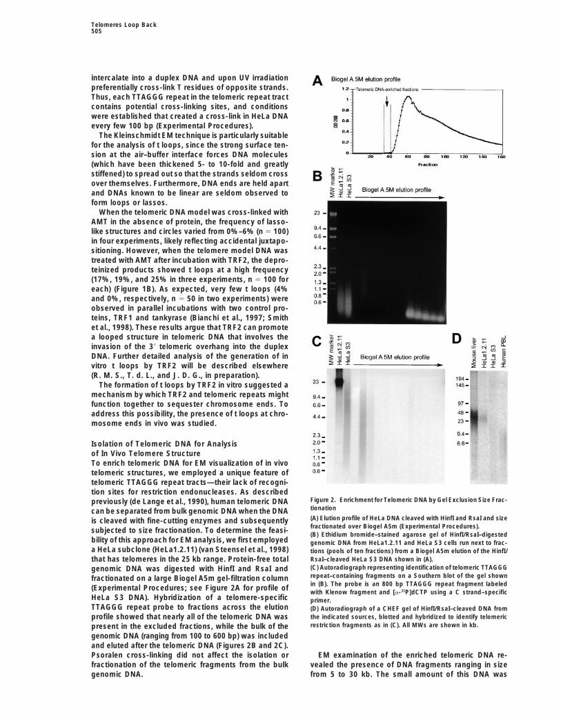
Isolation of Telomeric DNA for Analysisof In Vivo Telomere StructureTo enrich telomeric DNA for EM visualization of in vivotelomeric structures, we employed a unique feature oftelomeric TTAGGG repeat tracts—their lack of recogni-tion sites for restriction endonucleases. As described
Figure 2. Enrichment for Telomeric DNA by Gel Exclusion Size Frac-previously (de Lange et al., 1990), human telomeric DNAtionationcan be separated from bulk genomic DNA when the DNA(A) Elution profile of HeLa DNA cleaved with HinfI and RsaI and sizeis cleaved with fine-cutting enzymes and subsequentlyfractionated over Biogel A5m (Experimental Procedures).subjected to size fractionation. To determine the feasi-(B) Ethidium bromide–stained agarose gel of HinfI/RsaI–digested
bility of this approach for EM analysis, we first employed genomic DNA from HeLa1.2.11 and HeLa S3 cells run next to frac-a HeLa subclone (HeLa1.2.11) (van Steensel et al., 1998) tions (pools of ten fractions) from a Biogel A5m elution of the HinfI/
RsaI–cleaved HeLa S3 DNA shown in (A).that has telomeres in the 25 kb range. Protein-free total(C) Autoradiograph representing identification of telomeric TTAGGGgenomic DNA was digested with HinfI and RsaI andrepeat–containing fragments on a Southern blot of the gel shownfractionated on a large Biogel A5m gel-filtration columnin (B). The probe is an 800 bp TTAGGG repeat fragment labeled(Experimental Procedures; see Figure 2A for profile ofwith Klenow fragment and [a-32P]dCTP using a C strand–specific
HeLa S3 DNA). Hybridization of a telomere-specific primer.TTAGGG repeat probe to fractions across the elution (D) Autoradiograph of a CHEF gel of HinfI/RsaI–cleaved DNA from
the indicated sources, blotted and hybridized to identify telomericprofile showed that nearly all of the telomeric DNA wasrestriction fragments as in (C). All MWs are shown in kb.present in the excluded fractions, while the bulk of the
genomic DNA (ranging from 100 to 600 bp) was includedand eluted after the telomeric DNA (Figures 2B and 2C).Psoralen cross-linking did not affect the isolation or EM examination of the enriched telomeric DNA re-
vealed the presence of DNA fragments ranging in sizefractionation of the telomeric fragments from the bulkgenomic DNA. from 5 to 30 kb. The small amount of this DNA was
Cell506
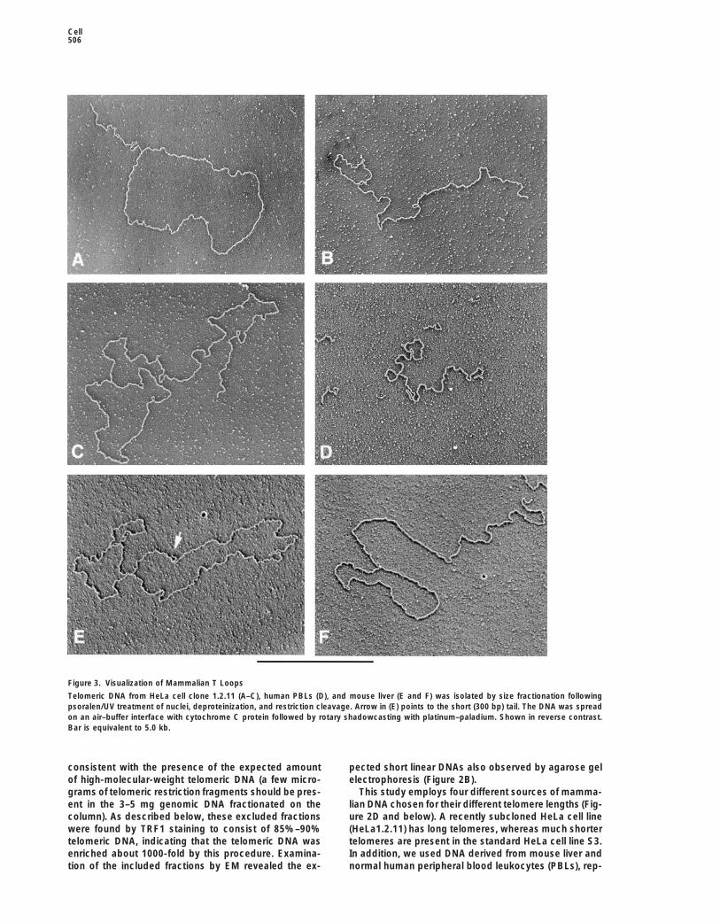
Figure 3. Visualization of Mammalian T Loops
Telomeric DNA from HeLa cell clone 1.2.11 (A–C), human PBLs (D), and mouse liver (E and F) was isolated by size fractionation followingpsoralen/UV treatment of nuclei, deproteinization, and restriction cleavage. Arrow in (E) points to the short (300 bp) tail. The DNA was spreadon an air–buffer interface with cytochrome C protein followed by rotary shadowcasting with platinum–paladium. Shown in reverse contrast.Bar is equivalent to 5.0 kb.
consistent with the presence of the expected amount pected short linear DNAs also observed by agarose gelelectrophoresis (Figure 2B).of high-molecular-weight telomeric DNA (a few micro-
grams of telomeric restriction fragments should be pres- This study employs four different sources of mamma-lian DNA chosen for their different telomere lengths (Fig-ent in the 3–5 mg genomic DNA fractionated on the
column). As described below, these excluded fractions ure 2D and below). A recently subcloned HeLa cell line(HeLa1.2.11) has long telomeres, whereas much shorterwere found by TRF1 staining to consist of 85%–90%
telomeric DNA, indicating that the telomeric DNA was telomeres are present in the standard HeLa cell line S3.In addition, we used DNA derived from mouse liver andenriched about 1000-fold by this procedure. Examina-
tion of the included fractions by EM revealed the ex- normal human peripheral blood leukocytes (PBLs), rep-

Telomeres Loop Back507
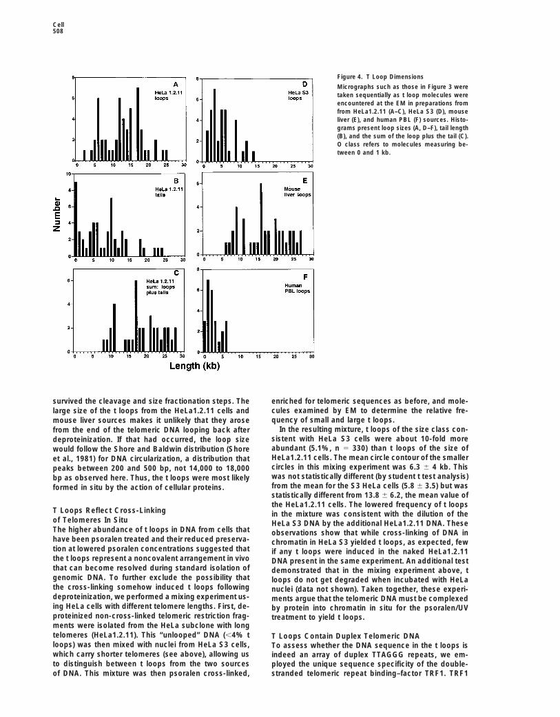
Table 1. T Loop Dimensions Correlate with Telomere Lengths
T Loop Dimensions (kb)Telomeric Restriction Fragments
Cell Type Range (Mean) in kb Loop Tail Loop 1 Tail
HeLa clone 15–40 (23) 13.8 6 6.2 8.5 6 6.6 22.4 6 6.9 n 5 581.2.11HeLa clone 3.5–20 (10) 5.8 6 3.5 5.2 6 3.0 10.1 6 2.8 n 5 37S3Mouse liver 10–50 (25) 18.2 6 6.2 0.92 6 1.7 19.2 6 5.9 n 5 38Human PBL 3.5–15 (7) 2.9 6 1.9 5.4 6 3.0 8.3 6 3.0 n 5 27
resenting untransformed mammalian cells with long mouse preparations ranged from 15% to 20% of theDNA $5 kb (n 5 300) in three experiments and wastelomeres and short telomeres, respectively.z15% (n 5 300) in the human PBL DNA. As describedbelow, however, the dimensions of these t loops wereFrequent T Loops in Telomere-Enriched DNAseveralfold smaller than those isolated from mouse liver.The telomere-enriched fractions from psoralen cross-
linked HeLa nuclei revealed a striking abundance of tloops (Figures 3A–3C). In some cases the tail was not T Loop Sizes Correlate with Telomere Lengthsvisible, resulting in a circle. The fraction of molecules If the t loops represent telomeres, the size of thesearranged into t loops was scored in 15 different experi- molecules might be expected to correlate with telomerements (using the two different HeLa lines) and found to length. To explore this possibility, we measured the di-range from 15%–40%. In this study, “percentage of t mensions of t loops in the four cell types with distinctlyloops” will be taken to mean the fraction of molecules different telomere lengths, shown in Figure 2D. Although$5 kb arranged into lassos or circles. In a titration exper- the exact lengths of human telomeric repeat tracts can-iment, the frequency of t loops was reduced severalfold not be determined accurately by genomic blotting (re-as the concentration of AMT was decreased 5- and 10- viewed in de Lange, 1995), HeLa1.2.11 telomeres appearfold. Less than 1 molecule in 1000 was arranged as to range from 15–40 kb (mean 23 kb), whereas HeLa S3a double lasso (both ends folded back to generate a and normal human PBL telomeres are shorter, dis-molecule consisting of two circles joined by a linear playing a mean telomeric restriction fragment lengthsegment), indicating that the t loops are not the result of 10 and 7 kb, respectively (Table 1). Mus musculusof DNA ends being sticky and fortuitously associating telomeric restriction fragments have a much wider sizewith an internal site during the purification or EM steps. distribution, ranging from 5–60 kb (Kipling and Cooke,
Although t loops were more abundant in psoralen 1990; Zijlmans et al., 1997), with the bulk migrating atcross-linked preparations, they could also be observed 25 kb in agarose (CHEF) gels (Figure 2D; Table 1).without this treatment. EM analysis of the excluded frac- Psoralen cross-linked, telomere-enriched DNAs fromtions of non-cross-linked HeLa preparations showed the four cell types were examined by EM, and sequentiallythat 95%–98% of the DNA was linear with no noteworthy encountered t loops were photographed and their lengthsstructures at either end. However, 2%–5% of the DNA determined. The data in Figures 4A–4C and Table 1molecules carried t loops (results from seven experi- present values obtained from 58 t loops derived fromments, each involving scoring .100 molecules). While the HeLa subclone with long telomeres (HeLa1.2.11).this is a relatively small number, when HeLa or mouse The contour length of the circular segment of the t loopsgenomic DNA was randomly sheared into 5–20 kb in these cells varied from 3 to 25 kb with approximatelylengths and then prepared for EM as above, less than two-thirds of the values lying between 12 and 18 kb (mean0.25% of the molecules (n 5 1000) were found arranged of 13.8 6 6). The t loop tails varied from 0 to 12 kb (meaninto lasso structures. Thus, telomeric DNA is highly en- 8.5 6 6 kb), and the length of the entire t loop (summationriched (10- to 20-fold) for t loops even without cross- of tails and circles) yielded a value of 22.4 6 7 kb.linking. Similar measurements were carried out for the HeLa
S3 cell t loops as well as for those from mouse liver andhuman PBLs. Histograms representing the sizes of theT Loops in Primary Human and Mouse Cells
To determine whether t loops occur in primary cells, circular t loop segments (Figures 4D–4F) reveal an obvi-ous difference that correlates with telomere lengths inmouse liver and human blood leukocytes were exam-
ined as above. Nuclei were prepared from fresh mouse these cells. The mouse liver t loops have the largestcircles, the HeLa S3 cells have substantially smaller cir-livers, psoralen cross-linked, and the DNA processed
to enrich for telomeric restriction fragments. Examina- cular segments, and the t loops from human PBLs havesmaller circles still. The mean lengths of the circulartion of telomere-enriched fractions revealed frequent
large t loops (Figures 3E and 3F) similar to those from segment plus the tail (entire t loops) also correlated withtelomere lengths, with the mouse liver t loops greaterHeLa cells, although the tails tended to be shorter in
the mouse liver DNA than in preparations from human than 20 kb, HeLa S3 t loops around 10 kb, and the PBLt loops measuring around 8 kb (Table 1).cells. T loops were also present in telomere-enriched
psoralen cross-linked DNA from human PBLs, isolated The good agreement between the length of the t loopsand the size of telomeres argued that t loops representfrom blood freshly drawn from a fairly normal donor
(J. D. G.) (Figure 3D). The abundance of t loops in the telomeric DNA, as contrasted to some other DNA that
Cell508
Figure 4. T Loop Dimensions
Micrographs such as those in Figure 3 weretaken sequentially as t loop molecules wereencountered at the EM in preparations fromfrom HeLa1.2.11 (A–C), HeLa S3 (D), mouseliver (E), and human PBL (F) sources. Histo-grams present loop sizes (A, D–F), tail length(B), and the sum of the loop plus the tail (C).O class refers to molecules measuring be-tween 0 and 1 kb.
survived the cleavage and size fractionation steps. The enriched for telomeric sequences as before, and mole-cules examined by EM to determine the relative fre-large size of the t loops from the HeLa1.2.11 cells andquency of small and large t loops.mouse liver sources makes it unlikely that they arose
In the resulting mixture, t loops of the size class con-from the end of the telomeric DNA looping back aftersistent with HeLa S3 cells were about 10-fold moredeproteinization. If that had occurred, the loop sizeabundant (5.1%, n 5 330) than t loops of the size ofwould follow the Shore and Baldwin distribution (ShoreHeLa1.2.11 cells. The mean circle contour of the smalleret al., 1981) for DNA circularization, a distribution thatcircles in this mixing experiment was 6.3 6 4 kb. Thispeaks between 200 and 500 bp, not 14,000 to 18,000was not statistically different (by student t test analysis)bp as observed here. Thus, the t loops were most likelyfrom the mean for the S3 HeLa cells (5.8 6 3.5) but wasformed in situ by the action of cellular proteins.statistically different from 13.8 6 6.2, the mean value ofthe HeLa1.2.11 cells. The lowered frequency of t loops
T Loops Reflect Cross-Linking in the mixture was consistent with the dilution of theof Telomeres In Situ HeLa S3 DNA by the additional HeLa1.2.11 DNA. TheseThe higher abundance of t loops in DNA from cells that observations show that while cross-linking of DNA inhave been psoralen treated and their reduced preserva- chromatin in HeLa S3 yielded t loops, as expected, fewtion at lowered psoralen concentrations suggested that if any t loops were induced in the naked HeLa1.2.11the t loops represent a noncovalent arrangement in vivo DNA present in the same experiment. An additional testthat can become resolved during standard isolation of demonstrated that in the mixing experiment above, tgenomic DNA. To further exclude the possibility that loops do not get degraded when incubated with HeLathe cross-linking somehow induced t loops following nuclei (data not shown). Taken together, these experi-deproteinization, we performed a mixing experiment us- ments argue that the telomeric DNA must be complexeding HeLa cells with different telomere lengths. First, de- by protein into chromatin in situ for the psoralen/UVproteinized non-cross-linked telomeric restriction frag- treatment to yield t loops.ments were isolated from the HeLa subclone with longtelomeres (HeLa1.2.11). This “unlooped” DNA (,4% t T Loops Contain Duplex Telomeric DNAloops) was then mixed with nuclei from HeLa S3 cells, To assess whether the DNA sequence in the t loops iswhich carry shorter telomeres (see above), allowing us indeed an array of duplex TTAGGG repeats, we em-to distinguish between t loops from the two sources ployed the unique sequence specificity of the double-
stranded telomeric repeat binding–factor TRF1. TRF1of DNA. This mixture was then psoralen cross-linked,

Telomeres Loop Back509
contains a Myb-related C-terminal domain and binds to telomeric DNA based on TRF1 binding (n 5 100), andtelomeric DNA as a dimer (Chong et al., 1995; Bianchi 40% of this fraction was comprised of t loops. Thus, inet al., 1997; van Steensel and de Lange, 1997). The the latter experiment, a minimum of 63% (and most likelyspecificity of TRF1 for double-stranded TTAGGG repeat all) of the t loops contained telomeric DNA.arrays is well documented, and closely related se-quences (such as TTTAGGG or TTAGGC repeats) are SSB Reveals a Displacement Loopgenerally a very poor binding substrate, as is single- at the Loop–Tail Junctionstranded telomeric DNA (Zhong et al., 1992; Hanish et The t loops can be explained by a model in which theal., 1994; Chong et al., 1995). single-stranded TTAGGG repeat overhang of the telo-
Previous EM analysis of TRF1 DNA complexes re- mere folds back and undergoes limited strand invasionvealed that short arrays of telomeric DNA (e.g., 6 or 12 to form a displacement loop with the duplex part of therepeats) recruit a single ball of protein, representing a telomeric repeat tract. The resulting displacement loopTRF1 tetramer (Griffith et al., 1998). When TRF1 is al- at the junction is expected to contain up to 300 nt oflowed to bind to long telomeric tracts at saturating pro- single-stranded TTAGGG repeats. To explore the pres-tein concentrations, the protein coats the telomeric DNA ence of single-stranded DNA in the t loop junctions, wealong its length, forming a 10 nm thick smooth array of employed the E. coli SSB. HeLa t loops were incubatedbound proteins. Even at high TRF1 to DNA mass ratios, with SSB, and the complexes were prepared for EMno TRF1 binding was observed by EM on nontelomeric by directly adsorbing them onto thin carbon foils andsequences, making EM visualization of TRF1 binding a shadowcasting with tungsten. Inspection of the sampleswell-defined direct assay for the presence of telomeric showed that 35% of the t loops (n 5 30) had one orDNA. several SSB protein complexes at the loop–tail junction
To apply this test, t loop–containing fractions from (Figures 5E and 5F). Since each SSB complex (tetramersHeLa cells were incubated with baculovirus-derived pu- and octamers) would be expected to associate withrified human TRF1, cross-linked with glutaraldehyde, 75–150 nt (Chrysogelos and Griffith, 1982), this suggestsand visualized by EM (Experimental Procedures). At sat- that SSB-bearing sites contain a single-stranded seg-urating TRF1 concentrations, TRF1-coated telomeric ment of possibly 75 to 200 nucleotides. It is possibleDNA filaments were visible and readily distinguishable that the junctions that did not stain with SSB containedfrom protein-free DNA on the same grids (Figure 5A). some single-stranded character but not enough to bindThe appearance of naked and TRF1-bound DNAs side a tetramer or octamer of SSB.by side argued that the TRF1-coated species containedsubstantial regions of TTAGGG repeats. Discussion
At low protein concentrations, scattered TRF1 com-plexes were observed on some molecules (Figure 5B), Based on chromosome analysis in X-ray irradiated flies,whereas higher TRF1 concentrations yielded a large Muller proposed the term telomeres and surmised thatnumber of thick filaments. Some of the thick filaments they are required to “seal” chromosome ends (Muller,were spread well enough to reveal a t loop arrangement 1938; see Gall, 1995 for discussion). The data presented(Figures 5C and 5D), and in the examples shown, the here reveal an unexpected structure present in vivo atcircular segment of the t loops is thick and TRF1 coated, mammalian telomeres, called the t loop, that is proposedwhile the tail is thin, protein free–appearing DNA. Pre-
to represent the “sealed” chromosome ends that Mullersumably, in these molecules the telomeric DNA had
referred to. The t loop is a large duplex loop-back struc-looped back to anneal close to the junction of the telo-
ture most likely formed through the invasion of the sin-meric and subtelomeric sequences, resulting in a tailgle-stranded telomeric 39 overhang into the duplex telo-devoid of telomeric repeats and hence not bound bymeric repeat array. In vitro, t loops can be formed byTRF1. In the majority of TRF1-coated molecules, theTRF2, a telomeric protein known to be required for thethick filament was twisted about itself, making it difficultprotection of mammalian chromosome ends. We pro-to trace a clear contour even though the presence of apose that t loops represent the basic mechanism bycircular form was highly suggestive. Addition of saturat-which the telomeric nucleoprotein complex sequestersing amounts of TRF2 to such large molecules led tothe natural ends of chromosomes from DNA damagesevere collapse and multimolecule tangles. The specificcheckpoints, DNA repair enzymes, and telomerase.binding of TRF1 to t loops was further substantiated by
an immunodepletion experiment in which the presenceThe Structure of T Loopsof t loops was reduced z7-fold upon incubation of theA proposal for the in vivo configuration of telomeric DNADNA–TRF1 complexes with an anti-TRF1 antibody (Ex-at mammalian chromosome ends based on the dataperimental Procedures).reported here is presented in Figure 6A. Inspection ofAlthough their twisted appearance made scoring thethe in situ configuration of human and mouse telomericfraction of TRF1-bound t loops difficult, it was simpleDNA indicates the presence of large loops formed byto score the fraction of thick protein-containing DNA asfolding back the end of the telomere. The circular seg-contrasted with thin, uncoated DNA. In HeLa DNA highlyment of the loops is composed of duplex telomeric DNA,enriched for t loops, 80% of the large molecules judgedas identified by binding of TRF1. The 39 overhang ofto be $5 kb were bound by TRF1 (n 5 100). When ansingle-stranded TTAGGG repeats appears to be insertedaliquot of the same fraction was spread for EM in theinto the duplex telomeric tract, resulting in displacementabsence of TRF1, 35% of the DNA $5 kb was in t loops.of the TTAGGG repeat strand at the loop–tail junction.In an identical but independent experiment, the fraction
most enriched for telomeric DNA scored as 85%–90% Based on binding with SSB, the displacement loop of

Cell510
Figure 5. Staining T Loops with TRF1 and SSB Protein
A telomere-enriched fraction of DNA from a size fractionation of psoralen- and UV-treated HeLa1.2.11 cell DNA that contained z40% t loopswas incubated with human TRF1 protein at high (A, C, and D) or low (B) concentration. The same DNA was independently incubated with E.coli SSB protein (arrows point to SSB particles) (E and F). Samples were prepared for EM by fixation followed by either direct adsorption tothin carbon foils and tungsten shadowcasting (A–C, E, and F) or surface spreading with cytochrome C (D) and shadowcasting with platinum–paladium. Shown in reverse contrast. Bar is equal to 1 kb.
TTAGGG repeats is deduced to be in the order of a few Formation of T LoopsIn vitro data indicates that TRF2 promotes t loop forma-hundred nucleotides in many of the t loops. Although
the exact site of the 39 end invasion point was not estab- tion. A model telomeric substrate composed of a largesegment of duplex TTAGGG repeats terminating in alished, in most molecules the loop is very large (many
kilobases) and in some cases clearly encompasses the 100–200 nt 39 overhang of the same sequence was usedhere to study TRF2–telomere complexes. The telomericwhole telomere. There is a close correlation between
the length of the telomeric repeat array and the size of DNA model was converted into a large loop upon incu-bation with purified TRF2, and all of the resulting t loopsthe t loops. The question of whether t loops always
reach back to the “base” of the telomere (the junction carried TRF2 protein at the loop–tail junction. A chal-lenge will be to determine what type of biochemicalbetween telomeric and subtelomeric DNA) in primary
cells and how their lengths vary with age needs to be activity allows TRF2 to promote in vitro telomeric loopformation. While TRF2 is known to bind to duplex telo-addressed further.

Telomeres Loop Back511
These studies illuminate the possibility that TRF2 pro-tects human telomeres through the catalysis of an archi-tectural change involving the invasion of the 39 telomereterminus into the duplex part of the telomere (Figure6B). In this regard, it is of interest that TRF1 also hasthe ability to engineer the conformation of telomericDNA. TRF1 was inferred to induce a shallow bend induplex TTAGGG repeats (Bianchi et al., 1997), but per-haps more pertinent to the current results is the abilityof TRF1 to pair telomeric tracts in vitro (Griffith et al.,1998). Since telomeres are not paired or clustered inhuman somatic cells (Luderus et al., 1996), intratelo-meric synapsing of TTAGGG repeat arrays was sug-gested to induce a coiled structure at telomeres (Griffithet al., 1998) (illustrated in Figure 6B). The presence of tloops at telomeres in vivo now suggests a role for theTRF1-induced intratelomeric pairing. A synaptic com-plex of parallel or antiparallel paired TTAGGG repeatarrays could facilitate the strand invasion of the 39 over-hang and stabilize t loops. A scenario for a possiblesynergistic role of TRF1 and TRF2 in t loop formation ispresented in Figure 6B. According to this model, bothTRF1 and TRF2 stimulate t looping at telomeres, withTRF1 coiling the duplex telomeric tract on itself, allowingTRF2 to promote the invasion of the 39 end. A varietyof other cellular proteins might be expected to promotet loop formation (e.g., helicases) or stabilize/regulatetheir persistence (e.g., single-stranded binding pro-teins).
T Loop Model for Telomere FunctionA major function of telomeres is to sequester chromo-some ends from the DNA damage response pathwayand to prevent inappropriate DNA repair at these sites(for instance, ligation). Recent progress reveals a keyrole for TRF2 in this capping function (van Steensel et al.,1998; Karlseder et al., 1999). Inhibition of TRF2 resultsin activation of a double strand break checkpoint thatincludes signaling through ATM and p53 and inducesapoptosis in some cells. In addition, loss of TRF2 ren-ders chromosome ends sensitive to a nonhomologous
Figure 6. Proposed Structure, Formation, and Function of T Loops end-joining pathway resulting in dicentric chromosomes(A) The DNA structure at the ends of mammalian chromosomes and and anaphase bridges. Interestingly, one of the earliesta description of the proposed configuration of t loops. events in TRF2-deficient cells is the disappearance of(B) Speculative scheme depicting a possible mode of t loop forma- the G strand overhang from telomere termini, suggestingtion based on the in vitro biochemical activities of TRF1 and TRF2.
that TRF2 is required for the maintenance of these telo-T loops are proposed to mask telomere termini from cellular activi-meric tails. These results also raise the possibility thatties that can act on DNA ends. See text for discussion.the G strand overhang is critical to the protective activityof telomeres. The presence of t loops at mammalianmeric tracts (Broccoli et al., 1997; Bianchi, 1999), a DNAtelomeres now provides an explanation for these diverseend is not required for this interaction, and single-findings.stranded TTAGGG repeat arrays (representative of the
We propose a t loop–based model for the sequestra-39 telomeric overhang) have not been found to form ation of natural chromosome ends (Figure 6B). Accordingcomplex with TRF2 under incubation conditions similarto this proposal, the telomere terminus is normally notto that used here (Broccoli et al., 1997; Bianchi, 1999;exposed to the cell but rather embedded within theA. B. and T. d. L., unpublished data; R. M. S. and J. D. G.,double-stranded part of the telomere through formationunpublished data). One possibility is that TRF2 bindingof the t loop. Cellular activities that might act on thecauses partial unwinding of the duplex telomeric repeatends of linear DNAs are proposed to be incapable ofarray, thus allowing strand invasion by the 39 terminus.acting on telomeric ends that are sequestered in t loops.However, the finding of TRF2 bound at the tail–loopOpened t loops, perhaps resulting from inhibition ofjunction suggests that its binding might stabilize theTRF2 or through the loss of the G tail, would be expecteddisplacement loop, and this activity could also contrib-to induce the activation of DNA checkpoints, as ob-ute to the induction and maintenance of t loops in vitro
and in vivo. served in cells expressing a dominant-negative allele of

Cell512
TRF2 (Karlseder et al., 1999). T loops may also fail to be inhibited by tankyrase, a telomeric poly(ADP)ribosepolymerase (Smith et al., 1998), providing a possibleform in cells with very short telomeres, and this defi-
ciency may contribute to genome instability as well as regulatory pathway for t loop formation or maintenance.In addition, t loops could be opened by the replicationthe cellular phenotypes (senescence and crisis) associ-
ated with telomere shortening (reviewed in de Lange, forks, allowing transient access of telomerase to thetelomere end. It will be of interest to determine the cell1995). This model explains how cells can distinguish
randomly broken DNA from natural chromosome ends, cycle regulation of t loops and the timing of telomereelongation by telomerase.since random breaks are unlikely to form t loops effi-
ciently. Other mechanisms for the maintenance of telomericDNA, not involving telomerase or other reverse tran-One aspect to be explored further is the similarity of
the t loop junction to a DNA replication intermediate, a scriptases, have been proposed to explain observationson telomerase-negative eukaryotic cells. One feature offeature that might be crucial to the mechanism by which
checkpoints ignore this structure. Specifically, the inva- t loops that is particularly interesting in this regard isthe invasion of a 39 end into duplex DNA. Work in T4sion of the 39 telomere terminus in the duplex telomeric
tracts could create a configuration resembling a (stalled) phage (Luder and Mosig, 1982; Kreuzer and Morrical,1994) has shown how strand invasion of exactly the formreplication fork. In this structure, the 39 telomere termi-
nus would be topologically equivalent to the end of lead- illustrated here for telomeric DNA (Figure 6A) can primenew DNA synthesis. Such an event, followed by appro-ing strand synthesis. Additional invasion of the 59 end
of the telomere terminus (the end of the C-rich strand) priate cleavages and gap filling, could give rise to alarge increase in telomere size. The t loop junction alsowould create a structure analogous to the end of lagging
strand synthesis. Whether DNA replication factors (e.g., resembles half of a Holliday junction, and branch migra-tion in the centromeric direction followed by degradationPCNA, DNA polymerases, RPA) are a component of the
telomeric complex and their possible role at telomeres of the single strand DNA segments generated could giverise to substantial losses of telomeric sequences.in mammals warrants further consideration.
Although mammalian systems may have limited pre-dictive value for telomere biology in other organisms, Conclusionthe possibility that t loops are a more general mode bywhich chromosome ends are sequestered is not ex- The t loop model for telomere function (Figure 6B) pro-
poses an architectural solution to many of the problemscluded. T loop formation critically depends on the onlycommon essential feature of telomeres, the presence posed by chromosome ends. Further experimental test-
ing of this proposal should shed light on the role of tof tandem repeat arrays. In this regard, structures re-sembling t loops were observed more than 20 years loops in protection and synthesis of telomeres in mam-
mals and other eukaryotes.ago (Goldbach et al., 1979) at the ends of Tetrahymenamitochondrial DNA, later shown to be composed of tan-
Experimental Proceduresdem repeats (Morin and Cech, 1988). Thus, it is a possi-bility that t loops are not confined to the ends of eu-
Preparation of Nuclei, Psoralen Photocross-Linking,karyotic chromosomes. An interesting exception isand DNA Purification
found in the macronuclei of hypotrichous ciliates, where Mouse liver (10 g) was homogenized in 100 ml homogenizationtelomeres are probably too short to loop back (,50 bp; buffer (10 mM Tris [pH 7.4], 1 mM EDTA, 0.1 mM EGTA, 15 mM
NaCl, 60 mM KCl, 0.15 mM spermine, 0.5 mM spermidine, 0.2% NP-Klobutcher et al., 1981) and are tightly covered by a40 [Sigma Inc.], and 5% sucrose) using a Waring Blendor for 2 minprotein that buries their 39 termini (Horvath et al., 1998).at full speed. The homogenate was filtered through fine mesh cloth,Since homologs of this telomere protein are not foundand the nuclei were collected at 5000 g for 10 min, washed severalin other eukaryotes, this radically different mode of telo-times, and resuspended in 3 ml of cross-linking buffer (15 mM Tris
meric protection may be related to the remarkable abun- [pH 7.4], 15 mM NaCl, 60 mM KCl, 1 mM EDTA, 0.1 mM EGTA, anddance of telomeres (10 million per nucleus) in these 0.25 M sucrose). HeLa cell nuclei were isolated by suspending 3 3
108 cells in 20 ml of homogenization buffer for 10 min on ice followedorganisms (reviewed in Price, 1999).by centrifugation for 15 min at 1300 g, washed in homogenizationbuffer, and suspended in 3 ml of cross-linking buffer. Human PBLThe Role of T Loops in Telomere Maintenancenuclei were isolated from buffy coat cells by douncing on ice in 9
The embedded telomere terminus as present in t loops ml of homogenization buffer, collected at 1300 g for 15 min, andis not expected to be a good substrate for telomerase. then washed and resuspended as above.
To 3 ml of nuclei in cross-linking buffer, AMT (or HMT as specified)As such, t loop formation may provide a mechanism for(Sigma Inc.; 10 mg/ml stock dissolved in DMSO) was added to athe regulation of telomere maintenance by telomerase.concentration of 250 mg/ml. The mixture was spread on a 100 mmIn this regard, TRF1 is known to be a negative regulatorplastic petri dish on ice and stirred for 30 min while exposed to aof telomere maintenance in human cells (van Steensel365 nm UV light bulb at a distance of 2 cm. Nuclear suspensions
and de Lange, 1997), and TRF2 was recently found to were treated with proteinase K in the presence of SDS, and DNA washave a similar activity (A. Smogorzewska, A. B., B. van isolated by phenol/chloroform extraction and ethanol precipitation.
The deproteinized sample was suspended in 9 ml of 10 mM TrisSteensel, and T. d. L., in preparation). These findings(pH 7.5), 1 mM EDTA (TE) and cleaved with RsaI (1250 units) andcould be explained if t loops limit the access of telo-HinFI (1250 units) in a buffer of 10 mM Tris, 10 mM MgCl2, 30 mMmerase to the 39 end of the telomere (Figure 6B). If tNaCl, 1 mM DTT, and 100 mg/ml of bovine serum albumin for 12 hrloops prevent telomerase from elongating the telomere,at 378C. During the final hour, RNase (Pharmacia) was added to 20
their transient resolution and unfolding could be essen- mg/ml. The sample was then extracted one time with phenol:chloro-tial for telomerase-mediated telomere maintenance. In- form:isoamyl alcohol, precipitated with ethanol, and suspended in
3 ml of TE. The sample was applied to a 2.5 3 100 cm Biogel A5Mterestingly, the binding of TRF1 to telomeric DNA can

Telomeres Loop Back513
column (Biorad) and eluted at a flow rate of 0.2 ml/min. Fractions Acknowledgmentsof 0.6 ml were taken and the OD260 of each determined. Genomicblotting for telomeric DNA was done as described (Broccoli et al., This work was supported by grants from the National Institutes of
Health to J. D. G. (GM31819, CA19043) and T. d. L. (GM49046,1996; Luderus et al., 1996).To verify that the psoralen/UV treatment cross-linked the DNA in CA76027), a Human Frontier Science Program grant to T. d. L.
(RG0323), and a Burroughs Wellcome award to T. d. L. We wish tositu, aliquots of HeLa1.2.11 or S3 DNA from the Biogel A5m sizefractionations were heated to 958C, quick-cooled, and electropho- thank Drs. Randy Thresher and Oliver Smithies for providing mouse
liver tissue and Jason Lue for baculovirus protein preparations.resed on agarose gels. A single interstrand cross-link in a DNAfragment allows the two strands to rapidly reanneal after heating. Agata Smogorzewska is thanked for help with telomere length analy-
sis, Alice Tinker for the statistical analysis, and Susan Smith forWith no psoralen/UV treatment, DNA from all Biogel A5m fractionswas reduced to fast-migrating single-stranded DNA following heat- crucial comments on this manuscript. Tom Meier suggested the
term t loop, and Manolo Blahnik is thanked for basic support.ing. When the nuclei were treated with 250 mg/ml AMT, the DNAappeared heat resistant until the average size was z100 bp, atwhich point it was rendered single stranded (data not shown). With Received March 25, 1999; revised April 12, 1999.50 and 25 mg/ml AMT, this transition occurred at 100–200 bp andz300 bp, respectively.
References
Generation of a Telomeric DNA Model and the Formation Bianchi, A. (1999). Characterization of DNA binding activities at ver-of Model DNA–Protein Complexes tebrate telomeres. Doctoral Dissertation. The Rockefeller University.A model telomere DNA consisting of 1–2 kb of repeating TTAGGG
Bianchi, A., Smith, S., Chong, L., Elias, P., and de Lange, T. (1997).sequences grown from one end of a 3 kb unique sequence DNATRF1 is a dimer and bends telomeric DNA. EMBO J. 16, 1785–1794.was generated (R. M. S., J. Merker, S. Michalowski, and J. D. G., inBilaud, T., Brun, C., Ancelin, K., Koering, C.E., Laroche, T., andpreparation). The 59 ends of the DNA were resected 100–200 nt byGilson, E. (1997). Telomeric localization of TRF2, a novel humanincubation with T7 gene 6 exonuclease (USB) for 8 min at 168C.telobox protein. Nat. Genet. 17, 236–239.When the model DNA was incubated with SSB protein (purified by
the method of Chase et al. [1980]), 40% showed a short SSB tract Bodnar, A.G., Ouellette, M., Frolkis, M., Holt, S.E., Chiu, C.P., Morin,G.B., Harley, C.B., Shay, J.W., Lichtsteiner, S., and Wright, W.E.equivalent to a 100 to 200 nt overhang; the remaining 60% did
not stain with SSB, suggesting that they had overhangs less than (1998). Extension of life-span by introduction of telomerase intonormal human cells. Science 279, 349–352.50–75 nt.
DNA–protein complexes were assembled in 20 mM HEPES (pH Broccoli, D., Godley, L.A., Donehower, L.A., Varmus, H.E., and de7.5), 100 mM KCl, 0.5 mM dithiothreitol, 0.1 mM EDTA. Telomeric Lange, T. (1996). Telomerase activation in mouse mammary tumors:DNA (2 mg/ml) was incubated with TRF2 (4 mg/ml) (Bianchi, 1999), lack of telomere shortening and evidence for regulation of telo-TRF1 (4 mg/ml) (Bianchi et al., 1997), or tankyrase (8 mg/ml) (Smith merase RNA with cell proliferation. Mol. Cell. Biol. 16, 3765–3772.et al., 1998). Each of the proteins contained a His tag and was Broccoli, D., Smogorzewska, A., Chong, L., and de Lange, T. (1997).purified by Ni1 chromatography of baculovirus-produced protein. Human telomeres contain two distinct Myb-related proteins, TRF1All incubations were for 30 min on ice. and TRF2. Nat. Genet. 17, 231–235.
Bryan, T.M., Englezou, A., Dalla-Pozza, L., Dunham, M.A., and Red-Immunodepletion of T Loops del, R.R. (1997). Evidence for an alternative mechanism for main-A DNA fraction from HeLa cells with 35% t loops was incubated taining telomere length in human tumors and tumor-derived cellwith TRF1 and a TRF1-specific, affinity-purified polyclonal antiserum lines. Nat. Med. 3, 1271–1274.(#371, van Steensel and de Lange, 1997). This resulted in the TRF1– Cech, T.R., and Pardue, M.L. (1976). Electron microscopy of DNADNA filaments being sequestered into large aggregates, presumably crosslinked with trimethylpsoralen: test of the secondary structurethrough antibody cross-linking. Incubation with the antibody alone of eukaryotic inverted repeat sequences. Proc. Natl. Acad. Sci. USAdid not generate such aggregates. There remained some protein- 73, 2644–2648.free DNA on the grids, which contained only 5% t loops as con-
Chase, J.W., Whittier, R.F., Auerbach, J., Sancar, A., and Rupp,trasted to the 35% prior to addition of TRF1 and antisera.W.D. (1980). Amplification of single-strand DNA binding protein inEscherichia coli. Nucleic Acids Res. 8, 3215–3227.
Electron Microscopy Chong, L., van Steensel, B., Broccoli, D., Erdjument-Bromage, H.,TRF1–DNA complexes were formed by incubation of t loop– Hanish, J., Tempst, P., and de Lange, T. (1995). A human telomericcontaining fractions with 1 to 10 mg/ml of human TRF1 in a buffer protein. Science 270, 1663–1667.containing 20 mM HEPES (pH 7.5), 75 mM KCl, 0.1 mM EDTA for Chrysogelos, S., and Griffith, J. (1982). Escherichia coli single-strand20 min at 218C, followed by the addition of glutaraldehyde to 0.5% binding protein organizes single-stranded DNA in nucleosome-likefor 10 min. T loop DNA was stained with SSB protein by incubating units. Proc. Natl. Acad. Sci. USA 79, 5803–5807.the DNA with SSB at a concentration of 1 mg/ml for 20 min on ice
Counter, C.M., Hirte, H.W., Bacchetti, S., and Harley, C. (1994). Telo-followed by addition of glutaraldehyde to 0.6% for an additionalmerase activity in human ovarian carcinoma. Proc. Natl. Acad. Sci.10 min.USA 91, 2900–2904.The droplet variation of the Kleinschmidt method (Kleinschmidtde Lange, T. (1995). Telomere dynamics and genome instability inand Zahn, 1959) was used for surface-spreading DNA. A 50 ml aliquothuman cancer. In Telomeres, E.H. Blackburn and C.W. Greider, eds.of DNA (or DNA bound by TRF1) in TE was mixed with ammonium(Cold Spring Harbor, NY: Cold Spring Harbor Press), pp. 265–295.acetate (pH 7.9) to a final concentration of 0.25 M. Cytochrome C
(Sigma Inc.) was added to 4 mg/ml and the drop placed on Parafilm de Lange, T. (1996). In search of vertebrate telomeric proteins. Semi-for 90 s. A parlodion-covered EM grid was touched to the drop and nars Cell Biol. 7, 23–29.then dehydrated through two washes of 80% ethanol followed by de Lange, T., and DePinho, R.A. (1999). Unlimited mileage fromair drying and rotary shadowcast with platinum–paladium (80:20). telomerase? Science 283, 947–949.To directly visualize DNA with bound proteins, the samples were
de Lange, T., Shiue, L., Myers, R.M., Cox, D.R., Naylor, S.L., Killery,prepared as described (Griffith and Christiansen, 1978).A.M., and Varmus, H.E. (1990). Structure and variability of humanSamples were examined in a Philips CM12 instrument. Lengthschromosome ends. Mol. Cell. Biol 10, 518–527.were measured by projecting images onto a Summagraphics digitiz-Gall, J.G. (1995). Beginning of the end: origins of the telomere con-ing tablet coupled to a Macintosh computer programmed with soft-cept. In Telomeres, E.H. Blackburn and C.W. Greider, eds. (Coldware developed by J. D. G. Images for publication were scannedSpring Harbor, NY: Cold Spring Harbor Press), pp.1–11.from film using a Nikon LS4500 film scanner and the contrast ad-
justed using Abobe Photoshop. Goldbach, R.W., Bollen-de Boer, J.E., van Bruggen, E.F.J., and

Cell514
Borst, P. (1979). Replication of the linear mitochondrial DNA of Tetra- Price, C. (1999). Telomeres. Capping off the ends. Nature 397,hymena pyriformis. Biochim. Biophys. Acta 562, 400–417. 213–214.
Gottschling, D.E., and Zakian, V.A. (1986). Telomere proteins: spe- Rudolph, K.L., Chong, S., Lee, H.W., Blasco, M., Gottlieb, G.J.,cific recognition and protection of the natural termini of Oxytricha Greider, C., and DePinho, R.A. (1999). Longevity, stress response,macronuclear DNA. Cell 47, 195–205. and cancer in aging telomerase-deficient mice. Cell 96, 701–712.Greider, C.W., and Blackburn, E.H. (1985). Identification of a specific Sandell, L., and Zakian, V. (1993). Loss of a yeast telomere: arrest,telomere terminal transferase activity in Tetrahymena extracts. Cell recovery, and chromosome loss. Cell 75, 729–741.43, 405–413. Shore, D., Langowski, J., and Baldwin, R.L. (1981). DNA flexibilityGriffith, J.D., and Christiansen, G. (1978). Electron microscope visu- studied by covalent closure of short fragments into circles. Proc.alization of chromatin and other DNA-protein complexes. Annu. Rev. Natl. Acad. Sci. USA 78, 4833–4837.Biophys. Bioeng. 7, 19–35. Smith, S., Giriat, I., Schmitt, A., and de Lange, T. (1998). Tankyrase,Griffith, J., Bianchi, A., and de Lange, T. (1998). TRF1 promotes a poly(ADP-ribose) polymerase at human telomeres. Science 282,parallel pairing of telomeric tracts in vitro. J. Mol. Biol. 278, 79–88. 1484–1487.Hanish, J.P., Yanowitz, J., and de Lange, T. (1994). Stringent se- van Steensel, B., and de Lange, T. (1997). Control of telomere lengthquence requirements for telomere formation in human cells. Proc. by the human telomeric protein TRF1. Nature 385, 740–743.Natl. Acad. Sci. USA 91, 8861–8865.
van Steensel, B., Smogorzewska, A., and de Lange, T. (1998). TRF2Hanson, C.V., Shen, C.K., and Hearst, J.E. (1976). Cross-linking of protects human telomeres from end-to-end fusions. Cell 92,DNA in situ as a probe for chromatin structure. Science 193, 62–64. 401–413.Harley, C.B., Futcher, A.B., and Greider, C.W. (1990). Telomeres Weinert, T.A., and Hartwell, L.H. (1988). The RAD9 gene controls theshorten during ageing of human fibroblasts. Nature 345, 458–460. cell cycle response to DNA damage in Saccharomyces cerevisiae.Hastie, N.D., Dempster, M., Dunlop, M.G., Thompson, A.M., Green, Science 241, 317–322.D.K., and Allshire, R.C. (1990). Telomere reduction in human colo-
Williamson, J.R., Raghuraman, M.K., and Cech, T.R. (1989). Monova-rectal carcinoma and with ageing. Nature 346, 866–868.
lent cation-induced structure of telomeric DNA: the G-quartet model.Horvath, M.P., Schweiker, V.L., Bevilacqua, J.M., Ruggles, J.A., and Cell 59, 871–880.Schultz, S.C. (1998). Crystal structure of the Oxytricha nova telomere
Zhong, Z., Shiue, L., Kaplan, S., and de Lange, T. (1992). A mamma-end binding protein complexed with single strand DNA. Cell 95,lian factor that binds telomeric TTAGGG repeats in vitro. Mol. Cell.963–974.Biol. 13, 4834–4843.
Karlseder, J., Broccoli, D., Dai, Y., Hardy, S., and de Lange, T. (1999).Zijlmans, J.M., Martens, U.M., Poon, S.S., Raap, A.K., Tanke, H.J.,p53- and ATM-dependent apoptosis induced by telomeres lackingWard, R.K., and Lansdorp, P.M. (1997). Telomeres in the mouseTRF2. Science 283, 1321–1325.have large inter-chromosomal variations in the number of T2AG3
Kipling, D., and Cooke, H.J. (1990). Hypervariable ultra-long telo- repeats. Proc. Natl. Acad. Sci. USA 94, 7423–7428.meres in mice. Nature 347, 400–402.
Kleinschmidt, A.K., and Zahn, R.K. (1959). Uber desoxyribonuclein-saure-molekuln in protein mischfilmen. Z. Naturforsch. B 14,770–779.
Klobutcher, L.A., Swanton, M.T., Donini, P., and Prescott, D.M.(1981). All gene-sized DNA molecules in four species of hypotrichshave the same terminal sequence and an unusual 39 terminus. Proc.Natl. Acad. Sci. USA 78, 3015–3019.
Kreuzer, K.N., and Morrical, S. (1994). Initiation of DNA replication. InMolecular Biology of Bacteriophage T4, J.D. Karan, ed. (Washington,D.C.: ASM Press), pp. 28–42.
Lee, H.W., Blasco, M.A., Gottlieb, G.J., Horner, J.W., 2nd, Greider,C.W., and DePinho, R.A. (1998). Essential role of mouse telomerasein highly proliferative organs. Nature 392, 569–574.
Luder, A., and Mosig, G. (1982). Two alternative mechanisms forinitiation of DNA replication forks in bacteriophage T4: priming byRNA polymerase and by recombination. Proc. Natl. Acad. Sci. USA79, 1101–1105.
Luderus, M.E., van Steensel, B., Chong, L., Sibon, O.C., Cremers,F.F., and de Lange, T. (1996). Structure, subnuclear distribution, andnuclear matrix association of the mammalian telomeric complex. J.Cell Biol. 135, 867–881.
Lundblad, V., and Szostak, J.W. (1989). A mutant with a defect intelomere elongation leads to senescence in yeast. Cell 57, 633–643.
Makarov, V., Hirose, Y., and Langmore, J.P. (1997). Long G tails atboth ends of human chromosomes suggest a C strand degradationmechanism for telomere shortening. Cell 88, 657–666.
McClintock, B. (1941). The stability of broken ends of chromosomesin zea mays. Genetics 26, 234–282.
Morin, G.B., and Cech, T.R. (1988). Mitochondrial telomeres: surpris-ing diversity of repeated telomeric DNA sequences among six speci-ies of Tetrahymena. Cell 52, 367–374.
Muller, H.J. (1938). The remaking of chromosomes. The CollectingNet—Woods Hole 13, 181–195.
Naito, T., Matsuura, A., and Ishikawa, F. (1998). Circular chromo-some formation in a fission yeast mutant defective in two ATMhomologues. Nat. Genet. 20, 203–206.
Nugent, C.I., and Lundblad, V. (1998). The telomerase reverse tran-scriptase: components and regulation. Genes Dev. 12, 1073–1085.