cell reports report - stanford university
TRANSCRIPT

Cell Reports
Report
Recurrent Somatic Structural VariationsContribute to Tumorigenesisin Pediatric OsteosarcomaXiang Chen,1,12 Armita Bahrami,2,12 Alberto Pappo,3 John Easton,1 James Dalton,2 Erin Hedlund,1 David Ellison,2
Sheila Shurtleff,2 Gang Wu,1 Lei Wei,1 Matthew Parker,1 Michael Rusch,1 Panduka Nagahawatte,1 Jianrong Wu,4
Shenghua Mao,4 Kristy Boggs,1 Heather Mulder,1 Donald Yergeau,1 Charles Lu,6 Li Ding,6 Michael Edmonson,1
Chunxu Qu,1 Jianmin Wang,1 Yongjin Li,1 Fariba Navid,3 Najat C. Daw,5 Elaine R. Mardis,6,7,8 Richard K. Wilson,6,7,9
James R. Downing,3 Jinghui Zhang,1,* and Michael A. Dyer,10,11,* for the St. Jude Children’s ResearchHospital–Washington University Pediatric Cancer Genome Project1Department of Computational Biology2Department of Pathology3Department of Oncology4Department of Biostatistics
St. Jude Children’s Research Hospital, Memphis, TN 38105, USA5University of Texas MD Anderson Cancer Center, Houston, TX 77030, USA6The Genome Institute7Department of Genetics8Department of Medicine9Siteman Cancer Center
Washington University School of Medicine in St. Louis, St. Louis, MO 63108, USA10Department of Developmental Neurobiology, St. Jude Children’s Research Hospital, Memphis, TN 38105, USA11Howard Hughes Medical Institute, Chevy Chase, MD 20815, USA12These authors contributed equally to this work
*Correspondence: [email protected] (J.Z.), [email protected] (M.A.D.)http://dx.doi.org/10.1016/j.celrep.2014.03.003
This is an open access article under the CC BY-NC-ND license (http://creativecommons.org/licenses/by-nc-nd/3.0/).
SUMMARY
Pediatric osteosarcoma is characterized by multiplesomatic chromosomal lesions, including structuralvariations (SVs) and copy number alterations(CNAs). To define the landscape of somatic muta-tions in pediatric osteosarcoma, we performedwhole-genome sequencing of DNA from 20 osteosar-coma tumor samples and matched normal tissue in adiscovery cohort, as well as 14 samples in a valida-tion cohort. Single-nucleotide variations (SNVs) ex-hibited a pattern of localized hypermutation calledkataegis in 50% of the tumors. We identified p53pathway lesions in all tumors in the discovery cohort,nine of which were translocations in the first intron ofthe TP53 gene. Beyond TP53, the RB1, ATRX, andDLG2 genes showed recurrent somatic alterationsin 29%–53% of the tumors. These data highlight thepower of whole-genome sequencing for identifyingrecurrent somatic alterations in cancer genomesthat may be missed using other methods.
INTRODUCTION
Osteosarcoma is the most common malignant bone tumor in
children and adolescents, with approximately 400 new cases
each year in the United States (Ottaviani and Jaffe, 2009).
104 Cell Reports 7, 104–112, April 10, 2014 ª2014 The Authors
Although most cases are sporadic, the risk of osteosarcoma is
increased in patients with various genetic diseases, including
hereditary retinoblastoma, Li Fraumeni syndrome, and germline
mutations of RecQL4 (Hicks et al., 2007; Kleinerman et al., 2005;
McIntyre et al., 1994). Current multimodal therapies that
incorporate surgical excision and combination chemotherapy
(i.e., doxorubicin, methotrexate, and cisplatin) cure approxi-
mately 70% of patients (Meyers et al., 2005). However, clinical
outcomes and therapeutic strategies have remained virtually
unchanged over the past 20 years (Smith et al., 2010).
In this study, we characterized the genomic landscape of
osteosarcoma by performing whole-genome sequencing (WGS)
on34osteosarcomatumorandmatchednontumor tissuesamples
from 32 patients. Our results demonstrate that pediatric osteosar-
comas haveone of the highest rates of SVs of any pediatric cancer
sequenced to date (Downing et al., 2012), but relatively few recur-
rent single-nucleotide variations (SNVs). However, when SVs and
SNVs were combined, inactivating mutations were identified in
several cancer pathways. Taken together, our results provide in-
sights into the molecular pathology of pediatric osteosarcoma
and demonstrate that comprehensive WGS is required to eluci-
date the complete genetic landscape of osteosarcoma.
RESULTS
WGS of Primary and Metastatic OsteosarcomasUsing a paired-end sequencing approach, we generated 10,265
Gb of sequence data for DNA in 20 osteosarcomas andmatched
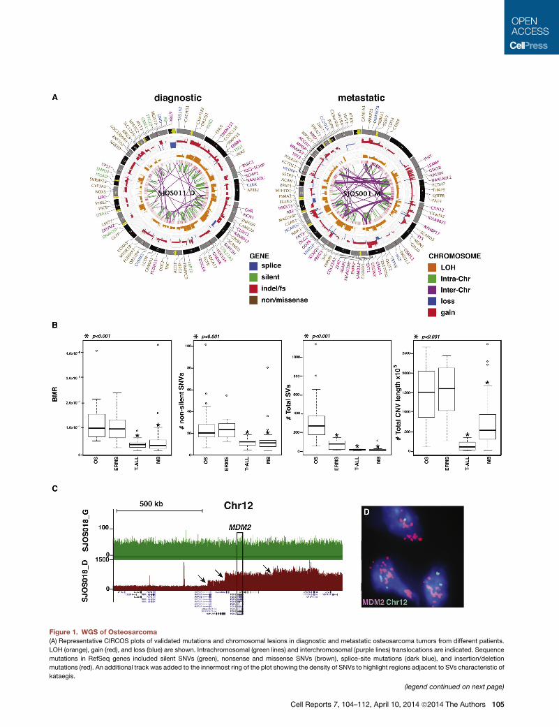
Figure 1. WGS of Osteosarcoma(A) Representative CIRCOS plots of validated mutations and chromosomal lesions in diagnostic and metastatic osteosarcoma tumors from different patients.
LOH (orange), gain (red), and loss (blue) are shown. Intrachromosomal (green lines) and interchromosomal (purple lines) translocations are indicated. Sequence
mutations in RefSeq genes included silent SNVs (green), nonsense and missense SNVs (brown), splice-site mutations (dark blue), and insertion/deletion
mutations (red). An additional track was added to the innermost ring of the plot showing the density of SNVs to highlight regions adjacent to SVs characteristic of
kataegis.
(legend continued on next page)
Cell Reports 7, 104–112, April 10, 2014 ª2014 The Authors 105

normal DNA from 19 osteosarcoma patients in a discovery
cohort, and 14 tumor specimens and matched normal DNA
from 13 patients in a validation cohort (Table S1); 9,671 Gb
(94%) were successfully mapped to the reference genome
(Table S2). In the discovery cohort, the samples included 17 pre-
treatment diagnostic samples (16 primary and one metastatic),
one recurrent metastatic sample (SJOS001), and two tumor
specimens (SJOS010_D and SJOS010_M) from the same
patient with metachronous osteosarcoma (Table S1).
The average genome coveragewas 443 and the average exon
coverage was 393; 99% of SNPs detected across all 39
genomes showed concordance with their corresponding SNP
array genotype calls (Table S2). Validation was carried out using
custom liquid capture for all SNVs, SVs, and insertions or dele-
tions (indels) identified in the original sequence data. Combining
the discovery and validation cohorts, we identified 50,426
validated somatic sequence mutations and 10,806 SVs
(Table S3). These included 856 nonsilent tier 1 mutations in
genes, 4,651 tier 2 mutations in evolutionarily conserved regions
of the genome, and 43,782 tier 3 mutations in nonrepetitive
regions of the genome that are not part of tier 1 or tier 2
(Table S3). The average number of sequence mutations was
1,483.1 per case (range 610–5,178), with 25.2 mutations per
case (range 5–103) resulting in amino acid changes (Table S3).
The estimated mean mutation rate was 1.15 3 10�6 per base
(range 4.90 3 10�7–3.99 3 10�6). Among the validated SVs,
377 were predicted to produce an in-frame fusion protein
(Table S3). Good-quality RNA sequencing (RNA-seq) data were
available for five tumors with 64 predicted fusion SVs. Among
them, 15 SVs (23%) were expressed (Table S3).
Primary and metastatic osteosarcomas had high rates of
validated SVs (Figures 1A and S1). The number of SVs and
CNVs, background mutation rate, and number of nonsilent tier
1mutations were significantly higher in osteosarcoma compared
with medulloblastoma and T-ALL (Robinson et al., 2012; Zhang
et al., 2012; Figure 1B). However, only the number of SVs was
significantly higher in osteosarcoma compared with another
pediatric solid tumor with high rates of somatic alterations
(embryonal rhabdomyosarcoma) (Chen et al., 2013; Figure 1B).
The global patterns revealed by the WGS analysis of osteosar-
coma suggest that themajority of SVs and CNVswere generated
by sequential accumulation of SVs (Figures 1C and 1D), but
chromothripsis (Stephens et al., 2011) was detected at specific
genomic regions in four samples (chr14 in SJOS002_D, chr17q
in SJOS003_D, chr6q in SJOS005_D, and chr13 in SJOS010_M;
Supplemental Experimental Procedures). We used a modified
version of GISTIC analysis to identify regions of the osteosar-
coma genome with recurrent copy number alterations in the
discovery cohort. The TP53, RB1, MYC, and PTEN pathways,
as well as ATRX, LSAMP-AS3, CCNE1, and a genomic region
(B) Boxplots of validated basal mutation rate (BMR) and numbers of nonsilent SN
alveolar rhabdomyosarcoma (ARMS) tumors in the discovery cohort. * represe
genomes.
(C) Representative plot of sequence reads on chromosome 12 for the matched ge
change are identified (arrows) spanning the MDM2 gene consistent with sequen
(D) MDM2 FISH of SJOSO18 showing amplification (red) relative to the probe f
medulloblastoma.
See also Figures S1–S3 and Tables S1–S3.
106 Cell Reports 7, 104–112, April 10, 2014 ª2014 The Authors
on chromosome 16 containing COPS3, PMP22, MAPK7,
NCOR1, and UBB, were recurrently mutated (Figure S1C).
Among SNVs with sufficient coverage in both SJOS010 samples
(203), we validated 673 SNVs in both samples, 1,686 in
diagnostic-only samples, and 1,408 in metastasis-only samples,
indicating that these two tumors shared a limited amount of com-
mon mutations and were divergent early in the progression.
Applying the GRIN method (Pounds et al., 2013) on functional
mutations (including SNVs and indels) and SVs, we identified
TP53 (false discovery rate [FDR] = 3.6E-51, mutated in 28/34
samples) RB1 (FDR = 1.1E-5, mutated in 10/34 samples),
ATRX (FDR = 2.4E-4, mutated in 10/34 samples), and DLG2
(FDR = 0.044, mutated in 18/34 samples) as significantly
mutated genes. All genes except DLG2 were mutated by point
mutations (nine for TP53, three for RB1, and five for ATRX) and
SVs in multiple tumors (18 for TP53, seven for RB1, and five for
ATRX). DLG2 was exclusively mutated by SVs.
Osteosarcoma Tumor Purity and Tumor HeterogeneityUsing the purity-adjusted mutant allele fraction (MAF)
derived from deep sequencing of all SNVs by capture enrich-
ment and Illumina sequencing, we analyzed intratumor hetero-
geneity. Eleven tumors (SJOS001_M, SJOS004, SJOS005,
SJOS008, SJOS012, SJOS013, SJOS015, SJOS001103_D1,
SJOS001105_D1, and SJOS001123_D1, and SJOS001125_D1)
were excluded from quantitative heterogeneity analysis due to
an insufficient number of SNVs in copy-neutral regions.
Statistical modeling demonstrated that 61% (14/23) of osteosar-
comas in this group had evidence of multiple clones, includ-
ing metastatic samples SJOS010_M, SJOS001107_M1 and
SJOS001107_M2 (Figure S2).
Kataegis in OsteosarcomaTo determine whether there was any relationship between the
SVs and location, distribution, or type of SNV in the osteosar-
coma genomes, we plotted the validated SVs and SNVs for
each sample andanalyzed the intermutationdistance (FigureS2).
Hypermutable regions with the five hallmarks of kataegis (Nik-
Zainal et al., 2012) were identified in 17 of the osteosarcoma
tumors (Figure 2A). These five hallmarks of kataegis are (1)
enriched C->T and C->G substitutions at TpCpX trinucleotides
(Figures 2B and 2C), (2) the same class of nucleotide mutation
occurring for contiguous stretches before switching to a different
class (Figure 2D), (3) mutations within short stretches of the
genome occurring on the same parental chromosome (Fig-
ure S2), (4) clustering of heavily mutated short stretches of the
genome at multiple scales (Figure 2E), and (5) association of
the hypermutated region with SV breakpoints (Figure 2E). The
regions of the genome with kataegis were not recurrent in our
cohort and were not associated with recurrently mutated genes
Vs, total SVs, and total CNVs in embryonal rhabdomyosarcoma (ERMS) and
nts statistical significance of p < 0.001 as compared with the osteosarcoma
rmline (green) and tumor (red) sample. Several distinct regions of copy number
tial chromosomal lesions.
or chromosome 12 (green). T-ALL, T cell acute lymphoblastic leukemia; MB,

Figure 2. Kataegis in Osteosarcoma
(A) Rainfall plot showing the Log10 of the intermutation distance versus genomic position for a representative osteosarcoma sample (SJOS005) with evidence of
kataegis. The chromosomes are demarcated by gray shading and the number of SVs in each chromosome is shown in brown at the bottom. The validated SNVs
are plotted and color-coded by the type of mutation.
(legend continued on next page)
Cell Reports 7, 104–112, April 10, 2014 ª2014 The Authors 107

81 7653 112 1094
ATG TGA
1 2
transactivationdomain
prolinedomain
DNA bindingdomain
oligomerizationdomain
1 393
R24
8Q
R27
3H
R33
7H (
ger
mlin
e)
T21
1fs
P58
sp
lice
ATGdeletion
interchromosomaltranslocation
intrachromosomaltranslocation
frameshift
splice site
missense
A
B
C
Figure 3. Validated Mutations in TP53
(A) Structure of the TP53 gene showing the trans-
activation, proline, DNA binding, and oligomeriza-
tion domains with splice-site, frameshift, and
missense mutations in tumors of the 19 patients in
the discovery cohort.
(B) Structure of the genomic locus of the TP53 gene
showing the exon boundaries color-coded in
accordance with the protein domains shown in (A).
Sites of interchromosomal translocations are
indicated by black arrowheads between exons 9
and 10. The sizes of the introns and exons are
scaled proportionally except for intron 1, which is
much larger than the other introns in human TP53.
(C) A magnified view of intron 1 of TP53 showing the
deletions (blue arrowheads), intrachromosomal
translocations (red arrowheads), and interchromo-
somal translocations (black arrowheads).
See also Figure S3 and Table S4.
in osteosarcoma (Figure S2). Tier 1 SNVs in kataegis regions
were not significantly associated with the expression status
(p = 0.16 by Fisher’s exact test).
Chronology of Kataegis, SVs, and Aneuploidy inSJOS005SJOS005 had the highest proportion (11%) of kataegis SNVs
in our cohort. The large number of kataegis SNVs (n = 212)
coupled with the accurate measurement of the MAFs of all
SNVs derived from deep sequencing allowed us to analyze
the chronology of kataegis in relation to other mutational
events in this tumor. First, we examined MAFs of SNVs in
kataegis microclusters containing five or more consecutive
kataegis SNVs within 10 kb. The MAF variance was relatively
small (6.7% of overall variance) within a microcluster, although
there was a wide range of MAFs across microclusters (range
0.142–0.839, median 0.364; Figure S2). This pattern, along
with the observation that SNVs in a microcluster occurred on
the same parental chromosome, supports the hypothesis
that SNVs in a kataegis microcluster originated from a single
event. MAF analysis of SVs flanking ‘‘kataegis’’ clusters (range
0.132–0.866, median 0.396) also showed a significant positive
correlation (p = 4.56E-5) with those of ‘‘kataegis’’ SNVs,
and there was no significant difference between them (p =
0.143 by Wilcoxon signed rank test), indicating that the neigh-
boring SVs likely arise simultaneously with kataegis SNVs
(Figure S2).
(B) The proportion of each type of validated SNV in osteosarcomas with evidence of kataegis versus those
(C) The distribution of each nucleotide sequence 50 to the C mutations in tumors with kataegis and those w
(D) A rainfall plot in a representative regions of chromosome 3 in SJOS005 with kataegis showing the stra
sequence clusters.
(E) A macrocluster of hypermutation with evidence of kataegis on chromosome 3 of SJOS005, with two sequ
of microclusters within a single macrocluster.
See also Figure S2.
108 Cell Reports 7, 104–112, April 10, 2014 ª2014 The Authors
SJOS005, like most osteosarcomas, ex-
hibits aneuploidy with copy number gains
spanning more than 50% of the genome.
Kataegis SNVs were significantly enriched
in regions of the genomewith four or more copies comparedwith
nonkataegis SNVs (p < 2.2e-16 by Fisher’s exact test; Figure S2).
However, the MAF distribution of kataegis SNVs showed a large
fraction of SNVs with multiple copies of the mutant allele in
amplified regions, whereas only a single copy of mutant alleles
was found in the majority of the nonkataegis SNVs (Figure S2).
Taken together, these data suggest that kataegis likely occurs
before global aneuploidy, and nonkataegis SNVs occur primarily
after the aneuploidy.
SVs in TP53
The p53 pathway was mutated in all 20 tumor samples from
the 19 patients in our discovery cohort. The majority (95%,
19/20) had either sequence mutations or SVs in the TP53
gene, and one (SJOS018) had an MDM2 amplification (see Fig-
ures 1C and 1D; Table S3). Surprisingly, 55% of the tumors
(11/20) had SVs in the TP53 gene, and the majority of those
were translocations with breakpoints that were confined to
the first intron of the gene (90%, 19/21 SV breakpoints; Figures
3A–3C; Table S4). Indeed, some tumors had rearrangements in
both alleles of TP53, resulting from two or more independent
translocations (Table S4). One patient’s tumor (SJOS006) had
a germline SNV (R337H), one (SJOS012) had a somatic
splice-site mutation, and two (SJOS004 and SJOS010) had
somatic missense SNVs (Figures 3A–3C; Table S4). The
remaining four patients had tumors that harbored indels in
the TP53 gene. Loss of heterozygosity (LOH) at the TP53 locus
without kataegis.
ithout kataegis.
nd of the hypermutation based on the C>T or G>A
ential magnifications (boxes) showing the existence

SJO
S00
8S
JOS
010
B
K
H
E
C
L
I
F
D
M
J
G
bia
llelic
rea
rran
gem
ent
mo
no
alle
lic r
earr
ang
emen
t
CHI 35PTE&H TP53 FISH
A
10 kb
TP53
3’ probe (169,150 bp)
5’ probe (213,405 bp)
full length probe (167,314 bp)
Chr17
SJO
S00
1S
JOS
002
po
lyso
my
mis
sen
sem
on
oal
lelic
del
etio
n
5’ probe 3’ probe
5’ probe 3’ probe
5’ probe 3’ probe
full length Chr 17
Figure 4. FISH and Immunohistochemistry
for TP53 in Osteosarcoma
(A) Genomic location of the 50 (green) and 30 (red)break-apart FISH probes showing their position
relative to the TP53 gene. The full-length probe used
to identify deletions at the TP53 locus is shown
in black.
(B–D) Images of hematoxylin and eosin (H&E), TP53
immunohistochemistry (IHC), and break-apart FISH
for SJOS001 with biallelic rearrangement of the
TP53 gene.
(E–G) Images of H&E, TP53 IHC, and break-apart
FISH for SJOS002 with monoallelic rearrangement
of the TP53 gene.
(H–J) Images of H&E, TP53 IHC, and break-apart
FISH for SJOS010 with polysomy and a missense
mutation leading to elevated accumulation of
nuclear TP53 protein.
(K–M) Images of H&E, TP53 IHC, and FISH using the
full-length probe (green) for SJOS008 with mono-
allelic deletion of TP53.
See also Table S5.
was evident in 40% (8/20) of the osteosarcoma tumors. In
total, 15 tumors had biallelic inactivation of TP53, four had
monoallelic inactivation of TP53, and one had MDM2 amplifi-
cation (Figure 1C; Table S4).
To further validate the translocations in the TP53 gene identi-
fied by WGS, we developed a break-apart fluorescence in situ
hybridization (FISH) assay with separate probes spanning the
50 and 30 regions of the gene (Figure 4A). We also developed a
FISH assay with a probe spanning the entire TP53 gene
(Figure 4A) to assess ploidy and determine whether the gene
was deleted. To complement the FISH analysis, we performed
p53 immunostaining to verify that the tumors with missense
mutations had accumulated high levels of nuclear p53 protein.
We successfully performed FISH in 18 of 20 tumors and p53
immunostaining on all 20 tumors (Table S4). Overall, there was
perfect concordance between the WGS data and the FISH
data (Figures 4B–4M; Table S4).
Cell Reports 7, 104–
In an additional cohort of patient tumor
samples, we found that 50% (16/32)
had TP53 rearrangements, 22% (7/32)
had missense mutations, 16% (5/32) had
nonsense mutations, 6% (2/32) had a
TP53 deletion, and 3% (1/32) had an
MDM2 amplification (Table S5). Three
patients with tumor showed no evidence
of a p53 pathway mutation.
We did not find any significant difference
in CNV (p = 0.20 by Wilcoxon rank sum
test), SV (p = 0.85), SNV (p = 0.43), nonsi-
lent tier 1 mutations (p = 0.66), or back-
ground mutation rate (p = 0.43) in the
osteosarcoma samples with mutant p53
versus those with inactivating (nonsense,
deletion and truncation) mutations in
TP53. Survival analysis, including event-
free survival and overall survival, did not
show a significant difference in outcome for the patients whose
tumors carried TP53-missense mutations (ten patients) versus
those with TP53-truncating mutations (34 patients), with log
rank test p values of 0.88 and 0.64, respectively.
RB1, ATRX, and DLG2 Are Recurrently Mutated inOsteosarcomaATRX is part of a multiprotein complex that regulates chromatin
remodeling, nucleosome assembly, and telomere maintenance.
It was recently shown that ATRXmutations in neuroblastoma are
associated with age at diagnosis (Cheung et al., 2012). Most
neuroblastomas with ATRXmutations show evidence of alterna-
tive lengthening of telomeres (ALT), as measured by WGS, telo-
mere FISH, and telomere quantitative PCR (qPCR) (Cheung
et al., 2012). In our osteosarcoma discovery cohort, we identified
five tumors (SJOS001, SJOS002, SJOS007, SJOS001112-M2,
and SJOS001117-D1) with point mutations in ATRX, and five
112, April 10, 2014 ª2014 The Authors 109

Y26
6*
ATRXNM_000489.3
0 2492
0 200 400 600 800 1000 1200 1400 1600 1800 2000 2200 2400 2600
Zinc Finger/RING (predicted)
SNF2 family N-terminal domain
DEXDc-DEAD-like
HELICc-Helicase
Missense
Nonsense
NLS (predicted)Frameshift
DE
LE
TIO
NS
NV
R18
03C
A
B
DC
E
D13
83-f
s
SJOS016 (M6-fs) SJOS004 (wt ATRX)
* **
SJOS001SJOS014SJOS012
SJOS006 (T1885-fs)
SJOS011 (M6R*)SJOS018 (M6-R-S1406)
SJOS016 (M6-fs)
SJOS001_M
SJOS010_M
SJOS002_D
SJOS007_D
SJOS006_D
SJOS005_D
SJOS004_D
SJOS003_D
SJOS012_D
SJOS011_D
SJOS010_D
SJOS009_D
SJOS008_D
SJOS017_D
SJOS016_D
SJOS015_D
SJOS014_D
SJOS013_D
SJOS019_D
SJOS018_D
Difference in Telomeric Reads
-1.5 0.50.0-0.5 0.20.1- 1.51.0
DiagnosticTelomere Status
Gain
Loss
No Change
No
rmal
ized
Tel
om
eric
Rea
ds
SampleG D
6,000
0
2,000
4,000
10,000
8,000
12,000
SJO
S00
1_M
SJO
S01
0_M
SJO
S00
2_D
SJO
S00
7_D
SJO
S00
6_D
SJO
S00
5_D
SJO
S00
4_D
SJO
S00
3_D
SJO
S01
2_D
SJO
S01
1_D
SJO
S01
0_D
SJO
S00
9_D
SJO
S00
8_D
SJO
S01
7_D
SJO
S01
6_D
SJO
S01
5_D
SJO
S01
4_D
SJO
S01
3_D
SJO
S01
9_D
SJO
S01
8_D
10
-5
0
5
Lo
g2
(tu
mo
r/g
erm
line)
S21
3*
L17
55V
SJOS015
Figure 5. ATRX Mutations Correlate with ALT in Osteosarcoma
(A) Diagram of the five SNVs, four deletions, and one interchromosomal SV found in the ATRX genes of the osteosarcoma cohort. Three of the samples with ATRX
SVs (SJOS006, SJOS018, and SJOS011) had matching RNA-seq data. SJ006 has a short deletion at exon 23 and the RNA-seq data confirmed a readthrough
(legend continued on next page)
110 Cell Reports 7, 104–112, April 10, 2014 ª2014 The Authors

with focal deletions or SVs affecting the coding region of the
gene (Figure 5A; Table S6). There was no significant gender
bias in ATRX mutations (p = 0.25 by Fisher’s exact test) even
though it is located on the X chromosome. By immunohisto-
chemistry, 31% (6/19) of the tumors in the discovery cohort
were ATRX negative (Figure 5B; Table S6). The sample with a
missense mutation (SJOS007-R1803C) and one with an SV
(SJOS018) were heterogeneous for ATRX protein expression.
Analysis of telomere sequence reads from the WGS data
and qPCR of telomeres showed that the majority of osteosar-
comas had longer telomeres (Figures 5C and 5D), and ALT
was found in 85% (12/14) of the samples using telomere FISH
(Table S6).
Beyond TP53 and ATRX, there were significant recurrent
mutations in RB1 (10/34, FDR q = 1.1E-5) and DLG2 (18/34,
FDR q = 0.044). DLG2 encodes a multi-PDZ domain protein
that is involved in epithelial polarity during cell division and has
been implicated in cancer cell invasion. In Drosophila, DLG is a
tumor suppressor, but a clear tumor-suppressor function has
not yet been confirmed for DLG2 in human cancer.
SVs in Cancer GenesSVs contributed 91% (9,605/10,523) of all functional genetic
lesions in our osteosarcoma cohort. In total, 122 cancer genes
had at least one SV breakpoint (Table S7) and all but one tumor
(SJOS001118_D1) had at least one breakpoint (range 1–40) in a
cancer gene. SV breakpoint enrichment in the cancer genes
was highly significant even when we excluded TP53 from the
list (p = 2.5E-6). Twelve of the 34 tumors (35%) achieved signif-
icant enrichment of SV breakpoints in cancer genes individually.
In addition, some tumors have ‘‘fold-back intrachromosomal
translocations’’ (Campbell et al., 2010) to inactivate tumor-
suppressor genes (Figure S3). These results further support the
hypothesis that genomic instability leads to lesions in various
cancer genes.
DISCUSSION
WGS of osteosarcoma demonstrated that the rate of SNVs
was similar to that in other pediatric solid tumors, and only a
few recurrent SNVs were detected. Approximately half of the
osteosarcomas in our discovery cohort had a pattern of hyper-
mutation associated with SVs, called kataegis (Nik-Zainal
et al., 2012). The regions of the genome with kataegis were
not recurrent, and none of the most recurrently mutated genes
were found in regions of kataegis. Chromosomal lesions,
rather than SNVs, were the major mechanism of recurrent
mutations, and many of the most significant chromosomal
event that would result in a T1885 frameshift. For SJOS011, the RNA-seq andWG
mutation (M6R*). For SJOS018, the RNA-seq and WGS data supported a deleti
(M6RS1406). The WGS for SJOS016 predicts a deletion that connects exon 1 to
(B) Representative IHC for ATRX showing nuclear ATRX in a sample with intense s
ATRX (SJOS014), and a sample with a nonsense mutation (SJOS001). Arrows ind
immunopositive vascular endothelial cells among the tumor cells that are negativ
(C and D) Relative telomere length in the osteosarcomas compared with that in t
(E) Representative telomere FISH showing characteristics of ALT (arrow) in an os
See also Tables S6 and S7.
lesions were found in known cancer genes, including TP53,
RB1, and ATRX.
Genomic Stability and Osteosarcoma Initiation andProgressionThe most frequent mutation in osteosarcoma is in TP53. By our
estimates, both alleles aremutated in asmany as 80%of tumors,
and at least one allele was mutated in >90% of tumors. These
data suggest that p53 mutations are a major oncogenic driver
in osteosarcoma. Although this finding is not novel, what is
surprising is the mechanism of inactivation. Most TP53 muta-
tions are SVs in intron 1, which suggests that either the TP53
locus is particularly susceptible to SVs or SVs occur at a high
rate in the osteosarcoma tumor-initiating cell. Aside from osteo-
sarcomas and prostate cancers (Baca et al., 2013; Berger et al.,
2011), there is no evidence of TP53 SVs in any other cancer, so
the locus is probably not uniquely susceptible to chromosomal
rearrangements. These data raise an intriguing possibility:
genomic instability characterized by high rates of CNVs and
SVs may precede TP53 inactivation, and may be the underlying
mechanism that initiates and promotes osteosarcoma.
Kataegis in OsteosarcomaIn a recent WGS study, Nik-Zainal et al. (2012) described a
distinct hypermutation phenomenon in breast cancer that they
termed kataegis. Here, we found SNV clusters with the same
five characteristics of kataegis in 50% of the osteosarcomas
analyzed by WGS. Interestingly, genomic regions encoding
TP53 and ATRX, the two most frequently mutated genes in oste-
osarcoma, did not exhibit this pattern of local hypermutation.
Furthermore, there was no association between kataegis and
TP53 mutation type (i.e., SNV, indel, or SV).
TP53-Mutant or -Null OsteosarcomasPrevious studies have estimated that 20%–70% of osteosar-
comas carry mutations in the p53 pathway (Lonardo et al.,
1997; Wunder et al., 2005), but our data suggest that the propor-
tion is much higher. For example, Wunder et al. (2005)
sequenced exons 4–10 of the TP53 gene in 196 osteosarcoma
samples and found that 19.4% (38/196) had TP53 SNVs. The
investigators concluded that the remaining 80.6% (158/196)
had wild-type TP53 (Wunder et al., 2005). They went on to
show that event-free survival was indistinguishable between
the two groups (wild-type and mutant TP53) (Wunder et al.,
2005). SVs in the first intron of TP53 were not analyzed in that
study, even though such lesions had previously been reported
in osteosarcoma (Miller et al., 1990). Our data suggest that the
majority of the tumors identified as TP53 wild-type in the study
S data supported a junction connecting exon 1 to exon 28, creating a nonsense
on connecting exon 1 to exon 13, thereby creating an in-frame fusion protein
exon 16, producing a frameshift (M6fs).
taining and wild-type ATRX (SJOS012), a sample with fainter nuclear localized
icate representative nuclei stained positive for ATRX. Asterisks indicate ATRX
e for ATRX IHC.
he matched germline DNA, as analyzed by WGS and qPCR.
teosarcoma with an ATRX deletion.
Cell Reports 7, 104–112, April 10, 2014 ª2014 The Authors 111

by Wunder et al. (2005) actually had inactivating SVs in TP53.
Therefore, it may be useful to revisit the association of TP53
pathway inactivation with osteosarcoma outcome in a large
cohort of patient samples.
EXPERIMENTAL PROCEDURES
Full details regarding sample acquisition, molecular and biochemical proce-
dures, informatics, and WGS are provided in the Supplemental Information.
All tumors in this study were obtained from St. Jude Children’s Research
Hospital (SJCRH) patients. The SJCRH IRB approved experiments involving
human subjects and informed consent was obtained from all subjects.
ACCESSION NUMBERS
The European Bioinformatics Institute accession number for the sequencing
data reported in this paper is EGAS00001000263.
SUPPLEMENTAL INFORMATION
Supplemental Information includes Supplemental Experimental Procedures,
three figures, and seven tables and can be found with this article online at
http://dx.doi.org/10.1016/j.celrep.2014.03.003.
ACKNOWLEDGMENTS
This work was supported, in part, by Cancer Center Support (CA21765) from
the NCI, grants to M.A.D from the NIH (EY014867, EY018599, and CA168875),
and the American Lebanese Syrian Associated Charities (ALSAC). M.A.D. is an
HHMI Investigator. The whole-genome sequencing was supported as part of
the St. Jude Children’s Research Hospital -Washington University Pediatric
Cancer Genome Project.
Received: May 10, 2013
Revised: November 22, 2013
Accepted: March 3, 2014
Published: April 3, 2014
REFERENCES
Baca, S.C., Prandi, D., Lawrence, M.S., Mosquera, J.M., Romanel, A., Drier,
Y., Park, K., Kitabayashi, N., MacDonald, T.Y., Ghandi, M., et al. (2013).
Punctuated evolution of prostate cancer genomes. Cell 153, 666–677.
Berger, M.F., Lawrence, M.S., Demichelis, F., Drier, Y., Cibulskis, K.,
Sivachenko, A.Y., Sboner, A., Esgueva, R., Pflueger, D., Sougnez, C., et al.
(2011). The genomic complexity of primary human prostate cancer. Nature
470, 214–220.
Campbell, P.J., Yachida, S., Mudie, L.J., Stephens, P.J., Pleasance, E.D.,
Stebbings, L.A., Morsberger, L.A., Latimer, C., McLaren, S., Lin, M.L., et al.
(2010). The patterns and dynamics of genomic instability in metastatic pancre-
atic cancer. Nature 467, 1109–1113.
Chen, X., Steward, E., Shelat, A., Qu, C., Bahrami, A., Hatley, M.,Wu, G., Brad-
ley, C., McEvoy, J., Pappo, A., et al.; St. Jude Children’s Research Hospital–
Washington University Pediatric Cancer Genome Project (2013). Targeting
oxidative stress in embryonal rhabdomyosarcoma. Cancer Cell 24, 710–724.
Cheung, N.K., Zhang, J., Lu, C., Parker, M., Bahrami, A., Tickoo, S.K., Heguy,
A., Pappo, A.S., Federico, S., Dalton, J., et al. (2012). Association of age at
112 Cell Reports 7, 104–112, April 10, 2014 ª2014 The Authors
diagnosis and genetic mutations in patients with neuroblastoma. JAMA 307,
1062–1071.
Downing, J.R., Wilson, R.K., Zhang, J., Mardis, E.R., Pui, C.H., Ding, L., Ley,
T.J., and Evans, W.E. (2012). The Pediatric Cancer Genome Project. Nat.
Genet. 44, 619–622.
Hicks, M.J., Roth, J.R., Kozinetz, C.A., and Wang, L.L. (2007). Clinicopatho-
logic features of osteosarcoma in patients with Rothmund-Thomson
syndrome. J. Clin. Oncol. 25, 370–375.
Kleinerman, R.A., Tucker, M.A., Tarone, R.E., Abramson, D.H., Seddon, J.M.,
Stovall, M., Li, F.P., and Fraumeni, J.F., Jr. (2005). ). Risk of new cancers after
radiotherapy in long-term survivors of retinoblastoma: an extended follow-up.
J. Clin. Oncol. 23, 2272–2279.
Lonardo, F., Ueda, T., Huvos, A.G., Healey, J., and Ladanyi, M. (1997). p53
and MDM2 alterations in osteosarcomas: correlation with clinicopathologic
features and proliferative rate. Cancer 79, 1541–1547.
McIntyre, J.F., Smith-Sorensen, B., Friend, S.H., Kassell, J., Borresen, A.L.,
Yan, Y.X., Russo, C., Sato, J., Barbier, N., Miser, J., et al. (1994). Germline
mutations of the p53 tumor suppressor gene in children with osteosarcoma.
J. Clin. Oncol. 12, 925–930.
Meyers, P.A., Schwartz, C.L., Krailo, M., Kleinerman, E.S., Betcher, D.,
Bernstein, M.L., Conrad, E., Ferguson, W., Gebhardt, M., Goorin, A.M., et al.
(2005). Osteosarcoma: a randomized, prospective trial of the addition of ifos-
famide and/or muramyl tripeptide to cisplatin, doxorubicin, and high-dose
methotrexate. J. Clin. Oncol. 23, 2004–2011.
Miller, C.W., Aslo, A., Tsay, C., Slamon, D., Ishizaki, K., Toguchida, J., Yama-
muro, T., Lampkin, B., and Koeffler, H.P. (1990). Frequency and structure of
p53 rearrangements in human osteosarcoma. Cancer Res. 50, 7950–7954.
Nik-Zainal, S., Alexandrov, L.B., Wedge, D.C., Van Loo, P., Greenman, C.D.,
Raine, K., Jones, D., Hinton, J., Marshall, J., Stebbings, L.A., et al.; Breast
Cancer Working Group of the International Cancer Genome Consortium
(2012). Mutational processes molding the genomes of 21 breast cancers.
Cell 149, 979–993.
Ottaviani, G., and Jaffe, N. (2009). The epidemiology of osteosarcoma. Cancer
Treat. Res. 152, 3–13.
Pounds, S., Cheng, C., Li, S., Liu, Z., Zhang, J., and Mullighan, C. (2013). A
genomic random interval model for statistical analysis of genomic lesion
data. Bioinformatics 29, 2088–2095.
Robinson, G., Parker, M., Kranenburg, T.A., Lu, C., Chen, X., Ding, L., Phoenix,
T.N., Hedlund, E., Wei, L., Zhu, X., et al. (2012). Novel mutations target distinct
subgroups of medulloblastoma. Nature 488, 43–48.
Smith,M.A., Seibel, N.L., Altekruse, S.F., Ries, L.A., Melbert, D.L., O’Leary,M.,
Smith, F.O., and Reaman, G.H. (2010). Outcomes for children and adolescents
with cancer: challenges for the twenty-first century. J. Clin. Oncol. 28, 2625–
2634.
Stephens, P.J., Greenman, C.D., Fu, B., Yang, F., Bignell, G.R., Mudie, L.J.,
Pleasance, E.D., Lau, K.W., Beare, D., Stebbings, L.A., et al. (2011). Massive
genomic rearrangement acquired in a single catastrophic event during cancer
development. Cell 144, 27–40.
Wunder, J.S., Gokgoz, N., Parkes, R., Bull, S.B., Eskandarian, S., Davis, A.M.,
Beauchamp, C.P., Conrad, E.U., Grimer, R.J., Healey, J.H., et al. (2005). TP53
mutations and outcome in osteosarcoma: a prospective, multicenter study.
J. Clin. Oncol. 23, 1483–1490.
Zhang, J., Ding, L., Holmfeldt, L., Wu, G., Heatley, S.L., Payne-Turner, D.,
Easton, J., Chen, X., Wang, J., Rusch, M., et al. (2012). The genetic basis of
early T-cell precursor acute lymphoblastic leukaemia. Nature 481, 157–163.