ce book - midcentral district health board · 2017-02-17 · ce book acknowledgements: central...
TRANSCRIPT
Clinical Nurse Specialist (Lead) Intravenous & Related Therapies – Pager: 060. Ext. 8206 Page 1
Intravenous & Related Therapy
Resource for Students
MIDCENTRAL DISTRICT HEALTH BOARD
INTRAVENOUS & RELATED THERAPIES – PRACTICE DEVELOPMENT UNIT
2015
Acknowledgements: Central Region District Health Boards
Stu
de
nt
Res
ou
rce
bo
ok
Clinical Nurse Specialist (Lead) Intravenous & Related Therapies – Pager: 060. Ext. 8206 Page 2
INTRODUCTION
This resource is designed to assist students in gaining knowledge, confidence, and skills in performing basic intravenous therapy. The content of this resource relates only to the Basic Principles of I.V. Therapy and DOES NOT INCLUDE;
Insertion of peripheral I.V. catheters.
Phlebotomy.
Management of central venous access devices (CVAD).
Management of patient controlled analgesia (PCA). IMPORTANT POINTS
This resource is a supplement for student learning and skill development.
Completion of this resource does not equate to being certified in Fundamental I.V. & Related Therapies.
A student is not allowed to manage any I.V. therapy unsupervised.
A student must work under the direction and delegation of a registered health practitioner when providing I.V. & related therapy care.
This document is best utilized in conjunction with other resources. Resources can be your nurse lecturer, clinical nurse educator, ward based I.V. Resource or Preceptor, Notes on Injectable Drugs, New Zealand Formulary.
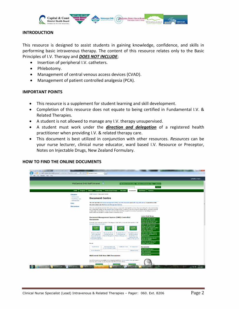
HOW TO FIND THE ONLINE DOCUMENTS
Clinical Nurse Specialist (Lead) Intravenous & Related Therapies – Pager: 060. Ext. 8206 Page 3
SUPPLEMENTAL WEB SITES http://www.ins1.org/ - Recognized as the global authority in infusion therapy, INS is dedicated to exceeding the public’s expectations of excellence by setting the standard for infusion care. http://www.ivteam.com/ - IVTEAM is an online resource for the practitioner working in the field of IV therapy. IVTEAM provide free IV news and updates to the whole of the world via the World Wide Web.
http://www.iv-therapy.net/ - Focus on vascular therapy. Includes Chat forums, Great photos of IV related problems, Numerous IV related articles
http://www.ivaccess.com/ - A quick and easy reference to find the pH and Osmolarity of medicines.
DEFINITION OF TERMS (Source: Nursing Council New Zealand)
DELEGATION - The transfer of the responsibility for the performance of a task/activity from one person to another, with the former person retaining accountability for the process and the outcome.
DIRECTION - The active process of directing, guiding, monitoring and influencing the outcome of an individual’s performance of a task/activity related to assigned aspects of patient care.
DIRECT DIRECTION – Direction is provided directly when the registered health professional is actually present, observes, works with and directs the person who is being supervised. Direct direction is required when assessing the skill or competency level of another.
INDIRECT DIRECTION - Direction is provided indirectly when the registered health professional in the same facility or organisation as the supervised person but does not constantly observe his/her activities. The registered health professional must be available for reasonable access (i.e. on-site and accessible within a few minutes).
DOUBLE CHECKING - Double checking must be performed in situations where it is appropriate or legally necessary; - e.g. when checking controlled drugs, blood products, dosage calculations, etc.
Clinical Nurse Specialist (Lead) Intravenous & Related Therapies – Pager: 060. Ext. 8206 Page 4
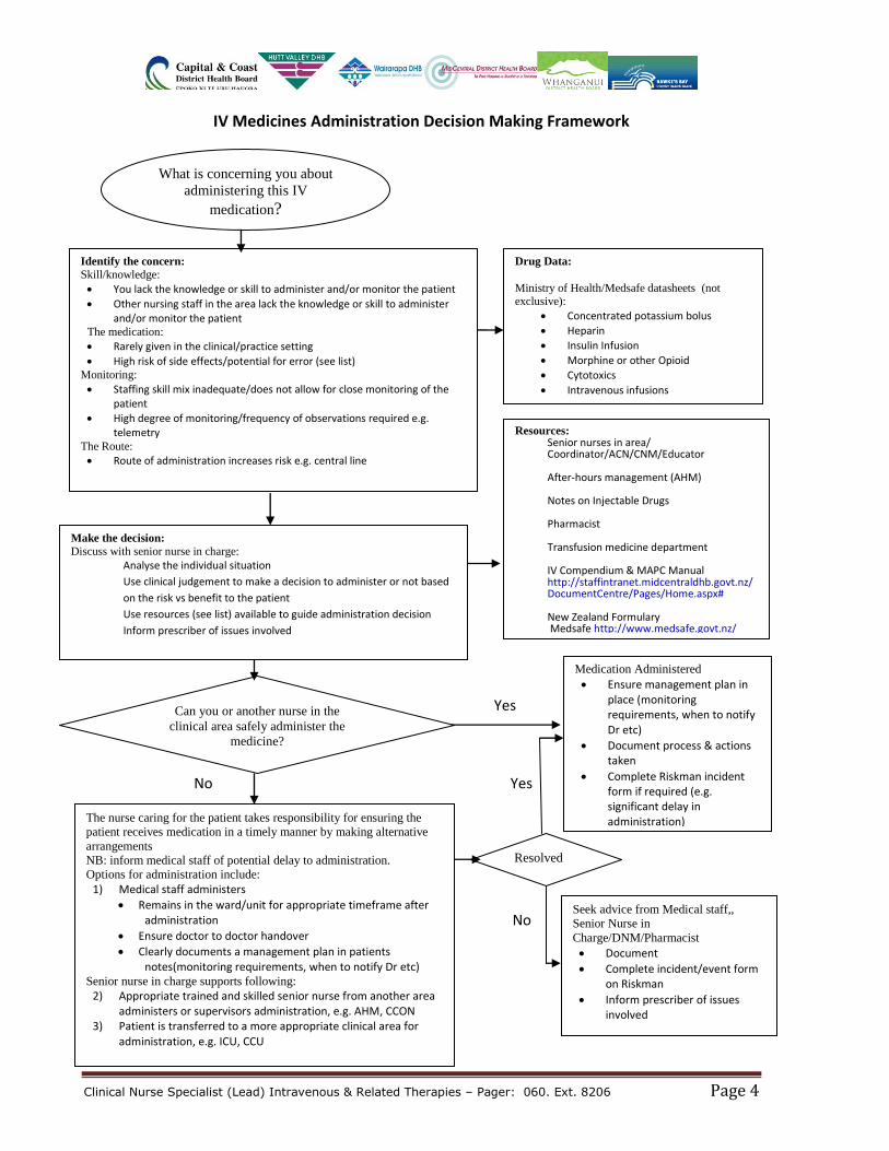
IV Medicines Administration Decision Making Framework
No
Yes No Yes No
What is concerning you about
administering this IV
medication?
Identify the concern:
Skill/knowledge:
You lack the knowledge or skill to administer and/or monitor the patient
Other nursing staff in the area lack the knowledge or skill to administer and/or monitor the patient
The medication:
Rarely given in the clinical/practice setting
High risk of side effects/potential for error (see list) Monitoring:
Staffing skill mix inadequate/does not allow for close monitoring of the patient
High degree of monitoring/frequency of observations required e.g. telemetry
The Route:
Route of administration increases risk e.g. central line
Drug Data:
Ministry of Health/Medsafe datasheets (not exclusive):
Concentrated potassium bolus
Heparin
Insulin Infusion
Morphine or other Opioid
Cytotoxics
Intravenous infusions
Make the decision:
Discuss with senior nurse in charge:
Analyse the individual situation
Use clinical judgement to make a decision to administer or not based
on the risk vs benefit to the patient
Use resources (see list) available to guide administration decision
Inform prescriber of issues involved
Resources: Senior nurses in area/ Coordinator/ACN/CNM/Educator After-hours management (AHM) Notes on Injectable Drugs Pharmacist Transfusion medicine department IV Compendium & MAPC Manual http://staffintranet.midcentraldhb.govt.nz/DocumentCentre/Pages/Home.aspx# New Zealand Formulary Medsafe http://www.medsafe.govt.nz/
The nurse caring for the patient takes responsibility for ensuring the
patient receives medication in a timely manner by making alternative
arrangements
NB: inform medical staff of potential delay to administration.
Options for administration include:
1) Medical staff administers
Remains in the ward/unit for appropriate timeframe after administration
Ensure doctor to doctor handover
Clearly documents a management plan in patients notes(monitoring requirements, when to notify Dr etc)
Senior nurse in charge supports following:
2) Appropriate trained and skilled senior nurse from another area administers or supervisors administration, e.g. AHM, CCON
3) Patient is transferred to a more appropriate clinical area for administration, e.g. ICU, CCU
Can you or another nurse in the
clinical area safely administer the
medicine?
Medication Administered
Ensure management plan in place (monitoring requirements, when to notify Dr etc)
Document process & actions taken
Complete Riskman incident form if required (e.g. significant delay in administration)
Seek advice from Medical staff,,
Senior Nurse in
Charge/DNM/Pharmacist
Document
Complete incident/event form on Riskman
Inform prescriber of issues involved
Resolved
Clinical Nurse Specialist (Lead) Intravenous & Related Therapies – Pager: 060. Ext. 8206 Page 5
DOSAGES & CALCULATIONS
Know Your Basic Drug Calculations
1 kilogram (kg) = 1000 grams (g)
1 gram (g) = 1000 milligrams (mg)
1 milligram (mg) = 1000 micrograms (mcg)
1 liter (L) = 1000 milliliters (ml)
To convert grams into milligrams multiply the grams by 1000. To convert milligrams to micrograms multiply the milligrams by 1000. To convert micrograms into milligrams divide the micrograms by 1000. To convert milligrams to grams divide the milligrams by 1000.
Example: Express 4200 grams in kilograms… Answer: 4200 g ÷ 1000 = 4.2 kg Express 1.5 grams in milligrams… Answer: 1.5 g x 1000 = 1500 milligrams Express 3000 milligrams in grams… Answer: 3000 mg ÷ 1000 = 3 grams
Drug Dosage Formula
(What you want ÷ What you have) x Volume in ml.
Example: Prescribed dose = 20 mg. Stock dose = 80 mg / 10 ml Answer: (20 mg ÷ 80 mg) x 10 ml = Give 2.5 ml
Using Body Weight mg/kg - mg x patient mass in kg
mcg/kg - mcg x patient mass in kg
Example: The dose for drug “A” is 10 mg / kg / every 8 hours. The patient weighs 48 kg. How milligrams of the drug should the patient receive every 8 hours? Answer: 10 mg x 48 kg = Give 480 milligrams every 8 hours
Clinical Nurse Specialist (Lead) Intravenous & Related Therapies – Pager: 060. Ext. 8206 Page 6
Percentages % Percentages as expressed in intravenous solutions refer to the number of grams dissolved in 100 ml of solution.
Example Glucose 5% means there is 5 grams of glucose / 100 mL Glucose 10% means there is 10 grams of glucose / 100 mL Glucose 50% means there is 50 grams of glucose / 100 mL
Fluid Rate Formula (ml/hr)
Volume to be infused ÷ Hours to be infused
Example Administer 1000 ml of 0.9% Sodium Chloride over 8 hours Answer: 1000 ml ÷ 8 hours = Rate of infusion is 125 ml/hr
Fluid Rate formula (drops/ min)
Volume to be infused (ml) x Drop Factor Time (min.)
Example Administer 1000 ml of 0.9% Sodium Chloride over 8 hours. The drop factor of the IV administration set is 20 drops/ml. Answer: (1000 ml x 20) ÷ 480 minutes = 41.6 drops /minute (round-off to 42)
What is a “DROP FACTOR”? A “drop factor” is the number of drops needed to make up 1ml. The drop factor may vary depending on the infusion line being used. The drop factor is always printed on the packaging. What’s the use of calculating DROP RATES when we have electronic pumps? Drop rate calculation is a basic calculation skill in IV therapy. You may need to calculate drop rates when an electronic infusion device is unavailable.
Clinical Nurse Specialist (Lead) Intravenous & Related Therapies – Pager: 060. Ext. 8206 Page 7
Body Surface Area / BSA (m²): Mosteller formula.
Get the square root (√) of:
Height (cm) x Weight (kg) ÷ 3600
Example Patient “A” is 170 cm tall and weighs 85 kg. What is the patient’s BSA? Answer: (170 cm x 85 kg) ÷ 3600 = 4.013
Press the square root √ function for 4.013, the BSA is = 2.003
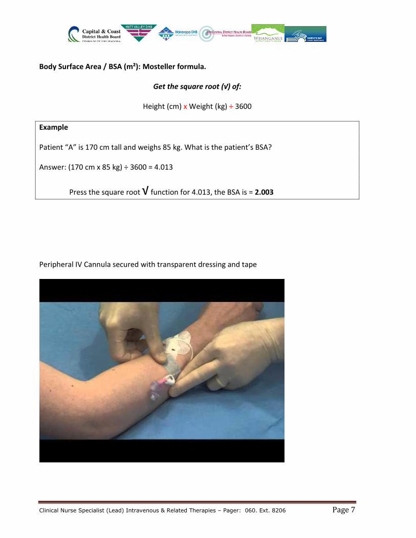
Peripheral IV Cannula secured with transparent dressing and tape
Clinical Nurse Specialist (Lead) Intravenous & Related Therapies – Pager: 060. Ext. 8206 Page 8
FUNDAMENTAL INTRAVENOUS KNOWLEDGE
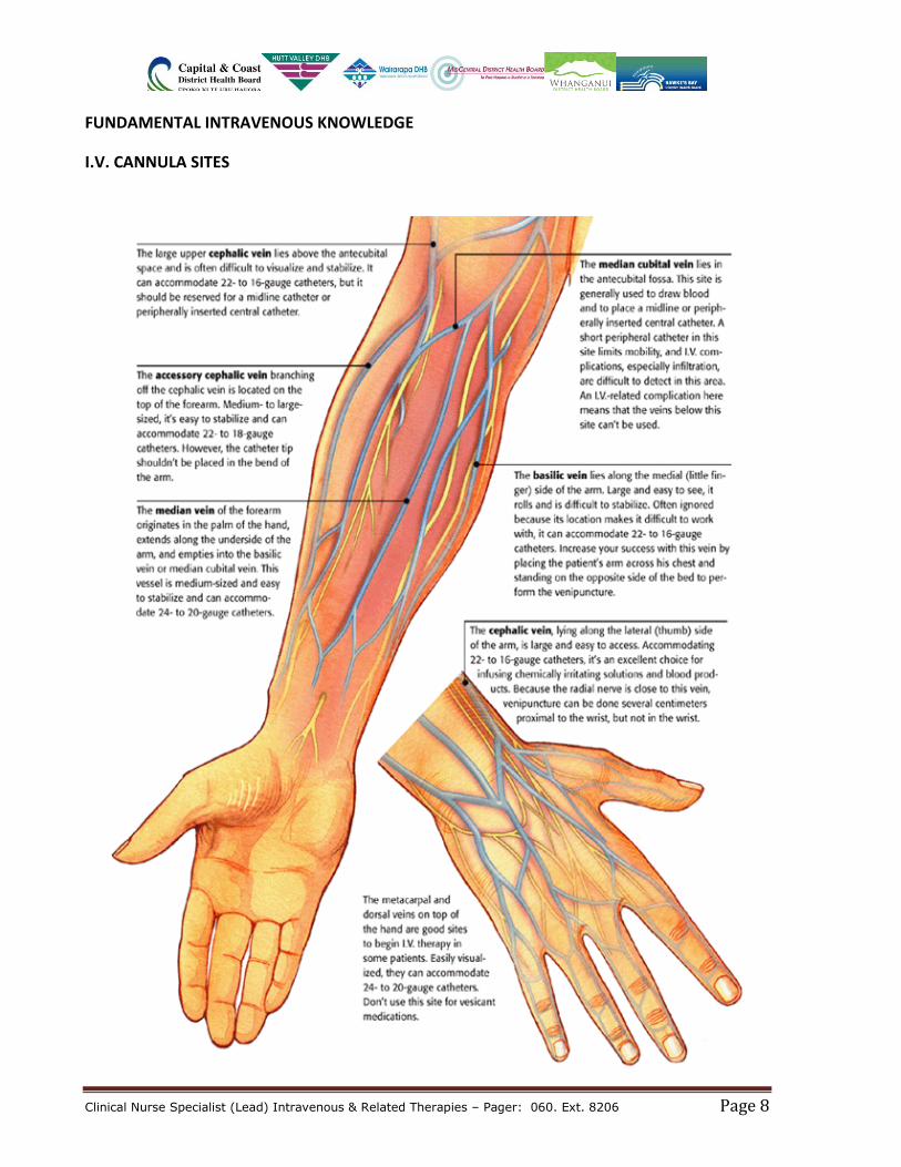
I.V. CANNULA SITES
Clinical Nurse Specialist (Lead) Intravenous & Related Therapies – Pager: 060. Ext. 8206 Page 9
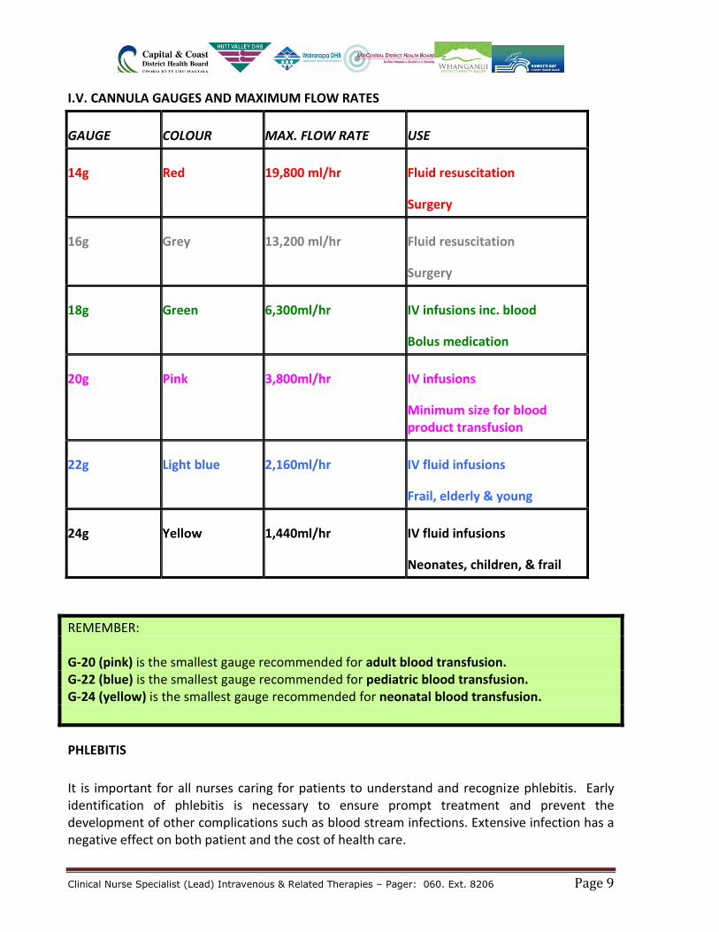
I.V. CANNULA GAUGES AND MAXIMUM FLOW RATES
GAUGE COLOUR MAX. FLOW RATE USE
14g Red 19,800 ml/hr Fluid resuscitation
Surgery
16g Grey 13,200 ml/hr Fluid resuscitation
Surgery
18g Green 6,300ml/hr IV infusions inc. blood
Bolus medication
20g Pink 3,800ml/hr IV infusions
Minimum size for blood product transfusion
22g Light blue 2,160ml/hr IV fluid infusions
Frail, elderly & young
24g Yellow 1,440ml/hr IV fluid infusions
Neonates, children, & frail
REMEMBER: G-20 (pink) is the smallest gauge recommended for adult blood transfusion. G-22 (blue) is the smallest gauge recommended for pediatric blood transfusion. G-24 (yellow) is the smallest gauge recommended for neonatal blood transfusion.
PHLEBITIS
It is important for all nurses caring for patients to understand and recognize phlebitis. Early identification of phlebitis is necessary to ensure prompt treatment and prevent the development of other complications such as blood stream infections. Extensive infection has a negative effect on both patient and the cost of health care.
Clinical Nurse Specialist (Lead) Intravenous & Related Therapies – Pager: 060. Ext. 8206 Page 10
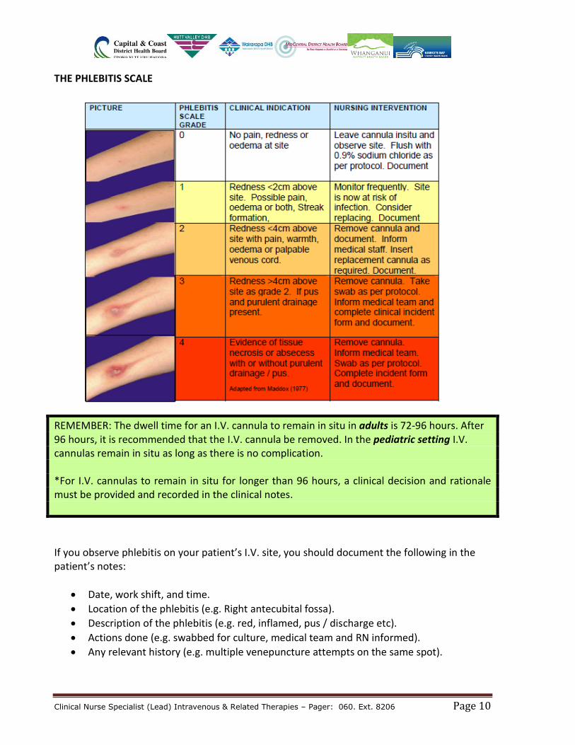
THE PHLEBITIS SCALE
REMEMBER: The dwell time for an I.V. cannula to remain in situ in adults is 72-96 hours. After 96 hours, it is recommended that the I.V. cannula be removed. In the pediatric setting I.V. cannulas remain in situ as long as there is no complication. *For I.V. cannulas to remain in situ for longer than 96 hours, a clinical decision and rationale must be provided and recorded in the clinical notes.
If you observe phlebitis on your patient’s I.V. site, you should document the following in the patient’s notes:
Date, work shift, and time.
Location of the phlebitis (e.g. Right antecubital fossa).
Description of the phlebitis (e.g. red, inflamed, pus / discharge etc).
Actions done (e.g. swabbed for culture, medical team and RN informed).
Any relevant history (e.g. multiple venepuncture attempts on the same spot).
Clinical Nurse Specialist (Lead) Intravenous & Related Therapies – Pager: 060. Ext. 8206 Page 11
MANAGEMENT AND CARE of A PERIPHERAL I.V. CANNULA The S-A-S Protocol When accessing a peripheral I.V. cannula to give fluid or a medication, always follow the S-A-S protocol.
S - Sodium chloride 0.9% flush (3 – 5 ml is sufficient). *To test the I.V.C. patency.
A - Access / Administer the drug or fluid.
S - Sodium chloride 0.9% flush (3 – 5 ml is sufficient). *To flush the medication and maintain patency.
REMEMBER: When flushing an I.V. cannula, ALWAYS lock it using the positive pressure technique. This is done by simultaneously engaging the clamp on the I.V. cannula while flushing the last few milliliters of Sodium chloride 0.9%. This “locking under positive pressure” will prevent blood from drawing back into the I.V. cannula thus, reducing the chances of occlusion.
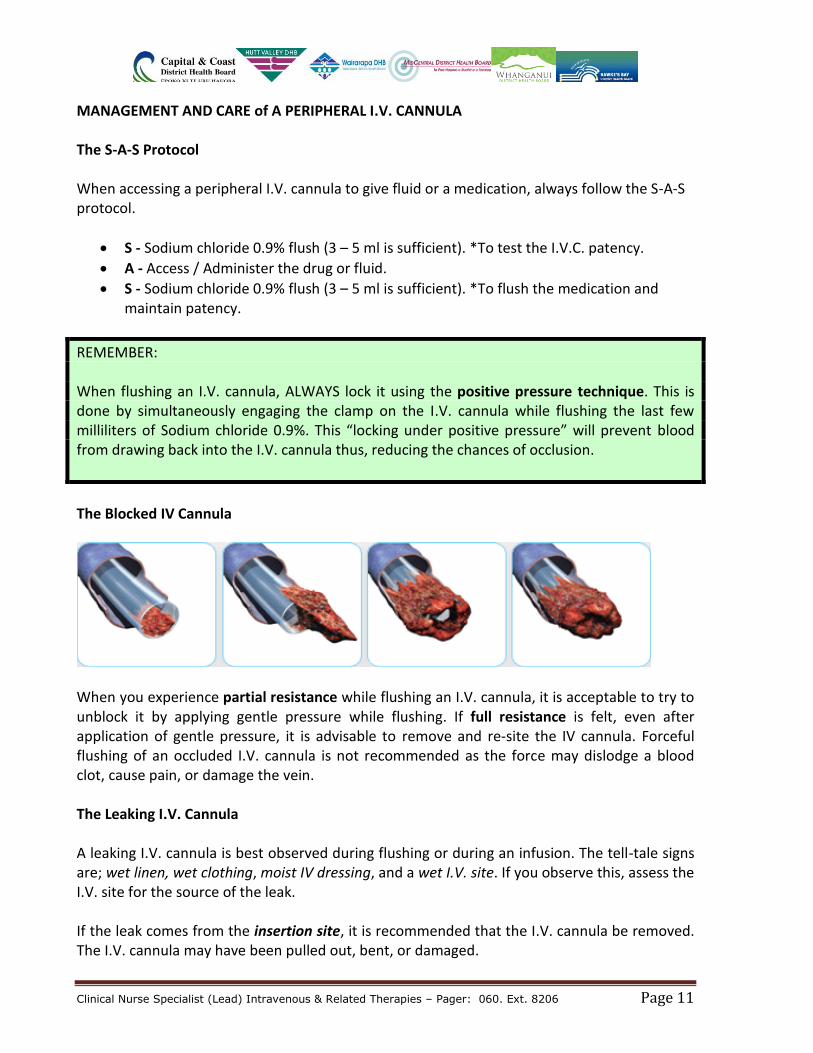
The Blocked IV Cannula
When you experience partial resistance while flushing an I.V. cannula, it is acceptable to try to unblock it by applying gentle pressure while flushing. If full resistance is felt, even after application of gentle pressure, it is advisable to remove and re-site the IV cannula. Forceful flushing of an occluded I.V. cannula is not recommended as the force may dislodge a blood clot, cause pain, or damage the vein. The Leaking I.V. Cannula A leaking I.V. cannula is best observed during flushing or during an infusion. The tell-tale signs are; wet linen, wet clothing, moist IV dressing, and a wet I.V. site. If you observe this, assess the I.V. site for the source of the leak. If the leak comes from the insertion site, it is recommended that the I.V. cannula be removed. The I.V. cannula may have been pulled out, bent, or damaged.
Clinical Nurse Specialist (Lead) Intravenous & Related Therapies – Pager: 060. Ext. 8206 Page 12
If the leak is coming from the connection (where the I.V. cannula is connected to the extension set or access valve), simply secure the connection by ensuring the luer-lock connection is tight. If no leak is observed, you may re-secure the I.V. cannula by putting on a new dressing. Removing an I.V. Cannula Removing an I.V. Cannula is simple and easy. It is also relatively painless if done using a proper technique. What you need for removing an I.V. Cannula:
1 x small sterile gauze or pressure pad dressing.
Tape roll.
REMOVE wipes (adhesive remover).
Alcohol or Clorhexidine swabs (*if needed for cleaning).
Clean pair of gloves.
Procedure
1. Make sure that the cannula is no longer clinically indicated. 2. Explain the procedure to the patient. 3. Cleanse hands. 4. Wear non-sterile gloves. 5. Carefully remove the dressing and tape from the skin to expose the skin and cannula. 6. Avoid manipulation of the device to prevent contamination and pain. 7. Cover site with gauze square then gently remove the cannula. 8. Apply digital pressure until bleeding has stopped. Secure the dressing in place. 9. Dispose of surplus equipment. 10. Cleanse hands. 11. Document removal in clinical notes and record tissue integrity. 12. Monitor ongoing healing and advise patient to report any symptoms of discomfort,
bleeding, pain, swelling or redness. 13. Alert the medical staff if the area is reddened 14. If infection is suspected complete an incident report on Riskman. 15. Advise patient to keep dressing on for at least 8 hours unless it is a pressure dressing,
which must be removed within one hour of application (otherwise persistent pressure may result in damage to the vein).
16. On discharge, inform patient to notify GP if ongoing discomfort, swelling, heat or discharge from site.
Clinical Nurse Specialist (Lead) Intravenous & Related Therapies – Pager: 060. Ext. 8206 Page 13
ADVERSE EFFECTS OF TRANSFUSION (Source: New Zealand Blood Service, 2008) Transfusions can both benefit and harm the patient. Good clinical practice depends on understanding the benefits and risks that each treatment can provide for patients, such as not using a blood product or of using an alternative. Blood transfusion has become steadily safer over the years as various viruses and infectious hazards have been identified through donor selection procedures and blood screening tests. Though much effort is put into ensuring the safety of transfusion particularly in preventing viral transmission, mistransfusion and non-infectious hazards of transfusion still account for a significant proportion of the reported transfusion-related adverse events. Data on the occurrence of adverse reactions and incidents are collected as part of NZBS’ haemovigilance activities. REPORTING ADVERSE REACTIONS OR INCIDENTS Serious or acute life-threatening reactions are rare so any new or unexpected symptoms during transfusion must be taken seriously as they may be an early warning of a serious reaction.
Severe reactions are most likely to occur within 15 minutes of starting transfusion of each individual unit of blood products.
It is extremely important to closely monitor the patient during this initial 15 minute period and then at regular intervals.
Serious events must be discussed with a NZBS Transfusion Medicine Specialist or clinical haematologist for advice on further management of the patient, laboratory investigations and future transfusion requirements.
Clinical Nurse Specialist (Lead) Intravenous & Related Therapies – Pager: 060. Ext. 8206 Page 14
Signs and Symptoms of Anaphylactoid/Anaphylactic Reactions (Transfusion Medicine Handbook, NZBS, 2008)
Symptoms Signs
Dyspnoea Chest pain Abdominal pain Nausea
Hypotension Bronchospasm Periorbital and laryngeal oedema Vomiting Erythema Urticaria Conjunctivitis
Signs and Symptoms of Acute Haemolytic Reaction (Transfusion Medicine Handbook, NZBS, 2008)
Symptoms Signs
Feeling of apprehension or “sense of impending doom” Agitation Pain at the venepuncture site Pain in the abdomen, flank or chest Difficulty breathing
Unexplained fever Flushing Hypotension, or sudden onset of hypertension General oozing from wounds or puncture sites Haemoglobinaemia Haemoglobinuria Skin rash
Bacterial Contamination: Signs and symptoms of septic reaction includes: (Transfusion Medicine Handbook, NZBS, 2008):
Fever (temperature ≥ 38.5°C or a rise from baseline of ≥ 1.5°C) Rigors Nausea/vomiting Diarrhoea Hypothermia Tachycardia (≥ 120 beats per minute or change from baseline of ≥ 40 beats per minute) Hypotension or hypertension (rise / fall in systolic blood pressure of ≥ 30 mm Hg) Haemolysis Shock Multiple organ failure during or immediately after the transfusion
Clinical Nurse Specialist (Lead) Intravenous & Related Therapies – Pager: 060. Ext. 8206 Page 15
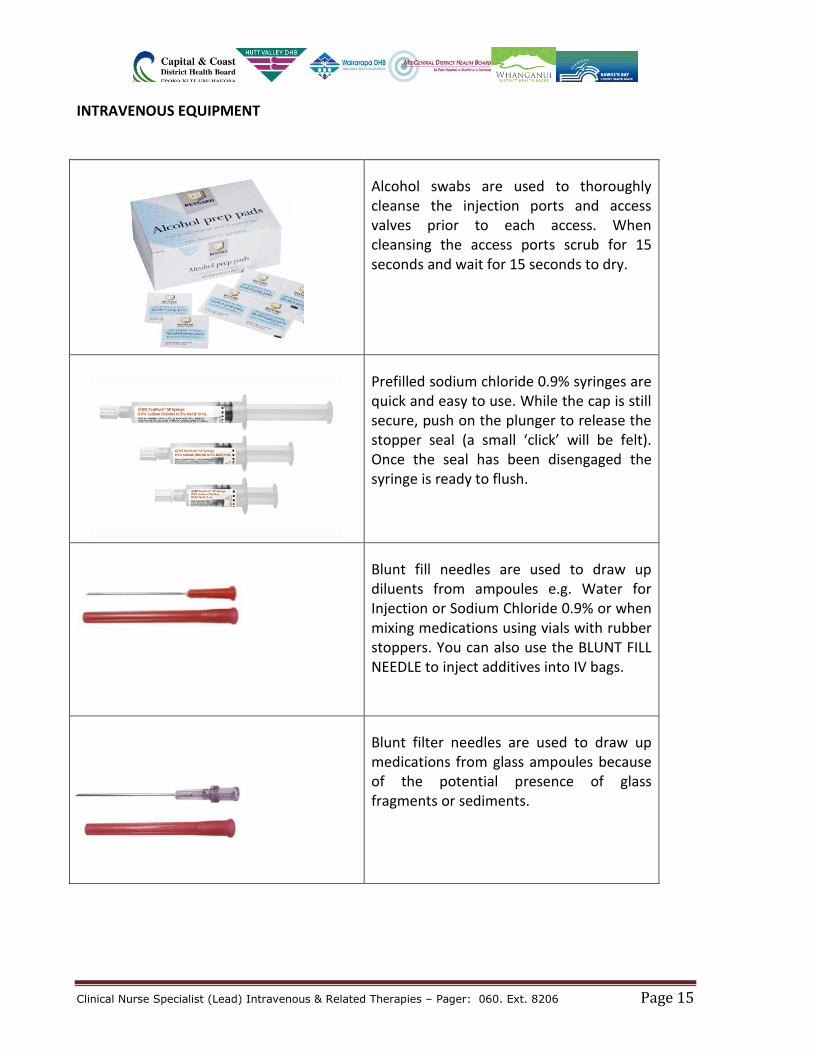
INTRAVENOUS EQUIPMENT
Alcohol swabs are used to thoroughly cleanse the injection ports and access valves prior to each access. When cleansing the access ports scrub for 15 seconds and wait for 15 seconds to dry.
Prefilled sodium chloride 0.9% syringes are quick and easy to use. While the cap is still secure, push on the plunger to release the stopper seal (a small ‘click’ will be felt). Once the seal has been disengaged the syringe is ready to flush.
Blunt fill needles are used to draw up diluents from ampoules e.g. Water for Injection or Sodium Chloride 0.9% or when mixing medications using vials with rubber stoppers. You can also use the BLUNT FILL NEEDLE to inject additives into IV bags.
Blunt filter needles are used to draw up medications from glass ampoules because of the potential presence of glass fragments or sediments.
Clinical Nurse Specialist (Lead) Intravenous & Related Therapies – Pager: 060. Ext. 8206 Page 16
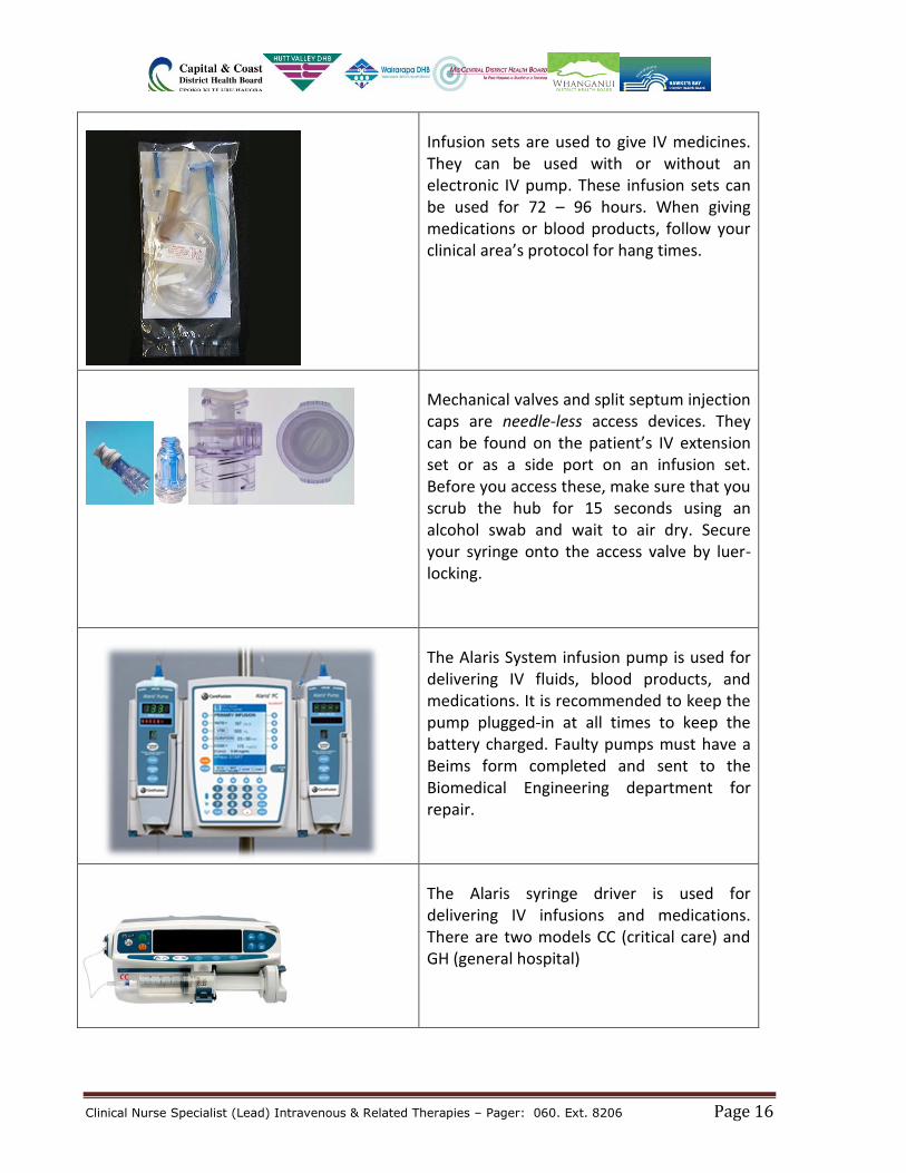
Infusion sets are used to give IV medicines. They can be used with or without an electronic IV pump. These infusion sets can be used for 72 – 96 hours. When giving medications or blood products, follow your clinical area’s protocol for hang times.
Mechanical valves and split septum injection caps are needle-less access devices. They can be found on the patient’s IV extension set or as a side port on an infusion set. Before you access these, make sure that you scrub the hub for 15 seconds using an alcohol swab and wait to air dry. Secure your syringe onto the access valve by luer-locking.
The Alaris System infusion pump is used for delivering IV fluids, blood products, and medications. It is recommended to keep the pump plugged-in at all times to keep the battery charged. Faulty pumps must have a Beims form completed and sent to the Biomedical Engineering department for repair.
The Alaris syringe driver is used for delivering IV infusions and medications. There are two models CC (critical care) and GH (general hospital)
Clinical Nurse Specialist (Lead) Intravenous & Related Therapies – Pager: 060. Ext. 8206 Page 17
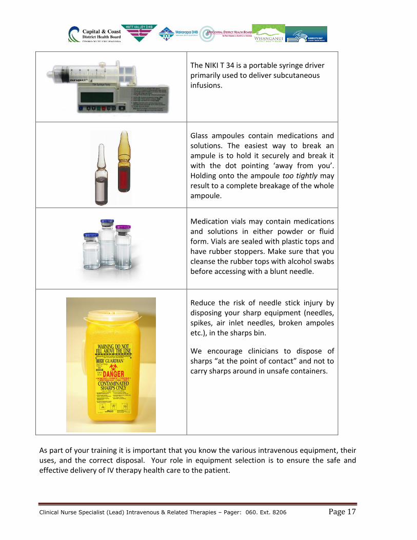
The NIKI T 34 is a portable syringe driver primarily used to deliver subcutaneous infusions.
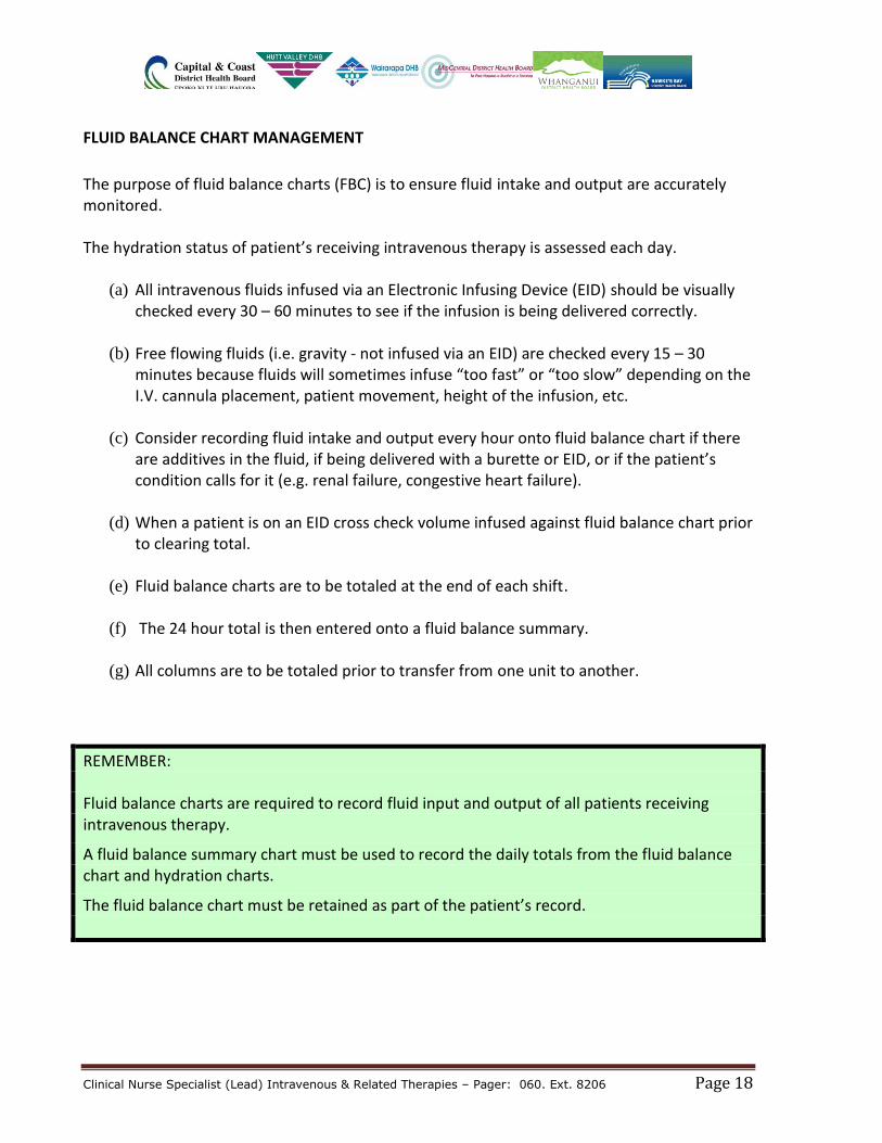
Glass ampoules contain medications and solutions. The easiest way to break an ampule is to hold it securely and break it with the dot pointing ‘away from you’. Holding onto the ampoule too tightly may result to a complete breakage of the whole ampoule.
Medication vials may contain medications and solutions in either powder or fluid form. Vials are sealed with plastic tops and have rubber stoppers. Make sure that you cleanse the rubber tops with alcohol swabs before accessing with a blunt needle.
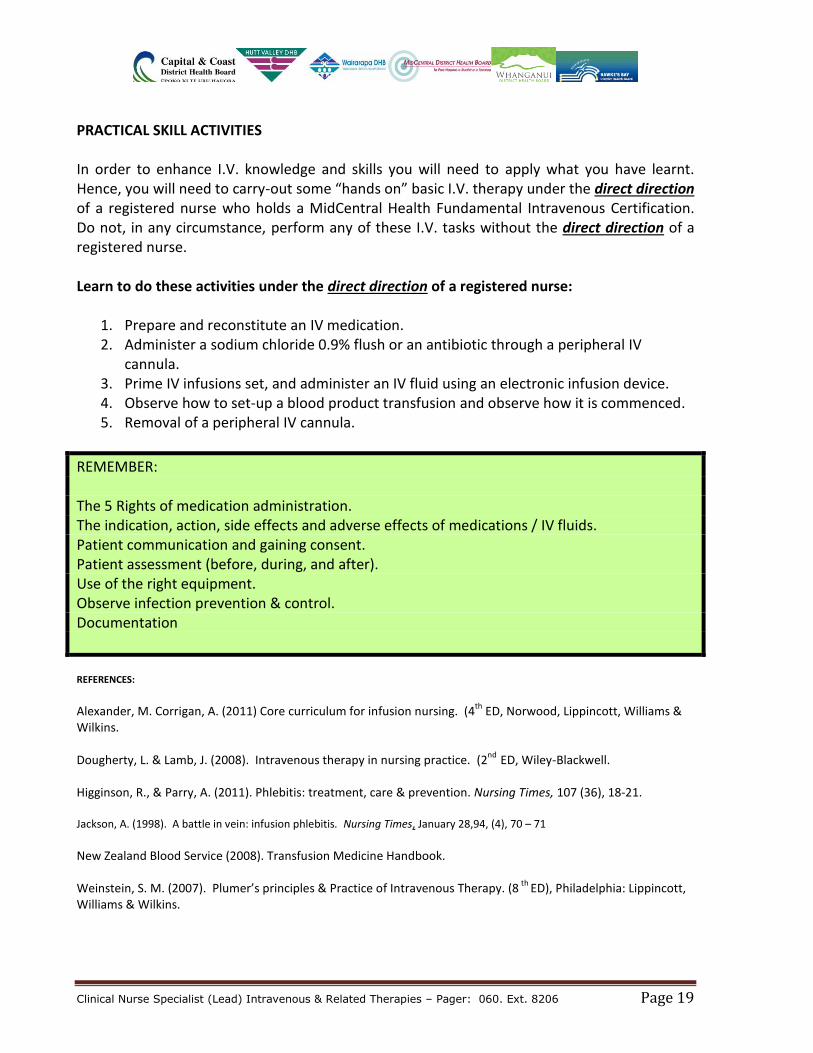
Reduce the risk of needle stick injury by disposing your sharp equipment (needles, spikes, air inlet needles, broken ampoles etc.), in the sharps bin.
We encourage clinicians to dispose of sharps “at the point of contact” and not to carry sharps around in unsafe containers.
As part of your training it is important that you know the various intravenous equipment, their uses, and the correct disposal. Your role in equipment selection is to ensure the safe and effective delivery of IV therapy health care to the patient.
Clinical Nurse Specialist (Lead) Intravenous & Related Therapies – Pager: 060. Ext. 8206 Page 18
FLUID BALANCE CHART MANAGEMENT
The purpose of fluid balance charts (FBC) is to ensure fluid intake and output are accurately monitored. The hydration status of patient’s receiving intravenous therapy is assessed each day.
(a) All intravenous fluids infused via an Electronic Infusing Device (EID) should be visually checked every 30 – 60 minutes to see if the infusion is being delivered correctly.
(b) Free flowing fluids (i.e. gravity - not infused via an EID) are checked every 15 – 30
minutes because fluids will sometimes infuse “too fast” or “too slow” depending on the I.V. cannula placement, patient movement, height of the infusion, etc.
(c) Consider recording fluid intake and output every hour onto fluid balance chart if there
are additives in the fluid, if being delivered with a burette or EID, or if the patient’s condition calls for it (e.g. renal failure, congestive heart failure).
(d) When a patient is on an EID cross check volume infused against fluid balance chart prior
to clearing total.
(e) Fluid balance charts are to be totaled at the end of each shift.
(f) The 24 hour total is then entered onto a fluid balance summary.
(g) All columns are to be totaled prior to transfer from one unit to another.
REMEMBER: Fluid balance charts are required to record fluid input and output of all patients receiving intravenous therapy.
A fluid balance summary chart must be used to record the daily totals from the fluid balance chart and hydration charts.
The fluid balance chart must be retained as part of the patient’s record.
Clinical Nurse Specialist (Lead) Intravenous & Related Therapies – Pager: 060. Ext. 8206 Page 19
PRACTICAL SKILL ACTIVITIES In order to enhance I.V. knowledge and skills you will need to apply what you have learnt. Hence, you will need to carry-out some “hands on” basic I.V. therapy under the direct direction of a registered nurse who holds a MidCentral Health Fundamental Intravenous Certification. Do not, in any circumstance, perform any of these I.V. tasks without the direct direction of a registered nurse. Learn to do these activities under the direct direction of a registered nurse:
1. Prepare and reconstitute an IV medication. 2. Administer a sodium chloride 0.9% flush or an antibiotic through a peripheral IV
cannula. 3. Prime IV infusions set, and administer an IV fluid using an electronic infusion device. 4. Observe how to set-up a blood product transfusion and observe how it is commenced. 5. Removal of a peripheral IV cannula.
REMEMBER: The 5 Rights of medication administration. The indication, action, side effects and adverse effects of medications / IV fluids. Patient communication and gaining consent. Patient assessment (before, during, and after). Use of the right equipment. Observe infection prevention & control. Documentation
REFERENCES:
Alexander, M. Corrigan, A. (2011) Core curriculum for infusion nursing. (4th
ED, Norwood, Lippincott, Williams & Wilkins. Dougherty, L. & Lamb, J. (2008). Intravenous therapy in nursing practice. (2
nd ED, Wiley-Blackwell.
Higginson, R., & Parry, A. (2011). Phlebitis: treatment, care & prevention. Nursing Times, 107 (36), 18-21. Jackson, A. (1998). A battle in vein: infusion phlebitis. Nursing Times, January 28,94, (4), 70 – 71 New Zealand Blood Service (2008). Transfusion Medicine Handbook. Weinstein, S. M. (2007). Plumer’s principles & Practice of Intravenous Therapy. (8
th ED), Philadelphia: Lippincott,
Williams & Wilkins.