catalytic mechanism of the glycyl radical enzyme 4
TRANSCRIPT
Catalytic Mechanism of the Glycyl Radical Enzyme
4-Hydroxyphenylacetate Decarboxylase from Continuum
Electrostatic and QC/MM Calculations
Supplementary Materials
Mikolaj Feliks,1 Berta M. Martins,2 G. Matthias Ullmann1,†
1) Computational Biochemistry Group, University of Bayreuth, Universitatsstr. 30, BGI,
95447 Bayreuth, Germany
2) Institut fur Biologie, Strukturbiologie/Biochemie – Radikalenzyme, Humboldt-Universitat
zu Berlin, Unter den Linden 6, 10099 Berlin, Germany
†) to whom correspondence should be addressed;
e-mail: [email protected]
S1
Contents
S1 NEB reaction profiles for models M1, M2 and M3 . . . . . . . . . . . . . . . . . . . S3
S2 Titration curves . . . . . . . . . . . . . . . . . . . . . . . . . . . . . . . . . . . . . S6
S3 QC/MM-optimized geometries . . . . . . . . . . . . . . . . . . . . . . . . . . . . . S10
S4 Ruling out of the possibility that the substrate can be differently orientated in the
binding pocket . . . . . . . . . . . . . . . . . . . . . . . . . . . . . . . . . . . . . S11
S2
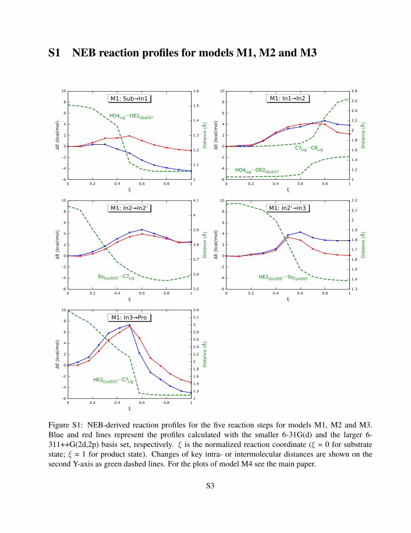
S1 NEB reaction profiles for models M1, M2 and M3
Figure S1: NEB-derived reaction profiles for the five reaction steps for models M1, M2 and M3.Blue and red lines represent the profiles calculated with the smaller 6-31G(d) and the larger 6-311++G(2d,2p) basis set, respectively. ξ is the normalized reaction coordinate (ξ = 0 for substratestate; ξ = 1 for product state). Changes of key intra- or intermolecular distances are shown on thesecond Y-axis as green dashed lines. For the plots of model M4 see the main paper.
S3

Figure S1 continued
S4
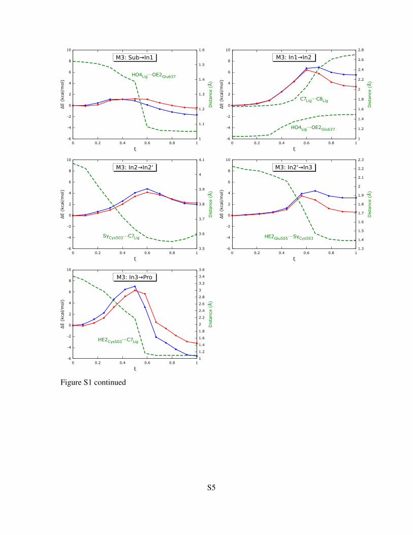
Figure S1 continued
S5
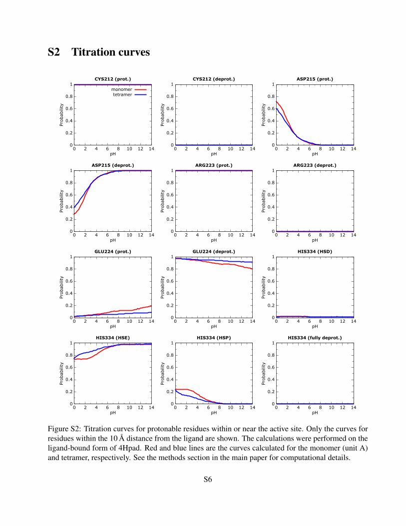
S2 Titration curves
Figure S2: Titration curves for protonable residues within or near the active site. Only the curves forresidues within the 10 A distance from the ligand are shown. The calculations were performed on theligand-bound form of 4Hpad. Red and blue lines are the curves calculated for the monomer (unit A)and tetramer, respectively. See the methods section in the main paper for computational details.
S6
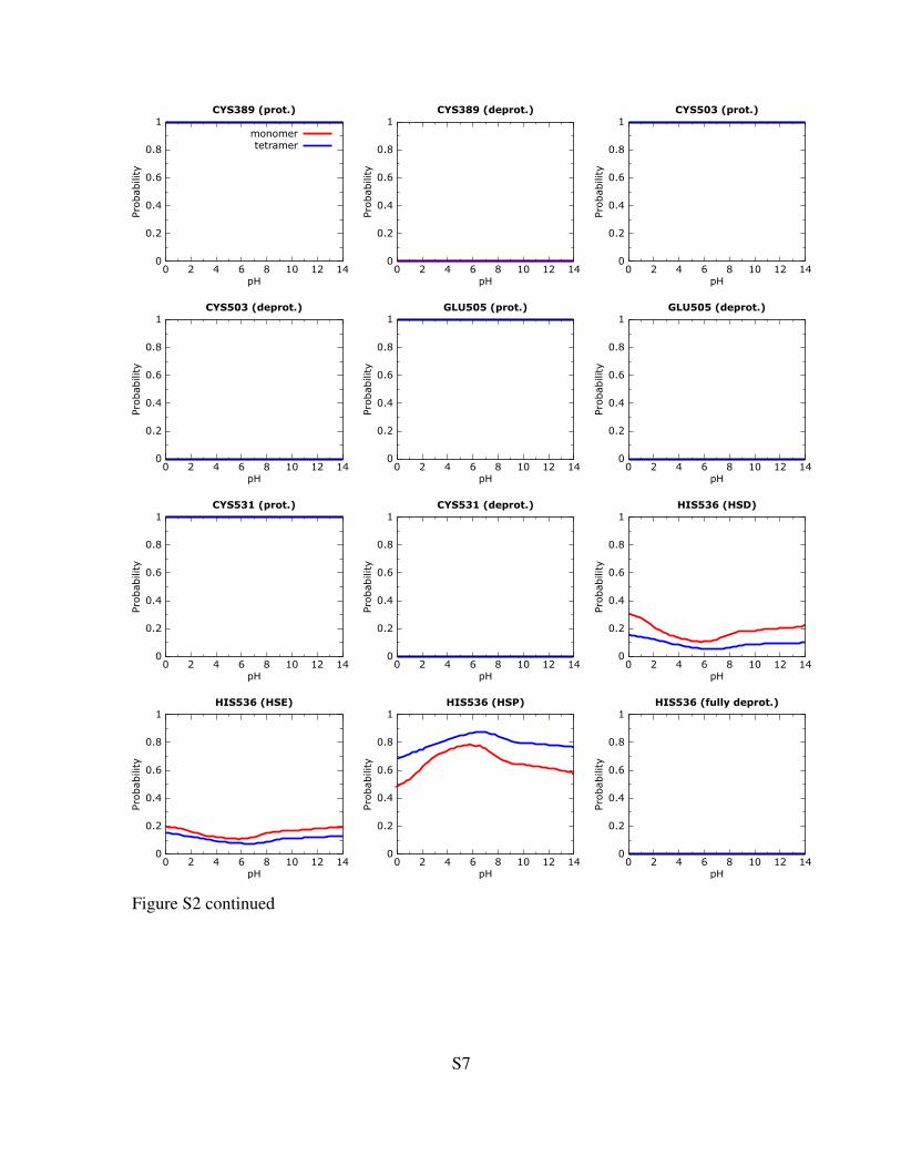
Figure S2 continued
S7

Figure S2 continued
S8
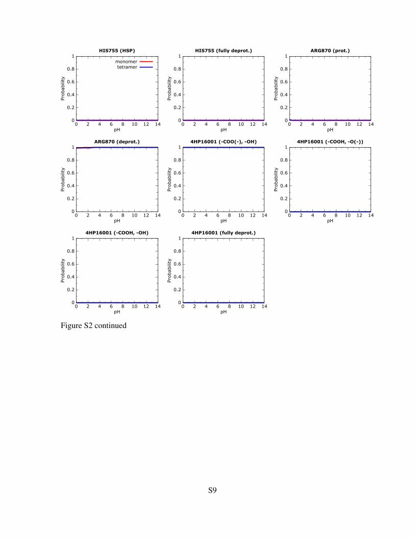
Figure S2 continued
S9

S3 QC/MM-optimized geometries
The QC/MM-optimized geometries of reactants, intermediates, transition states and products for mod-
els M1, M2, M3 and M4 are given in the gzipped tar-file Geometries Hartree.tar.gz. The
geometries are in the PDB format using an easily understandable naming scheme. For instance, the
name M4 ts1.pdb means that it is the model of the transition state ts1 using our model M4. The
QC, MM and QC/MM energies are given in the header of the file in Hartrees. The two columns that
usually contain occupancy and temperature factor are the atomic charges (in case of QC atoms Mul-
liken charges) and Mulliken spin populations, respectively. Mulliken charges and spin densities were
obtained by single-point calculations using the 6-311++G(2d,2p) basis set on top of the geometries
optimized using the 6-31G(d) basis set. For both types of calculations, the B3LYP density functional
was used as a QC-potential and the CHARMM27 force field was used as a MM-potential. The last
column shows weather the atom is a part of the MM region (M) or QC region (Q).
S10
S4 Ruling out of the possibility that the substrate can be differ-
ently orientated in the binding pocket
For the reaction mechanism that was proposed before the structure was known,1 the substrate would
have to bind in a different binding mode, namely 180◦ rotated. To test whether this substrate binding
mode is possible, we tried to find a reasonable alternative ligand orientation in the protein. First, we
tried to dock the substrate to the substrate-free structure of 4Hpad (PDB code 2y8n)2 using Autodock.3
With this software, we always found the ligand orientation seen in the substrate-bound structure of
4Hpad (PDB code: 2yaj). Thus, we rotated the substrate manually inside the binding pocked (using
Coot4). In the rotated conformation, the hydroxyl group of the substrate is located in the vicinity of
the radical center on Cys503 and the carboxylic group is positioned close to His536 and Glu637. This
alternative binding mode could allow the catalytic mechanism that was proposed for 4Hpad before the
determination of the crystal structure. The rotated conformation was energetically optimized using a
5ps molecular dynamics simulations followed by an energy minimization in pDynamo. During the
simulation, the outer parts of the model were kept restrained using the same set of restraints as for the
M4 QC/MM model. As a reference, we also performed an equivalent simulation for the orientation
found in the crystal structure. The resulting two geometries were geometry optimized and their MM-
energies were compared. The energy of the geometry representing the rotated substrate was calculated
to be about 24 kcal/mol higher than the energy of the geometry found in the crystal structure, which
makes this orientation a very unlikely starting point for the reaction.
The far less favorable binding of the rotated substrate can be explained by the distorted network
of hydrogen bonds in the active site. In the crystal, the substrate is stabilized in the active site by four
hydrogen bonds, namely from Ser344, the backbone fragment at Cys503, Glu505 and Glu637. In the
rotated conformation, the carboxylic group of the substrate is partly stabilized by Arg223 and a water
molecule between this group and Glu637. The hydroxyl group is only stabilized by a weakly negative
radical center at Cys503. Also, this hydrophobic group is now located close to the hydrophobic ring
of Phe405. The Ser344 side-chain has moved away from the active site. The backbone fragment at
Cys503 is now involved in hydrogen bonds only with Glu505 and not with the substrate.
S11
Bibliography
[1] Selmer, T.; Andrei, P. Eur. J. Biochem., 2001, 268, 1363–1372.
[2] Martins, B. M.; Blaser, M.; Feliks, M.; Ullmann, G. M.; Buckel, W.; Selmer, T. J. Am. Chem.
Soc., 2011, 133, 14666–14674.
[3] Morris, G. M.; Huey, R.; Lindstrom, W.; Sanner, M. F.; Belew, R. K.; Goodsell, D. S.; Olson,
A. J. J. Comp. Chem., 2009, 16, 2785–91.
[4] Emsley, P.; Lohkamp, B.; Scott, W.; Cowtan, K. Acta Cryst. D, 2010, 66, 486–501.
S12