casey - gait
TRANSCRIPT
5/15/2018 Casey - Gait - slidepdf.com
http://slidepdf.com/reader/full/casey-gait 1/22
•• •CHAPTER 8
Gait Analysis
Rehabil itat ion Medicine: Principles and Pract ice, Third Edition,edited by Joel A. DeLisa and Bruce M. Gans.Lippincott-Raven Publishers. Philadelphia © 1998.
D. Casey Kerrigan, Michael Schaufele, and Marco N. Wen
Gait, referring in humans to walking and running, is one of
the most fundamental actions in life. Rehabilitation clini-
cians can especially appreciate the complexity of gait in theface of impairment or functional limitation. Often, an indi-
vidual has difficulty walking, and for some, gait may be
functionally impossible. It is the physiatrist's task to deter-
mine the specific causes of why a person cannot walk well,
not only at the pathophysiology level, but also at the impair-
ment and functional limitation levels as well. The effective-
ness of any physiatric treatment relies heavily on the ability
to accurately determine these causes.
NOMENCLATURE
An understanding of gait analysis first requires familiari-
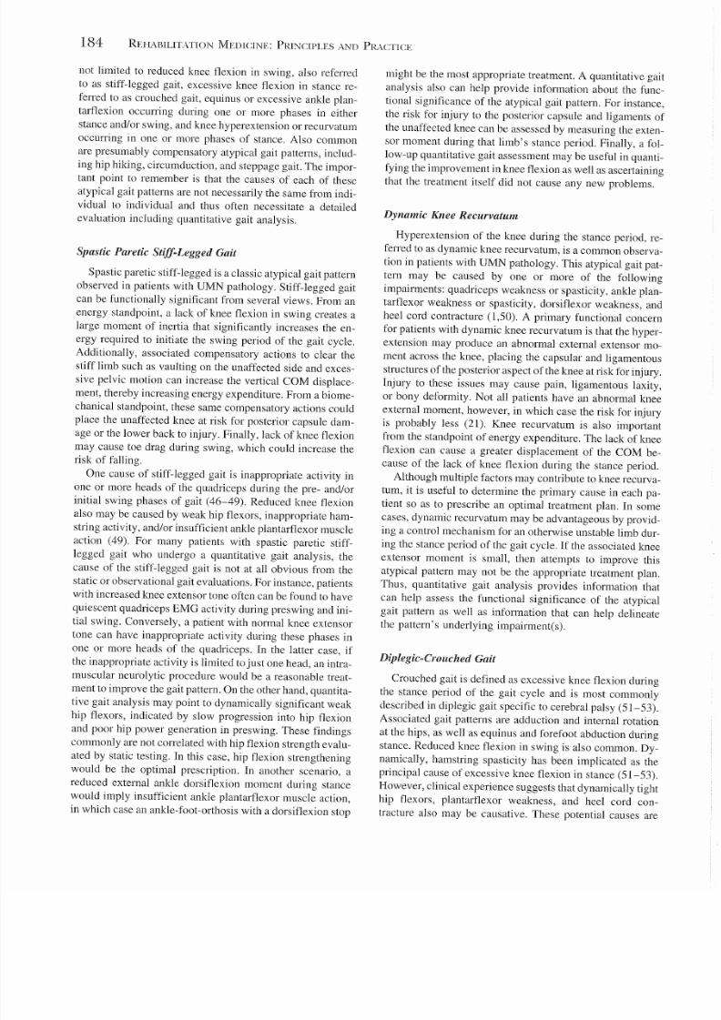
zation with the currently accepted terminology. Because gaitis habitual in nature, we often focus our analysis on the
functional unit of gait, called the gait cycle, or stride. Various
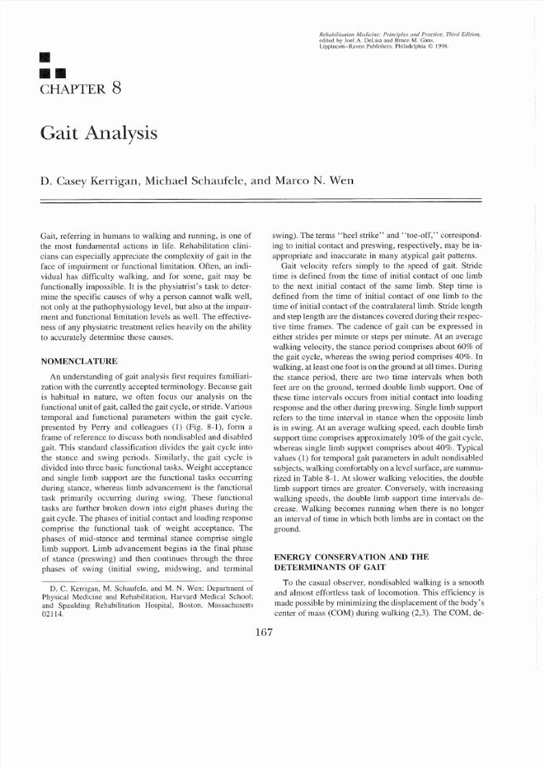
temporal and functional parameters within the gait cycle.
presented by Perry and colleagues (1) (Fig. 8-1), form a
frame of reference to discuss both nondisabled and disabled
gait. This standard classification divides the gait cycle into
the stance and swing periods. Similarly, the gait cycle is
divided into three basic functional tasks. Weight acceptance
and single limb support are the functional tasks occurring
during stance, whereas limb advancement is the functional
task primarily occurring during swing. These functional
tasks are further broken down into eight phases during the
gait cycle. The phases of initial contact and loading responsecomprise the functional task of weight acceptance. The
phases of mid-stance and terminal stance comprise single
limb support. Limb advancement begins in the final phase
of stance (preswing) and then continues through the three
phases of swing (initial swing, midswing, and terminal
D. C. Kerrigan, M. Schaufele, and M. N. Wen: Department of
Physical Medicine and Rehabilitation, Harvard Medical School;
and Spaulding Rehabil ita tion Hospital , Boston. Massachusetts
02114.
swing). The terms "heel strike" and "toe-off," correspond-
ing to initial contact and preswing, respectively, may be in-
appropriate and inaccurate in many atypical gait patterns.Gait velocity refers simply to the speed of gait. Stride
time is defined from the time of initial contact of one limb
to the next initial contact of the same limb. Step time is
defined from the time of initial contact of one limb to the
time of initial contact of the contralateral limb. Stride length
and step length are the distances covered during their respec-
tive time frames. The cadence of gait can be expressed in
either strides per minute or steps per minute. At an average
walking velocity, the stance period comprises about 60% of
the gait cycle, whereas the swing period comprises 40%. In
walking, at least one foot is on the ground at all times. During
the stance period, there are two time intervals when both
feet are on the ground, termed double limb support. One ofthese time intervals occurs from initial contact into loading
response and the other during preswing. Single limb support
refers to the time interval in stance when the opposite limb
is in swing. At an average walking speed, each double limb
support time comprises approximately 10% of the gait cycle,
whereas single limb support comprises about 40%. Typical
values (1) for temporal gait parameters in adult nondisabled
subjects, walking comfortably on a level surface, are summa-
rized in Table 8-1. At slower walking velocities, the double
limb support times are greater. Conversely, with increasing
walking speeds, the double limb support time intervals de-
crease. Walking becomes running when there is no longer
an interval of time in which both limbs are in contact on theground.
ENERGY CONSERVAnON AND THE
DETERMINANTS OF GAIT
To the casual observer, nondisabled walking is a smooth
and almost effortless task of locomotion. This efficiency is
made possible by minimizing the displacement of the body's
center of mass (COM) during walking (2,3). The COM, de-
167
5/15/2018 Casey - Gait - slidepdf.com
http://slidepdf.com/reader/full/casey-gait 2/22
168 REHABILITATION MEDICINE: PRINCIPLES AND PRACTICE
Periods:Stance
Tasks:
Single Limb
Support
Weight
Acceptance
Phases:Loading
Response
Mid
Stance
FIG. 8-1. Periods of the gait cycle. The gait cycle is separated into two distinct periods of stance and
swing. Functional tasks include weight acceptance and single limb support during stance and limb
advancement during swing. The stance period of the gait cycle includes initial contact, loading response,
midstance, terminal stance, and preswing. The swing period includes initial swing, midswing, and
terminal swing.
fined as the hypothetical point at which all mass can be
considered to be concentrated, is located just anterior to the
second sacral vertebrae in the average human lying in the
anatomic position (4). During walking, the COM normally
travels along a sinusoidal up-and-down and side-to-side path
with each step. It reaches its highest point during single limb
support and its lowest point during double limb support.
With regard to efficiency of walking, the vertical displace-
ment of the COM is far more relevant than the lateral dis-
placement (5). The major mechanisms by which the body
minimizes the displacement of the COM during walking are
via a series of maneuvers described as the determinants of
gait by Saunders and colleagues (6):
• Pelvic rotation in the transverse plane
• Pelvic obliquity in the coronal plane
• Lateral displacement in the coronal plane
• Interchange between knee, ankle, and foot motion
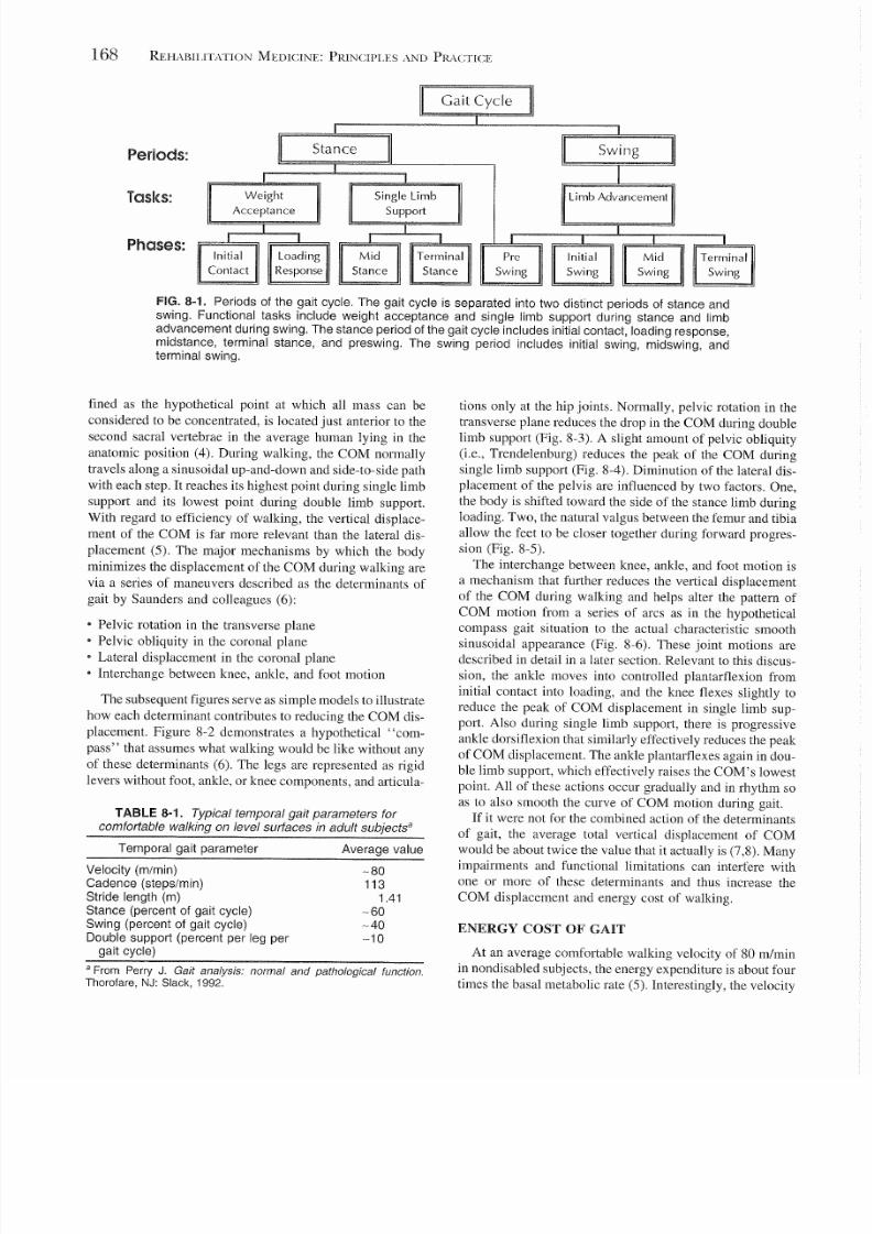
The subsequent figures serve as simple models to illustrate
how each determinant contributes to reducing the COM dis-
placement. Figure 8-2 demonstrates a hypothetical "com-
pass" that assumes what walking would be like without any
of these determinants (6). The legs are represented as rigid
levers without foot, ankle, or knee components, and articula-
TABLE 8-1. Typical temporal gait parameters for
comfortable walking on level surfaces in adult subjectsa
Temporal gait parameter Average value
Velocity (m/min)
Cadence (steps/min)
Stride length (m)
Stance (percent of gait cycle)
Swing (percent of gait cycle)
Double support (percent per leg per
gait cycle)
-80113
1.41
-60-40
-10
a From Perry J. Gait analysis: normal and pat hological function.
Thorofare, NJ: Slack, 1992.
tions only at the hip joints. Normally, pelvic rotation in the
transverse plane reduces the drop in the COM during double
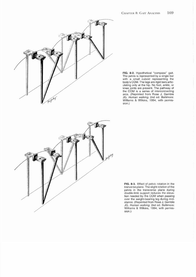
limb support (Fig. 8-3). A slight amount of pelvic obliquity
(i.e., Trendelenburg) reduces the peak of the COM during
single limb support (Fig. 8-4). Diminution of the lateral dis-
placement of the pelvis are influenced by two factors. One,
the body is shifted toward the side of the stance limb during
loading. Two, the natural valgus between the femur and tibia
allow the feet to be closer together during forward progres-
sion (Fig. 8-5).
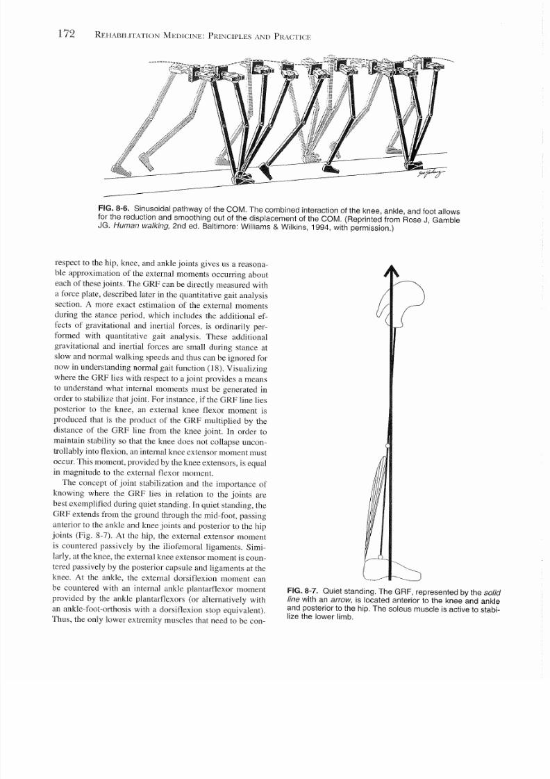
The interchange between knee, ankle, and foot motion is
a mechanism that further reduces the vertical displacement
of the COM during walking and helps alter the pattern of
COM motion from a series of arcs as in the hypothetical
compass gait situation to the actual characteristic smooth
sinusoidal appearance (Fig. 8-6). These joint motions are
described in detail in a later section. Relevant to this discus-
sion, the ankle moves into controlled plantarflexion from
initial contact into loading, and the knee flexes slightly to
reduce the peak of COM displacement in single limb sup-
port. Also during single limb support, there is progressive
ankle dorsiflexion that similarly effectively reduces the peak
of COM displacement. The ankle plantarflexes again in dou-
ble limb support, which effectively raises the COM's lowest
point. All of these actions occur gradually and in rhythm so
as to also smooth the curve of COM motion during gait.
If it were not for the combined action of the determinants
of gait, the average total vertical displacement of COMwould be about twice the value that it actually is (7,8). Many
impairments and functional limitations can interfere with
one or more of these determinants and thus increase the
COM displacement and energy cost of walking.
ENERGY COST OF GAIT
At an average comfortable walking velocity of 80 m/min
in nondisabled subjects, the energy expenditure is about four
times the basal metabolic rate (5). Interestingly, the velocity
5/15/2018 Casey - Gait - slidepdf.com
http://slidepdf.com/reader/full/casey-gait 3/22
~~"' .. .
CHAPTER 8: GAIT ANALYSIS 169
FIG. 8-2. Hypothetical "compass" gait.
The pelvis is represented by a single bar
with a small cuboid representing the
body's COM. The legs are rigid bars artic-
ulating only at the hip. No foot, ankle, or
knee joints are present. The pathway of
the COM is a series of interconnecting
arcs. (Reprinted from Rose J, Gamble
JG. Human walking, 2nd ed. Baltimore:
Williams & Wilkins, 1994, with permis-
sion.)
FIG. 8-3. Effect of pelvic rotation in the
tranverse plane. The slight rotat ion ofthe
pelvis in the transverse plane during
double-l imb support reduces the eleva-
tion needed by the COM when passing
over the weight-bearing leg during mid-
stance. (Reprinted from Rose J, Gamble
JG. Human walking, 2nd ed. Baltimore:
Williams & Wilkins, 1994, with permis-
sion.)
5/15/2018 Casey - Gait - slidepdf.com
http://slidepdf.com/reader/full/casey-gait 4/22
170 REHABILITATION MEDICINE: PRINCIPLES AND PRACTICE
/
that subjects choose as their comfortable speed is also the
velocity that requires the least energy per unit distance (9).
Walking faster or running usually requires anaerobic metab-
olism. On the other hand, walking slower requires extra en-
ergy, probably for balance support, rather than for propelling
the body forward (10). Importantly, the rate of energy ex-
pended during comfortable walking is consistent across thenondisabled and disabled gait populations (5). A person with
a gait disability tends to walk slower than a person without
a gait disability (11). Thus, although the energy expenditure
per unit time is consistent in subjects with gait disability,
increases in energy expenditure per unit distance are com-
mon. For instance, patients with hemiplegia affecting their
gait spend the same amount of energy per time during com-
fortable walking as subjects without gait disability, but they
walk slower and spend 37% (12) to 62% (13) more energy
per unit distance.
An important aim of improving gait disability may be to
reduce the energy required to walk. To this end, the effective-
ness of a particular type of rehabilitation treatment can be
assessed by evaluating the energy expended during walking.
The most direct method to evaluate energy expended is via
measuring the oxygen that is consumed during walking. This
involves having the subject breathe into a mask that is linked
to a gas analyzer. The analyzer determines how much oxy-
gen is being used, and from this, a calculation of energy
expenditure is given, based on the knowledge that about
4.83 kcal of energy is expended for every liter of oxygen
consumed (5). Alternatively, an estimate of energy expended
FIG. 8-4. Pelvic obliquity during single-
limb support. A drop in the pelvis on the
non-weight-bearing side allows for a re-
duction in the peak height of the COMduring mid-stance. (Reprinted from Rose
J, Gamble JG. Human walking, 2nd ed.
Baltimore: Williams & Wilkins, 1994, with
permission.)
can be obtained by measuring heart rate before and during
walking because the change in heart rate that occurs with
walking is linearly correlated with oxygen consumption
measurements (14). An easier, although more indirect,
method to evaluate the energy required to walk is to measure
the comfortable walking speed. This can be performed using
a stopwatch and a designated walking distance. This simplemeasure rests on the fact noted above that subjects with gait
disability tend to walk at a consistent energy rate, just slower.
Thus, comfortable walking speed relates indirectly to the
energy required to walk. Unfortunately, all of these mea-
sures, including oxygen consumption, heart rate, and com-
fortable walking speed, relate not only to biomechanical as-
pects of walking, but to cardiopulmonary conditioning and
psychological factors including mood as well. Quantitative
gait measures described in a later section, although useful
in determining the mechanisms of the gait impairment, are
insufficient in evaluating the efficiency of walking. A so-
called biomechanical efficiency quotient was proposed
(8,15) based on the concept of minimizing the COM dis-
placement through the determinants of gait. This measure
was introduced as a means to specifically evaluate biome-
chanical walking efficiency in subjects with gait disability,
independent of cardiopulmonary conditioning and psycho-
logical factors. The quotient is the measured vertical dis-
placement of the COM divided by the predicted vertical dis-
placement, the latter being a function of the subject's average
stride length and height of the pelvis from the ground. Pa-
tients with gait disability tend to have higher biomechanical
5/15/2018 Casey - Gait - slidepdf.com
http://slidepdf.com/reader/full/casey-gait 5/22
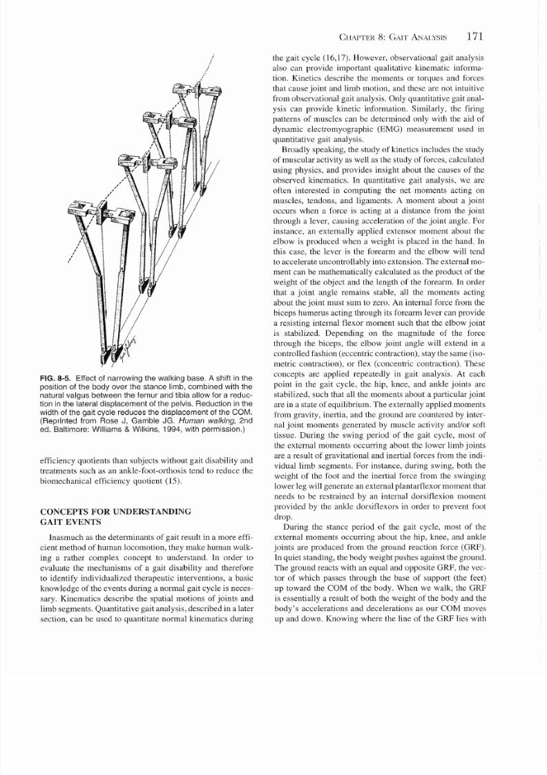
FIG. 8-5. Effect of narrowing the walking base. A shift in the
position of the body over the stance limb, combined with thenatural valgus between the femur and tibia allow for a reduc-
tion in the lateral displacement of the pelvis. Reduction in the
width of the gait cycle reduces the displacement of the COM.
(Reprinted from Rose J, Gamble JG. Human walking, 2nd
ed. Baltimore: Williams & Wilkins, 1994, with permission.)
efficiency quotients than subjects without gait disability and
treatments such as an ankle-foot-orthosis tend to reduce the
biomechanical efficiency quotient (15).
CONCEPTS FOR UNDERSTANDING
GAIT EVENTS
Inasmuch as the determinants of gait result in a more effi-
cient method of human locomotion, they make human walk-
ing a rather complex concept to understand. In order to
evaluate the mechanisms of a gait disability and therefore
to identify individualized therapeutic interventions, a basic
knowledge of the events during a normal gait cycle is neces-
sary. Kinematics describe the spatial motions of joints and
limb segments. Quantitative gait analysis, described in a later
section, can be used to quantitate normal kinematics during
CHAPTER 8: GAIT ANALYSIS 171
the gait cycle (16,17). However, observational gait analysis
also can provide important qualitative kinematic informa-
tion. Kinetics describe the moments or torques and forces
that cause joint and limb motion, and these are not intuitive
from observational gait analysis. Only quantitative gait anal-
ysis can provide kinetic information. Similarly, the firing
patterns of muscles can be determined only with the aid of
dynamic electromyographic (EMG) measurement used inquantitative gait analysis.
Broadly speaking, the study of kinetics includes the study
of muscular activity as well as the study of forces, calculated
using physics, and provides insight about the causes of the
observed kinematics. In quantitative gait analysis, we are
often interested in computing the net moments acting on
muscles, tendons, and ligaments. A moment about a joint
occurs when a force is acting at a distance from the joint
through a lever, causing acceleration of the joint angle. For
instance, an externally applied extensor moment about the
elbow is produced when a weight is placed in the hand. In
this case, the lever is the forearm and the elbow will tend
to accelerate uncontrollably into extension. The external mo-
ment can be mathematically calculated as the product of the
weight of the object and the length of the forearm. In order
that a joint angle remains stable, all the moments acting
about the joint must sum to zero. An internal force from the
biceps humerus acting through its forearm lever can provide
a resisting internal flexor moment such that the elbow joint
is stabilized. Depending on the magnitude of the force
through the biceps, the elbow joint angle will extend in a
controlled fashion (eccentric contraction), stay the same (iso-
metric contraction), or flex (concentric contraction). These
concepts are applied repeatedly in gait analysis. At each
point in the gait cycle, the hip, knee, and ankle joints are
stabilized, such that all the moments about a particular joint
are in a state of equilibrium. The externally applied moments
from gravity, inertia, and the ground are countered by inter-
nal joint moments generated by muscle activity and/or soft
tissue. During the swing period of the gait cycle, most of
the external moments occurring about the lower limb joints
are a result of gravitational and inertial forces hom the indi-
vidual limb segments. For instance, during swing, both the
weight of the foot and the inertial force from the swinging
lower leg will generate an external plantarflexor moment that
needs to be restrained by an internal dorsiflexion moment
provided by the ankle dorsiflexors in order to prevent foot
drop.During the stance period of the gait cycle, most of the
external moments occurring about the hip, knee, and ankle
joints are produced from the ground reaction force (GRF).
In quiet standing, the body weight pushes against the ground.
The ground reacts with an equal and opposite GRF, the vec-
tor of which passes through the base of support (the feet)
up toward the COM of the body. When we walk, the GRF
is essentially a result of both the weight of the body and the
body's accelerations and decelerations as our COM moves
up and down. Knowing where the line of the GRF lies with
5/15/2018 Casey - Gait - slidepdf.com
http://slidepdf.com/reader/full/casey-gait 6/22
172 REHABILITATION MEDICINE: PRINCIPLES AND PRACTICE
FIG. 8-6. Sinusoidal pathway of the COM. The combined interaction of the knee, ankle, and foot allows
for the reduction and smoothing out of the displacement of the COM. (Reprinted from Rose J, Gamble
JG. Human walking, 2nd ed. Baltimore: Williams & Wilkins, 1994, with permission.)
respect to the hip, knee, and ankle joints gives us a reasona-
ble approximation of the external moments occurring about
each of these joints. The GRF can be directly measured with
a force plate, described later in the quantitative gait analysis
section. A more exact estimation of the external moments
during the stance period, which includes the additional ef-
fects of gravitational and inertial forces, is ordinarily per-
formed with quantitative gait analysis. These additional
gravitational and inertial forces are small during stance at
slow and normal walking speeds and thus can be ignored for
now in understanding normal gait function (18). Visualizing
where the GRF lies with respect to a joint provides a means
to understand what internal moments must be generated in
order to stabilize that joint. For instance, if the GRF line lies
posterior to the knee, an external knee flexor moment is
produced that is the product of the GRF multiplied by the
distance of the GRF line from the knee joint. In order to
maintain stabili ty so that the knee does not collapse uncon-
trollably into flexion, an internal knee extensor moment must
occur. This moment, provided by the knee extensors, is equal
in magnitude to the external flexor moment.
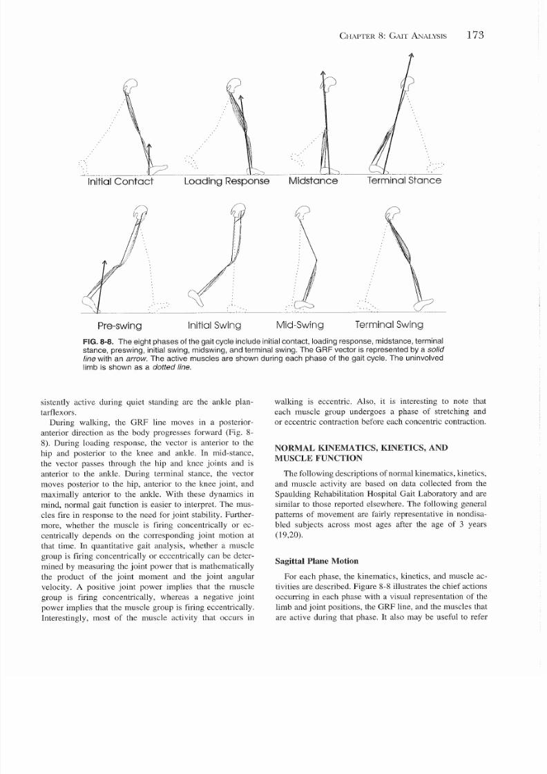
The concept of joint stabilization and the importance of
knowing where the GRF lies in relation to the joints are
best exemplified during quiet standing. In quiet standing, the
GRF extends from the ground through the mid-foot, passing
anterior to the ankle and knee joints and posterior to the hipjoints (Fig. 8-7). At the hip, the external extensor moment
is countered passively by the iliofemoral ligaments. Simi-
larly, at the knee, the external knee extensor moment is coun-
tered passively by the posterior capsule and ligaments at the
knee. At the ankle, the external dorsiflexion moment can
be countered with an internal ankle plantarflexor moment
provided by the ankle plantart1exors (or alternatively with
an ankle-foot-orthosis with a dorsiflexion stop equivalent).
Thus, the only lower extremity muscles that need to be con-
FIG. 8-7. Quiet standing. The GRF, represented by the solid
line with an arrow, is located anterior to the knee and ankle
and posterior to the hip. The soleus muscle is active to stabi-
lize the lower limb.
5/15/2018 Casey - Gait - slidepdf.com
http://slidepdf.com/reader/full/casey-gait 7/22
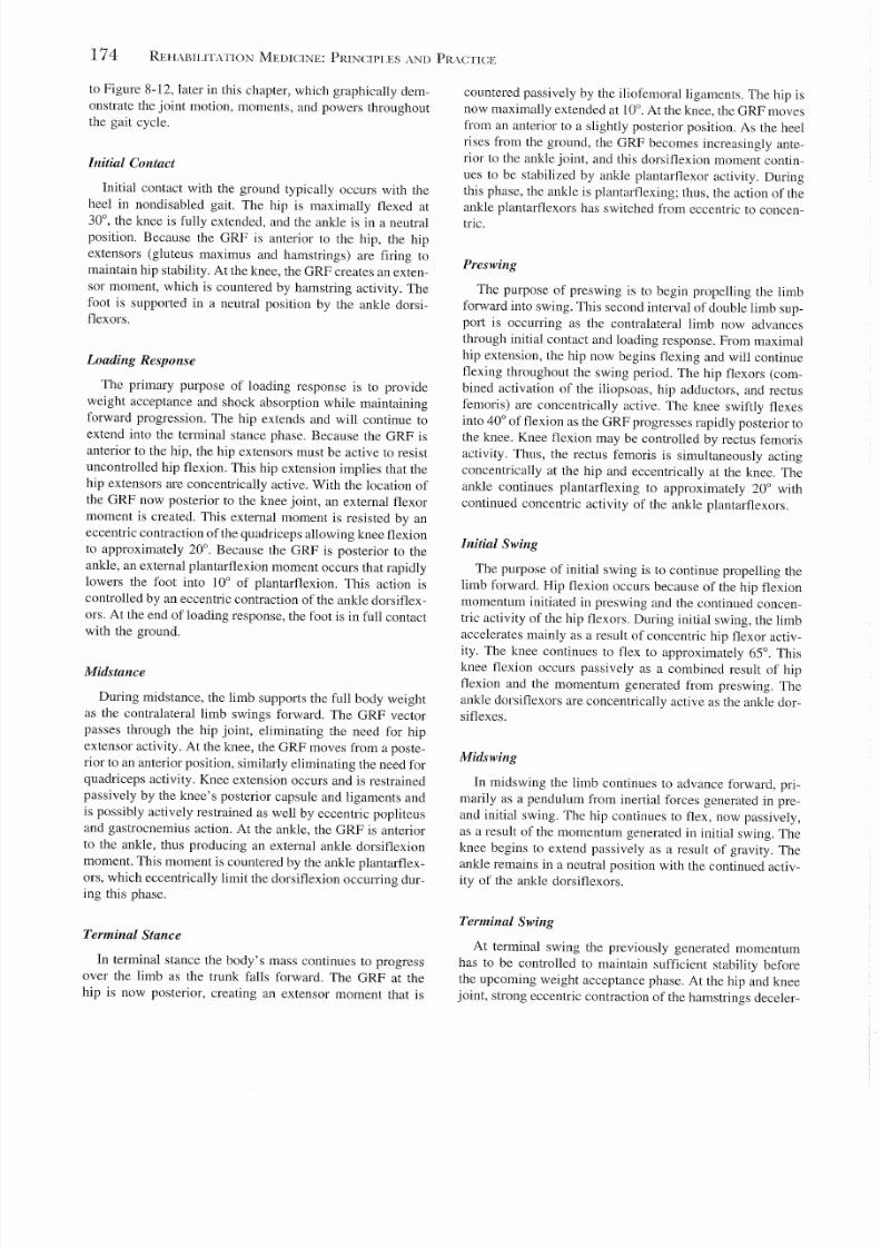
Initial Contact Loading Response
_. .
~~~--.--.----~-~-----~-
Pre-swing Initial Swing
CHAPTER 8: GAIT A."\JALYSIS 173
__ '---.db-J.
Midstance Terminal Stance
Mid-Swing Terminal Swing
FIG.8-8. The eight phases of the gait cycle include initial contact, loading response, midstance, terminal
stance, preswing, initial swing, midswing, and terminal swing. The GRF vector is represented by a solid
line with an arrow. The active muscles are shown during each phase of the gait cycle. The uninvolved
limb is shown as a dotted line.
sistently active during quiet standing are the ankle plan-
tarflexors.
During walking, the GRF line moves in a posterior-
anterior direction as the body progresses forward (Fig. 8-
8). During loading response, the vector is anterior to the
hip and posterior to the knee and ankle. In mid-stance,
the vector passes through the hip and knee joints and is
anterior to the ankle. During terminal stance, the vector
moves posterior to the hip, anterior to the knee joint, and
maximally anterior to the ankle. With these dynamics in
mind, normal gait function is easier to interpret. The mus-
cles fire in response to the need for joint stability. Further-
more, whether the muscle is firing concentrically or ec-
centrically depends on the corresponding joint motion at
that time. In quantitative gait analysis, whether a muscle
group is firing concentrically or eccentrically can be deter-
mined by measuring the joint power that is mathematically
the product of the joint moment and the joint angular
velocity. A positive joint power implies that the muscle
group is firing concentrically, whereas a negative joint
power implies that the muscle group is firing eccentrically.
Interestingly, most of the muscle activity that occurs in
walking is eccentric. Also, it is interesting to note that
each muscle group undergoes a phase of stretching and
or eccentric contraction before each concentric contraction.
NORMAL KINEMATICS, KINETICS, AND
MUSCLE FUNCTION
The following descriptions of normal kinematics, kinetics,
and muscle activity are based on data collected from the
Spaulding Rehabilitation Hospital Gait Laboratory and are
similar to those reported elsewhere. The following general
patterns of movement are fairly representative in nondisa-
bled subjects across most ages after the age of 3 years
(19,20).
Sagittal Plane Motion
For each phase, the kinematics, kinetics, and muscle ac-
tivities are described. Figure 8-8 illustrates the chief actions
occurring in each phase with a visual representation of the
limb and joint positions, the GRF line, and the muscles that
are active during that phase. It also may be useful to refer
5/15/2018 Casey - Gait - slidepdf.com
http://slidepdf.com/reader/full/casey-gait 8/22
174 REHABILITATION MEDICINE: PRINCIPLES AND PRACTICE
to Figure 8-12, later in this chapter, which graphically dem-
onstrate the joint motion, moments, and powers throughout
the gait cycle.
Initial Contact
Initial contact with the ground typically occurs with the
heel in nondisabled gait. The hip is maximally flexed at300, the knee is fully extended, and the ankle is in a neutral
position. Because the GRF is anterior to the hip, the hip
extensors (gluteus maximus and hamstrings) are firing to
maintain hip stability. At the knee, the GRF creates an exten-
sor moment, which is countered by hamstring activity. The
foot is supported in a neutral position by the ankle dorsi-
flexors.
Loading Response
The primary purpose of loading response is to provide
weight acceptance and shock absorption while maintaining
forward progression. The hip extends and will continue toextend into the terminal stance phase. Because the GRF is
anterior to the hip, the hip extensors must be active to resist
uncontrolled hip flexion. This hip extension implies that the
hip extensors are concentrically active. With the location of
the GRF now posterior to the knee joint, an external flexor
moment is created. This external moment is resisted by an
eccentric contraction of the quadriceps allowing knee flexion
to approximately 200• Because the GRF is posterior to the
ankle, an external plantarflexion moment occurs that rapidly
lowers the foot into 100 of plantarflexion. This action is
controlled by an eccentric contraction of the ankle dorsiflex-
ors. At the end of loading response, the foot is in full contact
with the ground.
Midstance
During midstance, the limb supports the full body weight
as the contralateral limb swings forward. The GRF vector
passes through the hip joint, eliminating the need for hip
extensor activity. At the knee, the GRF moves from a poste-
rior to an anterior position, similarly eliminating the need for
quadriceps activity. Knee extension occurs and is restrained
passively by the knee's posterior capsule and ligaments and
is possibly actively restrained as well by eccentric popliteus
and gastrocnemius action. At the ankle, the GRF is anterior
to the ankle, thus producing an external ankle dorsiflexion
moment. This moment is countered by the ankle plantarflex-
ors, which eccentrically limit the dorsiflexion occurring dur-
ing this phase.
Terminal Stance
In terminal stance the body's mass continues to progress
over the limb as the trunk falls forward. The GRF at the
hip is now posterior, creating an extensor moment that is
countered passively by the iliofemoral ligaments. The hip is
now maximally extended at 100. At the knee, the GRF moves
from an anterior to a slightly posterior position. As the heel
rises from the ground, the GRF becomes increasingly ante-
rior to the ankle joint, and this dorsiflexion moment contin-
ues to be stabilized by ankle plantarflexor activity. During
this phase, the ankle is plantarflexing; thus, the action of the
ankle plantarflexors has switched from eccentric to concen-tric.
Pres wing
The purpose of preswing is to begin propelling the limb
forward into swing. This second interval of double limb sup-
port is occurring as the contralateral limb now advances
through initial contact and loading response. From maximal
hip extension, the hip now begins flexing and will continue
flexing throughout the swing period. The hip flexors (com-
bined activation of the iliopsoas, hip adductors, and rectus
femoris) are concentrically active. The knee swiftly flexes
into 400
of flexion as the GRF progresses rapidly posterior tothe knee. Knee flexion may be controlled by rectus femoris
activity. Thus, the rectus femoris is simultaneously acting
concentrically at the hip and eccentrically at the knee. The
ankle continues plantarflexing to approximately 200 with
continued concentric activity of the ankle plantarflexors.
Initial Swing
The purpose of initial swing is to continue propelling the
limb forward. Hip flexion occurs because of the hip flexion
momentum initiated in pres wing and the continued concen-
tric activity of the hip flexors. During initial swing, the limb
accelerates mainly as a result of concentric hip flexor activ-ity. The knee continues to flex to approximately 650• This
knee t1exion occurs passively as a combined result of hip
t1exion and the momentum generated from preswing. The
ankle dorsiflexors are concentrically active as the ankle dor-
sit1exes.
Midswing
In midswing the limb continues to advance forward, pri-
marily as a pendulum from inertial forces generated in pre-
and initial swing. The hip continues to flex, now passively,
as a result of the momentum generated in initial swing. The
knee begins to extend passively as a result of gravity. The
ankle remains in a neutral position with the continued activ-
ity of the ankle dorsiflexors.
Terminal Swing
At terminal swing the previously generated momentum
has to be controlled to maintain sufficient stability before
the upcoming weight acceptance phase. At the hip and knee
joint, strong eccentric contraction of the hamstrings deceler-
5/15/2018 Casey - Gait - slidepdf.com
http://slidepdf.com/reader/full/casey-gait 9/22
ate hip flexion and control knee extension. The ankle dorsi-
flexors remain active to ensure a neutral ankle position at
initial con tac t.
Coronal and Transverse Plane Motion
Most lower extremity motion during gait occurs in the
sagittal planes. The joint motions and kinetics about the hip
in the transverse plane and about the knee and ankle in both
the coronal and transverse planes are normally quite small.
Although significant motion and associated moments occur
in these planes in various gait disabilities, it is difficult to
reliably measure these parameters with current quantitative
gait analysis techniques. However, significant coronal plane
motion and kinetics do occur about the hip (and pelvis) nor-
mally and can be accurately evaluated with quantitative anal-
ysis. At initial contact both the pelvis and hip are in neutral
positions in the coronal plane. During loading response, GRF
passes medially to the hip joint center as the opposite limb
is unloading. This medial GRF causes an external adductor
moment, which tends to allow the contralateral side of thepelvis to drop slightly (the slight Trendelenburg noted previ-
ously as one of the determinants of gait). This motion is
controlled by eccentric contraction of the hip abductors. Dur-
ing mid-stance and terminal stance, the GRF is still medial
to the hip; however, now the contralateral side of the pelvis is
lifted concentrically by the hip abductors. During preswing,
unloading of the limb causes the ipsilateral side of the pelvis
to drop again.
GENERAL APPROACH TO EVALUATING A
PATIENT WITH AN ATYPICAL GAIT PATTERN
Although a number of atypical gait patterns have been
described, each patient has a unique set of impairments,
functional limitations, and associated compensations caus-
ing these patterns. Examples of atypical gait patterns associ-
ated with distinct diagnoses are described in the following
sections. Especially in the case of upper motor neuron
(UMN) pathology, a stereotypical description of the gait pat-
tern may be sufficient for an initial classification but is too
imprecise for determining the mechanisms in an individual
patient. It is important to determine these mechanisms in
individual patients because they are the basis for directing
optimal rehabilitation treatment.
It should be noted that an atypical gait pattern may or
may not be functionally significant and thus mayor may not
be considered a true gait disability. Thus, the atypical gait
pattern first should be evaluated with respect to each of the
following:
• Energy requirement
• Risk of falling
• Biomechanical injury
• Cosmesis
Treatment to change the gait pattern should be prescribed
if the pattern is functionally significant with respect to these
CHAPTER 8: GAIT A"lALYSIS 175
four criteria. For instance, the pattern of knee recurvatum
(or hyperextension) can be functionally significant if it in-
creases the energy required to walk by not allowing the peak
of the COM to be minimized during single limb support.
Alternatively, knee recurvatum mayor may not be associ-
ated with increased forces across the posterior capsule and
ligaments of the knee (21), which would predispose to bio-
mechanical injury. Another example is equinus during theswing period, which mayor may not predispose to falling
depending on the associated compensations. To this end,
the associated compensatory gait patterns also need to be
evaluated with respect to these four criteria. In the case of
equinus in swing, a compensation at the pelvis such as hip
hiking would interfere with the pelvic obliquity determinant
and thus increase the energy required to walk. Finally, an
atypical gait pattern should be evaluated with respect to
cosmesis. For this assessment, the patient's own perceptions
are far more important than the clinician's perceptions.
From the examples above, it is clear that a detailed evalua-
tion of the patient is required. The summary of the patient's
history and musculoskeletal examination, observational gaitanalysis, and information from a quantitative gait analysis
assist in determining the functional significance of the gait
patterns and help identify specific causes for each pattern.
Based on these results, a detailed treatment plan can be pre-
scribed.
STATIC EVALUATION
History
The initial part of a comprehensive gait evaluation should
include a focused history and physical examination. Based
on this evaluation, the underlying diagnosis (or diagnoses)
can be classified as a UMN pathology, lower motor neuron(LMN) pathology, orthopedic disorder, amputation (the
evaluation of which is described in another chapter), cerebel-
lar or basal ganglia related disorder, or psychogenic cause,
to name a few. Itis helpful to anticipate certain gait patterns
associated with these diagnoses as well as to anticipate the
need for various components of a quantitative gait analysis.
For example, the use of dynamic EMG is particularly useful
in detecting inappropriate firing patterns in patients with
UMN pathology but may not be necessary for every patient
with one of the other mentioned diagnostic categories. The
reason for referral should be identified, and the patient's
chief complaint with regard to walking should be considered.
Any previous medications, neurolytic procedures, or surger-ies affecting the lower extremities should be noted. Also,
a detailed history of strengthening and stretching exercises
previously and currently being performed should be ascer-
tained. Finally, the use of assistive devices andlor orthotics
should be recorded.
Physical Examination
The physical examination should focus on the neurologic
and musculoskeletal system and include a static evaluation
5/15/2018 Casey - Gait - slidepdf.com
http://slidepdf.com/reader/full/casey-gait 10/22
176 REHABILITATION MEDICINE: PRINCIPLES AND PRACTICE
of the patient's strength, joint range of motion, tone, and
proprioception. Although static evaluation is a routine part
of a gait consultation and should be included in the assess-
ment of every patient with an atypical gait pattern, it is gener-
ally agreed upon that, especially in the case of an UMN
pathology, the static evaluation has limited usefulness in
determining the underlying mechanisms responsible for the
atypical gait pattern (22-24). Thus, often the results fromthe static evaluation need to be combined with those obtained
from quantitative gait analysis to provide dynamically rele-
vant information on which to base treatment.
Strength
Classic evaluation of strength involves quantitative man-
ual muscle testing about each joint. It requires the ability
of the patient to cooperate with resistive movement of the
examiner (25),which often is difficult in patients presenting
with a UMN pathology. The patient with a UMN pathology
has impaired voluntary muscle control in the affected limbs
so that selectively activating an agonist while simultaneouslyrelaxing the antagonist may be impossible. Thus, the result
is a limited relationship between static strength performance
and dynamic strength associated with gait. For example, a
patient with hemiplegia affecting his or her gait may not be
able to dorsiflex the foot during static examination, yet when
walking may be able to actively dorsiflex during the swing
period of the gait cycle (26,27), presumably under the control
of primitive reflexes. Conversely, a patient with normal dor-
siflexion strength of the ankle during static evaluation may
demonstrate an equinus gait during the swing period of gait.
Range of Motion
Determining the passive range of motion at each joint is
the traditional method to assess soft tissue contracture and
should be performed in at least the lower extremities in all
patients presenting with a gait disabili ty. However, it is im-
portant to note that this static testing is somewhat limited,
particularly in the case of UMN pathology. Differentiating
between contracture of a one-joint and a two-joint muscle
is difficult in patients with a UMN pathology, undoubtedly
because of impaired selective control of these muscles. Fur-
thermore, there seems to be a limited relationship between
static range of motion observed with passive ranging and
the dynamic range of motion that occurs during gait.
Three clinical tests are commonly performed in patientswith UMN pathology to screen for contractures of a two-
joint muscle. The Duncan-Ely-test differentiates between a
rectus femoris and a iliopsoas contracture given that the rec-
tus femoris is both a hip flexor and knee extensor. In this
test, the patient is placed in a prone position and the knee
is rapidly flexed. With a contracture of the rectus femoris,
the hips will flex and the buttocks will rise off the table.
Although this test is somewhat useful, EMG studies have
demonstrated that this test induces activity not only in the
rectus femoris, but also in the iliopsoas in some patients with
cerebral palsy affecting their gait (28). The Silverskiold test
is used to differentiate between a contracture of the soleus
and the gastrocnemius muscle. Whereas the soleus is a one-
joint muscle, the gastrocnemius is both a knee flexor and
ankle plantarflexor. With the patient in the sitting position,
the knee is flexed at 90° and the foot is brought to maximal
dorsiflexion. With a gastrocnemius contracture, some of theankle dorsiflexion will be lost when the knee is extended.
Ithas been shown that this test is not always clinically relia-
ble (29). The Phelps test differentiates a contracture of the
gracilis from the other hip abductors, given that the gracilis
is the only hip adductor that crosses the hip and the knee.
With the patient in a prone position, the knees are flexed
and the hips are brought into an abducted position. A gracilis
contracture is present when the hip adducts when one knee
is extended.
Structural deformities also may contribute to reduced
range of motion and gait. If indicated, further clinical tests
and x-rays are helpful to document common problems such
as femoral anteversion, knee valgus and varus, tibial torsion,and foot abnormalities. These structural problems are some-
times associated with other underlying diagnoses and im-
pairments and can have significant impact on the patient's
walking.
Tone
Tone in all muscle groups should be assessed in each
patient presenting with a gait disability. Clinical examination
of tone involves testing for resistance by passively moving
a joint through its range of motion. This assessment is fairly
subjective and is dependent on time of day, temperature, and
limb position. Thus, as in the other static tests, there is oftena limited association between what is observed statically and
what actually occurs during gait.
Proprioception
Joint sense position should be evaluated in all patients in
whom a neurologic diagnosis is suspected. Ifthis is impaired,
it is important to also evaluate the degree of impairment by
evaluating joint position sense not only at the great toe, but
at the ankle, knee, and hip as well.
OBSERVATIONAL GAIT ANALYSIS
Observational gait analysis is common practice for physi-
atrists. The observer describes the gait after watching the
patient walk without the aid of any electronic devices. How-
ever, it is often difficult to appreciate all limb segment and
joint motions throughout the different phases of gait because
of the difficulty in concurrently observing the multiple body
segments and joint motion (30). Videotaping can be an im-
portant part of observational gait analysis because it allows
repeated viewing of the patient's gait pattern without causing

5/15/2018 Casey - Gait - slidepdf.com
http://slidepdf.com/reader/full/casey-gait 11/22
undue patient fatigue. The patient should be observed from
the side and from behind. Stride and step length, width, and
symmetry should be noted. By concentrating on one joint at
a time, including hip, knee, and ankle, atypical motions may
be easier to identify. Having the patient walk at faster speed
sometimes exaggerates an atypical motion. Observational
gait analysis can identify obvious atypical gait patterns, such
as excessive ankle plantarflexion or reduced knee flexion inswing. However, in certain cases this approach may not show
all atypical patterns. For example, an increased lumbar lor-
dosis or anterior pelvic tilt due to a hip flexion contracture
may be apparent only via quantitative analysis. Moreover,
quantitative gait analysis can be quite helpful in delineating
the specific causes for each atypical pattern and thus help
direct the appropriate treatment.
A number of terms are commonly used to characterize
various atypical gait patterns that are obvious from observa-
tional assessment alone. For instance, antalgic gait has been
described as a pattern common to patients with pain in one
lower extremity. In this pattern, gait is modified to reduce
weight bearing on the involved side. The uninvolved limb
is rapidly advanced to shorten stance on the affected side.
Gait is often slow and steps are short in order to limit the
weight-bearing period. Steppage gait is a compensatory gait
pattern used to describe excessive hip and knee t1exion to
assist a "functionally long" lower leg to clear the ground
in swing. Festinating gait have been described as a character-
istic pattern of Parkinson's disease, in which there is a ten-
dency to take short accelerating steps. Shuffling gait is also
common in Parkinson's disease and refers to the feet shuf-
fling during swing. Ataxic gait, associated with cerebellar
pathologies, peripheral neuropathies, and dorsal column pa-
thologies, is a broad term used to describe a pattern of appar-ent poor balance, a wide base of support, and variable mo-
tions from stride to stride.
Various gait patterns associated with the use of assistive
gait devices are easily noted with observational analysis. The
specific indications and use of each of these type of devices
are described in detail in another chapter. A cane essentially
increases the base of support by providing an additional point
of contact with the ground. When pathology, imp ailment,
and functional limitation involve bilateral extremities, two
canes or crutches are occasionally used. In this situation, an
alternating two-point gait is commonly used in which one
cane and opposite lower limb are in contact with the ground
alternating with the opposite cane and lower limb in each
successive step. In three-point gait, contact with one limb
that fully bears weight onto the ground alternates with full
weight-bearing through two crutches that make simultane-
ous contact with the ground. In four-point gait, which pro-
vides maximal stability and base of support (at the cost of
reduced speed of locomotion), there is always three points
of support on the ground at all times. It is initiated by forward
movement by an upper extremity crutch, followed by for-
ward movement of the contralateral lower limb, then forward
CHAPTER 8: GAIT ANALYSIS 177
movement of the other crutch followed by forward move-
ment of the other lower limb.
QUANTITATIVE GAIT EVALUATION
Modern-day quantitative gait analysis systems typically
include measurement of three primary components: kine-
matics, kinetics, and muscle activity. Quantitative gait analy-sis also can include other components such as footswitches
and oxygen consumption monitoring to measure overall en-
ergy expenditure. To measure these various components, a
variety of equipment is used, including optoelectronic mo-
tion analysis systems to measure kinematics, force plates to
help measure kinetics, and a multi-channel dynamic EMG
apparatus to measure electrical muscle activity in multiple
muscles during gait. Given the previously described limita-
tions of static evaluations and of observational gait analysis,
quantitative gait analysis can be a particularly useful clinical
tool for developing a treatment plan.
Modern quantitative gait analysis is clearly recognized as
useful in outlining an effective orthopedic surgical treatmentplan in patients with spastic paretic gait from cerebral palsy
(23,31-33). Children with cerebral palsy often undergo ten-
don lengthening or transfer procedures to improve range of
motion in the lower extremities in an effort to improve gait
disability. The results from a detailed quantitative gait analy-
sis can help determine the best surgical plan (i.e., which
tendons should be lengthened or transferred) to provide the
most optimal gait. In the same way that quantitative gait
analysis is helpful in orthopedic decision making in patients
with UMN pathology, it should be similarly useful in direct-
ing these patients' rehabilitation management. Many physia-
tric treatments, as described in other chapters, include intra-
muscular neurolytic techniques, strengthening, bracing,
functional electrical stimulation, stretching, modalities, and
many other management techniques aimed at (a) strengthen-
ing or compensating for weakness, (b) stretching a con-
tracture, andlor (c) reducing tone in a spastic muscle. The
outcome of these treatments ultimately rely on the proper
determination of the specific underlying impairment or func-
tional limitation causing the gait disability. In some in-
stances, rehabilitation treatments are aimed at improving
motor control through, for example, EMG biofeedback or
neuromuscular re-education. In these instances, quantitative
gait analysis is especially helpful in determining which spe-
cific muscle groups are firing at inappropriate times.
Unfortunately, skepticism still persists about the value of
quantitative gait analysis in defining a physiatric therapeutic
plan because there have been few reports about the value
of quantitative gait analysis as a useful evaluation tool in
rehabilitation. Human gait is complex. Quantitative analysis
offers a clinical tool to better understand these complexities
and thus prescribe an optimal rehabilitation treatment pro-
gram (34). Some of the reluctance in using quantitative gait
analysis may be due to the heavy time commitment neces-
sary to understand and interpret the data and the necessity
5/15/2018 Casey - Gait - slidepdf.com
http://slidepdf.com/reader/full/casey-gait 12/22
178 RU-IABILITATION MEDICINE: PRINCIPLES AND PRACTICE
for teamwork between many disciplines, including medicine
and engineering. The cost for gait analysis systems is declin-
ing, and the technology required for acquiring and analyzing
the data is continually improving. A rapid expansion of com-
puter and optoelectronic technology has brought dramatic
changes in image-based motion analysis in the past 10 years.
It is anticipated that quantitative gait analysis will soon be-
come a routine clinical evaluation, much like electrodi-agnosis has become a routine clinical extension of our physi-
atric examination. Formal training in quantitative gait
analysis, which is already a mandatory part of our physiatric
residency curriculum, is likely to become the norm.
Systems to Evaluate Temporal Parameters of Gait
Common temporal parameters such as velocity, cadence,
and stride length can be measured to monitor a patient's
progress outside of a sophisticated gait laboratory. As noted
previously, velocity can be measured simply with a stop-
watch as a patient traverses a designated distance. Similarly,
step and stride length can be measured without sophisticatedequipment if the walkway is sprinkled with talcum powder.
Computerized stride analyzers may provide this same infor-
mation in a more automated fashion (35,36). They usually
consist of instrumented insoles with footswitches (i.e., pres-
sure sensitive transducers), typically attached to the heel,
toe, and occasionally the metatarsal region. They are con-
nected to data boxes worn by the patient either around the
waist or the ankle. These sensors measure the duration of
floor contact via opening and closing switches. After acqui-
sition, data transfer and analysis are typically performed
using a personal computer.
Footswitches are also commonly used in gait laboratories
to help determine the beginning and end of the stance period,allowing calculation of temporal gait parameters such as the
duration of the stance and swing periods, single and double
support time, and cadence. These parameters are useful in
interpreting the temporal relationships of kinematic, kinetic,
and particularly dynamic EMG data. Although this same
information can be obtained directly from force plate data,
footswitches are particularly helpful in the gait laboratory
when force plate data cannot be obtained.
Foot Pressure Systems
Foot pressure systems are electronic instruments to mea-
sure pressure distributions in the soles of the feet. The sys-
tems work via a large number of capacitive or force sensitive
sensors in foot insoles or platforms and are linked to a com-
puter by either cable or radiowave telemetry. Several com-
mercial systems are available and used clinically and for
research. These systems may help direct appropriate shoe
wear and orthotic prescriptions by providing information
about abnormal pressure distributions, particularly in pa-
tients with structural foot deformities or in patients at risk for
developing skin ulcerations in the feet because of diabetes
mellitus or other underlying vascular and peripheral neurop-
athy disorders.
Kinematics
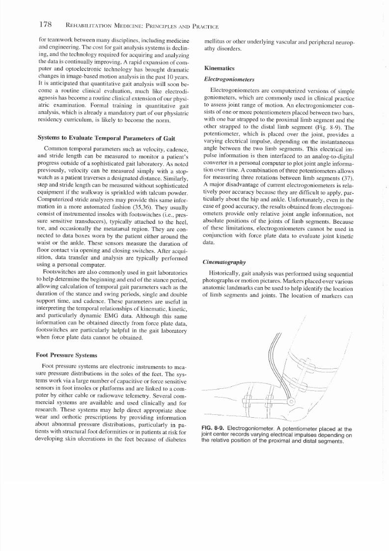
Electrogoniometers
Electrogoniometers are computerized versions of simplegoniometers, which are commonly used in clinical practice
to assess joint range of motion. An electro goniometer con-
sists of one or more potentiometers placed between two bars,
with one bar strapped to the proximal limb segment and the
other strapped to the distal limb segment (Fig. 8-9). The
potentiometer, which is placed over the joint, provides a
varying electrical impulse, depending on the instantaneous
angle between the two limb segments. This electrical im-
pulse information is then interfaced to an analog-to-digital
converter in a personal computer to plot joint angle informa-
tion over time. A combination of three potentiometers allows
for measuring three rotations between limb segments (37).
A major disadvantage of current electrogoniometers is rela-tively poor accuracy because they are difficult to apply, par-
ticularly about the hip and ankle. Unfortunately, even in the
case of good accuracy, the results obtained from electrogoni-
ometers provide only relative joint angle information, not
absolute positions of the joints of limb segments. Because
of these limitations, electrogoniometers cannot be used in
conjunction with force plate data to evaluate joint kinetic
data.
Cinematography
Historically, gait analysis was performed using sequential
photographs or motion pictures. Markers placed over variousanatomic landmarks can be used to help identify the location
of limb segments and joints. The location of markers can
FIG. 8-9. Electrogoniometer. A potentiometer placed at the
joint center records varying electrical impulses depending on
the relative position of the proximal and distal segments.
5/15/2018 Casey - Gait - slidepdf.com
http://slidepdf.com/reader/full/casey-gait 13/22
then be manually digitized, frame by frame, so that the
marker position in two dimensions can be determined. In
both cinematographic and optoelectronic systems, a singJe
camera provides two-dimensional information. By using two
cameras, triangulation can be performed to determine the
three-dimensional position of each marker. Although the ci-
nematographic system is theoretically as accurate as what
can be obtained with modern-day optoelectronic systems,the time necessary to manually digitize and process the data
is of such great magnitude that it makes this procedure unfea-
sible for routine clinical evaluation.
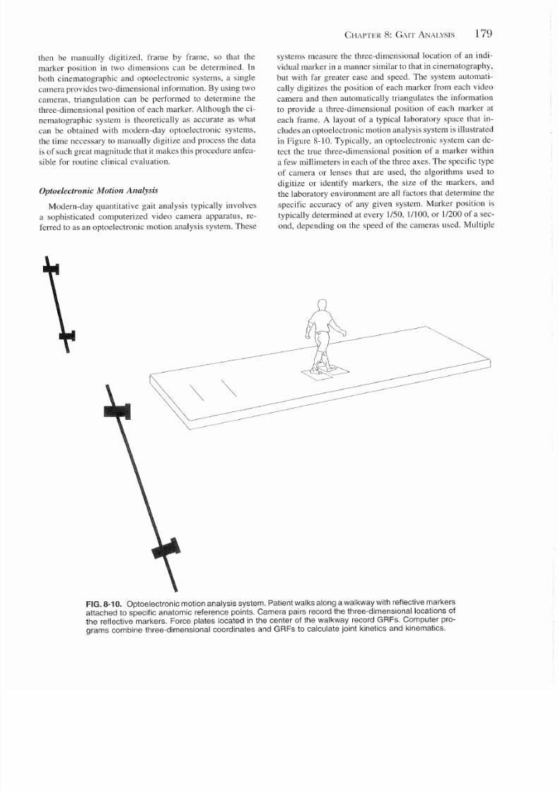
Optoelectronic Motion Analysis
Modern-day quantitative gait analysis typically involves
a sophisticated computerized video camera apparatus, re-
ferred to as an optoelectronic motion analysis system. These
CHAPTER 8: GAIT ANALYSIS 179
systems measure the three-dimensional location of an indi-
vidual marker in a manner similar to that in cinematography,
but with far greater ease and speed. The system automati-
cally digitizes the position of each marker from each video
camera and then automatically triangulates the information
to provide a three-dimensional position of each marker at
each frame. A layout of a typical laboratory space that in-
cludes an optoelectronic motion analysis system is illustratedin Figure 8-10. Typically, an optoelectronic system can de-
tect the true three-dimensional position of a marker within
a few millimeters in each of the three axes. The specific type
of camera or lenses that are used, the algorithms used to
digitize or identify markers, the size of the markers, and
the laboratory environment are all factors that determine the
specific accuracy of any given system. Marker position is
typically determined at every 1150 , 11100, or 1/200 of a sec-
ond, depending on the speed of the cameras used. Multiple
FIG. 8-10. Optoelectronic motion analysis system. Patient walks along a walkway with reflective markers
attached to specific anatomic reference points. Camera pairs record the three-dimensional locations of
the reflective markers. Force plates located in the center of the walkway record GRFs. Computer pro-
grams combine three-dimensional coordinates and GRFs to calculate joint k inetics and kinematics.
5/15/2018 Casey - Gait - slidepdf.com
http://slidepdf.com/reader/full/casey-gait 14/22
180 REHABILITATION MEDICINE: PRINCIPLES AND PRACTICE
markers are affixed to the skin of the pelvis and the lower
extremities in relationship to bony landmarks. Similar to the
cinematographic method, two cameras are necessary to visu-
alize each marker to obtain its three-dimensional position.
Often a camera cannot visualize a marker during a particular
part of a movement because of limb rotation or because
another limb segment gets in the way. For this reason. some-
times a laboratory uses more than two cameras to ensurethat at every given frame of movement, at least two cameras
can visualize each of the markers. In the case where three
or more cameras visualize a marker, an algorithm must be
used to determine the true position of the marker because
there is invariably some error such that not all cameras con-
verge on the identical three-dimensional position. Other lab-
oratories strategically position the markers so that the same
two cameras can visualize a particular marker throughout
the movement.
Currently, there are two different types of optoelectronic
systems used for quantitative gait evaluation: (a) active
marker systems, where the markers are actively illuminated
by a computer, and (b) passive marker systems. A built-inadvantage of an active marker system is that the computer
knows in advance which marker it is illuminating at any
given frame so that the markers are automatically identified
as the lateral femoral epicondyle marker, the lateral malleo-
lus marker, etc. The main disadvantage of current active
marker systems, however, is that the illuminators require
power; thus, multiple wires connected to a power source
need to be attached to the patient, which tend to encumber
the patient's gait. In contrast. passive marker systems require
only that a small infrared reflective piece of material be
placed over each anatomic landmark. Although passive
markers do not encumber the patient, they do require some
additional type of system to determine which marker is
which. Fortunately, sophisticated computer software pro-grams have been developed that automate this procedure.
Thus, passive marker optoelectronic systems have become
the preferred systems for routine clinical practice and are
readily commercially available with all necessary software
programs.
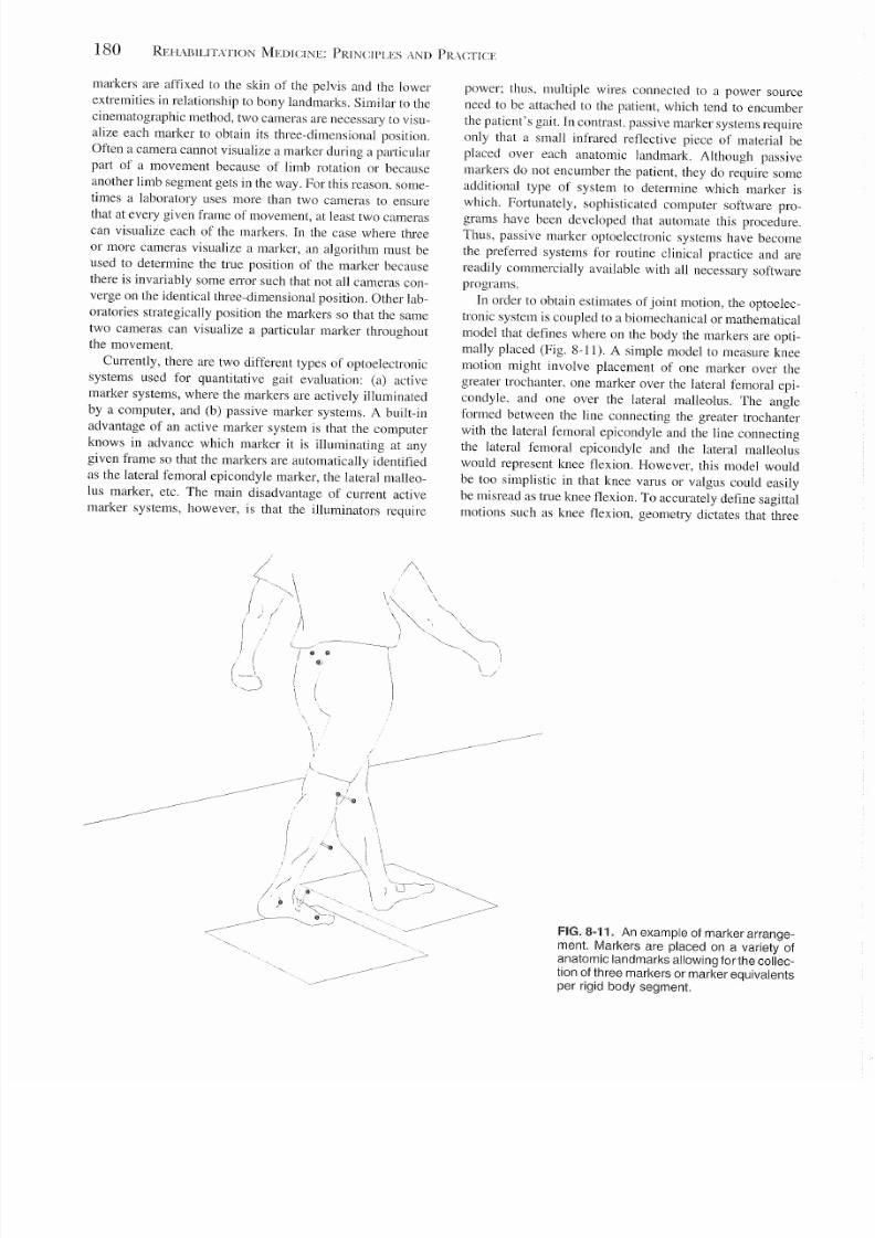
In order to obtain estimates of joint motion, the optoelec-
tronic system is coupled to a biomechanical or mathematical
model that defines where on the body the markers are opti-
mally placed (Fig. 8-11). A simple model to measure knee
motion might involve placement of one marker over the
greater trochanter, one marker over the lateral femoral epi-
condyle. and one over the lateral malleolus. The angle
formed between the line connecting the greater trochanterwith the lateral femoral epicondyle and the line connecting
the lateral femoral epicondyle and the lateral malleolus
would represent knee flexion. However, this model would
be too simplistic in that knee varus or valgus could easily
be misread as true knee flexion. To accurately define sagittal
motions such as knee flexion, geometry dictates that three
FIG.8-11. An example of marker arrange-
ment. Markers are placed on a variety of
anatomic landmarks allowing for the collec-
tion of three markers or marker equivalents
per rigid body segment.
5/15/2018 Casey - Gait - slidepdf.com
http://slidepdf.com/reader/full/casey-gait 15/22
c40
0
'§ 30iI:
20 . . . . .w
~ 10[;,
" 0
c -1010
0
.~
s -20
Jl-30
0.6c0
.~ 0.4iI:
0.2E'"
: . : :
E 10
Z -0.2
s -0.4
.~-0.6
~-0.8
Hip Motion
CHAPTER 8: GAIT ANALYSIS 181
Knee Motion Ankle Motion
70 ,
: ~ 140 i I '/
30 I '/20 .--...... I.i > <, /, '/10 I ...-.._ < ,./"/
o l~ ---,-~---,~~- -
-10 6 10 20 30 40 50 60
-20 I
80 90 100
--Average data - -±1SD
Hip Moment
--Average data - -±1SD
Hip Power
(\I
0.6 / \ I\
::,' '. II \\ I
: ~ f i . ~ ~ ~ ~ ~ ~ ~ : ~ ~ ~ ~ ~ oi
--Average data - - 11 SO
------_------- ---~
0.8
70 80 90 100
0.4
--Average Data - -±1SD
Ankle Moment
markers (or marker equivalents) be placed on each limb seg-
ment, assumed to be rigid, to define the three-dimensional
coordinate system for that segment (Fig. 8-12). A marker
equivalent could be some imaginary anatomic point calcu-
lated on the basis of the position of real markers. For in-
stance, three markers could be used to define a plane in the
pelvis. From this and the known geometry of the pelvis, the
location of the hip joint center can be calculated. The hip
joint center then becomes an imaginary marker equivalent
and can be used in defining the thigh segment coordinate
system. Marker locations are often chosen in order to facili-
tate estimating joint centers as well as to ensure that themarkers can be visualized by the camera system.
Typically, markers are placed over bony landmarks to
ensure consistent applications as well as to reduce skin
movement artifact. With three markers or marker equivalents
for each body segment, the segment can be represented in
the form of a local coordinate system whose orientation is
determined with respect to a global coordinate system. The
local coordinate system is defined by three mutually perpen-
dicular vectors. Joint angle information then can be ascer-
0.3
0.2
0.1
-0.1
-0.2
-0.3
0.4
0.2
-0.2
-0.4
-0.6
-0.8
-1
-12
-1.4
--Average data - -11 SD
I \
11""'\
,..-I \\
/ / \ \/ / \ ~
1_,..-/ ~~~ _
1l i 20 30 40 50 60 70
--Average data - -t1SD
80 90 100 I
I
- - - - - - - 1I
% Gait Cycle
----_-------Average data- -±1 SD
FIG. 8-12. Kinetics and kinematics at the hip, knee, and ankle. Sagittal joint motion, moments, and
powers are shown.
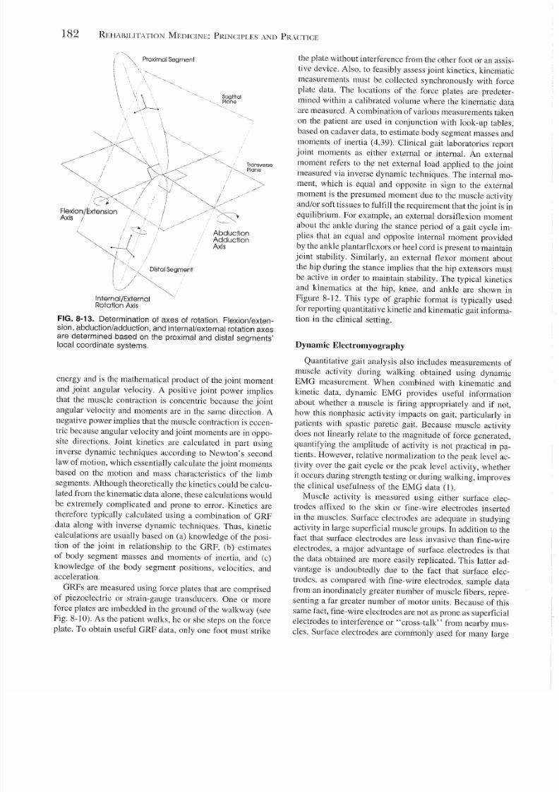
tained from the proximal and distal limb segment local coor-
dinate systems. Several methods exist for determining joint
angle information. Commonly, one axis is chosen to be par-
allel to the proximal segment local coordinate system axis,
and a second axis is chosen to be parallel to the distal seg-
ment local coordinate system axis (38). In this way, a medial/
lateral axis is selected from the proximal segment local coor-
dinate system and is considered to be the axis about which
joint flexion/extension occurs. A longitudinal axis chosen
from the distal segment local coordinate system represents
the axis about which internal/external rotation occurs. Fi-
nally, an axis formed mutually perpendicular to these twoaxes is considered the axis about which abduction/adduction
occurs (Fig. 8-13).
Kinetics
Joint moments and power are commonly measured with
quantitative gait analysis. The concept of a joint moment
has already been described. A joint power, also referred to
previously, represents the net rate of generating or absorbing
5/15/2018 Casey - Gait - slidepdf.com
http://slidepdf.com/reader/full/casey-gait 16/22
182 REHABILITATION MEDICINE: PRINCIPLES AND PR\CTICE
Proximal Segment
SagittalPlane
Transverse
Plane
AbductionAdductionAxis
FIG. 8-13. Determination of axes of rotation. Flexion/exten-
sion, abduction/adduction, and internal/external rotation axes
are determined based on the proximal and distal segments'
local coordinate systems.
energy and is the mathematical product of the joint moment
and joint angular velocity. A positive joint power implies
that the muscle contraction is concentric because the jointangular velocity and moments are in the same direction. A
negative power implies that the muscle contraction is eccen-
tric because angular velocity and joint moments are in oppo-
site directions. Joint kinetics are calculated in part using
inverse dynamic techniques according to Newton's second
law of motion, which essentially calculate the joint moments
based on the motion and mass characteristics of the limb
segments. Although theoretically the kinetics could be calcu-
lated from the kinematic data alone, these calculations would
be extremely complicated and prone to error. Kinetics are
therefore typically calculated using a combination of GRF
data along with inverse dynamic techniques. Thus, kinetic
calculations are usually based on (a) knowledge of the posi-tion of the joint in relationship to the GRF, (b) estimates
of body segment masses and moments of inertia, and (c)
knowledge of the body segment positions, velocities, and
acceleration.
GRFs are measured using force plates that are comprised
of piezoelectric or strain-gauge transducers. One or more
force plates are imbedded in the ground of the walkway (see
Fig. 8-10). As the patient walks, he or she steps on the force
plate. To obtain useful GRF data, only one foot must strike
the plate without interference from the other foot or an assis-
tive device. Also, to feasibly assess joint kinetics, kinematic
measurements must be collected synchronously with force
plate data. The locations of the force plates are predeter-
mined within a calibrated volume where the kinematic data
are measured. A combination of various measurements taken
on the patient are used in conjunction with look-up tables,
based on cadaver data, to estimate body segment masses andmoments of inertia (4,39). Clinical gait laboratories report
joint moments as either external or internal. An external
moment refers to the net external load applied to the joint
measured via inverse dynamic techniques. The internal mo-
ment, which is equal and opposite in sign to the external
moment is the presumed moment due to the muscle activity
and/or soft tissues to fulfill the requirement that the joint is in
equilibrium. For example, an external dorsiflexion moment
about the ankle during the stance period of a gait cycle im-
plies that an equal and opposite internal moment provided
by the ankle plantarflexors or heel cord ispresent to maintain
joint stability. Similarly, an external flexor moment about
the hip during the stance implies that the hip extensors mustbe active in order to maintain stability. The typical kinetics
and kinematics at the hip, knee, and ankle are shown in
Figure 8-12. This type of graphic format is typically used
for reporting quantitative kinetic and kinematic gait informa-
tion in the clinical setting.
Dynamic Electromyography
Quantitative gait analysis also includes measurements of
muscle activity during walking obtained using dynamic
EMG measurement. When combined with kinematic and
kinetic data, dynamic EMG provides useful information
about whether a muscle is firing appropriately and if not,how this nonphasic activity impacts on gait, particularly in
patients with spastic paretic gait. Because muscle activity
does not linearly relate to the magnitude of force generated,
quantifying the amplitude of activity is not practical in pa-
tients. However, relative normalization to the peak level ac-
tivity over the gait cycle or the peak level activity, whether
it occurs during strength testing or during walking, improves
the clinical usefulness of the EMG data (1).
Muscle activity is measured using either surface elec-
trodes affixed to the skin or fine-wire electrodes inserted
in the muscles. Surface electrodes are adequate in studying
activity in large superficial muscle groups. In addition to the
fact that surface electrodes are less invasive than fine-wireelectrodes, a major advantage of surface electrodes is that
the data obtained are more easily replicated. This latter ad-
vantage is undoubtedly due to the fact that surface elec-
trodes. as compared with fine-wire electrodes, sample data
from an inordinately greater number of muscle fibers, repre-
senting a far greater number of motor units. Because of this
same fact, fine-wire electrodes are not as prone as superficial
electrodes to interference or "cross-talk" from nearby mus-
cles. Surface electrodes are commonly used for many large
5/15/2018 Casey - Gait - slidepdf.com
http://slidepdf.com/reader/full/casey-gait 17/22
superficial muscles in the lower extremity. Fine-wire elec-
trodes are necessary for analyzing activity from smaller,
deeper muscles, such as the iliopsoas and posterior tibialis.
In addition, fine-wire electrodes are useful for differentiating
activity from overlapping muscles such as the rectus femoris
and vastus intermedius.
Surface EMG is typically recorded using disposable,
gelled electrodes attached to the patient's skin overlying themuscle to be sampled. Usually, bipolar electrodes are used
and the signal recorded is the potential difference between
the two electrodes. Fine-wire EMG is often recorded using
a wire bipolar electrode consisting of two thin, insulated
wires with bared tips. The wires are placed through the shaft
of a 25-gauge needle with the two ends bent over the needle
and the bared tips staggered so as to avoid contact between
them. The needle is inserted through the skin into the muscle.
and then quickly removed, leaving the fine-wire in place.
When in place, the bend in the wires provides a means for
the electrodes to "catch" on the muscle fascicles. Again,
the signal recorded is the potential difference between the
two electrode ends. At the end of the study, the wires areremoved with a gentle pull.
Preamplified EMG signals, either from fine-wire elec-
trodes or surface electrodes, can be transmitted by cable
or radiowave telemetry to a receiver that is connected to a
computer system. The EMG signals are usually filtered to
remove artifacts created by the mechanical movement. The
signals are displayed and the gait cycle events identified.
Some laboratories report raw EMG signals, whereas others
report rectified and smooth EMG activity as well. The timing
of the activity is typically what is important in the assess-
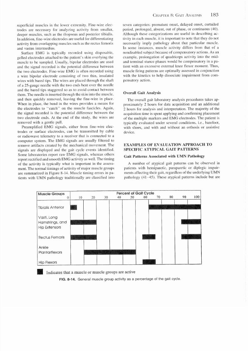
ment. The normal timings of activity of major muscle groups
are summarized in Figure 8-14. Muscle timing errors in pa-
tients with UMN pathology traditionally are classified into
CHAPTER 8: GAlT ANALYSIS 183
seven categories: premature onset, delayed onset, curtailed
period, prolonged, absent, out of phase, or continuous (40).
Although these categorizations are useful in describing ac-
tivity in each muscle, it is important to note that they do not
necessarily imply pathology about that particular muscle.
In some instances, muscle activity differs from that of a
nondisabled subject because of compensatory actions. As an
example, prolongation of quadriceps activity into the mid-and terminal stance phases would be compensatory in a pa-
tient with an excessive external knee flexor moment. Thus,
muscle firing patterns are optimally assessed in conjunction
with the kinetics to help dissociate impairment from com-
pensatory action.
Overall Gait Analysis
The overall gait laboratory analysis procedures takes ap-
proximately 2 hours for data acquisition and an additional
2 hours for analysis and interpretation. The majority of the
acquisition time is spent applying and confirming placement
of the multiple markers and EMG electrodes. The patient istypically evaluated under several conditions, i.e., barefoot,
with shoes, and with and without an orthosis or assistive
device.
EXAMPLES OF EVALUA TION APPROACH TO
SPECIFIC ATYPICAL GAIT PATTERNS
Gait Patterns Associated with UMN Pathology
A number of atypical gait patterns can be observed in
patients with hemiparetic, paraparetic or diplegic impair-
ments affecting their gait, regardless of the underlying UMN
pathology (41-45). These atypical patterns include but are
10 30 80
Muscle Groups20
TibialisAnterior
vostl. Long
Hamstrings, and
Hip Extensors
Rectus Femoris
Ankle
Plantarflexors
Hip Flexors
Percent of Gait Cycle40 50 60 70
• Indicates that a muscle or muscle groups are active
FIG. 8-14. General muscle group activity as a percentage of the gait cycle.
5/15/2018 Casey - Gait - slidepdf.com
http://slidepdf.com/reader/full/casey-gait 18/22
184 REHABILITATION MEDICINE: PRINCIPLES AND PRACTICE
not limited to reduced knee flexion in swing, also referred
to as stiff-legged gait, excessive knee flexion in stance re-
ferred to as crouched gait, equinus or excessive ankle plan-
tarflexion occurring during one or more phases in either
stance and/or swing, and knee hyperextension or recurvatum
occurring in one or more phases of stance. Also common
are presumably compensatory atypical gait patterns, includ-
ing hip hiking, circumduction, and steppage gait. The impor-tant point to remember is that the causes of each of these
atypical gait patterns are not necessarily the same from indi-
vidual to individual and thus often necessitate a detailed
evaluation including quantitative gait analysis.
Spastic Paretic Stiff-Legged Gait
Spastic paretic stiff-legged is a classic atypical gait pattern
observed in patients with UMN pathology. Stiff-legged gait
can be functionally significant from several views. From an
energy standpoint, a lack of knee flexion in swing creates a
large moment of inertia that significantly increases the en-
ergy required to initiate the swing period of the gait cycle.Additionally, associated compensatory actions to clear the
stiff limb such as vaulting on the unaffected side and exces-
sive pelvic motion can increase the vertical COM displace-
ment, thereby increasing energy expenditure. From a biome-
chanical standpoint, these same compensatory actions could
place the unaffected knee at risk for posterior capsule dam-
age or the lower back to injury. Finally, lack of knee flexion
may cause toe drag during swing, which could increase the
risk of falling.
One cause of stiff-legged gait is inappropriate activity in
one or more heads of the quadriceps during the pre- and/or
initial swing phases of gait (46-49). Reduced knee flexion
also may be caused by weak hip flexors, inappropriate ham-string activity, and/or insufficient ankle plantarflexor muscle
action (49). For many patients with spastic paretic stiff-
legged gait who undergo a quantitative gait analysis, the
cause of the stiff-legged gait is not at all obvious from the
static or observational gait evaluations. For instance, patients
with increased knee extensor tone often can be found to have
quiescent quadriceps EMG activity during preswing and ini-
tial swing. Conversely, a patient with normal knee extensor
tone can have inappropriate activity during these phases in
one or more heads of the quadriceps. In the latter case, if
the inappropriate activity is limited tojust one head, an intra-
muscular neurolytic procedure would be a reasonable treat-
ment to improve the gait pattern. On the other hand, quantita-tive gait analysis may point to dynamically significant weak
hip flexors, indicated by slow progression into hip flexion
and poor hip power generation in pres wing. These findings
commonly are not correlated with hip flexion strength evalu-
ated by static testing. In this case, hip flexion strengthening
would be the optimal prescription. In another scenario, a
reduced external ankle dorsiflexion moment during stance
would imply insufficient ankle plantarflexor muscle action,
in which case an ankle-foot-orthosis with a dorsiflexion stop
might be the most appropriate treatment. A quantitative gait
analysis also can help provide information about the func-
tional significance of the atypical gait pattern. For instance,
the risk for injury to the posterior capsule and ligaments of
the unaffected knee can be assessed by measuring the exten-
sor moment during that limb's stance period. Finally, a fol-
low-up quantitative gait assessment may be useful in quanti-
fying the improvement in knee flexion as well as ascertainingthat the treatment itself did not cause any new problems.
Dynamic Knee Recurvatum
Hyperextension of the knee during the stance period, re-
ferred to as dynamic knee recurvatum, is a common observa-
tion in patients with UMN pathology. This atypical gait pat-
tern may be caused by one or more of the following
impairments: quadriceps weakness or spasticity, ankle plan-
tart1exor weakness or spasticity, dorsiflexor weakness, and
heel cord contracture (1,50). A primary functional concern
for patients with dynamic knee recurvatum is that the hyper-
extension may produce an abnormal external extensor mo-ment across the knee, placing the capsular and ligamentous
structures of the posterior aspect of the knee at risk for injury.
Injury to these issues may cause pain, ligamentous laxity,
or bony deformity. Not all patients have an abnormal knee
external moment, however, in which case the risk for injury
is probably less (21). Knee recurvatum is also important
from the standpoint of energy expenditure. The lack of knee
flexion can cause a greater displacement of the COM be-
cause of the lack of knee t1exion during the stance period.
Although multiple factors may contribute to knee recurva-
tum, it is useful to determine the primary cause in each pa-
tient so as to prescribe an optimal treatment plan. In some
cases, dynamic recurvatum may be advantageous by provid-ing a control mechanism for an otherwise unstable limb dur-
ing the stance period of the gait cycle. Ifthe associated knee
extensor moment is small, then attempts to improve this
atypical pattern may not be the appropriate treatment plan.
Thus, quantitative gait analysis provides information that
can help assess the functional significance of the atypical
gait pattern as well as information that can help delineate
the pattern's underlying impairment(s).
Diplegic-Crouched Gait
Crouched gait is defined as excessive knee flexion during
the stance period of the gait cycle and is most commonlydescribed in diplegic gait specific to cerebral palsy (51-53).
Associated gait patterns are adduction and internal rotation
at the hips, as well as equinus and forefoot abduction during
stance. Reduced knee flexion in swing is also common. Dy-
namically, hamstring spasticity has been implicated as the
principal cause of excessive knee flexion in stance (51-53).
However, clinical experience suggests that dynamically tight
hip flexors, plantarflexor weakness, and heel cord con-
tracture also may be causative. These potential causes are
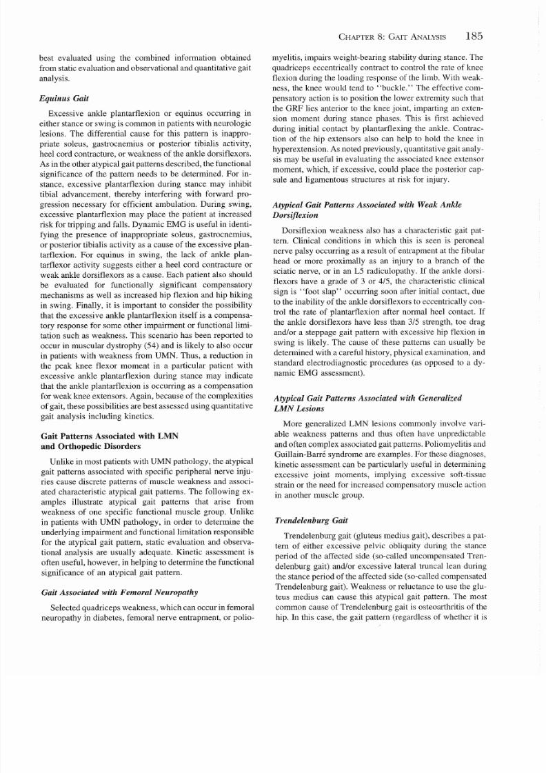
5/15/2018 Casey - Gait - slidepdf.com
http://slidepdf.com/reader/full/casey-gait 19/22
best evaluated using the combined information obtained
from static evaluation and observational and quantitative gait
analysis.
Equinus Gait
Excessive ankle plantarflexion or equinus occurring in
either stance or swing is common in patients with neurologic
lesions. The differential cause for this pattern is inappro-
priate soleus, gastrocnemius or posterior tibialis activity,
heel cord contracture, or weakness of the ankle dorsiflexors.
As in the other atypical gait patterns described, the functional
significance of the pattern needs to be determined. For in-
stance, excessive plantarflexion during stance may inhibit
tibial advancement, thereby interfering with forward pro-
gression necessary for efficient ambulation. During swing,
excessive plantarflexion may place the patient at increased
risk for tripping and falls. Dynamic EMG is useful in identi-
fying the presence of inappropriate soleus, gastrocnemius,
or posterior tibialis activity as a cause of the excessive plan-
tarflexion. For equinus in swing, the lack of ankle plan-
tarflexor activity suggests either a heel cord contracture or
weak ankle dorsiflexors as a cause. Each patient also should
be evaluated for functionally significant compensatory
mechanisms as well as increased hip flexion and hip hiking
in swing. Finally, it is important to consider the possibility
that the excessive ankle plantarflexion itself is a compensa-
tory response for some other impairment or functionallimi-
tation such as weakness. This scenario has been reported to
occur in muscular dystrophy (54) and is likely to also occur
in patients with weakness from UMN. Thus, a reduction in
the peak knee flexor moment in a particular patient with
excessive ankle plantarflexion during stance may indicate
that the ankle plantarflexion is occurring as a compensationfor weak knee extensors. Again, because of the complexities
of gait, these possibilities are best assessed using quantitative
gait analysis including kinetics.
Gait Patterns Associatedwith LMN
and Orthopedic Disorders
Unlike in most patients with UMN pathology, the atypical
gait patterns associated with specific peripheral nerve inju-
ries cause discrete patterns of muscle weakness and associ-
ated characteristic atypical gait patterns. The following ex-
amples illustrate atypical gait patterns that arise from
weakness of one specific functional muscle group. Unlike
in patients with UMN pathology, in order to determine the
underlying impairment and functional limitation responsible
for the atypical gait pattern, static evaluation and observa-
tional analysis are usually adequate. Kinetic assessment is
often useful, however, in helping to determine the functional
significance of an atypical gait pattern.
Gait Associated with Femoral Neuropathy
Selected quadriceps weakness, which can occur in femoral
neuropathy in diabetes, femoral nerve entrapment, or polio-
CHAPTER 8: GAIT ANALYSIS 185
myelitis, impairs weight-bearing stability during stance. The
quadriceps eccentrically contract to control the rate of knee
flexion during the loading response of the limb. With weak-
ness, the knee would tend to "buckle." The effective com-
pensatory action is to position the lower extremity such that
the GRF lies anterior to the knee joint, imparting an exten-
sion moment during stance phases. This is first achieved
during initial contact by plantarflexing the ankle. Contrac-tion of the hip extensors also can help to hold the knee in
hyperextension. As noted previously, quantitative gait analy-
sis may be useful in evaluating the associated knee extensor
moment, which, if excessive, could place the posterior cap-
sule and ligamentous structures at risk for injury.
Atypical Gait Patterns Associated with Weak Ankle
Dorsiflexion
Dorsiflexion weakness also has a characteristic gait pat-
tern. Clinical conditions in which this is seen is peroneal
nerve palsy occurring as a result of entrapment at the fibular
head or more proximally as an injury to a branch of thesciatic nerve, or in an L5 radiculopathy. If the ankle dorsi-
flexors have a grade of 3 or 4/5, the characteristic clinical
sign is "foot slap" occurring soon after initial contact, due
to the inability of the ankle dorsiflexors to eccentrically con-
trol the rate of plantarflexion after normal heel contact. If
the ankle dorsiflexors have less than 3/5 strength, toe drag
and/or a steppage gait pattern with excessive hip flexion in
swing is likely. The cause of these patterns can usually be
determined with a careful history, physical examination, and
standard electrodiagnostic procedures (as opposed to a dy-
namic EMG assessment).
Atypical Gait Patterns Associated with Generalized
LMN Lesions
More generalized LMN lesions commonly involve vari-
able weakness patterns and thus often have unpredictable
and often complex associated gait patterns. Poliomyelitis and
Guillain-Barre syndrome are examples. For these diagnoses,
kinetic assessment can be particularly useful in determining
excessive joint moments, implying excessive soft-tissue
strain or the need for increased compensatory muscle action
in another muscle group.
Trendelenburg Gait
Trendelenburg gait (gluteus medius gait), describes a pat-
tern of either excessive pelvic obliquity during the stance
period of the affected side (so-called uncompensated Tren-
delenburg gait) and/or excessive lateral truncal lean during
the stance period of the affected side (so-called compensated
Trendelenburg gait). Weakness or reluctance to use the glu-
teus medius can cause this atypical gait pattern. The most
common cause of Trendelenburg gait is osteoarthritis of the
hip. In this case, the gait pattern (regardless of whether it is
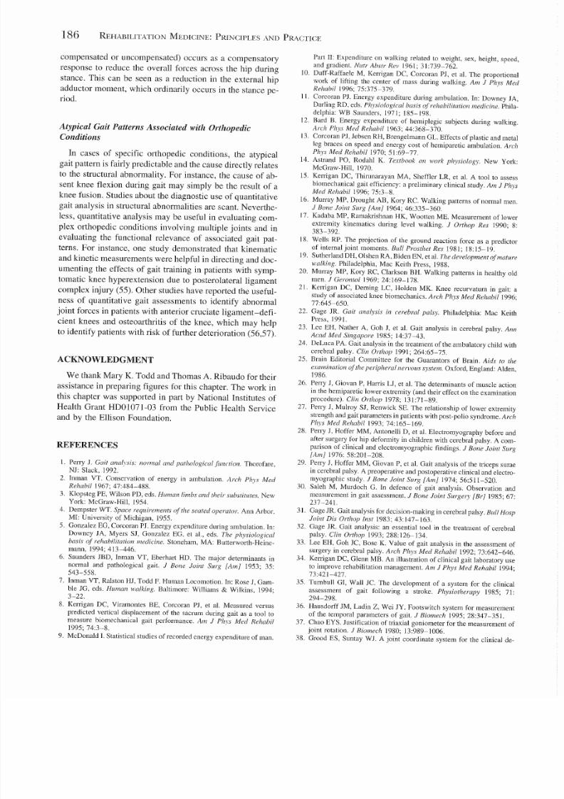
5/15/2018 Casey - Gait - slidepdf.com
http://slidepdf.com/reader/full/casey-gait 20/22
186 REHABILITATION MEDICINE: PRINCIPLES AND PRACTICE
compensated or uncompensated) occurs as a compensatory
response to reduce the overall forces across the hip during
stance. This can be seen as a reduction in the external hip
adductor moment, which ordinarily occurs in the stance pe-
riod.
Atypical Gait Patterns Associated with Orthopedic
Conditions
In cases of specific orthopedic conditions, the atypical
gait pattern is fairly predictable and the cause directly relates
to the structural abnormality. For instance, the cause of ab-
sent knee flexion during gait may simply be the result of a
knee fusion. Studies about the diagnostic use of quantitative
gait analysis in structural abnormalities are scant. Neverthe-
less, quantitative analysis may be useful in evaluating com-
plex orthopedic conditions involving multiple joints and in
evaluating the functional relevance of associated gait pat-
terns. For instance, one study demonstrated that kinematic
and kinetic measurements were helpful in directing and doc-
umenting the effects of gait training in patients with symp-
tomatic knee hyperextension due to posterolateral ligament
complex injury (55). Other studies have reported the useful-
ness of quantitative gait assessments to identify abnormal
joint forces in patients with anterior cruciate ligament-defi-
cient knees and osteoarthritis of the knee, which may help
to identify patients with risk of further deterioration (56,57).
ACKNOWLEDGMENT
We thank Mary K. Todd and Thomas A. Ribaudo for their
assistance in preparing figures for this chapter. The work in
this chapter was supported in part by National Institutes of
Health Grant HD01071-03 from the Public Health Service
and by the Ellison Foundation.
REFERENCES
1. Perry J. Gait analysis: normal and pathological function. Thorofare,
NJ: Slack, 1992.
2. Inman VT. Conservation of energy in ambulation. Arch Phys Med
Rehabil 1967; 47:484-488.
3. Klopsteg PE, Wilson PD, eds. Human limbs and their substitutes. New
York: McGraw-Hill, 1954.
4. Dempster WT. Space requirements of the seated operator. Ann Arbor.
MI: University of Michigan, 1955.
5. Gonzalez EG, Corcoran PJ. Energy expenditure during ambulation. In:
Downey JA, Myers SJ, Gonzalez EG, et aJ., eds. The physiological
basis of rehabilitation medicine. Stoneham, MA: Butterworth-Heine-
mann, 1994; 413-446.
6. Saunders JED, Inman VT, Eberhart HD. The major determinants in
normal and pathological gait. J Bone Joint Surg [Am] 1953; 35:
543-558.
7. Inman VT, Ralston HJ, Todd F. Human Locomotion. In: Rose J, Gam-
ble JG, eds. Human walking. Baltimore: Williams & Wilkins, 1994;
3-22.
8. Kerrigan DC, Viramontes BE, Corcoran PJ, et al. Measured versus
predicted vertical displacement of the sacrum during gait as a tool to
measure biornechanical gait performance. Am J Phvs Med Rehabil
1995; 74:3-8.
9. McDonald I.Statistical studies of recorded energy expenditure of man.
Part II: Expenditure on walking related to weight. sex, height, speed,
and gradient. Nutr Abstr Rev 1961; 31:739-762.
10. Duff-Raffaele M, Kerrigan DC, Corcoran PJ, et al. The proportional
work of lifting the center of mass during walking. Am J Phys Med
Rehabil 1996; 75:375-379.
1 I. Corcoran PJ. Energy expenditure during ambulation. In: Downey JA,
Darling RD. cds. Physiological basis of rehabilitation medicine. Phila-
delphia: WB Saunders, 1971; 185-198.
12. Bard B. Energy expenditure of hemiplegic subjects during walking.
Arch Phys Med Rehabil 1963; 44:368-370.
13. Corcoran PJ, Jebsen RH, Brengelmann GL. Effects of plastic and metal
leg braces on speed and energy cost of hemiparetic ambulation. Arch
Phvs Med Rehabil 1970; 51:69-77.
14. Astrand PO, Rodahl K. Textbook on work physiology. New York:
McGraw-Hill, 1970.
15. Kerrigan DC, Thirunarayan MA, Sheffler LR, et al. A tool to assess
biomechanical gait efficiency: a preliminary clinical study. Am J Phvs
Med Rehabil 1996; 75:3-8.
16. Murray MP, Drought AB, Kory RC. Walking patterns of normal men.
J Bone Joint Surg [Am] 1964; 46:335-360.
17. Kadaba MP, Ramakrishnan HK, Wootten ME. Measurement of lower
extremity kinematics during level walking. J Orthop Res 1990; 8:
383-392.
IS. Wells RP. The projection of the ground reaction force as a predictor
of internal joint moments. Bull Prosthet Res 1981; 18:15-19.
19. Sutherland DH, Olshen RA, Biden EN, et al. The development of mature
walking. Philadelphia, Mac Keith Press, 1988.
20. Murray MP, Kory RC, Clarkson BH. Walking patterns in healthy old
men. J Gerontol 1969; 24:169-178.
21. Kerrigan DC, Deming LC, Holden MK. Knee recurvatum in gait: a
study of associated knee biomechanics. Arch Phys M ed R ehabil 1996;
77:645-650.
22. Gage JR. Gait analysis in cerebral pals) '. Philadelphia: Mac Keith
Press, 1991.
23. Lee EH, Nather A, Goh J, et al. Gait analysis in cerebral palsy. Ann
Acad Med Singapore 1985; 14:37-43.
24. DeLuca PA. Gait analysis in the treatment of the ambulatory child with
cerebral palsy. Clin Orthop 1991; 264:65-75.
25. Brain Editorial Committee for the Guarantors of Brain. Aids to the
examination of the peripheralnervous system. Oxford, England: Alden,
1986.
26. Perry J, Giovan P, Harris LJ, et al. The determinants of muscle action
in the hemiparetic lower extremity (and their effect on the examination
procedure). cu « Orthop 1978; 131:71-89.
27. Perry J, Mulroy SJ, Renwick SE. The relat ionship of lower ext remity
strength and gait parameters in patients with post-polio syndrome. Arch
Phys Med Rehabil 1993; 74:165-169.
28. Perry J, Hoffer MM, Antonelli D, et al. Electromyography before and
after surgery for hip deformity in children with cerebral palsy. A com-
parison of clinical and electromyographic findings. J Bone Joint Surg
[Am] 1976: 58:201-208.
29. Perry J, Hoffer MM, Giovan P, et al . Gait analysis of the triceps surae
in cerebral palsy. A preoperative and postoperative clinical and electro-
myographic study. J Bone Joint Surg [Am] 1974; 56:511-520.
30. Saleh M, Murdoch G. In defence of gait analysis. Observation and
measurement in gait assessment. J Bone Joint Surgery [Br] 1985; 67:
237-241.
31. Gage JR. Gait analysis for decision-making in cerebral palsy. Bull Hosp
Join/ Dis Orthop Inst 1983: 43:147-163.
32. Gage JR. Gai t analysis: an essential tool in the treatment of cerebral
palsy. Clin Orthop 1993; 288:126-134.
33. Lee EH, Goh JC, Bose K. Value of gait analysis in the assessment ofsurgery in cerebral palsy. Arch Phvs Med Rehabil 1992; 73:642-646.
34. Kerrigan DC, Glenn ME. An illustration of clinical gait laboratory use
to improve rehabilitation management. Am J Phys Med Rehabil 1994;
73:421-427.
35. Turnbull GI, Wall JC. The development of a system for the clinical
assessment of gait following a stroke. Physiotherapy 1985; 71:
294-298.
36. Hausdorff JM, Ladin Z, Wei JY. Footswitch system for measurement
of the temporal parameters of gait. J Biomecli 1995; 28:347-351.
37. Chao EYS. Justif ication of triaxial goniometer for the measurement of
joint rotation. J Biomecli 1980; 13:989-1006.
3S. Grood ES, Suntay WJ. A joint coordinate system for the clinical de-
5/15/2018 Casey - Gait - slidepdf.com
http://slidepdf.com/reader/full/casey-gait 21/22
scription of three-dimensional motions: applications to the knee. 1 Bio-
mech Eng 1983; 105:136-144.
39. Zatsiorsky VM, Se1uyanov VN. The mass and inertia characteristics
of the main segments of the human body. In: Matsui H, Kobayashi K,
eds. Human kinetics. Champaign, IL: 1983; 1152-1159.
40. Bekey GA, Chang CJP, et al. Pattern recognition of multiple EMG
signals applied to the description of human gait . Proc IEEE 1977; 65:
674-691.
41. Hirschberg GG, Nathanson M. Electromyographic recordings of mus-
cular activity in normal and spastic gaits. Arch Phvs Med Rehabill947:
33:217-224.
42. Peat M, Dubo HI, Winter DA, et al. Electromyographic temporal analy-
sis of gait: hemiplegic locomotion. Arch Phys Med Rehabi l 1976; 57:
421-425.
43. Knutsson E, Richards e. Different types of disturbed motor control in
gait of hemiparetic patients. Brain 1979; 102:405-430.
44. Shiavi R, Bugle HI, Limbird T. Elect romyographic gai t assessment.
Pm1 2: Preliminary assessment of hemiparetic synergy patterns. 1 Re-
habi l Res Dev 1987; 24:24-30.
45. Winters TF Jr, Gage IR, Hicks R. Gai t patterns in spastic hemiplegia in
children and young adults. 1 Bone Joint Surg [Am] 1987; 69:437-441.
46. Waters RL, Garland DE, Perry I, et al. Stiff -legged gait in hemiplegia:
surgical correction. 1 Bone Joint Surg [Am] 1979; 61:927-933.
47. Treanor WI. The role of physical medicine treatment in stroke rehabili-
tation. Clin Orthop 1969; 63: 14-22.
48. Sutherland DH, Santi M, Abel MF. Treatment of stiff-knee gait in
cerebral palsy: a comparison by gait analysis of distal rectus femoris
CHAPTER 8: GAIT ANALYSIS 187
t ransfer versus proximal rectus release. 1 Pediatr Orthop 1990; 10:
433-441.
49. Kerrigan DC, Gronley I, Perry J. Stiff- legged gait in spastic paresis:
a study of quadriceps and hamstrings muscle activity. Am 1 Phvs Med
Rehabil 1991; 70:294-300.
50. Simon SR, Deutsch SD, Nuzzo RM, et al . Genu recurvatum in spast ic
cerebral palsy. Repor t on findings by gai t analysis. 1 Bone Joint Surg
[Am] 1978; 60:882-894.
51. Gage IR, Perry J, Hicks RR, et al. Rectus femoris transfer to improve
knee function of children with cerebral palsy. Del' Med Child Neurol
1987; 29:159-166.
52. Sutherland DH, Cooper L. The pathomechanics of progressive crouch
gait in spastic diplegia. Orthop c/in North Am 1978; 9:143-154.
53. Thometz I, Simon S, Rosenthal R. The effect on gait of lengthening
of the medial hamstrings in cerebral palsy. 1 Bone Joint Surg [Am]
1989; 71:345-353.
54. Iohnson EW, Walter I. Zeiter lecture: pathokinesiology of Duchenne
muscular dystrophy: implications for management. Arch Phys Med Re-
habil1977; 58:4-7.
55. Noyes F, Dunwor th L, Andriacchi T, et al . Knee hyperextension; gait
abnormalities in unstable knees. Am 1 Sports Med 1996; 24:35-45.
56. Noyes FR, Schipplein OD, Andriacchi TP, et al. The anter ior cruciate
ligament-deficient knee with varus alignment: an analysis of gait adap-
tations and dynamic joint loadings. Am 1 Sports Med 1992; 20:
707-716.
57. Goh ICB, Bose K, Khoo BCe. Gait analysis study on patients with
varus osteoarthrosis of the knee. Clin Orthop 1993; 294:223-231.

5/15/2018 Casey - Gait - slidepdf.com
http://slidepdf.com/reader/full/casey-gait 22/22