case study: liver transplantation karim qumosani, md ... · case study: liver transplantation karim...
TRANSCRIPT

Case Study: Liver Transplantation
Karim Qumosani, MD, FRCPC, ABIM, MdMEd
2017 CST-Astellas Canadian Transplant Fellows Symposium
Dr. Qumosani is Assistant Professor, Medical Director of theAdvanced Hepatology and Liver Transplantation Fellowship, andis now Program Director of Gastroentrology Training (startingJuly 2017) at Western University. He attended medical school atKing Abdulaziz University, Saudi Arabia; and he studied internalmedicine, as well as received his gastroenterology training andan advanced hepatology and liver transplantation fellowship, atWestern University. He also has a Diploma of Medical Educationfrom the University of Dundee, UK. His research interests includehepatocellular carcinoma and nonalcoholic steatohepatitis.

Liver Transplant Cases Discussion
Karim Qumosani MD, FRCPC, ABIM, MdMEd
Multi-organ Transplant Unit, London

Clinical Scenario
Mr ML 58 years old gentleman
– HCV diagnosed 2000
• Liver biopsy 2004 (Staging) showed stage 4/4 fibrosis
• Peg-interferon and Ribavirin = Null responder
– Well compensated
– No other comorbidities

Clinical Scenario
PMHx
– No other chronic medical illness
PSHx
– Unremarkable
Drugs
– None

Clinical Scenario
Social Hx
– Remote IVDU, no EtOH misuse
– Smoker 1 p/day for 25 years
Family HX
– Brother had liver transplant for HCV cirrhosis, died within first year after transplant

Clinical Scenario


HCC – Staging

Clinical Scenario

Clinical Scenario

Clinical Scenario

Clinical Scenario

Clinical Scenario

Clinical Scenario
August 2007; Liver Transplant Assessment
– No medical or psychosocial contraindication for liver transplantation
– Down staged to Milan criteria

Clinical Scenario
September 2007; Liver Transplantation
– NDD
– Cold ischemia time 5 hours
– No intraoperative complications
– Hospital stay was uneventful
– Discharged home 9 days after transplant

Clinical Scenario

Discussion
High Risk Features of HCC recurrence
– Outside Milan criteria
– HCC more than 5 cm
– Microvascular invasion
– Moderately – Poorly differentiated HCC
– AFP more than 200
Sotiropoulos GC, et al Eur J Med Res. 2007;12:527–534


Clinical Scenario – Follow up
October 2007; Transplant Clinic
– No major complains
– Unremarkable post operative course
– Discussion about Xeloda

Clinical Scenario – Follow up
April 2008; CT Chest/Abdomen/Pelvis
– No evidence of HCC recurrence
– Normal Liver Enzymes and Function

Clinical Scenario – Follow up
October 2008; CT Chest/Abdomen/Pelvis
– No evidence of HCC recurrence
July 2009; CT Chest/Abdomen/Pelvis
– No evidence of HCC recurrence

Clinical Scenario – Follow up
February 2010; CT Chest/Abdomen/Pelvis
– Liver; Clear
– No lymph nodes
– Lungs; 3 new nodules in the left upper lobe measuring 11, 7, and 5 mm. Also new 4 mm nodule in the left lower lobe
– AFP 9.3


Clinical Scenario – Follow up
March 2010; FNA
– Left upper lobe
– Suspicious for malignancy
May 2010; CT Guided Biopsy
– Left upper lobe
– HCC

Clinical Scenario – Follow up
August 2010; Medical Oncology
– No chemotherapy
– Surgical resection would be ideal
September 2010; Thoracic Surgery
– Resection feasible

Clinical Scenario – Follow up
October 2010; Surgical Resection
– Wedge resection of apical nodule
– Wedge resection of 2 nodules in the left upper lobe
– Wedge excision of the lung nodule in the posterior basal segment
– Wedge resection of lung nodule in the superior segment of the left lower lobe

Clinical Scenario – Follow up
October 2010; Hospital Stay
– No post operative complications
– Total hospital stay 5 days
– No changes in his immunosuppressive medication

Clinical Scenario – Follow up
Discussion
Bodzit, A et al; Annals of surgery; 2016

Discussion

Discussion

Clinical Scenario – Follow up
December 2011; CT Chest
– No recurrence
March 2012; CT Chest/Abdomen/Pelvis
– No recurrence
September 2012; CT Chest
– No recurrence

Clinical Scenario – Follow up
March 2013; CT Chest/Abdomen/Pelvis
– No recurrence
October 2013; CT Chest
– No recurrence
May 2014; CT Chest
– No recurrence

Clinical Scenario – Follow up
December 2014; CT Chest
– No recurrence
May 2015; CT Chest/Abdomen/Pelvis
– No recurrence
December 2015; CT Chest
– No recurrence

Clinical Scenario – Follow up
May 2016; CT Chest/Abdomen/Pelvis – No recurrence
May 2017; CT Chest/Abdomen/Pelvis – No recurrence
May 2017; Transplant Clinic – Doing well
– Working full time


Clinical Scenario
Mr DT 43 year old male with hepatitis C virus (HCV) cirrhosis
– Refractory Ascites (LVP Biweekly)
– Esophegeal varices
– Muscle wasting

Clinical Scenario
PMHx
– No other chronic medical illness
PSHx
– Unremarkable
Social Hx
– He is married with 2 children and has been off work from his job as a carpenter for 10 months

Clinical Scenario
Current/previous medications
Duration Response, other relevant information
Nadolol 40 mg/day 12 months Pulse at target (60/min)
Spironolactone200 mg/day
6 months Stopped due to lack of effect
Furosemide 6 months Stopped due to lack of effect
Vitamin D 12 months
Multivitamin 12 months

Clinical Scenario Investigation Finding
Hb 110 gm/L
WBC count 3.8 x 10⁹/L
Platelets 85 x 109/L
Bilirubin 110 μmol/L
Creatinine 105 μmol/L
PT INR 2.1
MELD Na 28
US and MRI abdomen • Cirrhosis with portal HT• No HCC or portal venous thrombosis
Hb, hemoglobin; HCC, hepatocellular carcinoma; HT, hypertension; PT INR, prothrombin time, international normalized ratio; MELD Na, model for end-stage liver disease - sodium; MRI, magnetic resonance imaging;US, ultrasound; WBC, white blood cell count

Clinical Scenario
Liver Transplant • He undergoes an appropriately sized matched, ABO-identical
cadaveric liver transplant without technical complications
• Immunosuppression:
– Steriod for induction– Tacrolimus 3 mg BID – Mycophenolate mofetil
(MMF) 1 g BID

Clinical Scenario
Post Liver Transplant Course• Patient extubated Day 1, oriented and alert; off all pressors
• Good initial graft function
• Poor urine output on Day 1 (20 cc/hr)
• Creatinine 170 μmol/L
• Tacrolimus level 4.5 μg/L

Clinical Scenario
Did the patient have pre-op risk factors for renal dysfunction post liver transplant?

Clinical Scenario
Risk factors for post liver transplant renal dysfunction:
• Reduced glomerular filtration rate (GFR) pre-transplant, including hepatorenal syndrome
• Hepatitis C
• Use of full-dose calcineurin inhibitor
• Diabetes mellitus
• Hypertension
• Nonalcoholic steatohepatitis (NASH)

Clinical Scenario
Are liver transplant recipients more vulnerable to renal dysfunction?

Cardenas A. Am J Gastro 2005;100:460-467
CirrhosisPortal hypertension
Splanchnic arterial vasodilation
Decreased systemic vascular resistance
Arterial underfilling
Renal vasoconstriction
Early stages of cirrhosis
Increased systemic and local vasodilators
Preserved renal perfusion
Late stages of cirrhosis
Decrease in local vasodilators with increase of local vasoconstrictors
Hepatorenal syndrome
Clinical Scenario

Clinical Scenario
What could have been done differently?1. Reduced dose of tacrolimus
2. Delayed initiation of tacrolimus
3. Both 1 and 2
4. Alternate immunosuppression, e.g., sirolimus
5. Other

Change in creatinine clearance in liver allograft recipients
Neuberger JM, et al. Am J Transplant 2009;9(2):327-336
Mean change in creatinine clearance (Cockcroft–Gault) at 52 weeks
Group A: Standard dose TAC [target trough levels >10 ng/mL], corticosteroidsGroup B: MMF 2 g/day, reduced-dose TAC (target trough levels ≤8 ng/mL]), corticosteroidsGroup C: Daclizumab induction, MMF, reduced-dose TAC delayed until 5th day post-Tx, corticosteroids
-23.6
-21.2
-13.6
-35.0
-30.0
-25.0
-20.0
-15.0
-10.0
-5.0
0.0
n=181 n=168 n=168
Group A
Group B
Group C
Me
an c
han
ge f
rom
bas
elin
e
in c
reat
inin
e cl
ear
ance
(9
5%
CI)
∗p< 0.05 vs. Group A
*
Clinical Scenario
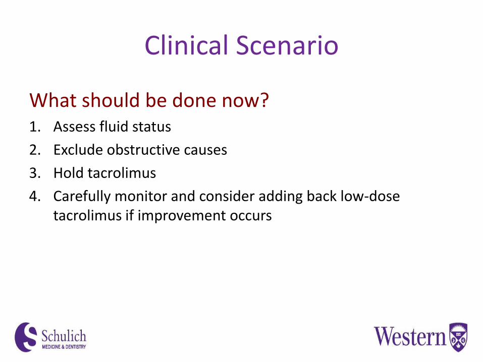
What should be done now?1. Assess fluid status
2. Exclude obstructive causes
3. Hold tacrolimus
4. Carefully monitor and consider adding back low-dose tacrolimus if improvement occurs

Clinical Scenario
Group 1 Group 2 Group 3 Group 4
Time Average GFR, mL/min (n)
Pre-transplant 47±13 (109) 80±9 (106) 108±7 (114) 140±20 (114)
3-month GFR 44±16 (54) 58±23 (50) 63±30 (53) 74±31 (46)
1-year GFR 45±16 (55) 57±20 (49) 68±29 (54) 68±23 (45)
2-year GFR 44±18 (31) 54±19 (26) 67±24 (38) 70±29 (30)
3-year GFR 35±16 (8) 57±34 (4) 86±37 (5) 78±27 (5)
4-year GFR 49±8 (10) 51±22 (11) 75±44 (8) 49±15 (4)
GFR determined by iothalamate clearance. Group 1 had pretransplant GFR ≤67 mL/min; Group 2,67–95 mL/min; Group 3, 96–120 mL/min; Group 4, ≥121 mL/min. Data are average 6 SEM (no. of patients)
Davis CL, et al. Liver Transpl 2002;8(3):193-211
Renal function post-transplant according to pre-transplant GFR

Chronic renal failure in nonrenal organ transplants
Ojo AO, el al. N Engl J Med 2003;349:931-940
Cumulative incidence of chronic renal failure among US nonrenal organ transplants
N = 69 321 persons who received nonrenal organ transplants in US (1990-2000)Renal function measurements obtained at 6-month intervals during the first year and annually thereafter
Cu
mu
lati
ve in
cid
en
ce o
f ch
ron
ic r
en
al f
ailu
re
No. at tiskHeart-lung 576 375 295 219 194 156 133 107 72 46 30Heart 24,024 19,885 17,238 14,687 12,341 10,022 7,997 6,104 4,526 3,096 1,991Intestine 228 152 110 84 57 33 23 13 8 5 5Liver 36,849 28,495 24,041 19,508 15,724 12,564 9,844 7,345 5,292 3,614 2,261Lung 7,643 5,633 4,316 3,184 2,327 1,629 1,136 745 468 258 133
Months since transplantation
LiverLungHeart
Heart-lung
0.000.050.100.150.200.250.300.35
0 12 24 36 48 60 72 84 96 108 120
Intestine
Clinical Scenario
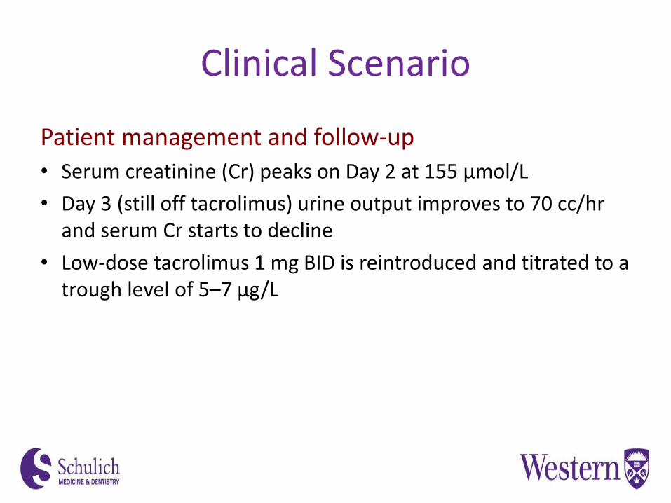
Patient management and follow-up
• Serum creatinine (Cr) peaks on Day 2 at 155 μmol/L
• Day 3 (still off tacrolimus) urine output improves to 70 cc/hrand serum Cr starts to decline
• Low-dose tacrolimus 1 mg BID is reintroduced and titrated to a trough level of 5–7 μg/L

Take Home Messages
• Renal function is often worse than it appears in patients with decompensated liver cirrhosis
• Early AKI is highly prevalent post liver transplant, but risk factors have been identified that help predict which patients are most vulnerable
• Liver transplant patients are uniquely sensitive to CNIs in the immediate post-op period
• Strategies exist to minimize post-op renal injury
