case report a case of giant uterine lipoleiomyoma...
TRANSCRIPT
Case ReportA Case of Giant Uterine Lipoleiomyoma Simulating Malignancy
Erbil Karaman,1 Numan Çim,1 Gülay Bulut,2 Gülhan Elçi,1 Esra AndJç,1
Mustafa Tekin,1 and Ali KolusarJ1
1Department of Obstetrics and Gynecology, Yuzuncu Yil University, 65000 Van, Turkey2Department of Pathology, Yuzuncu Yil University, 65000 Van, Turkey
Correspondence should be addressed to Erbil Karaman; [email protected]
Received 7 April 2015; Accepted 13 July 2015
Academic Editor: Kyousuke Takeuchi
Copyright © 2015 Erbil Karaman et al. This is an open access article distributed under the Creative Commons Attribution License,which permits unrestricted use, distribution, and reproduction in any medium, provided the original work is properly cited.
Introduction. Uterine leiomyoma is the most common benign pathology in women and lipoleiomyoma is an extremely rareand specific type of leiomyoma. Here, we report an unusual case of giant pedunculated subserous lipoleiomyoma misdiagnosedpreoperatively as leiomyosarcoma. Case. A 45-year-old woman admitted to our gynecology outpatient clinic for complaints ofabdominal distention, tiredness, and pelvic pain for the last 6 months. Sonography and abdominal magnetic resonance imaging(MRI) showed a giant semisolidmass that filled whole abdominal cavity from pelvis to subdiaphragmatic area. A primary diagnosisof uterine sarcoma or ovarian malignancy was made. On operation, total abdominal hysterectomy with a pedunculated mass ofsize 30 × 23 × 12 cm and weighing 5.4 kg and bilateral salpingo-oophorectomy were performed. The histopathology revealed alipoleiomyomawith extensive cystic and fatty degenerationwithout anymalignancy.Discussion.Thediagnosis of leiomyoma is doneusually with pelvic ultrasound but sometimes it is difficult to reach a correct diagnosis especially in cases of giant and pedunculatedlipoleiomyoma that included fatty tissue whichmaymimickmalignancy.Conclusion. Subserous pedunculated giant lipoleiomyomashould be kept in mind in the differential diagnosis of leiomyosarcoma or ovarian malignancy.
1. Introduction
Uterine leiomyomas are the most commonly seen gyneco-logic tumors and their prevalence is stated as 25–40% inreproductive age [1]. The fibroids originate from the smoothmuscle cells of uterinewall. Its size varies frommicroscopic togiant and they can be submucosal, intramural, or subserouslocation. Huge uterine myomas are exceedingly rare [2].Lipoleiomyoma is a benign variant of leiomyoma and iscomposed ofmature smoothmuscle cells and adipocyteswithan incidence ranging from 0.03% to 0.2%. The exact etiologyof fibroids is still unknown but it is linked with the role ofestradiol and growth factors [3].However, fattymetamorpho-genesis of the smooth muscle cells of leiomyomas is the mostlikely cause for the development of lipoleiomyoma [4]. Thecomplaints of fibroids can be menstrual disturbances, pelvicpain, constipation, micturition problem, or some effectson fertility such as miscarriage and preterm labour. Thediagnosis of fibroids is made with ultrasound or MRI witha good accuracy. However, in case of pedunculated giant
myoma with thin stalk and fatty cystic degeneration, thediagnosis is difficult and can be misdiagnosed as uterinesarcoma or ovarian malignancy. The treatment options varyfrom expectant management of small and asymptomaticfibroids to surgical therapy especially in case of giant ones.
2. Case
A 45-year-old premenopausal multiparous woman wasadmitted to our hospital’s outpatient gynecology clinic withcomplaints of lower abdominal pain and abdominal dis-tension for the last 6 months. On detailed anamnesis, thepatient had noticed a mass in her abdomen for 3 monthsand a gradually increasing pain with easy tiredness. Shehad four previous vaginal deliveries with no abdominalsurgical operation. Her medical history was remarkable for10 years of type II diabetes and hypercholesterolemia andhad no history for family member with genital malignancy.She had no complaints related with menstrual bleeding.On physical examination, her vital signs were normal and
Hindawi Publishing CorporationCase Reports in Obstetrics and GynecologyVolume 2015, Article ID 926961, 4 pageshttp://dx.doi.org/10.1155/2015/926961
2 Case Reports in Obstetrics and Gynecology
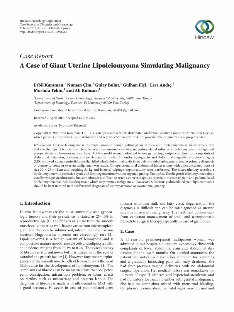
Figure 1: The figure shows MRI image of giant pelvic massfilling whole abdominal cavity with heterogenous and semisolidappearance (corresponding the image between four white callipers).
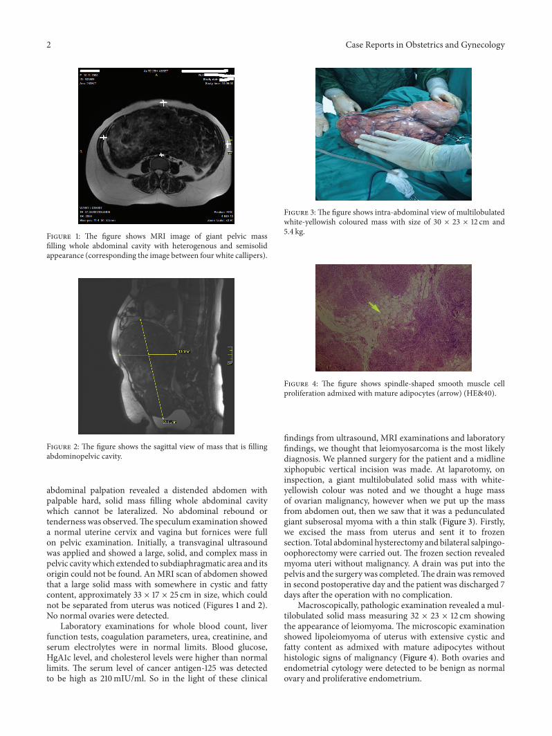
Figure 2: The figure shows the sagittal view of mass that is fillingabdominopelvic cavity.
abdominal palpation revealed a distended abdomen withpalpable hard, solid mass filling whole abdominal cavitywhich cannot be lateralized. No abdominal rebound ortenderness was observed.The speculum examination showeda normal uterine cervix and vagina but fornices were fullon pelvic examination. Initially, a transvaginal ultrasoundwas applied and showed a large, solid, and complex mass inpelvic cavitywhich extended to subdiaphragmatic area and itsorigin could not be found. AnMRI scan of abdomen showedthat a large solid mass with somewhere in cystic and fattycontent, approximately 33 × 17 × 25 cm in size, which couldnot be separated from uterus was noticed (Figures 1 and 2).No normal ovaries were detected.
Laboratory examinations for whole blood count, liverfunction tests, coagulation parameters, urea, creatinine, andserum electrolytes were in normal limits. Blood glucose,HgA1c level, and cholesterol levels were higher than normallimits. The serum level of cancer antigen-125 was detectedto be high as 210mIU/ml. So in the light of these clinical
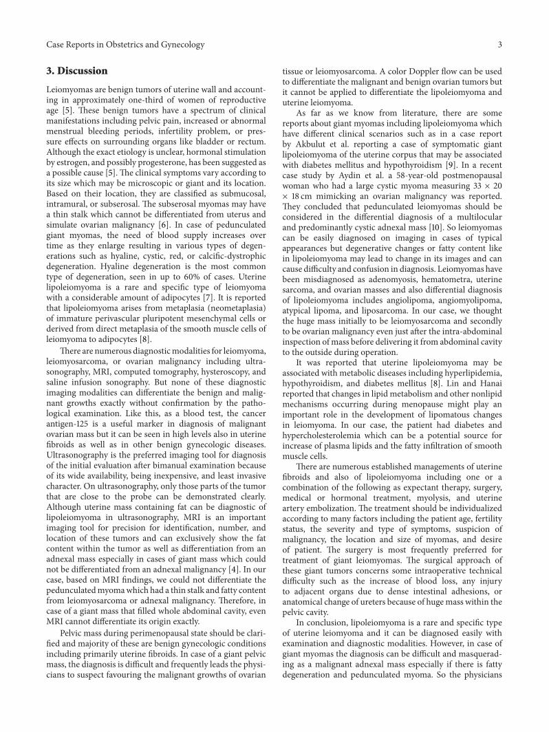
Figure 3: The figure shows intra-abdominal view of multilobulatedwhite-yellowish coloured mass with size of 30 × 23 × 12 cm and5.4 kg.
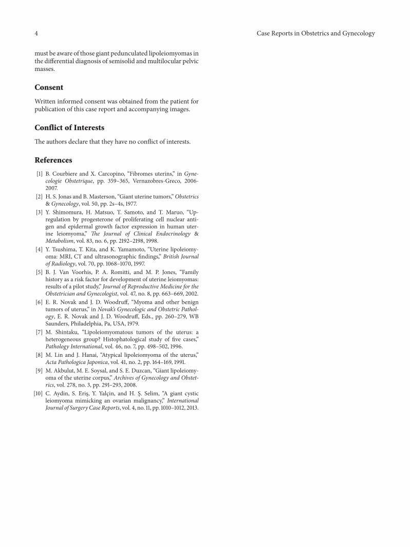
Figure 4: The figure shows spindle-shaped smooth muscle cellproliferation admixed with mature adipocytes (arrow) (HE&40).
findings from ultrasound, MRI examinations and laboratoryfindings, we thought that leiomyosarcoma is the most likelydiagnosis. We planned surgery for the patient and a midlinexiphopubic vertical incision was made. At laparotomy, oninspection, a giant multilobulated solid mass with white-yellowish colour was noted and we thought a huge massof ovarian malignancy, however when we put up the massfrom abdomen out, then we saw that it was a pedunculatedgiant subserosal myoma with a thin stalk (Figure 3). Firstly,we excised the mass from uterus and sent it to frozensection. Total abdominal hysterectomy andbilateral salpingo-oophorectomy were carried out. The frozen section revealedmyoma uteri without malignancy. A drain was put into thepelvis and the surgery was completed.The drainwas removedin second postoperative day and the patient was discharged 7days after the operation with no complication.
Macroscopically, pathologic examination revealed a mul-tilobulated solid mass measuring 32 × 23 × 12 cm showingthe appearance of leiomyoma. The microscopic examinationshowed lipoleiomyoma of uterus with extensive cystic andfatty content as admixed with mature adipocytes withouthistologic signs of malignancy (Figure 4). Both ovaries andendometrial cytology were detected to be benign as normalovary and proliferative endometrium.
Case Reports in Obstetrics and Gynecology 3
3. Discussion
Leiomyomas are benign tumors of uterine wall and account-ing in approximately one-third of women of reproductiveage [5]. These benign tumors have a spectrum of clinicalmanifestations including pelvic pain, increased or abnormalmenstrual bleeding periods, infertility problem, or pres-sure effects on surrounding organs like bladder or rectum.Although the exact etiology is unclear, hormonal stimulationby estrogen, and possibly progesterone, has been suggested asa possible cause [5]. The clinical symptoms vary according toits size which may be microscopic or giant and its location.Based on their location, they are classified as submucosal,intramural, or subserosal. The subserosal myomas may havea thin stalk which cannot be differentiated from uterus andsimulate ovarian malignancy [6]. In case of pedunculatedgiant myomas, the need of blood supply increases overtime as they enlarge resulting in various types of degen-erations such as hyaline, cystic, red, or calcific-dystrophicdegeneration. Hyaline degeneration is the most commontype of degeneration, seen in up to 60% of cases. Uterinelipoleiomyoma is a rare and specific type of leiomyomawith a considerable amount of adipocytes [7]. It is reportedthat lipoleiomyoma arises from metaplasia (neometaplasia)of immature perivascular pluripotent mesenchymal cells orderived from direct metaplasia of the smooth muscle cells ofleiomyoma to adipocytes [8].
There are numerous diagnosticmodalities for leiomyoma,leiomyosarcoma, or ovarian malignancy including ultra-sonography, MRI, computed tomography, hysteroscopy, andsaline infusion sonography. But none of these diagnosticimaging modalities can differentiate the benign and malig-nant growths exactly without confirmation by the patho-logical examination. Like this, as a blood test, the cancerantigen-125 is a useful marker in diagnosis of malignantovarian mass but it can be seen in high levels also in uterinefibroids as well as in other benign gynecologic diseases.Ultrasonography is the preferred imaging tool for diagnosisof the initial evaluation after bimanual examination becauseof its wide availability, being inexpensive, and least invasivecharacter. On ultrasonography, only those parts of the tumorthat are close to the probe can be demonstrated clearly.Although uterine mass containing fat can be diagnostic oflipoleiomyoma in ultrasonography, MRI is an importantimaging tool for precision for identification, number, andlocation of these tumors and can exclusively show the fatcontent within the tumor as well as differentiation from anadnexal mass especially in cases of giant mass which couldnot be differentiated from an adnexal malignancy [4]. In ourcase, based on MRI findings, we could not differentiate thepedunculatedmyomawhich had a thin stalk and fatty contentfrom leiomyosarcoma or adnexal malignancy. Therefore, incase of a giant mass that filled whole abdominal cavity, evenMRI cannot differentiate its origin exactly.
Pelvic mass during perimenopausal state should be clari-fied and majority of these are benign gynecologic conditionsincluding primarily uterine fibroids. In case of a giant pelvicmass, the diagnosis is difficult and frequently leads the physi-cians to suspect favouring the malignant growths of ovarian
tissue or leiomyosarcoma. A color Doppler flow can be usedto differentiate the malignant and benign ovarian tumors butit cannot be applied to differentiate the lipoleiomyoma anduterine leiomyoma.
As far as we know from literature, there are somereports about giant myomas including lipoleiomyoma whichhave different clinical scenarios such as in a case reportby Akbulut et al. reporting a case of symptomatic giantlipoleiomyoma of the uterine corpus that may be associatedwith diabetes mellitus and hypothyroidism [9]. In a recentcase study by Aydin et al. a 58-year-old postmenopausalwoman who had a large cystic myoma measuring 33 × 20× 18 cm mimicking an ovarian malignancy was reported.They concluded that pedunculated leiomyomas should beconsidered in the differential diagnosis of a multilocularand predominantly cystic adnexal mass [10]. So leiomyomascan be easily diagnosed on imaging in cases of typicalappearances but degenerative changes or fatty content likein lipoleiomyoma may lead to change in its images and cancause difficulty and confusion in diagnosis. Leiomyomas havebeen misdiagnosed as adenomyosis, hematometra, uterinesarcoma, and ovarian masses and also differential diagnosisof lipoleiomyoma includes angiolipoma, angiomyolipoma,atypical lipoma, and liposarcoma. In our case, we thoughtthe huge mass initially to be leiomyosarcoma and secondlyto be ovarian malignancy even just after the intra-abdominalinspection ofmass before delivering it from abdominal cavityto the outside during operation.
It was reported that uterine lipoleiomyoma may beassociated with metabolic diseases including hyperlipidemia,hypothyroidism, and diabetes mellitus [8]. Lin and Hanaireported that changes in lipid metabolism and other nonlipidmechanisms occurring during menopause might play animportant role in the development of lipomatous changesin leiomyoma. In our case, the patient had diabetes andhypercholesterolemia which can be a potential source forincrease of plasma lipids and the fatty infiltration of smoothmuscle cells.
There are numerous established managements of uterinefibroids and also of lipoleiomyoma including one or acombination of the following as expectant therapy, surgery,medical or hormonal treatment, myolysis, and uterineartery embolization. The treatment should be individualizedaccording to many factors including the patient age, fertilitystatus, the severity and type of symptoms, suspicion ofmalignancy, the location and size of myomas, and desireof patient. The surgery is most frequently preferred fortreatment of giant leiomyomas. The surgical approach ofthese giant tumors concerns some intraoperative technicaldifficulty such as the increase of blood loss, any injuryto adjacent organs due to dense intestinal adhesions, oranatomical change of ureters because of hugemass within thepelvic cavity.
In conclusion, lipoleiomyoma is a rare and specific typeof uterine leiomyoma and it can be diagnosed easily withexamination and diagnostic modalities. However, in case ofgiant myomas the diagnosis can be difficult and masquerad-ing as a malignant adnexal mass especially if there is fattydegeneration and pedunculated myoma. So the physicians
4 Case Reports in Obstetrics and Gynecology
must be aware of those giant pedunculated lipoleiomyomas inthe differential diagnosis of semisolid andmultilocular pelvicmasses.
Consent
Written informed consent was obtained from the patient forpublication of this case report and accompanying images.
Conflict of Interests
The authors declare that they have no conflict of interests.
References
[1] B. Courbiere and X. Carcopino, “Fibromes uterins,” in Gyne-cologie Obstetrique, pp. 359–365, Vernazobres-Greco, 2006-2007.
[2] H. S. Jonas and B. Masterson, “Giant uterine tumors,”Obstetrics& Gynecology, vol. 50, pp. 2s–4s, 1977.
[3] Y. Shimomura, H. Matsuo, T. Samoto, and T. Maruo, “Up-regulation by progesterone of proliferating cell nuclear anti-gen and epidermal growth factor expression in human uter-ine leiomyoma,” The Journal of Clinical Endocrinology &Metabolism, vol. 83, no. 6, pp. 2192–2198, 1998.
[4] Y. Tsushima, T. Kita, and K. Yamamoto, “Uterine lipoleiomy-oma: MRI, CT and ultrasonographic findings,” British Journalof Radiology, vol. 70, pp. 1068–1070, 1997.
[5] B. J. Van Voorhis, P. A. Romitti, and M. P. Jones, “Familyhistory as a risk factor for development of uterine leiomyomas:results of a pilot study,” Journal of Reproductive Medicine for theObstetrician and Gynecologist, vol. 47, no. 8, pp. 663–669, 2002.
[6] E. R. Novak and J. D. Woodruff, “Myoma and other benigntumors of uterus,” in Novak’s Gynecologic and Obstetric Pathol-ogy, E. R. Novak and J. D. Woodruff, Eds., pp. 260–279, WBSaunders, Philadelphia, Pa, USA, 1979.
[7] M. Shintaku, “Lipoleiomyomatous tumors of the uterus: aheterogeneous group? Histophatological study of five cases,”Pathology International, vol. 46, no. 7, pp. 498–502, 1996.
[8] M. Lin and J. Hanai, “Atypical lipoleiomyoma of the uterus,”Acta Pathologica Japonica, vol. 41, no. 2, pp. 164–169, 1991.
[9] M. Akbulut, M. E. Soysal, and S. E. Duzcan, “Giant lipoleiomy-oma of the uterine corpus,” Archives of Gynecology and Obstet-rics, vol. 278, no. 3, pp. 291–293, 2008.
[10] C. Aydin, S. Eris, Y. Yalcin, and H. S. Selim, “A giant cysticleiomyoma mimicking an ovarian malignancy,” InternationalJournal of Surgery Case Reports, vol. 4, no. 11, pp. 1010–1012, 2013.

Submit your manuscripts athttp://www.hindawi.com
Stem CellsInternational
Hindawi Publishing Corporationhttp://www.hindawi.com Volume 2014
Hindawi Publishing Corporationhttp://www.hindawi.com Volume 2014
MEDIATORSINFLAMMATION
of
Hindawi Publishing Corporationhttp://www.hindawi.com Volume 2014
Behavioural Neurology
EndocrinologyInternational Journal of
Hindawi Publishing Corporationhttp://www.hindawi.com Volume 2014
Hindawi Publishing Corporationhttp://www.hindawi.com Volume 2014
Disease Markers
Hindawi Publishing Corporationhttp://www.hindawi.com Volume 2014
BioMed Research International
OncologyJournal of
Hindawi Publishing Corporationhttp://www.hindawi.com Volume 2014
Hindawi Publishing Corporationhttp://www.hindawi.com Volume 2014
Oxidative Medicine and Cellular Longevity
Hindawi Publishing Corporationhttp://www.hindawi.com Volume 2014
PPAR Research
The Scientific World JournalHindawi Publishing Corporation http://www.hindawi.com Volume 2014
Immunology ResearchHindawi Publishing Corporationhttp://www.hindawi.com Volume 2014
Journal of
ObesityJournal of
Hindawi Publishing Corporationhttp://www.hindawi.com Volume 2014
Hindawi Publishing Corporationhttp://www.hindawi.com Volume 2014
Computational and Mathematical Methods in Medicine
OphthalmologyJournal of
Hindawi Publishing Corporationhttp://www.hindawi.com Volume 2014
Diabetes ResearchJournal of
Hindawi Publishing Corporationhttp://www.hindawi.com Volume 2014
Hindawi Publishing Corporationhttp://www.hindawi.com Volume 2014
Research and TreatmentAIDS
Hindawi Publishing Corporationhttp://www.hindawi.com Volume 2014
Gastroenterology Research and Practice
Hindawi Publishing Corporationhttp://www.hindawi.com Volume 2014
Parkinson’s Disease
Evidence-Based Complementary and Alternative Medicine
Volume 2014Hindawi Publishing Corporationhttp://www.hindawi.com