case keratitis linearis migrans* - bjo.bmj.com · brit. j. ophthal. (1963) 47, 504. case notes...
TRANSCRIPT
Brit. J. Ophthal. (1963) 47, 504.
CASE NOTES
KERATITIS LINEARIS MIGRANS*BY
M. HATFIELD WRIGHTSwindon
WITHIN the group of deep forms of keratitis, Duke-Elder (1938) includeskeratitis linearis migrans, a condition described by Fuchs (1926) in which aline of opacity in the deep layers of the stroma of the cornea, associated withkeratic precipitates, travels from side to side across the cornea. Vejdovsky(1952) described a line-like clouding in a transparent cornea which graduallymoved upwards until it reached the upper border of the cornea. Both Fuchsand Vejdovsky attributed the cause to syphilis. Engelbrecht (1927) alsodescribed a deep, and Collomb (1923) a superficial, migratory line in non-syphilitic subjects.
In the case under review, not only were the side-to-side migrations of theopacity (Fuchs) and the upward movement on the cornea (Vejdovsky)present, but also a migration backwards into the corneal stroma from Bow-man's membrane through Descemet's membrane to the endothelial surface.
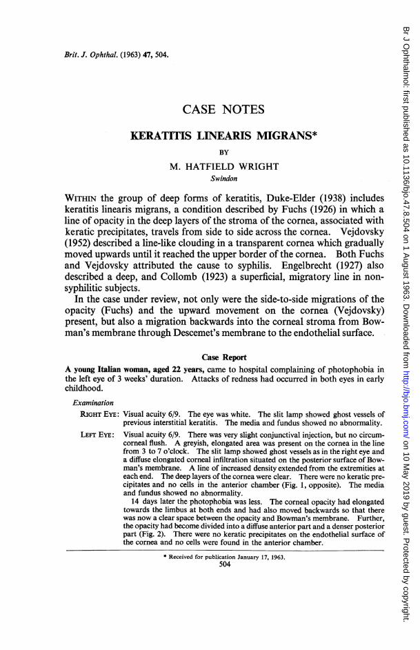
Case ReportA young Italian woman, aged 22 years, came to hospital complaining of photophobia inthe left eye of 3 weeks' duration. Attacks of redness had occurred in both eyes in earlychildhood.
ExaminationRIGHT EYE: Visual acuity 6/9. The eye was white. The slit lamp showed ghost vessels of
previous interstitial keratitis. The media and fundus showed no abnormality.LEFT EYE: Visual acuity 6/9. There was very slight conjunctival injection, but no circum-
corneal flush. A greyish, elongated area was present on the cornea in the linefrom 3 to 7 o'clock. The slit lamp showed ghost vessels as in the right eye anda diffuse elongated corneal infiltration situated on the posterior surface of Bow-man's membrane. A line of increased density extended from the extremities ateach end. The deep layers of the cornea were clear. There were no keratic pre-cipitates and no cells in the anterior chamber (Fig. 1, opposite). The mediaand fundus showed no abnormality.
14 days later the photophobia was less. The corneal opacity had elongatedtowards the limbus at both ends and had also moved backwards so that therewas now a clear space between the opacity and Bowman's membrane. Further,the opacity had become divided into a diffuse anterior part and a denser posteriorpart (Fig. 2). There were no keratic precipitates on the endothelial surface ofthe cornea and no cells were found in the anterior chamber.
* Received for publication January 17, 1963.504
on 10 May 2019 by guest. P
rotected by copyright.http://bjo.bm
j.com/
Br J O
phthalmol: first published as 10.1136/bjo.47.8.504 on 1 A
ugust 1963. Dow
nloaded from
KERATITIS LINEARIS MIGRANS
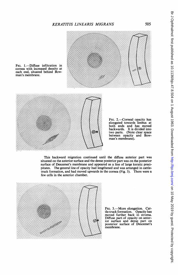
FIG. 1.-Diffuse infiltration incornea with increased density ateach end, situated behind Bow-man's membrane.
<.@t. FIG. 2.-Comeal opacity haselongated towards limbus atboth ends and has movedbackwards. It is divided intotwo parts. (Note clear spacebetween opacity and Bow-man's membrane).
This backward migration continued until the diffuse anterior part wassituated on the anterior surface and the dense posterior part was on the posteriorsurface of Descemet's membrane and appeared as a line of large keratic preci-pitates. The general line of opacity had lengthened and was arranged in cattle-truck formation, and had moved upwards in the cornea (Fig. 3). There were afew cells in the anterior chamber.
FIG. 3.-More elongation. Cat-tle-truck formation. Opacity hasmoved further back in stroma.Diffuse part of opacity on anter-ior surface and dense part onposterior surface of Descemet'smembrane.
."A0)P
,41.,'r.
505
...
......
on 10 May 2019 by guest. P
rotected by copyright.http://bjo.bm
j.com/
Br J O
phthalmol: first published as 10.1136/bjo.47.8.504 on 1 A
ugust 1963. Dow
nloaded from

M. HATFIELD WRIGHT
Within a few days there was further extension of the line almost to the limbusat each end. The corneal stroma was quite clear and the opacity was situatedon the endothelial surface of the cornea and had moved upwards (Fig. 4).During the next few weeks the line extended in length and became irregular
and at the same time continued migrating upwards on the cornea. The stromawas clear (Fig. 5).
FIG. 4.-Comeal stroma FIG. 5. Line of keratitis lengthened and irregular and hasclear. Opacity situated on also moved upwards. Corneal stroma clear.posterior surface of Desce-met's membrane.
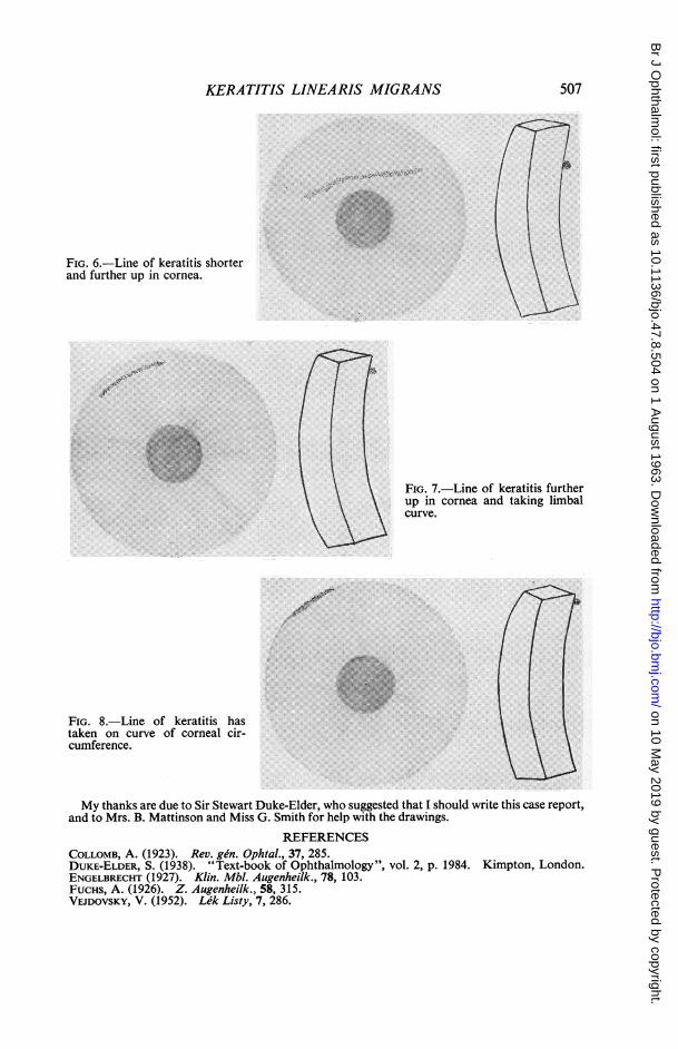
This upward progression continued for some weeks, the line becomingstraighter and shorter (Figs 6 and 7, opposite), until it reached the upper borderof the cornea near the angle of the anterior chamber between 10 and 12 o'clock,where it took on the curve of the comeal circumference (Fig. 8, opposite).The opacity remained in this position for some weeks, clearing slowly. There
were some fine dry keratic precipitates scattered over the posterior surface of thecornea. The keratitis finally cleared up about 10 months after onset.
Serological Examination.-Wassermann reaction + +; Kahn test + + +; Price's precipitationreaction + (diluted 1/4); treponemal immobilization test ±.
Summary
A case of keratitis linearis migrans has been described. In a white eyewith mild anterior uveitis the linear keratitis migrated in three directions:
(1) Towards the limbus at each end of the opacity;
(2) Backwards through the stroma from Bowman's membrane to the endothelialsurface of the cornea;
(3) Upwards towards the upper border of the cornea.
It was not possible to obtain satisfactory photographs and the diagramshave been drawn to elucidate the various stages in the course of thecondition.
506
on 10 May 2019 by guest. P
rotected by copyright.http://bjo.bm
j.com/
Br J O
phthalmol: first published as 10.1136/bjo.47.8.504 on 1 A
ugust 1963. Dow
nloaded from
KERATITIS LINEARIS MIGRANS
FIG. 6.-Line of keratitis shorterand further up in cornea.
FIG. 7.-Line of keratitis furtherup in cornea and taking limbalcurve.
.<.. -.OiX.
.:.r'. . a ; .. : a*: .X. Er^.: ... , . . m .s. 1..U;e.::.,., o.v,.,. ° '.. 2 o. - :':.'.? . :°'.:'8' . g..B <.:.4.Br..g.3.g* . A g;e
:..\. o.-ery..
FIG. 8.-Line of keratitis hastaken on curve of corneal cir-cumference.
My thanks are due to Sir Stewart Duke-Elder, who suggested that I should write this case report,and to Mrs. B. Mattinson and Miss G. Smith for help with the drawings.
REFERENCESCOLLOMB, A. (1923). Rev. gen. Ophtal., 37, 285.DUKE-ELDER, S. (1938). "Text-book of Ophthalmology", vol. 2, p. 1984. Kimpton, London.ENGELBRECHT (1927). Klin. Mbl. Augenheilk., 78, 103.FUCHS, A. (1926). Z. Augenheilk., 58, 315.VEJDOVSKY, V. (1952). Lek Listy, 7, 286.
507
"4,.-1.
.4it
Z,1401
-lb
on 10 May 2019 by guest. P
rotected by copyright.http://bjo.bm
j.com/
Br J O
phthalmol: first published as 10.1136/bjo.47.8.504 on 1 A
ugust 1963. Dow
nloaded from