case: breast cancer alegre. almora. alonzo. amaro. amolenda. anacta. andal. ang. ang. ang
TRANSCRIPT

CASE: BREAST CANCER
Alegre. Almora. Alonzo. Amaro. Amolenda. Anacta. Andal. Ang. Ang. Ang.

General Information
• 45-y/o F • Chief Complaint: Left breast mass.
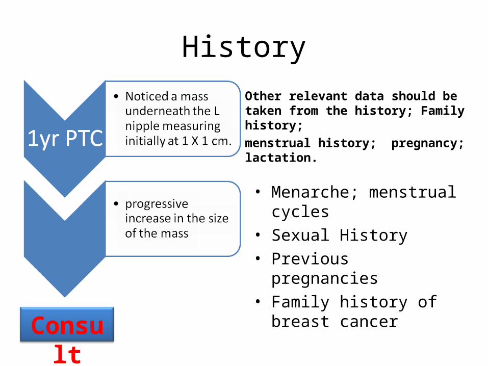
History
Other relevant data should be taken from the history; Family history;menstrual history; pregnancy; lactation.
• Menarche; menstrual cycles• Sexual History• Previous pregnancies• Family history of breast
cancer
Consult


Personal risk factors: • Gender
– Females have a 100-fold increase in risk as compared to males. • Age – breast cancer risk increases with age
– 96% of breast cancers occur in women age 40 and older (ACS, 2003-2004).
• Race – Caucasian women have a greater risk of breast cancer than
other racial groups. • Prolonged exposure to endogenous estrogen and
progestins • Exposure to exogenous combined estrogen and progestin
therapy in hormone replacement therapy for postmenopausal women
Breast Cancer Diagnostic Algorithms for Primary Care Providers
(Third Edition, June 2005)

• Alcohol use – greater than 27 drinks per week. (Gronbaek,
2004.)• Obesity
– obese women with BMI >30 had estrogen concentrations between 60% and 219% higher then thin women
• Radiation exposure to the upper torso – e.g. treatment of Hodgkin’s lymphoma.
Breast Cancer Diagnostic Algorithms for Primary Care Providers
(Third Edition, June 2005)

Risk Factors
• Increase number of menstrual cycles (increased estrogen exposure) – Early menarche, nulliparity late menopause
• Pregnant or Lactating Women– During pregnancy, breast grows secondary to
estrogen, progesterone, HPL, prolactin (low levels)– After pregnancy, progesterone drops, increasing
effects of prolactin

Family History
• One or more first- or second-degree relatives with breast cancer at an early age (less than 40-50 years of age).
• Breast cancer and a second primary cancer in a close relative, especially ovarian cancer. – Other cancers w/ a genetic risk include:
• thyroid, colorectal, prostate, endometrial, pancreatic, adrenocortical carcinoma, melanoma, childhood sarcoma, leukemia/lymphoma, and brain tumors.
• Male breast cancer in a close relative.• Two or more relatives with breast cancer at any age.
Breast Cancer Diagnostic Algorithms for Primary Care Providers
(Third Edition, June 2005)

Physical Exam• L breast
– 2 X 2 cm. hard, non-tender, & movable mass with irregular margins underneath the nipple.
• Axilla is negative for any masses.
• The R breast and the rest of the PE are normal.

Personal Risk Factors
• 45y/o F– 35% benign– 25% cyst– 9% fibroadenoma– 31% other benign lesion

Breast Mass
Benign • round or oval shape• circumscribed margins • width-to– AP dimension
ratio greater than 1.4
Malignant• irregular shape• microlobulated or
spiculated • width-to–AP dimension
ratio of 1.4 or less
Rhabar, et al. Benign versus Malignant Solid Breast Masses: US Differentiation. December 1999 Radiology, 213, 889-894.
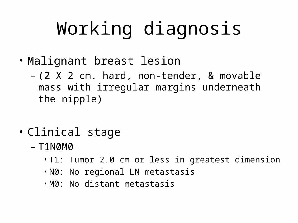
Working diagnosis
• Malignant breast lesion – (2 X 2 cm. hard, non-tender, & movable mass with
irregular margins underneath the nipple)
• Clinical stage– T1N0M0
• T1: Tumor 2.0 cm or less in greatest dimension • N0: No regional LN metastasis• M0: No distant metastasis