case 5: speaker notesmedu.s3.amazonaws.com/c788ebd8/core case 5 workshop... · web viewprompt...
TRANSCRIPT
Slide 1
Slide 2 No change
Slide 3 Changes as above
Slide 4
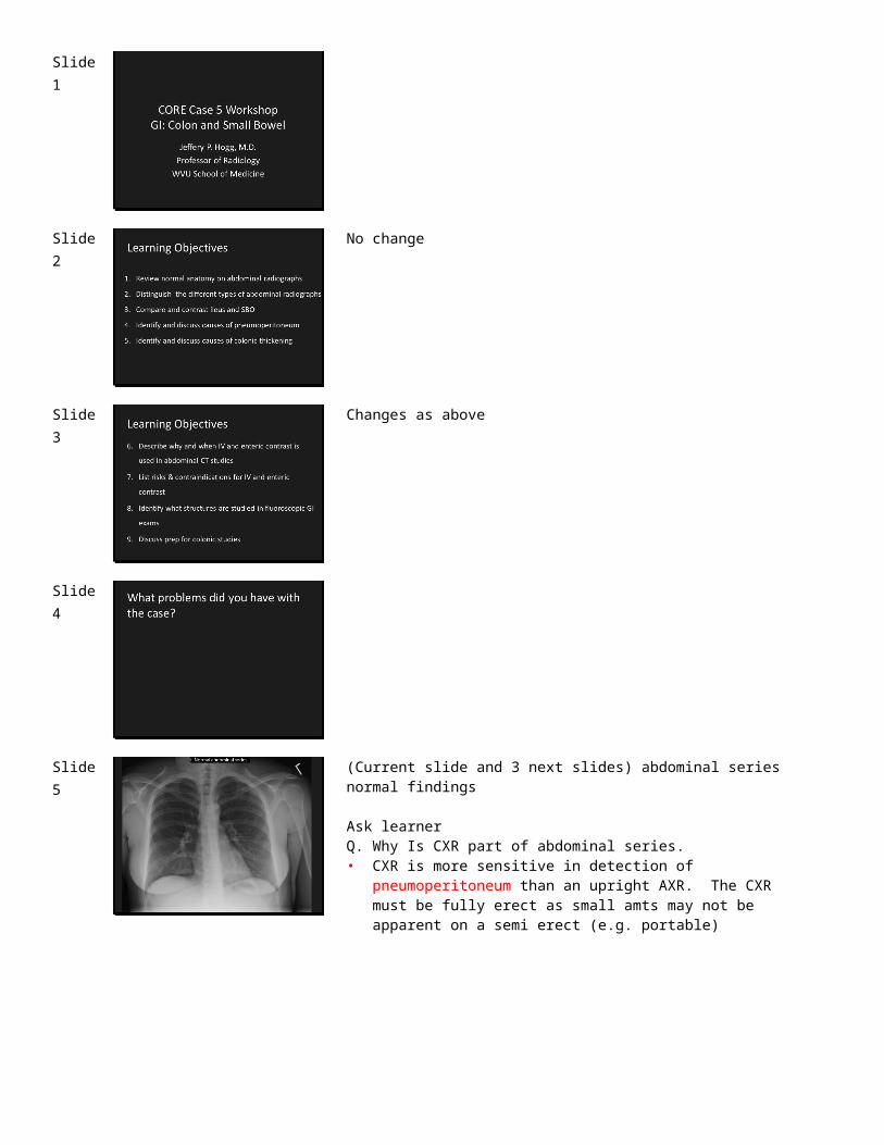
Slide 5 (Current slide and 3 next slides) abdominal series normal findings
Ask learnerQ. Why Is CXR part of abdominal series.• CXR is more sensitive in detection of pneumoperitoneum than an upright
AXR. The CXR must be fully erect as small amts may not be apparent on a semi erect (e.g. portable)

Slide 6 Q. In general, where is the colon?• Frames the central abdomen
Q. In general, where is the small bowel?A. Occupies the central abdomenCan they distinguish large and small bowel?
Slide 7 Q. What information is gotten from the upright abdominal image?• Can find air fluid levels, if present.Q. Why are both upright and supine needed?A: For full evaluation of where the air is, and if there are AFLsQ. How can you tell that this is an upright exam?A: Air-fluid level in stomach, movement of visceraPrompt learner to note position of transverse colon and vertebral levels in upright and compare to supine (soft tissues move esp transverse colon)
Slide 8
Slide 9 Use these and the 2 slides to see if they know what anatomical areas are studied for each one
Slide 10 Can use ipad deploying
Go through GI exams and have students draw which parts of anatomy are studied in each exam
copyright free image http://catalog.niddk.nih.gov/ImageLibrary

Slide 11 Q: What type of study is this?
A: Modified swallow
Series of slides for modified barium swallow, esophogram, UGI and SBFT: do kind of quickly more to introduce students to ways we can evaluate GI tract and what areas that we are visualizing with each type of study.
Slide 12
Slide 13
Slide 14
Q:Why is barium ‘white’ here and was ‘black’ on the prior imagesA: arbitrary choice of display, but typically fluoro spots are black on white
Q:What is this study?A: Esophagram.Have them identify the anatomy
Point out that in each of the various GI Tract studies with fluoroscopic techniques, the process involves making a LOT of radiographs of of the region in different projections (radiation dose) PLUS fluoroscopic observation (more radiation dose).
Slide 15
Q: What is this study? A: Upper GIHave them identify the anatomyAlso, to follow up the learners’ surprise that UGI was among more radiation intensive studies, point out that numerous images in multiple positions are made (each a radiographic exposure + fluoroscopy to position) of the abdomen, a thick region of soft tissue.Explain why we need to reposition them (to fill and empty structures)

Slide 16
Q: What is this study? Why?A: Small bowel follow throughIdentify the anatomyNotice that the initial image after ingestion of contrast is time stamped, as will be subsequent SBFT images.Q. Why? A. To document progress of ingested barium meal through the small bowel.
Q: What has this patient undergone before the SBFT?Show next image.
Slide 17
Scout.Q. What has this patient undergone before the SBFT?• Endovascular stenting of aorta and iliac arteries for PVD.
COULD OMIT IF WISH
Slide 18 Notice that the initial image after ingestion of contrast is time stamped, as will
be subsequent SBFT images.Q. Why? A. To document progress of ingested barium meal through the small bowel.
CAN GO QUICKLY THROUGH THE NEXT IMAGES OR OMIT SOME
Slide 19
This study is imaged at 30 min intervals to follow passage of ingested barium contrast through the small bowel. Again, multiple exposures + fluoroscopy.
Slide 20
How do we know this study is over? A: Barium has reached the colon

Slide 21 Q:What is this study? A: Single contrast barium enema.
Q: Who gets full column BE? A. Generally done if patient is not a candidate for air contrast BE, generally bedridden or debilitated patients. Sometimes in Olgivies using a water soluble contrast to stimulate the bowelQ: What other contrast might we use for an enema? A: Water soluble e.g., Gastrograffin or Dilute IV contrast.Q. When might we use that: If colonic ileus is a question (Olgivies)
Slide 22 Q. What are these densities? A. Could be retained stool, but cannot exclude
intraluminal tumor, hence the importance of proper prep.Q. How can this uncertainty in diagnosis be avoided? . A. Proper patient preparation – which is often the case in debilitated patients.
Q. How do we prep patients for double contrast diagnostic BE? A. Combination of restricted dietary intake (low to no fiber few days prior, to clear liquids day before + laxatives or other similar prep 12-8 hours prior) to make colon as free of residue as possible prior to enema study).
Just so they have an idea what the patients have to drink – make them aware that this can be an issue with dehydration in frail patients. Specifics vary between institutions
Slide 23
Q: What is this study? A: Air contrast or double contrast BESee if they can trace anatomyQ: When might we do this rather than a colonoscopy?A: Usually ‘failed’ colonoscopy.Now, ACR AC suggests a CT colonography over a double contrast BE.
Slide 24
Advantages of Colonoscopy – No radiation, ability to biopsyDisadvantages of colonoscopy – Sedation needed, more expensive, can’t see outside colon, more complicationsAll 3 exams need bowel prep (different for each), poor prep can result in failed exam.Remind the students that if a patient has a failed colonoscopy, can’t get a good barium enema immediately after the exam due to fluid in the colon (different kind of prep) and marked amount of gas in the colon.
Well-prepared patients may achieve very good diagnostic results with Double Contrast BE. However, newer techniques like CT colonography are increasingly in use, and the ACR AC recommends this above a BE, and may in future further supplant the fluoroscopic enema techniques for failed colonoscopies.

Slide 25
CT Colonography: Polyps found in transverse and ascending colon Q: What is this exam? Why might we use it rather than traditional colonoscopy?A:1. Failed colonoscopy2. Frail patient3. Large lesion so colonoscope can’t pass.
CT colonoscopy is assuming a larger role in screening for colon cancer. It is approaching parity in sensitivity and specificity to colonoscopy and is less invasive, but unfortunately is associated with a marked amount of radiation. Colonoscopy provides access to biopsy of lesions and/or removal of polyps.
Slide 26
Mod Ba Sw goes with aspiration PNA,Esophogram goes with atypical chest pain and dysphagiaUGI goes with dyspepsia epigastric pain and vomiting,SBFT goes with malabsorption,Constipation goes with BE.
This exercise addresses Key Objective related to Understanding and learning Imaging Algorithms (ADD Slides with lines that connect Exams to Misery)
Slide 27
This is somewhat institution dependant how these terms are used and I think that it is important to note that.
What does KUB stand for? A: Kidneys, Ureters and Bladder Try and get them not to use this term. Generally = supine AXR
Slide 28 • Upright and supine KUB
• Acute abdominal series (either +CXR or decub)• Supine AXR• Supine AXR
Slide 29

Slide 30
21 yf w epigastric pain.What do you see? Pneumoperitoneum.Patient had PUD with perforation from duodenal ulcer.
Slide 31
Upright or supine AXR? (upright)What do we see here that indicates free air?Continuous diaphragm sign with air visible continuously beneath Rt & Lt hemidiaphragms, and the “double wall” (Rigler’s) sign, with visibility of the inside and outside walls of the stomach.
Slide 32
55 year old man with severe abdominal pain after colonoscopyQ. What is the abnormal finding?A. Diffuse pneumoperitoneumQ. What is the smooth surface parallel to the course of the mucosa?• Serosal or peritoneal aspect of the bowel loops, a feature NEVER visible in
normals
Q. In what setting might this finding be expected?A. Only in post operative setting, when air may have been introduced by the surgery, but make sure this is the case before dismissing this finding as inconsequential.
Pneumoperitoneum after a colonoscopyOne of the risks of colonoscopy: This case can allow brief discussion of choices for colonic evaluation, with risks and benefits for each.This image nicely shows both the serosal surface and the mucosal surface of loops of small bowel, also known as Rigler’s sign.
Slide 33
Different Pneumoperitoneum case. 66 year old man with epigastric painQ. Where is this air located? A. In peritoneal cavity, NOT all air is in lumen of GI tract. Show how air is anterior on the sag imageQ. What is the structure extending from the upper liver to the undersurface of diaphragm? A. Falciform ligament.Proven case of perforated duodenal ulcer.Coronal image nicely shows falciform ligament outlined by air, similar to radiographic “Football Sign”.Sagittal shows air outlining both the inside (mucosal) and outside (serosal) surfaces of the bowel loops, similar to the radiographic “Rigler’s Sign”.

Slide 34
Same patientUse of lung viewing window helps make more conspicuous the diffuse abnormal air in the abdomen.Have them identify falciform ligament
COULD OMIT
Slide 35
47ym w ileus s/p opiates for renal stonesQ. What are the findings? A. gas filled and distended loops of colon and small bowel diffusely.Q. What is your differential diagnosis in this recently post-op patient? A. Ileus vs. possible early distal bowel obstruction with ieus preferred.Q. Why prefer Dx of Ileus? A. Gas distribution favors ileus, clinical context of recent medicationsQ. Describe what you think the bowel sounds would be like? A. Hypoactive.Q. Why?
Slide 36
Same patient.Q: What position is the patient in? How do you know?A: Llat decub (by AFLs)
Slide 37
SBO Case: 41ym with one prior similar episode of crampy abdominal pain and distention. (Had appendectomy in remote past. Small bowel obst, most likely due to adhesions. Resolved with NGT, npo and bowel rest. Nov 2010 upright and supine. )Q: Which image is upright and which supine?Q. What are the findings? A. Centrally located dilated small bowel loops with no dilatation in colon. Multiple air fluid levels in dilated small bowel.Q. How do you know whether this is small bowel or colon that is dilated? A. Mucosal folds in small bowel make lines that go all the way across the lumen in small bowel (valvulae conniventes) as seen here. Large bowel has haustra (which are interrupted by longitudinal muscle bands) which have a more saccular appearance.Q. What is in your DDx? A. SBO >> ileusQ. Describe what you think the bowel sounds would be like? A. Hyperactive.Q. What are typical SBO symptoms? A. Abdominal fullness and/or excessive gas, Pain and cramps in abdomen, vomiting, constipation, diarrhea, halitosis.Q. Why? A. Learner should integrate the pathophysiology of obstruction with the imaging findings.
o263 – WHAT DOES THIS MEAN?

Slide 38
Talk about how MOST of the time, we need IV contrast to do optimized abdominal imaging.Positive oral contrast not always required, but can be very helpful if abscess or inflammation is suspected, after surgery or in CT enterography. The ideal protocols are evolving, and the current literature states that the addition of oral contrast probably adds little other than delay in many cases. Still contraversial and many institutions use oral.Water is sometimes used as a negative contrast agent. Explain the concept of positive versus negative contrast.Rectal contrast is used infrequently, but talk about a couple of indications (after penetrating trauma or to evaluate surgical anatomosis).
Slide 39
41 year old man with abdominal pain, nausea, vomiting, abd distensionQ: What parts of the bowel are dilated here? dilated proximal and mid small bowel (mostly jejunum), small amount of fluid between loops, with normal caliber distal loops of ileum (can they see any decompressed small bowel – tricky). Good opportunity to ask learners to name off some anatomy (IVC heading to right atrium, a bit of aorta with celiac axis and SMA coming straight toward viewer).Q: What is the most likely diagnosis? A: SBO, why not ileus? A: decomp distal small bowel and colonQ. What are common causes of small bowel obstruction? A. adhesions commonest. Learners may also mention others in the Ddx, hernia, volvulus, impacted bodies (e.g, ingested foreign body, gallstone ileus), etcQ. How does the imaging help? A. May identify specific cause. May help alert where a zone of transition may be, and contribute to planning if medical or surgical care is needed.
Slide 40
Q. What are the abnormal findings? Can they point them out? A. Colonic wall thickening in descending colon. ‘Thumbprinting’• What is the pathological basis of this finding? A: Colonic wall edema or
hemorrhageQ: What are some potential causes? A: Ischemia, infection (esp C.Diff), hemorrhage, hypoproteinemia, inflammation (ulcerative colitis, less likely Crohns). Explain how all thicken the bowel wall and the haustra to produce the thumbprinting.This is C.difficile colits (pseudomembraneous colitis).
Slide 41
Same patient on CT, showing the mucosal enhancement and marked colonic thickening

Slide 42
Q: What do they see here? A: Distended cecum Have them try and work out what part of the bowel it is. Do they think it is proximal or distal large bowel and why.What could you do to confirm this? (CT or previously enema)
Slide 43
CT of cecal volulvus in a different patient. Get them to see how the cecum is in the wrong part of the abdomen as well as being distended. Could discuss vessel swirling in mesentary root, but probably beyond students.
Slide 44
Q. Can you distinguish this patient’s abnormality from the one you just saw in the last case (cecal volvulus) A. Learners should recognize that the ascending colon is properly situated in the right lower quadrant (strong evidence this case is NOT cecel volvulus).Q. Can you suggest the diagnosis? A. Sigmoid volvulus. They may describe the characteristic coffee bean shaped dilated loop low in abdomen and pelvis, with line directed superiorly and to right the shadow of the walls of the afferent and efferent loops of the sigmoid colonic loop pressed together.
The incidence of sigmoid volvulus is not well established (picture 1). In the United States, sigmoid volvulus is a relatively uncommon cause of intestinal obstruction, representing fewer than 10 percent of cases in most series [1]. In contrast, sigmoid volvulus is the underlying etiology in 50 to 80 percent of patients with intestinal obstruction in other parts of the world [5-9]. (See "Pathology and pathogenesis of Chagas disease".)Sigmoid volvulus usually occurs in older adults. Patients are often institutionalized and debilitated due to underlying neurologic or psychiatric disease and have a history of constipation [10-13]. Sigmoid volvulus has been reported in younger patients and in children in association with abnormal colonic motility [14-19]. (See 'Colonic dysmotility' below.)Although some series have reported a predominance in men, others have found no difference in incidence by gender [4,12,20]. Sigmoid volvulus has been reported in patients with Crohn disease, pregnancy, and Chagas disease [7,21-23].
Slide 45
Another sig volvulus. Ask them what is different here – many more loops of distended bowel.Q: Why? A: because this is a later presentation and the proximal bowel has had a chance to distend. Explain how many sig volvulus look like this.A: What patient groups do we see sig volvulus in? A: Elderly, bed ridden, developmentally delayed children and adults, chronic constipationQ: What can we do for diagnosis (and occasionally therapy) A: enema (ba or gastrograffin).CT can be difficult to intepret sometimes in these patients.

Slide 46
Barium enema finding of Bird-beak configuration in the sigmoid colon due to the volvulus. See if they can find the twist.
Slide 47
45ym had negative workup for trauma. Here, coax the students to find the appendix without telling them that is what they are looking for. Maybe ask them to name RLQ structures they know of that might cause troublesQ. What is this structure in the right abdomen? A. appendixQ. Is it normal? A. YesQ. How do you know? A. Thin walled, no stranding, measures 4.7mm across, so less than 6mm.
Slide 48
Child with w acute onset RLQ pain. Learner should find the inflammation of periappendiceal fat, enlarged appendix, and arrive at a DDx that has appendicitis as leading diagnosis.Q. What are the abnormal findings? A. Students should be able to say “thickened wall”, “enlarged size >6mm diameter”, “enhancement in wall of appendix”, and “periapeendical stranding”.Q. How old is this patient (child or adult)? A. Student should be able with some prompting to recognize that the femoral epiphyses remain open, and the triradiate cartilages have not ossified. A good fundamental observation exercise to help them orient to the patient characteristics on images.
Slide 49
Slide 50
CT IV (if possible) contrast and possibly oralThe ideal protocols are evolving, and the current literature states that the addition of oral contrast probably adds little other than delay in many cases. Still contraversial and many institutions use oral.

Slide 51
Slide 52
58ym w LLQ pain and leukocytosis sigmoid diverticulitis ct Dec 2011
• What are the abnormal findings? A. mesenteric stranding in LLQ surrounding region of diverticuli, indicating diverticulitis.
Q. Do you expect to see free air? Free fluid? A. No. and no. really unusual to see in diverticulitis and appendicitis. Where we see lots of free air is in duodenal perforation.
Slide 53
Q: What are you concerned about? A: non specific, but the ciinical hx is very suspicious for bowel ischemia (tho could be a perforated ulcer etc) Q: What should be done? A: CT with IV contrast. Oral probably not necessary (optional)
Slide 54
See if they can identify the pneumatosis here.Q: How do we know that the gas is not in the lumen A: gas rises if in lumen, this is dependantQ: What is this highly concerning for? A: bowel ischemiaDiscuss the seriousness of this when we see it on a CT
Slide 55
Ask where the abnormal air is here?Q: Biliary or PV? A: portal venous (peripheral not central)Q: How does it get into the liver? A: Via the mesenteric veins

Slide 56
For comparison if you wish: biliary air s/p Whipple
Slide 57
Slide 58
Slide 59
Slide 60

Slide 61
Slide 62
Slide 63
NB, colon deliberately moved to back here