cardiopulmonary response to - bmj
TRANSCRIPT

Br Heart J 1989;61:215-23
Cardiopulmonary response to dynamic exercise afterheart and combined heart-lung transplantationNICHOLAS R BANNER,*t M HUGH LLOYD*, RUSSELL D HAMILTON,tJ ALASTAIR INNES,t ABRAHAM GUZ,t MAGDI H YACOUB*From the *Cardiothoracic Unit, Harefield Hospital, Harefield, Middlesex and tDepartment of Medicine,Charing Cross and Westminster Medical School, London
SUMMARY The exercise capacity and cardiopulmonary response to progressive dynamic exerciseof eight healthy recipients of heart-lung transplants were compared with those of matchedrecipients of orthotopic cardiac transplants and normal controls. In both transplant groups themaximum workloads were lower than that in the normal group. The transplant recipients hadhigher pre-exercise heart rates and lower maximum heart rates than the normal controls.Ventilation during submaximal exercise was similar in the heart transplant group and the controls.The heart-lung group had an increased ventilatory response associated with lower end tidal carbondioxide concentrations.
Exercise capacity after combined heart-lung transplantation is similar to that after cardiactransplantation. Transplant recipients have an abnormal heart rate response during exerciserelated to cardiac denervation. The altered ventilatory response in heart-lung recipients may be theresult of pulmonary denervation.
Combined heart-lung transplantation is atherapeutic option for patients with end stage pul-monary vascular disease' and parenchymal lungdisease.2 Successful transplantation can relieve sym-ptoms and improve the patient's quality of life.'However, exercise capacity after transplantation isbelow predicted values4 and the physiological con-sequences of this extensive operation have not beenfully evaluated.We compared the exercise capacity and cardiopul-
monary response to exercise of a group of healthyrecipients of heart-lung transplants with that ofcardiac transplant recipients and normal controls.
Patients and methods
We studied eight recipients ofheart-lung transplantswho were free of cardiopulmonary complications(with the exception of treated hypertension). Theywere compared with eight normal individuals andeight recipients of cardiac transplants who werematched as closely as possible for age and sex. Thetransplant recipients were also matched for time after
Requests for reprints to Dr Nicholas R Banner, CardiothoracicUnit, Harefield Hospital, Harefield, Middlesex UB9 6JH.
Accepted for publication 13 September 1988
operation. Table 1 shows the characteristics of thethree groups; predicted values for lung functiontests, calculated for the anthropometric characteris-tics of the subjects, were obtained from Quanjer.' Allthe transplant recipients were well at the time of thestudy with no clinical evidence of rejection of theheart or lungs. All had clear chest radiographs andstable lung function tests. None had evidence ofcardiac rejection on the most recent endomyocardialbiopsy specimen. Table 1 shows the drug treatmentin the transplant recipients.The normal controls were well, had no symptoms
or previous history of cardiopulmonary disease, andwere not receiving any drug treatment. People whoundertook regular exercise training were excluded.The study protocol was approved by the Districtethics committee and informed consent was obtainedfrom all subjects.
METHODSExercise tests were performed on an electronicallybraked cycle ergometer (Lode, Groningen, the Neth-erlands). The subjects were kept at rest, seated on theexercise bike, for five minutes before data collectionstarted. The study consisted of two minutes' datacollection at rest followed by a period of exercise inwhich the workload was increased by 10 W every
215
on October 11, 2021 by guest. P
rotected by copyright.http://heart.bm
j.com/
Br H
eart J: first published as 10.1136/hrt.61.3.215 on 1 March 1989. D
ownloaded from

Banner, Lloyd, Hamilton, Innes, Guz, YacoubTable 1 Characteristics of study groups
Height Weight Donor age DotAge (yr) Sex (m) (kg) Pretransplant diagnosis (yr) sex
Heart-lung transplant1 44 F 1-63 67 Eisenmenger, VSD 28 F2 38 F 1-56 60 Lymphangioleiomyomatosis 14 F3 41 M 1-74 70 Emphysema 23 M4 21 M 1-67 54 Univentricular heart and pulmonary hypertension 27 M5 18 M 1-73 60 Complex pulmonary atresia 15 M6 47 M 1-71 65 Primary pulmonary hypertension 35 F7 33 M 1-69 60 Emphysema 25 M8 25 F 1-70 50 Primary pulmonary hypertension 26 MMean (SD) 33 (11) 1-68 (0-06) 61 (7) 24 (7)
Cardiac transplant1 51 F 1-65 63 DCM 20 F2 45 F 1-60 78 Ischaemic heart disease 35 F3 38 M 1-83 87 DCM 33 M4 18 M 1-56 42 Complex congenital heart disease 21 F5 23 M 1-78 69 DCM 17 M6 51 M 1-73 94 DCM 14 F7 32 M 1-77 74 DCM 17 F8 17 F 1-67 48 DCM 24 MMean (SD) 34 (14) 1-70 (0-10) 69 (18) 23 (8)
Controls1 45 F 1-68 652 40 F 1-65 573 37 M 1 85 844 21 M 1-68 665 17 M 1-70 656 48 M 1-80 757 30 M 1-60 768 24 F 1-56 49Mean (SD) 33 (12) 1-69 (0 10) 67 (11)
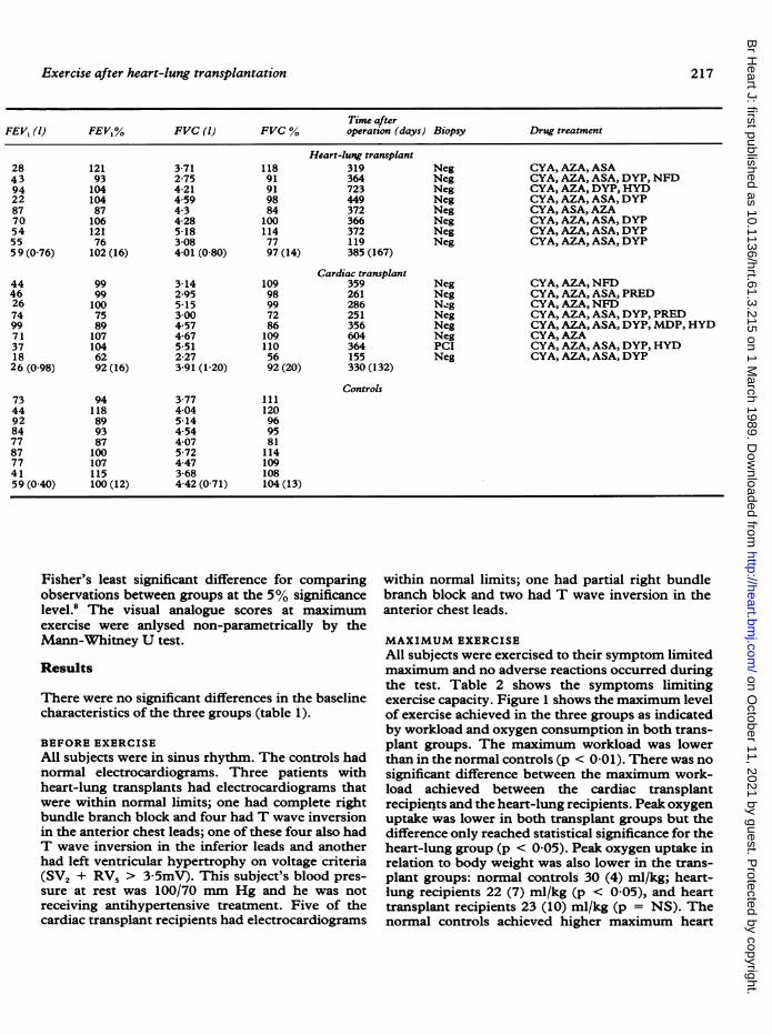
FEVI, forced expiratory volume in one second; FVC, forced vital capacity; FEV,% and FVC%, results expressed as percentage of predicted5;neg, no histological evidence of rejection; PCI, patchy cellular infiltrate, no myocytolysis (cyclosporin effect); CYA, cyclosporin; AZA, azathiASA, aspirin; DYP, dipyridamole; PRED, prednisone; NFD, nifedipine; HYD, hydralazine; MDP, methyldopa; DCM, dilated cardiomyVSD, ventricular septal defect.
minute until symptom limited maximum exercise.The subjects breathed via a mouthpiece through aHans Rudolph valve box (Hans Rudolph, KansasCity, USA). Tidal volume, respiratory frequency,and minute ventilation were measured by a turbineventilation monitor (PK Morgan, Rainham, UK).Mixed expired oxygen and carbon dioxide concen-trations were measured by sampling from a mixingchamber by a computer assisted mass spectrometer(Spectralab "M", VG Medical, Middlewich, UK)and used to calculate oxygen consumption andcarbon dioxide production.6 End tidal carbon dioxideconcentrations were measured at the mouth with acatheter probe connected to the mass spectrometer.The presence of a plateau in the expiratory carbondioxide tracing was confirmed by a chart recorder;the shape of the tracings was similar in the threegroups. Arterial oxygen saturation was recordedcontinuously with a ear oximeter (Biox III PulseOximeter, Ohmeda, Harlow, UK).Blood pressure was recorded with a sphygmoman-
ometer. Twelve lead electrocardiograms were recor-ded on a Marquette MAC I exercise electrocar-diograph system (Marquette, Manchester, UK),
which was also used to measure heart rate. Averagevalues for heart rate and each respiratory variablewere measured for each minute. Blood pressure and12 lead electrocardiograms were recorded at rest andat the end of each three minutes of exercise. Thesubjects quantified their sensation of breathlessnessat the end of each minute on a visual analogue scale.7Ahand controlled linear potentiometer, mounted in aconvenient position on the handlebars, allowed thepatients to indicate their degree of breathlessness bymoving a light along a 10 cm linear display, the endsof which were marked "not at all breathless" and"extremely breathless". At the end of the test eachperson was asked to report the symptoms limitingtheir exercise capacity.
STATISTICAL ANALYSISThe baseline characteristics ofthe three groups werecompared by analysis of variance. The results aregiven as mean (SD). Statistical analysis was perfor-med by Student's t test for unpaired data and twoway analysis ofvariance. The level ofsignificance wastaken asp < 0-05 in a two tailed test. Graphs showingthe data subjected to analysis of variance include
216
on October 11, 2021 by guest. P
rotected by copyright.http://heart.bm
j.com/
Br H
eart J: first published as 10.1136/hrt.61.3.215 on 1 March 1989. D
ownloaded from
Exercise after heart-lung transplantation
Time afterFEV, (1) FEV,% FVC (1) FVC % operation (days) Biopsy Drug treatment
Heart-lung transplant28 121 3-71 118 319 Neg CYA,AZA,ASA43 93 2-75 91 364 Neg CYA, AZA, ASA, DYP, NFD94 104 4 21 91 723 Neg CYA, AZA, DYP, HYD22 104 4.59 98 449 Neg CYA, AZA, ASA, DYP87 87 4-3 84 372 Neg CYA,ASA,AZA70 106 4-28 100 366 Neg CYA,AZA,ASA,DYP54 121 5-18 114 372 Neg CYA,AZA,ASA,DYP55 76 3-08 77 119 Neg CYA, AZA, ASA, DYP59(0.76) 102 (16) 4.01 (0.80) 97 (14) 385 (167)
Cardiac transplant44 99 3-14 109 359 Neg CYA, AZA, NFD46 99 2-95 98 261 Neg CYA, AZA, ASA, PRED26 100 5-15 99 286 Neg CYA,AZA, NFD74 75 3-00 72 251 Neg CYA, AZA, ASA, DYP, PRED99 89 4-57 86 356 Neg CYA, AZA, ASA, DYP, MDP, HYD71 107 4-67 109 604 Neg CYA,AZA37 104 5-51 110 364 PCI CYA, AZA, ASA, DYP, HYD18 62 2-27 56 155 Neg CYA, AZA, ASA, DYP26 (0.98) 92 (16) 3-91 (1.20) 92 (20) 330 (132)
Controls73 94 3.77 11144 118 4.04 12092 89 5-14 9684 93 4-54 9577 87 4 07 8187 100 5-72 11477 107 4-47 10941 115 3-68 10859 (0.40) 100(12) 4-42 (0-71) 104 (13)
Fisher's least significant difference for comparingobservations between groups at the 5% significancelevel.8 The visual analogue scores at maximumexercise were anlysed non-parametrically by theMann-Whitney U test.
Results
There were no significant differences in the baselinecharacteristics of the three groups (table 1).
BEFORE EXERCISEAll subjects were in sinus rhythm. The controls hadnormal electrocardiograms. Three patients withheart-lung transplants had electrocardiograms thatwere within normal limits; one had complete rightbundle branch block and four had T wave inversionin the anterior chest leads; one of these four also hadT wave inversion in the inferior leads and anotherhad left ventricular hypertrophy on voltage criteria(SV2 + RV5 > 3.5mV). This subject's blood pres-sure at rest was 100/70 mm Hg and he was notreceiving antihypertensive treatment. Five of thecardiac transplant recipients had electrocardiograms
within normal limits; one had partial right bundlebranch block and two had T wave inversion in theanterior chest leads.
MAXIMUM EXERCISEAll subjects were exercised to their symptom limitedmaximum and no adverse reactions occurred duringthe test. Table 2 shows the symptoms limitingexercise capacity. Figure 1 shows the maximum levelof exercise achieved in the three groups as indicatedby workload and oxygen consumption in both trans-plant groups. The maximum workload was lowerthan in the normal controls (p < 0.01). There was nosignificant difference between the maximum work-load achieved between the cardiac transplantrecipients and the heart-lung recipients. Peak oxygenuptake was lower in both transplant groups but thedifference only reached statistical significance for theheart-lung group (p < 0.05). Peak oxygen uptake inrelation to body weight was also lower in the trans-plant groups: normal controls 30 (4) ml/kg; heart-lung recipients 22 (7) ml/kg (p < 0 05), and hearttransplant recipients 23 (10) ml/kg (p = NS). Thenormal controls achieved higher maximum heart
217
on October 11, 2021 by guest. P
rotected by copyright.http://heart.bm
j.com/
Br H
eart J: first published as 10.1136/hrt.61.3.215 on 1 March 1989. D
ownloaded from

Banner, Lloyd, Hamilton, Innes, Guz, Yacoub
200'
3150-la0-x0
E 100-
xa
50-
Oa
3000-
** **
00
so00
HLT
Om
- 2500-
c 2000-
0.E' 1500.c0
:>. 1000-x0
a.. 500-
nJCT N
0
*
0
0
HLT
0~~0**
* 00
0
0 0
0
CT N
120-
0
*0 *
0000
100-
= w
C. _
_ 0
*. 60-
E.O-a
20-0
VHLT CT N
100-.
* *
0
0
0
0
_ 80-
'A 60-Inin
cIn
~ 40-n
20
-q20-0
HLT
i
O aCT N
0
0
*0
0 0
* o s
0
HLT CT
0
*00
N
HLT CT N
Fig 1 Maximum exercise response in heart-lung transplant recipients (HLT); orthotopic cardiac transplant recipients(CT); normal controls (N). *p < 0-05; **p < 0-01; ***p < 0-001 for unpaired t tests (comparison with normal controls).VAS, visual analogue scale.
200-
4,av
.0, 160--0
4l-a
E= 120-a
n0o
220-
I 200-EE-
SOe 180-Win
0f 160-00.0u 140-
OA 120-EE
100u-
80--
0*090
n,_
218
%F
on October 11, 2021 by guest. P
rotected by copyright.http://heart.bm
j.com/
Br H
eart J: first published as 10.1136/hrt.61.3.215 on 1 March 1989. D
ownloaded from
Exercise after heart-lung transplantation
Table 2 Symptoms limiting exercise capacity during test
Heart-lung Cardiactransplant transplant Normal
Fatigue 2 6 6Dyspnoea 2 1 1Both 4 1 1
rates than either ofthe transplant groups (p < 0-001).There was no significant difference in maximum
ventilation. Maximum systolic blood pressure was
higher in the normal subjects than in either trans-
plant group (p < 0-05). There was no significantdifference in breathlessness at maximum exercise, as
measured by the visual analogue scale, between thenormal subjects and either transplant group (Mann-Whitney U test).
PATTERN OF EXERCISE RESPONSEWe examined the differences in the incrementalresponse to exercise in the three groups by a two way
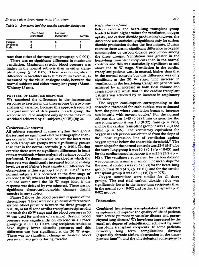
analysis of variance. Because this approach requiredan equal number of data points in each group, theresponse could be analysed only up to the maximumworkload achieved by all subjects (50 W) (fig 2).
Cardiovascular response
All subjects remained in sinus rhythm throughoutthe test and no significant electrocardiographic chan-ges occurred during exercise. At rest the heart ratesof both transplant groups were significantly greaterthan that in the normal controls (p < 0.01). Duringexercise there were no significant differences in heartrates at workloads where analysis ofvariance could beperformed. To determine the workload at which theheart rate was significantly increased from the restinglevel, we used Fisher's least significant difference forobservations within a group (for p < 0-05).8 In thenormal subjects this occurred at the first stage ofexercise (10 W) whereas in both transplant groups itdid not occur until the 30 W stage (that is theresponse was delayed by two minutes). There was nosignificant electrocardiographic changes duringexercise in any subject.
Figure 2 shows the blood pressure response of thethree groups. There were no significant differences insystolic blood pressure between the three groups atrest, 30, or 60W (one cardiac transplant recipient didnot reach the 60W stage and the blood pressure at 50W was used for analysis of variance). Systolic bloodpressure rose significantly on exercise in all threegroups (p < 0-001). Heart-lung recipients tended tohave slightly lower diastolic pressures and thisdifference was just significant at the 30 W stage.There was no significant change in diastolic bloodpressure in any group during exercise.
219
Respiratory responseBefore exercise the heart-lung transplant grouptended to have higher values for ventilation, oxygenuptake, and carbon dioxide production; however, thedifference was statistically significant only for carbondioxide production during the first minute. Duringexercise there was no significant difference in oxygenconsumption or carbon dioxide production amongthe three groups. Ventilation was greater in theheart-lung transplant recipients than in the normalcontrols and this was statistically significant at andabove the 30 W stage. Ventilation in the cardiactransplant patients was, in general, higher than thatin the normal controls but this difference was onlysignificant at the 30 W stage. The increase inventilation in the heart-lung transplant patients wasachieved by an increase in both tidal volume andrespiratory rate while that in the cardiac transplantpatients was achieved by an increase in respiratoryrate alone.The oxygen consumption corresponding to the
anaerobic threshold for each subject was estimatedfrom the point where ventilation began to increasenon-linearly with oxygen uptake.9 For the normalsubjects this was 1-43 (0-38) 1/min oxygen; for theheart-lung group it was 1-0 (0-32) 1/min (p = NS),and for the cardiac transplant group it was 1-2 (0A46)1/min (p = NS). The ventilatory equivalent foroxygen in each person was obtained from the slope ofthe linear regression line of ventilation againstoxygen uptake below the anaerobic threshold.'0 Themean slope for the normal controls was 23-9 (5 5); forthe heart-lung group it was 30 8 (6- 1) (p < 0 05), andfor the cardiac transplant group it was 25-8 (3 2) (p =NS). The ventilatory equivalent for carbon dioxidewas obtained in a similar manner. The mean slope forthe normal controls was 23-5 (3 2); for the heart-lunggroup it was 30 5 (4 7) (p < 0-01), and for the cardiactransplant group it was 27-1 (3.8) (p = NS).Oxygen saturations were similar for all three
groups. The end tidal carbon dioxide value wassignificantly lower in the heart-lung recipients thanin the normal (p < 0 02) and cardiac transplant (p <0-02) groups.
Discussion
Combined heart-lung transplantation can alleviatesymptoms and improve the quality of life of patientswith severe pulmonary vascular disease and paren-chymal lung disease.' We have been impressed by theexcellent degree of rehabilitation achieved by mostheart-lung transplant recipients. In some patients,however, long term complications develop(especially chronic airflow obstruction in the trans-planted lung"), and the physiological consequences
on October 11, 2021 by guest. P
rotected by copyright.http://heart.bm
j.com/
Br H
eart J: first published as 10.1136/hrt.61.3.215 on 1 March 1989. D
ownloaded from

Banner, Lloyd, Hamilton, Innes, Guz, Yacoub
0--z0Workload (W)
0 10 20 302 .
W.WL S DLSDp
.c 30
e 20-x-Sc. 10-
4.'I
0
"', ~0
0~~~~~~~~
aS
ll:~~~~ LSD
o -
4,.2
~0.0xOaLU
cm 45.0- O) O -~O-0-Q-. E
*t 40-LSD X.8
a 35
c 30-
2 3 4 5 6 7
Time (min)
0
8-g~ ILSD_O -
F-0- -
1 2 3 4Time (min)
5 6 7
Fig 2 Evolution of the exercise response in heart-lung transplant recipients ( *), cardiac transplant recipients ( A ), andnormal controls (0). LSD, Fisher's least significant differencefor observations between groups (p < 0-05).
220
120
.E.f 100-
.0
-Na80-
I
sn I
'1602
E
y 140-LSD in
n
C 120-
8D0 100-.,
aO80
40 52
0
L
eLSD~~~~~~~~~~S
i
01I
90
in4,
CU0.-o 70z.00-o
-57a 50C3c1000T
E 750-Q
E 5003
UAC0a 250-0IUxo o0
1500-
E
.' 1000'
-5*.
5O--
100
_95.Q0
-a 90U,
co0,.M 85K0
0
i
A
-.
on October 11, 2021 by guest. P
rotected by copyright.http://heart.bm
j.com/
Br H
eart J: first published as 10.1136/hrt.61.3.215 on 1 March 1989. D
ownloaded from

Exercise after heart-lung transplantationof the procedure have not been fully defined. Thisstudy was designed to compare the exercise capacityand pattem of exercise response ofa group ofhealthyheart-lung transplant recipients with those of cardiactransplant recipients and normal controls. We selec-ted transplant recipients who had no serious com-plications after transplantation.Our results confirm the findings of a previous
study which showed that recipients of heart-lungtransplants had an exercise capacity that was less thanthat predicted for normal controls.4 In addition, wehave established that their exercise capacity is similarto that ofmatched cardiac transplant recipients. Thereduced exercise capacity of patients with orthotopiccardiac transplants has already been shown.2 13Several factors may contribute to the reduced exer-cise capacity after heart and heart-lung transplanta-tion.
Illness before transplantation will have produced aperiod of deconditioning and muscle wasting thatmay persist after operation. A recent study showed adelayed recovery in the vasodilator response ofskeletal muscle after cardiac transplantation. Thismay reflect a persistent effect of preoperative cardiacfailure and deconditioning, which could affect exer-cise capacity.'4 A previous study of cardiac transplantrecipients at our hospital found them to have areduced lean body mass and exercise capacity com-pared with normal controls. A programme of regularexercise training improved exercise capacity andincreased lean body mass although their exercisecapacity was still less than that of controls.'3There is the possibility of residual left ventricular
dysfunction related to the process of brain death inthe donor,'5 the organ preservation at the time oftransplantation, and subsequent episodes of cardiacrejection. We found that the systolic ventricularfunction of cardiac transplant recipients treated withcyclosporin was, however, usually normal.'6The heart-lung transplant operation'7 has several
physiological consequences. The heart and lungs aretransplanted as a single block replacing the recipi-ent's diseased organs. Vascular anastomoses aremade between the donor right atrium and a remnantofthe recipient's right atrium and between donor andrecipient ascending aorta (the donor left atriumremains intact). The tracheal anastomosis is madebetween the lower part of the recipient's trachea andthe donor trachea just above the carina. The pro-cedure results in acute denervation of the transplan-ted heart and lungs. The recipient retains trachealinnervation down to the level of this anastomosis andthe recurrent laryngeal nerves remain intact. Thepulmonary lymphatic system and bronchial circula-tion are sacrificed.
Orthotopic cardiac transplantation results in com-
221plete denervation of the transplanted heart but thepulmonary innervation, lymphatic system, and thebronchial circulation are not affected. Cardiac rein-nervation has not been seen in patients after ortho-topic cardiac transplantation'8 although evidence ofefferent reinnervation has been found in recipients atheterotopic transplants.'9 In dogs cardiac and pul-monary reinnervation can occur after transplantationof the heart20 or lung.2' The sinus tachycardia beforeexercise together with the identical, abnormal, heartrate response during exercise of the heart-lung andheart transplant recipients studied here provideevidence of persistent cardiac denervation. It hasbeen difficult to show pulmonary denervation inheart-lung transplant recipients non-invasivelybecause of the presence of an innervated trachea.Recipients of heart-lung transplants do not have anincreased cough threshold when tested with a citricacid aerosol (Banner N R et al, unpublished). Theydo, however, have increased airways responsivenessto methacholine. This may represent a denervationhypersensitivity of muscarinic receptors in the air-ways of the transplanted lung.22 23The difference in exercise capacity between the
transplant groups and normal individuals was notcaused by altered mechanical or metabolic efficiencyas the relation of oxygen consumption and carbondioxide production to workload was the same in allthree groups. Because the two transplant groupsshowed a similar performance the overall limitationis likely to be related to the circulation and thetransplanted heart rather than to the transplantedlungs. This view is supported by the ventilatoryresponse, which provided indirect evidence ofanaerobic metabolism developing during exercise inall three groups in this study, and the increasedlactate concentrations found in recipients of heart-lung transplants at maximum exercise in anotherstudy.4The abnormal response of heart rate to exercise
may contribute to the reduced exercise capacity ofthe transplant recipient. The increased resting heartrate in the transplant recipients is caused by a loss ofvagal tone.'8 The delayed response ofheart rate at thestart of exercise is associate with an impaired initialcardiac output response.24 All three groups, however,had similar heart rates during the first few minutes ofexercise in this study. The lower peak heart ratesachieved by the transplant recipients might limitmaximum cardiac output or they may just reflect thelower workloads reached by the transplantrecipients. The cardiac output of heart transplantrecipients (measured under steady state conditionsduring submaximal exercise) is normal in relation tooxygen uptake.25 The slow response ofheart rate mayplace the transplant patient at a particular disadvan-
on October 11, 2021 by guest. P
rotected by copyright.http://heart.bm
j.com/
Br H
eart J: first published as 10.1136/hrt.61.3.215 on 1 March 1989. D
ownloaded from

222tage during an incremental exercise test. A previousstudy found that the cardiac output response ofrecipients of heart transplants to exercise was causedby a prominent rise in stroke volume and a slow risein heart rate; because cardiac output was low inrelation to oxygen uptake there was an increase in thearteriovenous oxygen difference.26 Exercise trainingimproves the response of heart rate to exercise incardiac transplant recipients and the improvement isassociated with an increased exercise capacity.'3Cardiac denervation may affect cardiac function inother ways. Contractility and the contractile reserveof the left ventricle in the transplanted heart seemnormal when tested pharmacologically27 but theeffects of chronic cardiac denervation on left ven-tricular performance during exercise are unknown.Hypertension is a common complication of cyclo-
sporin treatment in heart transplant recipients.' '9Four heart and two heart-lung recipients in thisstudy were on antihypertensive treatment. Thesystolic blood pressure response to exercise wassimilar in all three groups.
Cardiac transplant recipients have been reportedto have increased ventilation in relation to oxygenuptake during exercise." The cardiac transplantgroup in the present study had a slightly increasedventilation compared with the normal controls butthe difference was not statistically significant.Analysis by linear regression of ventilation againstoxygen uptake shows that the response was close tothe published values for a similar exercise protocol.'0The end tidal carbon dioxide concentrations werenormal in the cardiac transplant group, confirmingthat alveolar ventilation was normal. The relativelynormal ventilatory response in these patients may bebecause of the improvement in ventricular functionthat we saw in cardiac transplant recipients treatedwith cyclosporin compared with conventionalimmunosuppression.'6 In addition, the ventilatoryresponse of cardiac transplant recipients is related totheir physical fitness and can be altered by exercisetraining."
In contrast, both analysis of variance at equivalentworkloads or by linear regression of ventilationagainst oxygen below the anaerobic thresholdshowed an increased ventilatory response in theheart-lung group; this was owing to a combination ofincreased respiratory rate and tidal volume. A case ofincreased ventilation during exercise in a heart-lungtransplant recipient was reported while the presentwork was in progress.'0The increased ventilatory response in our study
did not seem to be the result of poor pulmonary gasexchange. Oximetry showed that the heart-lunggroup did not desaturate during exercise and endtidal carbon dioxide concentrations were low, sug-
Banner, Lloyd, Hamilton, Innes, Guz, Yacoubgesting that true alveolar hyperventilation was occur-ring. It may be that the increased ventilation and lowend tidal carbon dioxide concentrations were theresult ofregions ofhigh ventilation and low perfusionin the transplanted lungs. The heart-lung group hadgood baseline lung function, however, which makesthis explanation unlikely. Another study found lowarterial carbon dioxide tensions in heart-lung trans-plant recipients before and during exercise althoughit did not compare them with those in normalcontrols.4 The alveolar hyperventilation might becaused by the sluggish heart rate response with theearly development ofa lactic acidosis during exercise.The small increase in ventilation seen in the cardiactransplant group who had a similar exercise capacityand cardiovascular response to exercise does notsupport this idea. Drug treatment was similar in thetwo transplant groups so the increased ventilation isnot the result of medication.The mechanisms controlling exercise hyperpnoea
are complex and poorly understood. Most of theattention in this area has been focused on mechan-isms stimulating ventilation rather than those res-training it.3' Our results suggest that an increasedventilatory response to exercise occurs after pulmon-ary denervation, but not after cardiac denervation.We found that the ventilatory response to carbondioxide was increased in heart-lung transplantrecipients.32 A possible explanation is that theincreased ventilatory response to these stimuli iscaused by the loss ofnegative feedback from pulmon-ary afferents in the heart-lung recipients.The heart-lung recipients were still able to per-
ceive breathlessness during exercise and the max-imum breathlessness scores were similar in the threegroups. This indicates that pulmonary afferents donot make an essential contribution to the sensation ofexertional dyspnoea.
In conclusion, heart-lung and heart transplantrecipients have a similar exercise capacity and car-diovascular response to exercise. The altered car-diovascular response is partly the result of cardiacdenervation and their exercise capacity seemed to belimited by circulatory performance. The heart-lungtransplant recipients also show an increased ven-tilatory response during submaximal exercise, whichmay be caused by a loss ofnegative feedback from thepulmonary afferents. Exercise capacity after heart-lung transplantation is sufficient for everydayactivities and transplantation can provide excellentrehabilitation of patients with end stage cardiopul-monary disease.
We thank Dr Ken MacRae for statistical advice.
on October 11, 2021 by guest. P
rotected by copyright.http://heart.bm
j.com/
Br H
eart J: first published as 10.1136/hrt.61.3.215 on 1 March 1989. D
ownloaded from

Exercise after heart-lung transplantation 223
References
1 Reitz BA, Wallwork JL, Hunt SA, et al. Heart lungtransplantation: successful therapy for patients withpulmonary vascular disease. N Engl J Med1982;306:557-64.
2 Penketh A, Higenbottam T, Hakim M, Wallwork J.Heart-lung transplantation in patients with end stagelung disease. Br Med J 1987;295:311-4.
3 Yacoub MH, Banner NR, Gibson S, Thakrar SA,Khaghani A. A quantitative assessment of the qualityof life after combined heart-lung transplantation[Abstract]. JAm Coil Cardiol 1987;9:30A.
4 Theodore J, Morris AJ, Burke CM, et al. Cardiopul-monary function at maximal tolerable constant workrate exercise following human heart-lung transplan-tation. Chest 1987;92:433-9.
5 Quanjer Ph H, ed. Standardized lung function testing.Bull Eur Physiopath Resp 1983;19 (suppl 5):45-51.
6 Jones NL, Campbell EJM. Clinical exercise testing.Philadelphia, WB Saunders, 1982:231-9.
7 Adams L, Chronos N, Lane R, Guz A. Themeasurement of breathlessness in normal subjects:validity of two scaling techniques. Clin Sci 1985;69:7-16.
8 Fisher RA. The design ofexperiments. Edinburgh: Oliverand Boyd, 1935.
9 Wasserman K, Whipp BJ, Koyal SN, Beaver WL.Anaerobic threshold and respiratory gas exchangeduring exercise. J Appl Physiol 1973;35:236-43.
10 Spiro SG, Juniper E, Bowman P, Edwards RHT. Anincreasing work rate test for assessing thephysiological strain of submaximal exercise. Clin Sci1974;46:191-206.
11 Burke CM, Theodore J, Dawkins KD, et al. Posttransplant obliterative bronchiolitis and other latelung sequelae in human heart-lung transplantation.Chest 1984;86:824-9.
12 Savin WM, Haskell WL, Schroeder JS, Stinson EB.Cardiorespiratory responses of cardiac transplantpatients to graded, symptom limited exercise. Circ-ulation 1980;62:55-60.
13 Kavanagh T, Yacoub MH, Mertens DJ, Kennedy J,Campbell RB, Sawyer P. Cardiorespiratory responsesto exercise training after orthotopic transplantation.Circulation 1988;77:162-71.
14 Sinoway LI, Minotti JR, Davis D, et al. Delayedreversal of impaired vasodilation in congestive heartfailure after heart transplantation. Am J Cardiol1988;61:1076-9.
15 De Pasquale NP, Burch GE. How normal is the donorheart? Am Heart J 1969;77:719-20.
16 Reid CJ, Yacoub MH. Determinants of left ventricularfunction one year after cardiac transplantation. BrHeart J 1988;59:397-402.
17 Jamieson SW, Stinson EB, Oyer PE, Baldwin JC,Shumway NE. Operative technique for heart-lung
transplantation. J Thorac Cardiovasc Surg 1984;87:930-5.
18 Horak AR. Physiology and pharmacology of the trans-planted heart. In: Cooper DKC, Lanza RP, eds.Heart transplantation. Lancaster: MTP Press,1984: 147-56.
19 Mitchell AG, Yacoub M. Efferent autonomic rein-nervation after heterotopic cardiac transplantation inhumans [Abstract]. Br Heart J 1987;57:87.
20 Kontos HA, Thames MD, Lower RR. Response toelectrical and reflex autonomic stimulation in dogswith cardiac transplantation before and after rein-nervation. J Thorac Cardiovasc Surg 1970;59:382-92.
21 Edmunds LH, GrafPD, Nadel JA. Reinnervation ofthereimplanted canine lung. J Appl Physiol 1971;31:722-7.
22 Banner NR, Hollingshead L, Dear I, Heaton R, Guz A,YacoubMH. Increased bronchial reactivity to metha-choline following combined heart lung transplanta-tion [Abstract]. Thorax 1987;42:239-40.
23 Glanville AR, Burke CM, Theodore J, et al. Bronchialhyper-responsiveness after human cardiopulmonarytransplantation. Clin Sci 1987;73:299-303.
24 Banner NR, Guz A, Heaton R, Innes JA, Murphy K,Yacoub M. Ventilatory and circulatory responses atthe onset of exercise in man following heart or heart-lung transplantation. J Physiol (Lond) 1988;399:437-49.
25 Mertens D, Kavanagh T, Yacoub M. Long termexercise training and cardiac output following cardiactransplantation [Abstract]. J Am Coll Cardiol 1987;9:235.
26 Stinson EB, Griepp RB, Schroeder JS, Dong E, Shum-way NE. Haemodynamic observations one and twoyears after cardiac transplantation in man. Circulation1972;45:1183-94.
27 Borow KM, Neumann A, Arensman FW, Yacoub MH.Left ventricular contractility and contractile reservein humans after cardiac transplantation. Circulation1985;71:866-72.
28 Goldman MH, Barnhart G, Mohanakumar T, et al.Cyclosporine in cardiac transplantation. Surg Clin NAm 1985;65:637-59.
29 Banner NR, Fitzgerald M, Khaghani A, et al. Cardiactransplantation at Harefield Hospital. In: Terasaki P,ed. Clinical transplants 1987. Los Angeles: UCLATissue Typing Laboratory, 1987:17-26.
30 Estenne M, Primo G, Yernault JC. Cardiorespiratoryresponses to exercise after human heart-lung trans-plantation. Thorax 1987;42:629-30.
31 Whipp BJ. The control of exercise hyperpnea. InHornbein TF, ed. Regulation of breathing (part 2).New York, Marcel Dekker, 1981:1069-139.
32 Banner NR, Lloyd MH, Lane R, Guz A, Yacoub MH.Ventilatory response to progressive hypercapnicstimulation following combined heart lung transplan-tation in man [Abstract]. Clin Sci 1977;73 (suppl17):6P.
on October 11, 2021 by guest. P
rotected by copyright.http://heart.bm
j.com/
Br H
eart J: first published as 10.1136/hrt.61.3.215 on 1 March 1989. D
ownloaded from