cardiac arrest: vf/pvt
TRANSCRIPT

Cardiac Arrest: VF/pVT
Overview
To be successful, any resuscitation attempt needs a strong base of high-quality CPR and defibrillation when the patient’s ECG rhythm requires it. Leaders must also assess the performance of each system component, ensuring that system participants can effectively intervene to improve care. This process of quality improvement consists of an iterative and continuous cycle of
• Systematic evaluation of resuscitation care and outcome • Benchmarking with stakeholder feedback • Strategic efforts to address identified deficiencies
Another characteristic of high-quality CPR is minimal interruptions in chest compressions. Studies demonstrate that healthcare providers interrupt compressions far too often and for too long, in some cases spending 25% to 50% of a resuscitation attempt without delivering chest compressions.
Chest compression fraction (CCF) is the proportion of time during cardiac arrest resuscitation when the rescuer is performing chest compressions. CCF should be as high as possible: ideally greater than 80%. Data suggest lower CCF is associated with decreased ROSC and survival to hospital discharge.
Measurement
Quality improvement relies on valid assessment of resuscitation performance and outcome (refer to the Utstein guidelines in Part 1: Systems of Care).
• Share information among all links in the system of care, including o –Dispatch records o –EMS patient care report o –Hospital records
Benchmarking and Feedback
Systematically review and compare data internally to previous performance and externally to similar systems. Existing registries can help this benchmarking effort. Examples include the
• CARES for OHCA • Get With The Guidelines®-Resuscitation program for IHCA
Change
By simply measuring and benchmarking care, systems can positively influence outcome, but they’ll also need ongoing review and interpretation to identify areas for improvement, such as
• Citizen awareness • Citizen and healthcare professional education and training • Increased bystander CPR response rates • Improved CPR performance • Shortened time to defibrillation
Rhythms for VF/pVT • VF (example in Figure 40) • VT • ECG artifact that looks like VF • New LBBB

Figure 40. Example of VF.
Drugs for VF/pVT • Drugs for VF/pVT include • Epinephrine • Amiodarone • Lidocaine • Magnesium sulfate • Dopamine • Oxygen • Other medications, depending on the cause of the VT/pVT arrest
Managing VF/pVT: The Adult Cardiac Arrest Algorithm
You must know the most important algorithm for adult resuscitation: the Adult Cardiac Arrest Algorithm (Figure 41). This algorithm outlines all the steps to assess and manage a pulseless patient who does not initially respond to BLS interventions, including a first shock from an AED. The algorithm consists of the 2 pathways for cardiac arrest:
• A shockable rhythm, displayed on the VF/pVT pathway of the algorithm • A nonshockable rhythm, displayed on the asystole/PEA pathway of the algorithm


Figure 41. Adult Cardiac Arrest Algorithm, VF/pVT pathway.
Throughout the case discussion of the Adult Cardiac Arrest Algorithm, we will refer to Steps 1 through 12. These are the numbers assigned to the steps in the algorithm.
VF/pVT Path
Because many patients with sudden cardiac arrest demonstrate VF at some point in their arrest, most ACLS providers will often follow the VF/pVT pathway of the Adult Cardiac Arrest Algorithm (Figure 41). Rapidly treating VF according to this sequence is the best approach to restoring spontaneous circulation.
The algorithm includes pVT because it is treated as VF. VF and pVT require CPR until a defibrillator is available to deliver high-energy unsynchronized shocks.
Asystole/PEA Path
The asystole/PEA pathway of the algorithm outlines the sequence of actions to perform if the rhythm is nonshockable. You will practice this sequence in the Asystole and PEA Cases.
During the VF/pVT Case, you will practice performing rapid treatment on the VF/pVT pathway in the Adult Cardiac Arrest Algorithm.
Applying the Adult Cardiac Arrest Algorithm: VF/pVT Pathway
For this algorithm, healthcare providers should have already completed the BLS Assessment, including activating the emergency response system, performing high-quality CPR, attaching the manual defibrillator, and delivering the first shock (Steps 1 through 4). Now, the ACLS high-performance team intervenes and conducts the Primary Assessment. In this case, the team assesses the patient and takes actions as needed. The Team Leader coordinates the efforts of the high-performance team as they complete the steps listed in the VF/pVT pathway of the Adult Cardiac Arrest Algorithm.
Caution: Agonal Gasps • Agonal gasps may be present in the first minutes after sudden cardiac arrest. • Agonal gasps are not normal breathing.
A patient who gasps usually appears to be drawing air in very quickly. The mouth may be open and the jaw, head, or neck may move with gasps. Gasps may appear forceful or weak. Some time may pass between gasps because they usually happen at a slow, irregular rate. The gasp may sound like a snort, snore, or groan. Gasping is a sign of cardiac arrest.
Start CPR • Start CPR (Step 1)
The initial step in the Adult Cardiac Arrest Algorithm is to start CPR. As soon as the patient is found to be unresponsive with no breathing (or only gasping), shout for nearby help and activate the emergency response system, send for a defibrillator, check for a pulse, and start CPR, beginning with chest compressions. Attach the ECG monitor or AED pads as soon as they are available. Throughout the resuscitation attempt, provide high-quality CPR (give chest compressions of adequate rate and depth, allow complete chest recoil after each compression, minimize interruptions in compressions, and avoid excessive ventilation).
• Give oxygen. • Attach the monitor/defibrillator.
Once the monitor/defibrillator is attached, check the rhythm to determine whether it is shockable (VF/pVT) or nonshockable (asystole/PEA) and follow the appropriate cardiac arrest pathway.
Minimize Interruption of Chest Compressions
A team member should continue to perform high-quality CPR until someone brings the defibrillator and attaches it to the patient. The Team Leader assigns roles and responsibilities and organizes interventions to minimize interruptions in chest compressions. This accomplishes the most critical interventions for VF or pVT: CPR with minimal interruptions in chest compressions and defibrillation during the first minutes of arrest. CPR quality should be measured in real time with an audiovisual feedback device, including CCF and quantitative waveform capnography, that captures the following information:
• Rate: 100 to 120/min • Depth: at least 2 inches (5 cm) • Chest recoil • CCF: ideally greater than 80% • Time to first defibrillation • Time to first compression
Calculating CCF
Healthcare providers can calculate CCF using a feedback device, or they can calculate it manually by using 2 timers. Use one timer to measure the total code time, from code start until code stop, or until ROSC. Use a second timer to measure the total chest compression time. Each time chest compressions are stopped, pause the second timer until chest compressions are resumed. To calculate CCF, divide chest compression time by the total code time.
CCF = Actual chest compression time ÷ Total code time
The AHA does not recommend continued use of an AED (or the automatic mode) when a manual defibrillator is available and providers can adequately interpret rhythms. Rhythm analysis and shock administration with an AED may prolong the interruptions in chest compressions.
Additionally, while the manual defibrillator is charging, providers should resume CPR. Shortening the interval between the last compression and the shock by even a few seconds can improve shock success (defibrillation and ROSC), so practice efficient coordination between CPR and defibrillation.
For example, after you verify a shockable rhythm and initiate the charging sequence on the defibrillator, another provider should resume chest compressions and continue until the defibrillator is fully charged. You should deliver the shock as soon as the compressor removes his or her hands from the patient’s chest and all providers are “clear” of contact with the patient. The same compressor should resume compressions immediately after the shock is delivered.
Note: although manual defibrillators can shorten the interruption needed for rhythm analysis, providers who are inexperienced with rhythm analysis should use an AED instead to avoid delays or inappropriate shocks.
Figure 42 illustrates the need to minimize interruptions in compressions. Coronary perfusion pressure (CPP) is aortic relaxation (“diastolic”) pressure minus right atrial relaxation (“diastolic”) pressure. During CPR, CPP correlates with both myocardial blood flow and ROSC. In 1 human study, ROSC did not occur unless a CPP 15 mm Hg or greater was achieved during CPR.

Figure 42. Relationship of quality CPR to CPP demonstrating the need to minimize interruptions in compressions.
Defibrillate (Shockable Rhythm: VF/pVT)
As soon as you determine that the rhythm is shockable (VF or pVT), deliver 1 shock. The appropriate energy dose is determined by the identity of the defibrillator—monophasic or biphasic.
If you are using a monophasic defibrillator, give a single 360-J shock. Use the same energy dose for subsequent shocks.
Biphasic defibrillators use various waveforms that effectively terminate VF over a specific dose range. When using biphasic defibrillators, providers should use the manufacturer’s recommended energy dose (eg, initial dose of 120 to 200 J). Many biphasic defibrillator manufacturers display the effective energy dose range on the face of the device. If you do not know the effective dose range, deliver the maximal energy dose for the first and all subsequent shocks.
If the initial shock terminates VF but the arrhythmia recurs later in the resuscitation attempt, deliver subsequent shocks at the previously successful energy level.
For an AED, follow the device’s prompts or know your device-specific manufacturer’s recommendations. Healthcare providers should know how their defibrillator operates and limit pauses in chest compressions to rhythm analysis and shock delivery.
Immediately after the shock, resume CPR, beginning with chest compressions. Give 2 minutes of CPR. If there are available providers, IV or IO access should be established.
In adults experiencing sudden cardiac arrest due to VF or pVT, the heart is quivering but is not effectively pumping blood to vital organs. These patients have a much higher survival rate if they receive immediate chest compressions and early defibrillation. Timing is critical. Defibrillation stuns the heart—it doesn’t restart the heart—to briefly terminate all electrical activity, including VF and pVT. If the heart is still viable, defibrillation may help the heart’s normal pacemakers eventually resume electrical activity (return of spontaneous rhythm) that ultimately results in a perfusing rhythm (ROSC).
In the first 4 to 6 minutes after cardiac arrest, referred to as clinical death, no damage occurs to the brain. In the 6- to 10-minute period (biological death) after cardiac arrest, damage is likely to occur to the brain. Brain damage is usually irreversible after 10 minutes, except in special circumstances such as accidental hypothermia and cold-water drowning. Starting chest compressions immediately can delay these effects, and defibrillation can restore a perfusing rhythm. Again, time is critical. A defibrillator should be used as soon as it is available. If there are 2 or more providers present, CPR should be performed while the defibrillator pads are being attached to the patient’s chest.
In the first minutes after successful defibrillation, any spontaneous rhythm is typically slow and may not create pulses or adequate perfusion. The patient needs CPR (beginning with chest compressions) for several minutes until adequate heart function resumes. Moreover, not all shocks will lead to successful defibrillation, so resume high-quality CPR beginning with chest compressions immediately after a shock.

The interval from collapse to defibrillation is one of the most important determinants of survival from cardiac arrest, and early defibrillation is critical:
• A common initial rhythm in out-of-hospital witnessed sudden cardiac arrest is VF. • pVT rapidly deteriorates to VF, and then the heart quivers and does not pump blood. • Electrical defibrillation is the most effective way to treat VF (delivery of a shock to stop the VF) and pVT. • The probability of successful defibrillation decreases quickly over time. • VF deteriorates to asystole if not treated.
The earlier defibrillation occurs, the higher the survival rate. When VF is present, CPR can provide a small amount of blood flow to the heart and brain but cannot directly restore an organized rhythm. Restoring a perfusing rhythm is more likely with immediate CPR and defibrillation within a few minutes after the initial arrest (Figure 43).
Figure 43. Relationship between survival from VF sudden cardiac arrest and time from collapse to defibrillation.
For every minute that passes between collapse and defibrillation, the chance of survival from a witnessed VF sudden cardiac arrest declines by 7% to 10% per minute without bystander CPR.2 When bystanders perform CPR, the decline is more gradual and averages 3% to 4% per minute.2-5 Early CPR can double2,6 or triple7 survival from witnessed sudden cardiac arrest at most defibrillation intervals.
Lay rescuer AED programs increase the likelihood of early CPR and attempted defibrillation and shorten the time between collapse and defibrillation for more patients with sudden cardiac arrest.
To ensure safety during defibrillation, always announce the shock warning. State the warning firmly and in a forceful voice before delivering each shock (this entire sequence should take less than 5 seconds):
• “Clear. Shocking.” You do not need to use these exact words, but you must warn others that you are about to deliver shocks and that everyone must stand clear of the patient.
o –Check to make sure you are clear of contact with the patient, the stretcher, or other equipment.
o –Make a visual check to ensure that no one is touching the patient or stretcher. o –Be sure oxygen is not flowing across the patient’s chest.
• When pressing the shock button, the defibrillator operator should face the patient, not the machine. This helps to ensure coordination with the chest compressor and to verify that no one resumed contact with the patient.
Resume CPR, Establish IV/IO Access, and Check Rhythm • Perform CPR for 2 minutes.
o –Immediately resume CPR, beginning with chest compressions. Do not perform a rhythm or pulse check at this point unless the patient is showing signs of life, such as ROSC.
• Establish IV/IO access. o –While CPR is being performed, if you do not already have vascular access (IV/IO),
another member of the resuscitation team should establish vascular access to get ready for medications.
The Guidelines recommend that healthcare providers tailor the sequence of rescue actions based on the presumed etiology of the arrest. Moreover, ACLS providers can choose the best approach (functioning within a 2-minute cycle) for their high-performance team to minimize interruptions in chest compressions and improve CCF, including protocols such as
• Continuous chest compressions with asynchronous ventilation once every 6 seconds with the use of a bag-mask device
• Compression-only CPR in the first few minutes after arrest
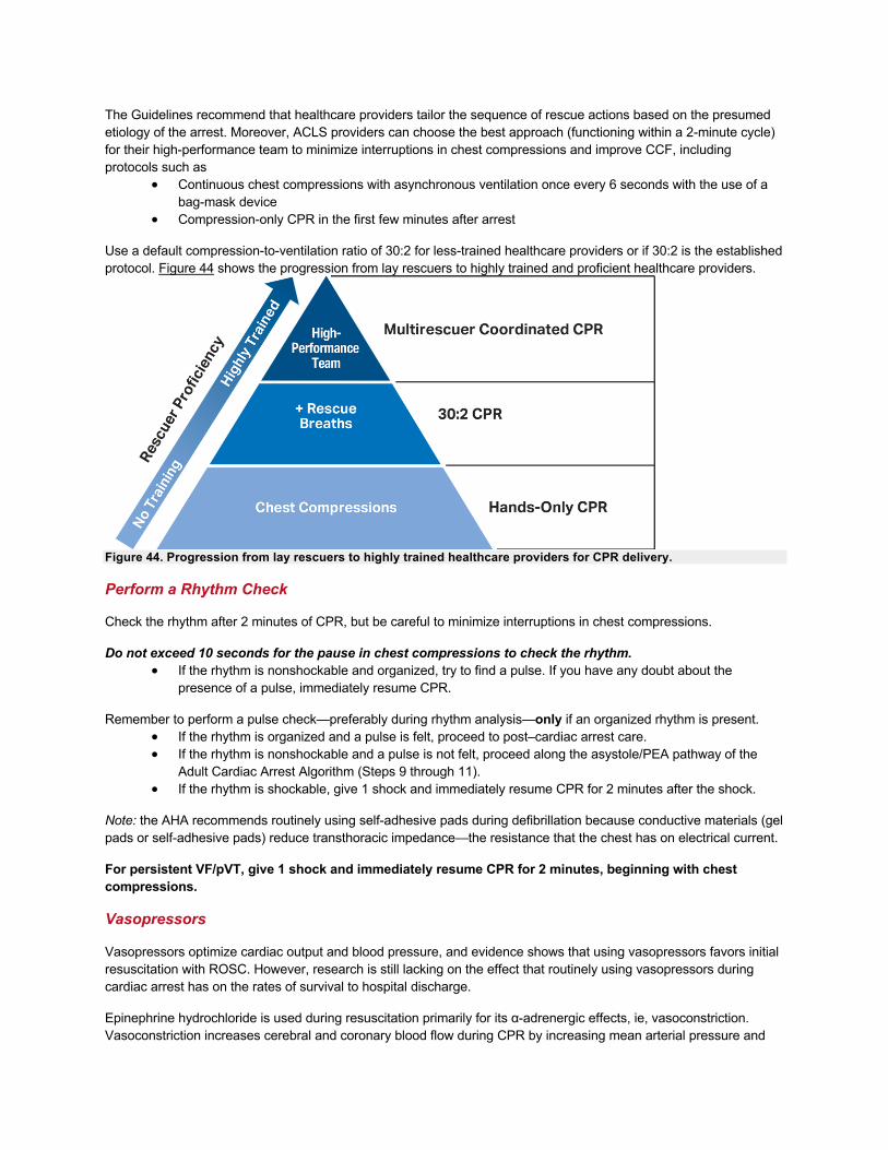
Use a default compression-to-ventilation ratio of 30:2 for less-trained healthcare providers or if 30:2 is the established protocol. Figure 44 shows the progression from lay rescuers to highly trained and proficient healthcare providers.
Figure 44. Progression from lay rescuers to highly trained healthcare providers for CPR delivery.
Perform a Rhythm Check
Check the rhythm after 2 minutes of CPR, but be careful to minimize interruptions in chest compressions.
Do not exceed 10 seconds for the pause in chest compressions to check the rhythm. • If the rhythm is nonshockable and organized, try to find a pulse. If you have any doubt about the
presence of a pulse, immediately resume CPR.
Remember to perform a pulse check—preferably during rhythm analysis—only if an organized rhythm is present. • If the rhythm is organized and a pulse is felt, proceed to post–cardiac arrest care. • If the rhythm is nonshockable and a pulse is not felt, proceed along the asystole/PEA pathway of the
Adult Cardiac Arrest Algorithm (Steps 9 through 11). • If the rhythm is shockable, give 1 shock and immediately resume CPR for 2 minutes after the shock.
Note: the AHA recommends routinely using self-adhesive pads during defibrillation because conductive materials (gel pads or self-adhesive pads) reduce transthoracic impedance—the resistance that the chest has on electrical current.
For persistent VF/pVT, give 1 shock and immediately resume CPR for 2 minutes, beginning with chest compressions.
Vasopressors
Vasopressors optimize cardiac output and blood pressure, and evidence shows that using vasopressors favors initial resuscitation with ROSC. However, research is still lacking on the effect that routinely using vasopressors during cardiac arrest has on the rates of survival to hospital discharge.
Epinephrine hydrochloride is used during resuscitation primarily for its α-adrenergic effects, ie, vasoconstriction. Vasoconstriction increases cerebral and coronary blood flow during CPR by increasing mean arterial pressure and

aortic diastolic pressure. In previous studies, escalating and high-dose epinephrine administration did not improve survival to discharge or neurologic outcome after resuscitation from cardiac arrest.
When IV/IO access is available, give epinephrine 1 mg IV/IO during CPR after the second shock, and repeat every 3 to 5 minutes, or every 4 minutes as a midrange (ie, every other rhythm check). If additional team members are available, they should anticipate the need for drugs and prepare them in advance.
No known vasopressor (epinephrine) increases survival from VF/pVT. But because these medications can improve aortic diastolic blood pressure, coronary artery perfusion pressure, and the rate of ROSC, the AHA continues to recommend their use.
Perform a Rhythm Check
Check the rhythm after 2 minutes of CPR, but be careful to minimize interruptions in chest compressions. If the rhythm is shockable, give 1 shock and immediately resume CPR for 2 minutes after the shock.
Antiarrhythmics
Healthcare providers may consider giving antiarrhythmic drugs, either before or after the shock. The focus should be on administering medications quickly and so that defibrillation is not delayed. Evidence is still lacking on whether giving antiarrhythmic drugs during cardiac arrest is associated with improved survival to hospital discharge. Amiodarone or lidocaine may be considered for VF/pVT that is unresponsive to defibrillation. These drugs may be particularly useful for patients with witnessed arrest, for whom time to drug administration may be shorter.8
In ROC-ALPS (Resuscitation Outcomes Consortium–Amiodarone, Lidocaine or Placebo Study), a large out-of-hospital randomized controlled trial that compared captisol-based amiodarone with lidocaine or placebo for patients with VF/pVT refractory after at least 1 shock, there was no overall statistically significant difference in survival with good neurologic outcome or survival to hospital discharge.9 In that study, ROSC was higher in patients receiving lidocaine compared with those receiving placebo but not for patients receiving amiodarone compared with patients receiving placebo. Among the subgroup of patients with bystander-witnessed cardiac arrest, survival to hospital discharge was higher for patients given amiodarone or lidocaine compared with those given placebo.8
Amiodarone or lidocaine may be considered for VF/pVT that is unresponsive to defibrillation. These drugs may be particularly useful for patients with witnessed arrest, for whom time to drug administration may be shorter.8
• Amiodarone: 300 mg IV/IO bolus, then consider 1 additional 150 mg IV/IO o –Amiodarone is considered a class III antiarrhythmic drug, but it possesses
electrophysiologic characteristics of the other classes. Amiodarone blocks sodium channels at rapid pacing frequencies (class I effect) and exerts a noncompetitive antisympathetic action (class II effect). One of the main effects of prolonged amiodarone administration is lengthening of the cardiac action potential (class III effect).
• Lidocaine: 1 to 1.5 mg/kg IV/IO first dose, then 0.5 to 0.75 mg/kg IV/IO at 5- to 10-minute intervals, to a maximum dose of 3 mg/kg
o –Lidocaine suppresses automaticity of conduction tissue in the heart by increasing the electrical stimulation threshold of the ventricle, His-Purkinje system, and spontaneous depolarization of the ventricles during diastole by a direct action on the tissues.
o –Lidocaine blocks permeability of the neuronal membrane to sodium ions, which inhibits depolarization and the blockade of conduction.
Providers should consider magnesium sulfate for torsades de pointes associated with a long QT interval. • Magnesium sulfate for torsades de pointes, loading dose 1 to 2 g IV/IO diluted in 10 mL (eg, D5W,
normal saline) given as IV/IO bolus, typically over 20 minutes o –Magnesium can be classified as a sodium/potassium pump agonist. Magnesium has
several electrophysiological effects, including suppression of atrial L- and T-type calcium channels, and ventricular after-depolarizations. Routinely administering magnesium sulfate in cardiac arrest is not recommended unless torsades de pointes is present.

Search for and treat any treatable underlying cause of cardiac arrest, such as the H’s and T’s.
Cardiac Arrest Treatment Sequences
The Adult Cardiac Arrest Circular Algorithm (Figure 45) summarizes the recommended sequence of CPR, rhythm checks, shocks, and delivery of drugs based on expert consensus. We don’t yet know the optimal number of CPR cycles and shocks to perform before starting pharmacologic therapy, but rhythm checks and shocks are organized around 5 cycles of CPR, or 2 minutes if a provider is timing the arrest. Do not delay shock. Continue CPR while preparing and administering drugs and charging the defibrillator. Interrupt chest compressions for only the minimum time required for ventilation (until an advanced airway is placed), rhythm check, and actual shock delivery.
Figure 45. Adult Cardiac Arrest Circular Algorithm.
Physiologic Monitoring During CPR
For intubated patients, the AHA recommends using quantitative waveform capnography to monitor CPR quality (Figure 46), optimize chest compressions, and detect ROSC during chest compressions (Figure 47). The capnography tracing in Figure 47 displays PETCO2 in millimeters of mercury on the vertical axis over time. This patient is intubated and receiving CPR. Note that the ventilation rate is approximately 10/min. Chest compressions are given continuously at a rate slightly faster than 100/min but are not visible with this tracing. The initial PETCO2 is less than 12.5 mm Hg during the first minute, indicating very low blood flow. PETCO2 increases to between 12.5 and 25 mm Hg during the second and third minutes, consistent with the increase in blood flow with ongoing resuscitation. ROSC

occurs during the fourth minute. ROSC is evident from the abrupt increase in PETCO2 (visible just after the fourth vertical line) to greater than 50 mm Hg, which is consistent with a substantial improvement in blood flow.
Figure 46A. Physiologic monitoring during CPR. A, High-quality compressions are shown through waveform capnography and intra-arterial relaxation pressure.

Figure 46B. Ineffective CPR compressions shown through intra-arterial relaxation pressure and waveform capnography.
Figure 47. Waveform capnography during CPR with ROSC.
Although invasive monitors are usually not needed during CPR, physiologic parameters such as intra-arterial relaxation pressures (Figures 46A and B) and central venous oxygen saturation (SCVO2) may help optimize CPR and detect ROSC.
Animal and human studies indicate that monitoring PETCO2, CPP, and SCVO2 provides valuable information on the patient’s condition and response to therapy.10-16 These physiologic parameters also correlate with cardiac output and myocardial blood flow during CPR, and when chest compressions fail to achieve identified threshold values, the patient rarely achieves ROSC. Furthermore, an abrupt increase in any of these parameters is a sensitive indicator of ROSC that you can monitor without interrupting chest compressions.
Although no clinical study has examined whether adjusting resuscitative efforts on the basis of physiologic parameters improves outcome, it is reasonable to use these parameters to optimize compressions and guide vasopressor therapy during cardiac arrest.
End-Tidal CO2
The main determinant of PETCO2 during CPR is blood delivery (cardiac output) to the lungs. Persistently low PETCO2 values less than 10 mm Hg during CPR in intubated patients (Figure 46B) suggest that ROSC is unlikely, and it is reasonable to try to improve chest compressions and vasopressor therapy. If PETCO2 abruptly increases to a normal value of 35 to 40 mm Hg or higher, it is reasonable to consider this an indicator of ROSC.
Coronary Perfusion Pressure or Arterial Relaxation Pressure
Increased coronary perfusion pressure (CPP) correlates with both myocardial blood flow and ROSC. A reasonable substitute for CPP during CPR is arterial relaxation (“diastolic”) pressure, which you can measure with an intra-arterial

catheter. If the arterial relaxation pressure is less than 20 mm Hg (Figure 46B), it is reasonable to try to improve chest compressions and vasopressor therapy.
Routes of Access for Drugs
The first priority during cardiac arrest is providing high-quality CPR and early defibrillation; inserting an advanced airway and administering drugs are secondary. There is no evidence that any drug used during cardiac arrest improves survival to hospital discharge with improved neurologic function.
Historically in ACLS, providers have administered drugs via IV or ET, but ET absorption of drugs is poor and optimal drug dosing is unknown. For these reasons, the IV route is preferred. When IV access is unsuccessful or not feasible, IO access may be considered.
Intravenous Route
Use a peripheral IV for drug and fluid administration unless central line access is already available. Central line access is not necessary during most resuscitation attempts, and it may cause interruptions in CPR and complications during insertion. These complications include vascular laceration, hematomas, bleeding, thrombosis, and infection. Inserting a central line in a noncompressible vessel is a relative (not absolute) contraindication to fibrinolytic therapy in patients with ACS.
You do not need to interrupt CPR to establish a peripheral IV line, but drugs typically take 1 to 2 minutes to reach the central circulation via the peripheral IV route. If you give a drug by the peripheral IV route, administer it as follows:
• Give the drug by bolus injection unless otherwise specified. • Follow with a 20-mL bolus of IV fluid. • Elevate the extremity for about 10 to 20 seconds to help deliver the drug to the central circulation.
Intraosseous Route
If IV access is not successful or feasible, you can safely and effectively deliver drugs and fluids during resuscitation via the IO route. Important points about IO access are that
• You can establish it in all age groups • You can achieve it in 30 to 60 seconds • It is preferable to ET and may be easier to establish in cardiac arrest • Any ACLS drug or fluid that you administer via IV can be given IO
IO cannulation provides access to a noncollapsible marrow venous plexus, which serves as a rapid, safe, and reliable route during resuscitation for administering drugs, crystalloids, colloids, and blood. The technique requires a rigid needle, preferably a specially designed IO or bone marrow needle from an IO access kit. For more information on IO access, see the Access for Medications section in the ACLS Student Resources.
Endotracheal Route
IV and IO routes are preferred over ET, but if you consider administering drugs via the ET route during CPR, keep these concepts in mind:
• The optimal dose of most drugs given by the ET route is unknown. • The typical dose of drugs administered via the ET route is 2 to 2½ times the IV route. • You will need to stop CPR briefly so that the drug does not regurgitate up the ET tube. • Drugs like epinephrine can negatively affect the colorimetric CO2 detector’s functionality.
Studies demonstrate that the circulatory system absorbs epinephrine, vasopressin, and lidocaine after administration via the ET route. When giving drugs via the ET route, dilute the dose in 5 to 10 mL of sterile water or normal saline, and inject it directly into the ET tube.
Fluid Administration

Adjust fluid administration and vasoactive or inotropic agents as needed to optimize blood pressure, cardiac output, and systemic perfusion. The optimal post–cardiac arrest blood pressure remains unknown; however, a mean arterial pressure 65 mm Hg or greater is a reasonable goal.
In hypovolemic patients, the ECF volume is typically restored with normal saline or lactated Ringer’s solution, and avoid D5W because it will reduce serum sodium too rapidly. Monitor serum electrolytes as appropriate.
Ultrasound for VF/pVT/Asystole/PEA
Ultrasound may be applied to patients receiving CPR to help assess myocardial contractility and identify potentially treatable causes of cardiac arrest, such as hypovolemia, pneumothorax, pulmonary thromboembolism, or pericardial tamponade. However, it is unclear whether routinely using ultrasound among patients experiencing cardiac arrest affects important clinical outcomes. If a qualified sonographer is present and use of ultrasound does not interfere with the standard cardiac arrest treatment protocol, then consider ultrasound as an adjunct to standard patient evaluation.
Return of Spontaneous Circulation
If resuscitative efforts successfully restore an organized rhythm (or you find other evidence of return of spontaneous circulation (ROSC), such as pulse and blood pressure, an abrupt and sustained increase in PETCO2 (typically 40 mm Hg or higher) or spontaneous arterial pressure waves with intra-arterial monitoring, go to the Adult Healthcare Provider Post–Cardiac Arrest Care Algorithm.
If no signs of ROSC, resume CPR, administer epinephrine, and treat reversible causes. Consider appropriateness of continued resuscitation.