caligus epidemicus (copepoda: caligidae), a …home.hiroshima-u.ac.jp/museum/siryou-data...caligus...
TRANSCRIPT
Caligus epidemicus (Copepoda: Caligidae), a Pathogenic Sea Louse of Wild and Captive Fish in Indo-West Pacific Region: a Review
Kazuya NAGASAWA1
Abstract: This paper compiles information on various aspects of the biology (i.e., taxonomy and morphology, geographical distribution and occurrence in relation to salinity, hosts, attachment sites, occurrence on wild and captive hosts, occurrence in plankton samples, life cycle, pathogenicity, and treatment and control) of the caligid copepod Caligus epidemicus based on the literature published in 1971–2013. The species attaches to the body surface of fish; it exhibits very low host specificity and has been found from 36 teleost species in 17 families and 5 orders and even from cultured shrimps. It is the most pathogenic parasite among the known caligid copepods from the Indo-West Pacific region, causing mortalities in captive fish as well as disease problems in wild and captive fish. Its infection level remains low in wild fish populations, but fish in captivity usually have higher levels of infection than those caught in the wild. Its life cycle was reported to comprise 10 stages (two naupliar, one copepodid, six chalimus stages, and adult) although Caligus spp. have typically eight stages in their life cycles. A rearing period of 17 days at 24–25ºC is necessary for the species to develop to an ovigerous female after hatching from the egg.Keywords: Caligus epidemicus, sea louse, fish parasite, biology, aquaculture, pathogenicity
I.Introduction Copepods of the siphonostomatoid family Caligidae, commonly called sea lice, often cause various problems, such as skin lesions, growth retardation, and mortalities, in farmed fish (Pike and Wadsworth, 1999; Johnson et al., 2004). Lepeophtheirus salmonis, Caligus elongatus, and C. roggercresseyi are such typical examples in salmon aquaculture conducted at higher latitudes in the Northern and Southern hemispheres. Since the salmon aquaculture supports a big industry and those caligids give a great economic loss to the industry (Costello, 2009), numerous basic and applied studies have been conducted to prevent their infection (Boxshall and Defaye, 1993; Costello, 2006; Boxaspen, 2006; Mordue (Luntz) and Birkett, 2008; Wagner et al., 2008; Jones and Beamish, 2011; Hayward et al., 2011). Caligid copepods also are problematic in finfish culture in East Asia and Australasia. For example, Caligus orientalis has been pathogenic to farmed fish in Japan, Taiwan, and China (Nagasawa, 2004). Caligus chiastos and C. amblygenitalis have become the pathogenic species in the southern bluefin tuna (Thunnus maccoyii) farming in South Australia (Hayward et al.,
2008, 2009). Caligus epidemicus has been regarded as the most dangerous parasite among the known caligid copepods in Asia (Ho, 2000). This species causes poor conditions in wild fish (Hewitt, 1971) and mortalities in captive fish (Lin and Ho, 1993; Lin et al., 1996). When Ho (2000) wrote a review on the sea lice related to cage aquaculture in Asia, the species was recorded only from Australia, New Zealand, Thailand, Philippines, and Taiwan. Its distribution, however, has since been confirmed in other countries of the Indo-West Pacific region, including India (Santhosh and Radhakrishnan, 2009; Vinoth et al., 2010; Rameshkumar et al., 2013), Malaysia (Venmathi Maran et al., 2009; Muhd-Faizul et al., 2012), Vietnam (Vo et al., 2008, 2012), and Indonesia (Yuniar et al., 2007). In Vietnam and Malaysia, the species was found infecting cultured marine fish (Vo et al., 2008, 2012; Venmathi Maran et al., 2009; Muhd-Faizul et al., 2012). Also, in the Philippines, more than 5,000 individuals of C. epidemicus were found on a single captive fish (elongate surgeonfish Acanthurus mata) (Ho et al., 2004). Therefore, recently, the significance of the species as a pathogenic parasite in finfish aquaculture in the
1 Graduate School of Biosphere Science, Hiroshima University
広島大学総合博物館研究報告 Bulletin of the Hiroshima University Museum 5: 71︲86, December 25, 2013
資料 Data

72 Kazuya NAGASAWA
広島大学総合博物館研究報告Bulletin of the Hiroshima University Museum 5: December 25, 2013 ©広島大学総合博物館Hiroshima University Museum
Indo-West Pacific region has been increasing more than before. This paper compiles information on various aspects of the biology of C. epidemicus based on the literature published from 1971–2013. It represents the second attempt of serial reviews on the biology of caligid copepods, especially those inducing economic losses in aquaculture and fisheries, in the Indo-West Pacific region (Nagasawa, 2004). In the present review, the scientific and common names of fish follow Froese and Pauly (2013). Terms used to show the occurrence of the parasi te in the host populat ion are prevalence (percentage of fish infected), mean intensity (average number of parasites per infected host) and mean abundance (average number of parasites per host regardless of whether the host is infected or not), as defined by Bush et al. (1997). For this paper, C. epidemicus were observed and newly illustrated (Figs. 1–3) using the specimens from the Philippines, because the same specimens had been reported but not described by Ho et al. (2004).
II.Taxonomy and morphology Caligus epidemicus was originally described by Hewitt (1971) based on adult female and male specimens as well as copepodid and chalimus specimens from southern black bream (Acanthopagrus butcheri) (as bream Mylio butcheri) and four species of the family Mugilidae (as mullet), i.e., flathead grey mullet (Mugil cephalus), yellow-eye mullet (Aldichetta forsteri, as Alorichetta forsteri), flat-tail mullet (Liza argentea), and sand grey mullet (Myxus elongatus, as M. elongans), caught in the lower Mitchell River, Victoria, Australia. Subsequently, Roubal (1981) and Byrnes (1987) briefly described this caligid species from surf bream (Acanthopagrus australis) and four species of seabreams (Acanthopagrus spp.), respectively, taken in the coastal waters of Australia. In their book, Ho and Lin (2004) reported the detailed morphology of female and male adults of C. epidemicus from Taiwan. Venmathi Maran et al. (2009) also briefly described the morphology of the species from Malaysia. In addition to these works, Lin and Ho (1998) studied the structure of legs 1 and 4 and the male antenna of C. epidemicus and five other species (C. acanthopagri, C. orientalis, C. oviceps, C. punctatus, and C. rotundigenitalis [as C. multispinosus]) from
Taiwan. There are records of “Caligus sp.” in the Philippines (Lio-Po and Barry, 1988) and Thailand (Chinabut, 1996). This unspecified caligid in both countries is herein regarded as C. epidemicus based on the photographed morphology of an ovigerous female. The illustrated morphology of Thai specimens also shows that they are identical to C. epidemicus. Caligus epidemicus is a small species among caligid copepods. Adult females do not exceed 3 mm in body length (BL) (excluding the setae on the caudal rami): 2.46–2.85 (mean 2.66, n = 10) or 2.362–2.649 (2.529, n = 15) mm BL from Australia (Hewitt, 1971; Roubal, 1981) and 2.23–2.33 (2.28, n = 10) mm BL from Malaysia (Venmathi Maran et al., 2009). Adult females from Taiwan are slightly smaller than Australian and Malaysian ones, being 1.97 mm BL (no data available for size variation) (Ho and Lin, 2004). Adult males do not exceed 2 mm BL: 1.52–1.72 (mean 1.59, n = 10) or 1.539–1.665 (1.562, n = 15) mm BL from Australia (Hewitt, 1971; Roubal, 1981), 1.58–1.65 (1.62, n = 8) mm BL from Malaysia (Venmathi Maran et al., 2009), and 1.51 mm BL from Taiwan (Ho and Lin, 2004). Byrnes (1987) collected many specimens of C. e p i d e m i c u s f r o m f o u r s p e c i e s o f s e a b r e a m s (Acanthopagrus spp.) in the coastal waters of Australia and presented the measurement data of these specimens by host species and sampling locality. While no particular comments were made by this author, the female specimens from southern black bream (A. butcheri) caught along the south coast of Australia were largest (2.442–2.480 mm in mean BL), followed by those specimens from surf bream (A. australis) from the southeast coast of Australia (2.030–2.309 mm in mean BL). In contrast, the female specimens from goldsilk seabream (A. berda) and yellowfin seabream (A. latus) collected along the north and northwest coasts of Australia were smaller, being 1.870–2.144 and 1.998–2.291 mm in mean BL, respectively. These data suggest that individuals of C. epidemicus can become larger or smaller at higher or lower latitudes, respectively, which may be due to the effect of water temperature on the growth rate of the parasite. The possibility that the host species affects the growth rate of C. epidemicus may be negligible in this case because Byrnes (1987) examined the four species of seabreams which are very closely

73Caligus epidemicus (Copepoda: Caligidae), a Pathogenic Sea Louse of
Wild and Captive Fish in Indo-West Pacific Region: a Review
広島大学総合博物館研究報告Bulletin of the Hiroshima University Museum 5: December 25, 2013 ©広島大学総合博物館Hiroshima University Museum
related. The best description of C. epidemicus was made by Ho and Lin (2004) using material from Taiwan. The following description of adult females and males is mostly based on it but is supplemented by the observation in this study using material from the Philippines. The female and male specimens illustrated in Figs. 1A and 3A were 2.04 and 1.44 mm BL, respectively, Female (Figs. 1–2): Cephalothoracic shield (1A) subcircular with well-developed frontal plate and medium-sized lunules. Fourth pediger (Fig. 1A) 2.78 times wider than long (3.27 times in the Philippine specimen). Genital complex (Fig. 1B) subrectangular with broadly rounded corners. Abdomen (Fig. 1B) short, subquadrate, and 1-segmented. Caudal ramus (Fig. 1C) short, about as long as wide, armed with 3 short, subterminal and 3 long, terminal setae. Egg sac about 2/3 length of body (about 1/2 length in the Philippine specimen). Antennule (Fig. 1D) 2-segmented; proximal segment with 25 setae on anterodistal surface; distal segment with 1 subterminal seta on posterior margin and 11 setae plus 2 aesthetascs on distal margin. Antenna (Fig. 1E) 3-segmented; proximal segment (coxa) with blunt posteromedial process; middle segment (basis) subrectangular and unarmed; distal segment (endopod) sharply pointed claw with one and another setae each in proximal and middle regions. Postantennal process (Fig. 1E) bluntly pointed, bearing 2 basal papillae with each carrying 4 setules (3 or 4 setules in the Philippine specimen), another similar papilla nearby on sternum carrying 4 setules (3 setules). Maxillule (Fig. 1E) consisting of straight dentiform process and basal papilla with 3 small setae. Maxilla (Fig. 1F) 2-segmented; proximal segment (lacertus) large and unarmed; distal segment (brachium) slender, carrying small, subterminal, hyaline membrane (flabellum) on outer edge and two spinulated elements, a short subterminal canna and a long te rmina l ca lamus . Maxi l l iped (F ig . 1G) 3-segmented; proximal segment (corpus) largest, with a basal rounded protrusion (such protrusion not distinguished but a small swelling in myxal area in the Philippine specimen), middle (shaft) and distal (claw) segments fused to form subchela, claw with long barbell at base. Sternal furca (Fig. 2A) with subquadrate box, bearing short tines. Armature on rami of legs 1–4 are as
follows (Roman numerals indicating spines and Arabic numerals, setae): 1-0; III,I,3 and vestigial for the exopod and endopod of leg 1; I-1, I-1; II,I,5 and 0-1; 0-2; 6 for those of leg 2; I-0, I-1; III,4 and 0-1; 6 for those of leg 3; and I-0; II and absent for those of leg 4. Leg 1 (Fig. 2B) protopod with long, plumose outer seta and another small, plumose inner seta, in addition to a papilla bearing 1 setule on outer margin of coxa; vestigial endopod a small process tipped with 1 setule; first segment of exopod with row of spinules on posterior edge and small spiniform seta at outer distal corner; middle 2 of 4 terminal elements (Fig. 2C) on last segment of exopod with accessory process, element 1 pinnate in distal half and element 4 naked and shorter than element 1. Leg 2 (Fig. 2D) coxa small, with setule-bearing papilla on ventral surface and large, plumose, inner seta on posterior edge; basis large, with small seta on outer edge; posterior margin of basis with long, narrow membrane and small marginal setule near midway of this membrane; outer edges of both basis and first segment of exopod fringed with large marginal membrane; Proximal 1 outer spine on terminal segment of exopod (Fig. 2E) naked. Leg 3 (Fig. 2F) protopod with small, plumose, outer seta and long, plumose inner seta, in addition to an outer and a posterior marginal membrane; ventral surface of protopod with setule-bearing papilla at both ends of latter membrane, outer margin corrugated anterior to former membrane; velum well developed and fringed with setules; proximal segment of exopod (Fig. 2G) with membrane on lateral margin and its spine also bearing lateral membrane. Leg 4 (Fig. 2H) protopod with plumose seta at outer distal corner; terminal segment of exopod with pecten at base of inner spine (pecten invisible in the Philippine specimen). Leg 5 (Fig. 1B) represented by 2 small, isolated papillae on posterolateral corner of genital complex, 1 tipped with small plumose seta and other with 3 similar setae. Male (Fig. 3): Cephalothoracic shield (Fig. 3A) suborbicular, with well-developed frontal plate and medium-sized lunules. Fourth pediger (Fig. 3B) 3.03 times wider than long (2.63 times in the Philippine specimen). Genital complex (3B) subrectangular, with posteriorly protruded posterolateral process. Abdomen (Fig. 3B) small and subquadrate. Caudal ramus (Fig. 3B) slightly wider than long and armed as in female. Antenna (Fig. 3C) 3-segmented; proximal segment slender and

74 Kazuya NAGASAWA
広島大学総合博物館研究報告Bulletin of the Hiroshima University Museum 5: December 25, 2013 ©広島大学総合博物館Hiroshima University Museum
Fig. 1 Caligus epidemicus, female. A, habitus, dorsal; B, genital complex and abdomen, dorsal; C, caudal ramus, dorsal; D, antennule; E, antenna, postantennary process and maxillule; F, maxilla; G, maxilliped. Scale bars (mm): 0.5 in A; 0.1 in B; 0.02 in C–D; 0.05 in E–G.

75Caligus epidemicus (Copepoda: Caligidae), a Pathogenic Sea Louse of
Wild and Captive Fish in Indo-West Pacific Region: a Review
広島大学総合博物館研究報告Bulletin of the Hiroshima University Museum 5: December 25, 2013 ©広島大学総合博物館Hiroshima University Museum
Fig. 2 Caligus epidemicus, female. A, sternal furca; B, leg 1; C, tip of leg 1 exopod; D, leg 2; E, outer distal part of leg 2 exopod; F, leg 3; G, rami of leg 3; H, leg 4. Scale bars (mm): 0.02 in A, C, G; 0.05 in B, E, F, H; 0.1 in D.

76 Kazuya NAGASAWA
広島大学総合博物館研究報告Bulletin of the Hiroshima University Museum 5: December 25, 2013 ©広島大学総合博物館Hiroshima University Museum
armed with long corrugated adhesive pad; middle segment largest, armed with 3 (1 large and 2 small) corrugated adhesion pads in central and distal regions; terminal segment (Fig. 3D) smallest, with 2 medial setae and a corrugated adhesion pad in basal region and a large tooth-like projection in middle region. Maxilliped (Fig. 3E) 3-segmented, bearing a spine in myxal area. Leg 5
(Fig. 3B) represented by 2 papillae on posterolateral side of genital complex, 1 tipped with seta and other with 3 setae. Leg 6 (Fig. 3B) represented by 2 short setae (3 setae in the Philippine specimen) on ventral surface at base of posterolateral process on genital complex. Sternal furca (Fig. 3F) with subquadrate box, bearing short tines.
Fig. 3 Caligus epidemicus, male. A, habitus, dorsal; B, genital complex, abdomen and caudal rami, ventral; C, antenna; D, terminal part of antenna; E, maxilliped; F, sternal furca. Scale bars (mm): 0.2 in A; 0.1 in B; 0.05 in C, E; 0.02 in D, F.

77Caligus epidemicus (Copepoda: Caligidae), a Pathogenic Sea Louse of
Wild and Captive Fish in Indo-West Pacific Region: a Review
広島大学総合博物館研究報告Bulletin of the Hiroshima University Museum 5: December 25, 2013 ©広島大学総合博物館Hiroshima University Museum
III. G e o g r a p h i c a l d i s t r i b u t i o n a n d occurrence in relation to salinity
Caligus epidemicus is widely distributed in the Indo-West Pacific region (Fig. 4) and has been reported so far from Australia, New Zealand, India, Indonesia, Malaysia, Thailand, Vietnam, Philippines, and Taiwan (see Table 1 for the literature). The known distribution of the species covers the tropical, subtropical, and temperate waters in the Southern and Northern hemispheres, ranging from Lakes Entrance (37°53′S), Victoria, southern Australia (Byrnes 1987; Byrnes and Rohde 1992) in south to Bei-men (23°17′N), Tainan, Taiwan (Ho and Lin, 2004) in north. The species may be found in other Asian countries, where no systematic investigations on caligid copepods have so far been conducted. The distribution of C. epidemicus in the coastal waters of Australia was reported by Byrnes and Rhode (1992) as divided into five zoogeographical regions (Banksian, Peronian, Maugean, Flindersian, and
Dampierian). The species occurs in all of these regions but it is abundant in two regions (Peronian and Maugean) located in southeast Australia, where surf bream (A. australis) and southern black bream (A. butcheri) serve as hosts, respectively. Caligus epidemicus is a brackish-water or coastal marine species found on fish in the lower reaches of rivers and the inshore and estuarine environments. As stated below (see the Treatment and control section), adults of C. epidemicus can tolerate a wide range of salinity (Roubal, 1997). In the lower Mitchell River, Australia (a type locality of the species), the salinity fluctuated between 4.5 and 28 ppt (Hewitt, 1971). The species was also recorded at different salinity levels ranging from 2–30 ppt (Regidor and Arthur, 1986), 12–28 ppt (Ruangpan and Kabata, 1984), 19.7–31.2 ppt (Yurinar et al., 2007), 30–32 ppt (Cruz-Lacierda et al., 2011) to 35–50 ppt (Lio-Po and Barry, 1988). Its life cycle also can be completed at 20 ppt in the laboratory (Lin and Ho, 1993).
Fig. 4 A map of the Indo-West Pacific region, showing the localities (closed circles) where Caligus epidemicus was collected.

78 Kazuya NAGASAWA
広島大学総合博物館研究報告Bulletin of the Hiroshima University Museum 5: December 25, 2013 ©広島大学総合博物館Hiroshima University Museum
IV.Hosts Since i ts original description in 1971, C. epidemicus has been reported from 36 teleost species, a tilapia hybrid, and unspecified and unidentified mugilids during the last four decades (Table 1). These fish hosts be long to 17 fami l ies (Chanidae , Mugi l idae , Acanthuridae, Ambassidae, Carangidae, Cichlidae, Gobiidae, Latidae, Lutjanidae, Monodactylidae, Rachcentridae, Scatophagidae, Serranidae, Siganidae, Sparidae, Teraponidae, and Pleuronectidae) in four orders (Gonorhynchiformes, Mugiliformes, Perciformes, and Pleuronectiformes). Most of these fish are coastal marine and brackish-water species. Interestingly, the species was accidentally found on tiger shrimps (Penaeus monodon) cultured in Thailand (Ruangpan and Kabata, 1984). Accordingly, it is evident that C.
epidemicus exhibits very low host specificity and can ut i l ize a wide variety of f infish. Nonetheless, elasmobranchs are not listed as the hosts of the species. Also, it has not been reported from offshore migratory fish, except for yellowtail amberjack (Seriola lalandi) from Australia (Hutson et al., 2007). The fact that some euryhaline fish species, such as mugilids and sparids of Acanthopagrus, are included in the list of the known hosts (Table 1) is one of the features in host utilization of C. epidemicus. Such euryhaline fish can move and carry the copepod between the lower reaches of rivers and the coastal sea area. In Far East Asia, Caligus orientalis also uses both brackish-water and coastal marine fish as hosts (Nagasawa, 2004), and these Caligus species are different from most of their congeners which infect only marine fish.
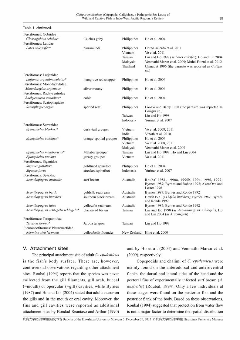
Table 1 Fish hosts of Caligus epidemicus reported between 1971 and 2013. The copepod was recorded from cultured individuals of the species with an asterisk (*). The classification and scientific and common names of fish are adopted from Froese and Pauly (2013).
Scientific name Common name Country or region Reference
Gonorynchiformes: Chanidae Chanos chanos* milkfish Philippines Regidor and Arthur 1986
Taiwan Lin and Ho 1998; Ho and Lin 2004Mugiliformes: Mugilidae Aldrichetta forsteri yellow-eye mullet Australia Hewitt 1971 (as Alorichetta forsteri) Liza argentea flat-tail mullet Australia Hewitt 1971 Liza macrolepis* largescale mullet Taiwan Lin and Ho 1998; Ho and Lin 2004 Mugil cephalus* flathead grey mullet Taiwan Lin et al., 1993; Lin and Ho 1998; Ho and Lin 2004
Australia Hewitt 1971 Myxus elongatus sand grey mullet Australia Hewitt 1971 (as Myxus elongans) Paramugil parmatus broad-mouthed mullet Philippines Ho et al. 2004 (as Liza parmata) Unspecified species India Santhosh and Radhakrishnan 2009 Unindentified species Thailand Chinabut 1996 (as mullet. The parasite was reported
as Caligus sp.)Perciformes: Acanthuridae Acanthurus mata elongate surgeonfish Philippines Ho et al. 2004Perciformes: Ambassidae Ambassis marianus estuary perchlet Australia Hallett and Roubal 1995Perciformes: Carangidae Carangoides malabaricus Malabar trevally India Rameshkumar et al. 2013 Parastromateus niger black pomfret India Rameshkumar et al. 2013 Seriola lalandi yellowtail amberjack Australia Hutson et al. 2007 Trachinotus blochii* snubnose pompano Philippines Cruz-Lacierda et al. 2011
Taiwan Lin and Ho 1998; Ho and Lin 2004Perciformes: Cichlidae Oreochromis aureus* blue tilapia Taiwan Lin and Ho 1998; Ho and Lin 2004 Oreochromis mossambicus* Mozambique tilapia Taiwan Lin and Ho 1993, 1998; Lin et al. 1996; Ho and Lin
2004Philippines Ho et al. 2004
Oreochromis niloticus niloticus* Nile tilapia Philippines Natividad et al. 1986 (as Oreochromis niloticus); Bondad-Reantaso and Arthur 1990 (as O. niloticus)
Oreochromis urolepis hornorum* Wami tilapia Philippines Ho et al. 2004 Oreochromis hybrid* Taiwan Lin and Ho 1998; Ho and Lin 2004
79Caligus epidemicus (Copepoda: Caligidae), a Pathogenic Sea Louse of
Wild and Captive Fish in Indo-West Pacific Region: a Review
広島大学総合博物館研究報告Bulletin of the Hiroshima University Museum 5: December 25, 2013 ©広島大学総合博物館Hiroshima University Museum
V.Attachment sites The principal attachment site of adult C. epidemicus is the fish 's body surface. There are, however, controversial observations regarding other attachment sites. Roubal (1994) reports that the species was never collected from the gill filaments, gill arch, buccal (=mouth) or opercular (=gill) cavities, while Byrnes (1987) and Ho and Lin (2004) stated that adults occur on the gills and in the mouth or oral cavity. Moreover, the fins and gill cavities were reported as additional attachment sites by Bondad-Reantaso and Arthur (1990)
and by Ho et al. (2004) and Venmathi Maran et al. (2009), respectively. Copepodids and chalimi of C. epidemicus were mainly found on the anterodorsal and anteroventral flanks, the dorsal and lateral sides of the head and the pectoral fins of experimentally infected surf bream (A. australis) (Roubal, 1994). Only a few individuals at these stages were found on the posterior fins and the posterior flank of the body. Based on these observations, Roubal (1994) suggested that protection from water flow is not a major factor to determine the spatial distribution
Table 1 cintinued.Perciformes: Gobiidae Glossogobius celebius Celebes goby Philippines Ho et al. 2004Perciformes: Latidae Lates calcarifer* barramundi Philippines Cruz-Lacierda et al. 2011
Vietnam Vo et al. 2011Taiwan Lin and Ho 1998 (as Lates calcifer); Ho and Lin 2004Malaysia Venmathi Maran et al. 2009; Muhd-Faizul et al. 2012Thailand Chinabut 1996 (the parasite was reported as Caligus
sp.)Perciformes: Lutjanidae Lutjanus argentimaculatus* mangrove red snapper Philippines Ho et al. 2004Perciformes: Monodactylidae Monodactylus argenteus silver moony Philippines Ho et al. 2004Perciformes: Rachycentridae Rachycentron canadum* cobia Philippines Ho et al. 2004Perciformes: Scatophagidae Scatophagus argus spotted scat Philippines Lio-Po and Barry 1988 (the parasite was reported as
Caligus sp.)Taiwan Lin and Ho 1998Indonesia Yurinar et al. 2007
Perciformes: Serranidae Epinephelus bleekeri* duskytail grouper Vietnam Vo et al. 2008, 2011
India Vinoth et al. 2010 Epinephelus coioides* orange-spotted grouper Philippines Ho et al. 2004
Vietnam Vo et al. 2008, 2011Malaysia Venmathi Maran et al. 2009
Epinephelus malabaricus* Malabar grouper Taiwan Lin and Ho 1998; Ho and Lin 2004 Epinephelus tauvina greasy grouper Vietnam Vo et al. 2011Perciformes: Siganidae Siganus guttatus* goldlined spinefoot Philippines Ho et al. 2004 Siganus javus streaked spinefoot Indonesia Yurinar et al. 2007Perciformes: Sparidae Acanthopagrus australis surf bream Australia Roubal 1981, 1990a, 1990b, 1994, 1995, 1997;
Byrnes 1987; Byrnes and Rohde 1992; Aken'Ova and Lester 1996
Acanthopagrus berda goldsilk seabream Australia Byrnes 1987; Byrnes and Rohde 1992 Acanthopagrus butcheri southern black bream Australia Hewit 1971 (as Mylio butcheri); Byrnes 1987; Byrnes
and Rohde 1992 Acanthopagrus latus yellowfin seabream Australia Byrnes 1987; Byrnes and Rohde 1992 Acanthopagrus schlegelii schlegelii* blackhead bream Taiwan Lin and Ho 1998 (as Acanthopagrus schlegeli); Ho
and Lin 2004 (as A. schlegeli)Perciformes: Terapontidae Terapon jarbua* Jarbua terapon Taiwan Lin and Ho 1998Pleuronectiformes: Pleuronectidae Rhombosolea leporina yellowbelly flounder New Zealand Hine et al. 2000

80 Kazuya NAGASAWA
広島大学総合博物館研究報告Bulletin of the Hiroshima University Museum 5: December 25, 2013 ©広島大学総合博物館Hiroshima University Museum
of the copepod of these stages. In the experimental infection with C. epidemicus using estuary perchlet (Ambassis marinus), a small coastal fish (1–4 cm long), most parasites of the copepodid and early chalimus stages attached to the pectoral fins but did not settle on other parts of the host (Hallett and Roubal, 1995).
VI.Occurrence on wild and captive hosts Infection level of C. epidemicus remains low in wild fish populations, but fish in captivity usually have higher levels of infection than those caught in the wild. Since this species of sea louse has a one-host life cycle, it can easily develop a large population at fish farms, where fish are held at high density in a limited water body. This section deals with the occurrence of C. epidemicus on various wild and captive fish by country and region (Australia, New Zealand, India, Indonesia, Malaysia, Thailand, Vietnam, Philippines, and Taiwan). Several investigations were conducted to determine the infection with C. epidemicus on four species of seabreams (A. butcheri, A. australis, A. berda, and A. latus) in Australia. Mean abundances of the parasite on these hosts from the coastal waters of this country were 0.13–2.3, 0.3–3.6, 0.18–7.8, and 0.38–1.9, respectively (Byrnes, 1987). In a population of surf bream (A. australis) from a small estuary in New South Wales, the species of caligid was rare: prevalence and mean intensity being only 3% and 1.0 and 1% and 1.0 for adult female and male copepods, respectively (Roubal, 1990a). On the other hand, when surf bream caught in a coastal pond, Queensland, were held in an experimental sea cage for up to six weeks, there was a marked increase in mean abundance (51.4) of the parasite, compared to wild fish (4.2) and pond fish (9.3) (Roubal, 1995). Udonellids (Udonella myliobati) are known to occur on C. epidemicus infecting surf bream from eastern Australia (Aken'Ova and Lester, 1996). In New Zealand, C. epidemicus was recorded from yellowbelly flounder (Rhombosolea leporina) in Manukau Harbour (Hine et al., 2000). No infection data were reported. In India, Santhosh and Radhakrishnan (2009) reported C. epidemicus for the first time and found it on three species of mullets in the estuarine habitats of Kerala. This parasite was also found on duskytail
grouper (Epinephelus bleekeri) from Vellar Estuary (Vinoth et al., 2010) and two species of carangids (Malabar trevally Caragoides malabaricus and black pomfret Parastromateus niger) from Parangipettai (Rameshkumar et al., 2013). Through a survey on the crustacean parasite fauna of coastal fish from Segara Anakan Lagoon, central Java, Indonesia, C. epidemicus was found on streaked spinefoot (Siganus javus) and spotted scat (Scatophagus argus) (Yurinar et al., 2007). Prevalence and mean intensity of the parasite on these fish species were 20% and 1.00 and 4.3% and 1.33, respectively. Two species of cage-cultured marine fish, barramundi (Lates calcarifer) and orange-spotted grouper (Epinephelus coioides), from Penang, Malaysia, were found to harbor C. epidemicus (Venmathi Maran et al., 2009). Barramundi harbored another caligid species, Caligus punctatus. Recently, five species of sea lice, C. chiastos , C. epidemicus , C. rotundigenitalis , C. punctatus, and Caligus sp., were found infecting barramundi cultured in Malaysia, but C. epidemicus constantly predominated among these species and at different sites (Muhd-Faizul et al., 2012). In Thailand, C. epidemicus was found to heavily infect tiger prawns (P. monodon) cultured at a farm in Chantaburi Province in 1977 (Ruangpan and Kabata, 1984). Despite the fact that this was a very unusual infection of the crustacean decapod by the fish-parasitizing C. epidemicus, when discovered, its infection level was high with prevalence and mean intensity of 72% and 19, respectively. Copepods were attached mainly to the telsons and legs of the prawns. While the authors could not assess whether C. epidemicus infection continued at the prawn farm, Chinabut (1996) later reported that fingerling of mullet (species unidentified) from prawn ponds in Rayong Province were heavily infected with C. epidemicus (as Caligus sp., see the Taxonomy and morphology section), which herein suggests that such fish serve as the reservoir hosts for the copepod in the ponds. Chinabut (1996) also found C. epidemicus on cage-cultured barramundi (L. calcarifer) from southern Thailand. Vietnam was recently added to the countries where C. epidemicus occurs. Vo et al. (2008) found this caligid on wild orange-spotted grouper (E. coioides) and duskytail grouper (E. bleekeri) caught in brackish waters

81Caligus epidemicus (Copepoda: Caligidae), a Pathogenic Sea Louse of
Wild and Captive Fish in Indo-West Pacific Region: a Review
広島大学総合博物館研究報告Bulletin of the Hiroshima University Museum 5: December 25, 2013 ©広島大学総合博物館Hiroshima University Museum
in Nha Trang. They also collected specimens of C. epidemicus from orange-spotted grouper cultured in brackish-water ponds. Prevalence and mean intensity of the parasite on these groupers were low, ranging from 6.3–11.1% and 1.9–2.5, respectively. The groupers were concurrently infected with another species of sea louse, Caligus multispinosus. Interestingly, no infection was observed on the groupers cultured in sea waters. Currently, Vo et al. (2012) listed four species of marine fish (E. coioides, E. bleekeri, Malabar grouper E. malabaricus , greasy grouper E. tauvina , and L. calcarifer) as the hosts of C. epidemicus. In the Philippines, C. epidemicus occurs on commercially important fish reared in brackish waters. The species infected Nile tilapia (Oreochromis niloticus niloticus, as O. niloticus) cultured in Leganes, Iloilo Province (Natividad et al., 1986; Bondad-Reantaso and Arthur, 1990). Up to 548 copepods were found on a single fish (Natividad et al. 1986). Prevalence and mean intensity of the parasite irregularly fluctuated at monthly intervals but reached up to 100% and 47.8, respectively. Infection level also increased with an increase in fish age (Bondad-Reantaso and Arthur, 1990). Juvenile milkfish (Chanos chanos) from brackish-water ponds in Pagbilao, Quezon Province and Molo, Iloilo Province, had infection with C. epidemicus (Regidor and Arthur, 1986). Prevalence and mean intensity ranged from 4.0–36.0% and 1.0–8.8, respectively. Recently, C. epidemicus was found infecting marine fish cultured in the coastal ponds, cages and tanks or occurring in the sea-water supply canals in various localities of the Philippines (Ho et al., 2004). The infected fish are orange-spotted grouper (E. coioides), mangrove red snapper (Lutjanus argentimaculatus), Mozambique tilapia (Oreochromis mossambicus), Wami tilapia (O. urolepis hornorum), cobia (Rachycentron canadum), goldlined spinefoot (Siganus guttatus), e longate surgeonfish (A. mata ) , Celebes goby (Glossogobius celebius), broad-mouthed mullet (Paramugil parmatus, as Liza parmata), and silver moony (Monodactylus argenteus). Prevalence of infection was usually high, often up to 100%. Over 5,000 individuals of C. epidemicus were found on a single elongate surgeonfish. The cultured fish often had infections with other species of sea lice, Caligus quadratus, Lepeophtheirus sigani and Pseudocaligus
uniartus, in addition to C. epidemicus. Furthermore, C. epidemicus caused disease problems in goldlined spinefoot (S. guttatus) and barramundi (L. calcarifer) experimentally reared at the Aquaculture Department of the Southeast Asian Fisheries Development Center (SEAFDEC-AQD) in Iloilo Province (Cruz-Lacierda et al., 2011). While two other caligids, P. uniartus and L. sigani , also parasitized goldlined spinefoot, C. epidemicus was the most predominant: up to 660 individuals of the species were found on a single fish. Prevalence and mean intensity on barramundi were 100% and 12.7, respectively. In Taiwan, C. epidemicus has been reported from 11 species and tilapia hybrid cultured in brackish and/or salt waters (Lin and Ho, 1998). These fish are milkfish (C. chanos), barramundi (L. calcarifer), Malabar grouper (E. malabaricus), Jarbua terapon (Terapon jarbua), snubnose pompano (Trachinotus blochii), blackhead seabream (Acanthopagrus schlegelii schlegelii, as A. schlegeli), spotted scat (S. argus), blue tilapia (Oreochromis aureus, as O. aurea), tilapia hybrid (O re o c h ro m i s s p . ) , M o z a m b i q u e t i l a p i a (O . mossambicus), largescale mullet (Liza macrolepis), and flathead grey mullet (M. cephalus). Later, however, Ho and Lin (2004: 174) did not list two species (T. jarbua and S. argus) as the Taiwanese hosts of C. epidemicus. Despite the great efforts of work on the caligid copepods of Taiwan, there is yet no record of C. epidemicus from wild fish. For this, Ho and Lin (2004: 61, 337) suspect that the species was introduced into Taiwan by means of human activities. No data on prevalence and intensity of the parasite on cultured fish from Taiwan are available.
VII.Occurrence in plankton samples When the population of C. epidemicus exploded in the lower Mitchell River, the species was found in plankton samples as well (Hewitt, 1971). While there was no record about the stage of individuals collected, it is most likely that adults detached from the fish host were sampled. Ho (2000) suggested that planktonic individuals of C. epidemicus are capable to disperse without “hitch hiking” on the wild fish.
VIII.Life cycle Hewitt (1971) was the first to describe the life history of C. epidemicus using specimens from the fish

82 Kazuya NAGASAWA
広島大学総合博物館研究報告Bulletin of the Hiroshima University Museum 5: December 25, 2013 ©広島大学総合博物館Hiroshima University Museum
in the lower Mitchell River, Australia. He recognized five instars in his specimens, which were called Stage A, B, C, D, and E, and gave descriptions for each of the stages. He regarded Stage A as “copepodite” and the remaining stages as “chalimus.” Subsequently, the species was reported to have 11 stages: two naupliar, one copepodid, six chalimus, one young adult, and adult stages (Lin and Ho, 1993; Lin et al., 1996; Ho and Lin, 2004). For the use of “young adult”, Ho and Lin (2004) recommended to use it rather than the “preadult” which had been used by Lin and Ho (1993) and Lin et al. (1996) for the species. The number of stages in the life history of C. epidemicus is very unusual among its congeners because most species of the genus have only four chalimus stages. Lin et al. (1996) explained that the delay in the development of legs 3 and 4 causes the addition of the fifth and sixth chalimus stages in this species. Moreover, these authors stressed that no moult was observed between the young adult and the adult, which indicates that both stages are identical, i.e., the adult. In other words, C. epidemicus has 10 stages in its life cycle. On the other hand, through a study on the life cycle of Lepephtheirus elegans and a review of the life cycles of caligid copepods, Venmathi Maran et al. (2013) concluded that the caligids have typically have eight stages (two naupliar, one copepodid, and four chalimus stages, and the adult). As stated above, the fifth and sixth chalimus stages were recognized in C. epidemicus. The life cycle of C. epidemicus was studied by Lin and Ho (1993) and Lin et al. (1996) at 24–25ºC in the laboratory. It takes approximately 28 h to hatch as nauplii after the appearance of eggs in the newly formed egg sac. It also needs a period of 17 days to develop to an ovigerous female after hatching. The duration for each of the 10 stages excluding the adult in the life cycle is 6 and 14.5 h each for the first and second nauplius stages, two days for the copepodid stage, 12 h for the first chalimus stage, one day each for the second to sixth chalimus stages and 5 days for the young adult stage. An ovigerous female of C. epidemicus has about 21 eggs in each of its egg sacs (Lin and Ho, 1993). The hatching in this species involves a two-step process: breaking of the chamber wall following swelling of the egg and rupture of the egg membrane. The newly hatched, first nauplius is motionless and weakly phototactic. After moulting, the second nauplius
gradually becomes less active and sinks to the bottom. The copepodid is initially very active, moves swiftly in the water but later spends much time on the bottom. However, when a fish is introduced in the water, the copepodid suddenly becomes active and attaches to the fish using the antennae. A frontal organ (including the frontal filament) later appears between the bases of the antennae. The attached copepodid moults into the first chalimus, which attaches to the fish by the frontal filament produced by the copepodid. Passing six moults, the chalimus develops into the young adult. The parasite of the latter stage uses the frontal filament to attach to the host but is easily detached. As mentioned above, copepodids of C. epidemicus are infective to fish hosts. They survived for up to 7, 8, and 5 days at 19, 22, and 26ºC, respectively, without food (Hallett and Roubal, 1995). Three- or 4-day-old copepodids were more infective than younger ones.
IX.Pathogenicity An epizootic of C. epidemicus occurred in the wild populations of southern black bream (A. butcheri), flathead grey mullet (M. cephalus), yellow-eye mullet (A. forsteri), flat-tail mullet (L. argentea), and sand grey mullet (M. elongatus) from January to March 1968 in the lower Mitchell River, Australia (Hewitt, 1971). In February, fish were found in highly poor condition, and prevalence was very high. Since the epizootic was associated with high water temperature and salinity during a period of severe drought, such environmental factors are considered to have contributed to the development of a large population of the species. While some authors (e.g., Roubal, 1994: 631, 1995: 423; Ho and Lin, 2004: 335) state that C. epidemicus killed many fish in this locality, no description is present in Hewitt (1971) about whether the infected fish were dead or not. No similar case of epizootic has happened since Hewitt's (1971) paper. Caligus epidemicus is known to cause mortalities in captive fish. For example, Mozambique tilapia (O. mossambicus) were killed by the heavy infection with the parasite in the experimental ponds at the Tainan Branch of the Fisheries Research Institute of Taiwan (Lin and Ho, 1993; Lin et al., 1996). This species of sea louse also killed ten other fish species, blackhead seabream (A. schlegelii schlegelii), milkfish (C. chanos), Malabar
83Caligus epidemicus (Copepoda: Caligidae), a Pathogenic Sea Louse of
Wild and Captive Fish in Indo-West Pacific Region: a Review
広島大学総合博物館研究報告Bulletin of the Hiroshima University Museum 5: December 25, 2013 ©広島大学総合博物館Hiroshima University Museum
grouper (E. malabaricus), barramundi (L. calcarifer), largescale mullet (L. macrolepis), flathead grey mullet (M. cephalus), spotted scat (S. argus), blue tilapia (O. aureus), Jarbua terapon (T. jarbua), and snubnose pompano (T. blochii), cultured in the coastal brackish-water ponds in Taiwan (Ho, 2000). At the SEAFDEC-AQD in the Philippines, infection with numerous C. epidemicus and a few individuals of two other caligids caused a 10% mortality in broodstocks of goldlined spinefoot (S. guttatus). The fish showed severe erosion and haemorrhaging of the body surface, fins, and eyes. Barramundi (L. calcarifer) infected with C. epidemicus also showed disease signs, such as loss of appetite and lethargy with reduced growth rate (Cruz-Lacierda et al., 2011). The mortality of fish infected with C. epidemicus depends on their size (Ho, 2000). In the infection experiment, larval Malabar grouper (E. malabaricus) (2–3 cm long) were killed within three minutes after attack by a single parasite, while young fish of the same species (20 cm long) did not die until four or five days later after attack by almost 100 parasites. When various numbers (one to 100) of either copepodids or adults of C. epidemicus were introduced into aquaria in which estuary perchlet (A. marinus) of two size groups (1 or 2–4 cm long) were kept, all of 2–4 cm long fish were killed within two days when exposed to 10 or more adults (Hallett and Roubal, 1995). However, fish of 2–4 cm and 1 cm long exposed to 25 or fewer copepodids and to five or fewer copepodids, respectively, were not killed during the five-day period of the experiment. Also, no 1–cm long fish exposed to five or fewer adult parasites were dead. These data indicate that heavy infection with adult C. epidemicus causes the mortality in small fish like estuary perchlet. It has been well documented that the feeding activity of adults of caligid copepods, such as the salmon louse Lepeophtheirus salmonis, causes severe damages (e.g. extensive epidermal and submucosal erosion, scale loss, and haemorrhage) in infected fish (Wooten et al., 1982; Pike and Wadsworth, 1999). Based on histopathological observations of the tissues affected by C. epidemicus from captive surf bream (A. australis), Roubal (1994) stated that no significant pathology could be attributed to either preadult or adult copepods, which is quite different from
the results (mentioned above) obtained by the experimental infection. The author observed that the impact associated with adult copepods were restricted to an imprint of the cephalothrax on the host's epidermis. As one reason for the lack of pathology, he explained that the thick scales of the host may prevent deep wounds. Moreover, he noted that copepodids eroded the host's epidermis and the most extensive tissue response was associated with the redundant frontal filament. Recently, udonellids have been reported not to take their nutrition from caligids but to feed directly on fish mucus and epithelium (Freeman, 2005). Udonella myliobati infecting C. epidemicus (Aken'Ova and Lester, 1996) may affect the health of surf bream surf bream (A. australis) in Australia. No work has been done to estimate an economic loss, due to, for example, mortalities and growth retardation caused by C. epidemicus, in the farming industry in Asia. Such estimation may be difficult in countries of Southeast Asia, where extensive fish culture using open, earthen ponds dug on the coast is commonly conducted. Moreover, since farmed fish often are not well managed even in the intensive culture cages in those countries, it may be very hard to estimate the loss.
X.Treatment and control Freshwater treatment may be used for infection with C. epidemicus in the intensive fish culture. At the SEAFDEC-AQD in the Philippines, no infection of the species was observed after a broodstock of goldlined spinefoot (S. guttatus) was subjected to flow-through fresh water for 4 h (Cruz-Lacierda et al., 2011). Roubal (1997) investigated the salinity tolerance in vitro of adults and free-living nauplii of the species. Adults can revive in normal salinity after short (15 min) exposure to fresh water but die in longer exposure. They also can survive in salinity from 10 to 50 ppt for at least 40 h. Eggs can develop at >5 ppt but no embryos develop in fresh water. No nauplii hatch or moult at <10 ppt. The author recommended that freshwater treatment is done repeatedly for extended periods. There is a short note (Chinabut, 1996) reporting that various types of insecticides have been used in the treatment of C. epidemicus (as Caligus sp., see the Taxonomy and morphology section) in both closed and open water system in Thailand. However, no detailed

84 Kazuya NAGASAWA
広島大学総合博物館研究報告Bulletin of the Hiroshima University Museum 5: December 25, 2013 ©広島大学総合博物館Hiroshima University Museum
information was published about this chemical treatment. In the same note (Chinabut, 1996), use of cleaner fish has been recommended as biological control to remove the parasite from farmed fish, but there has been no trial or practice at fish farms.
XI.Conclusions and future research Caligus epidemicus is a pathogenic parasite of wild and captive fish. In addition to its wide distribution in the tropical, subtropical and temperate waters of the Indo-West Pacific region in the Southern and Northern hemispheres, the species exhibits very low host specificity. It is very likely that the number of fish species as hosts of C. epidemicus increases through the faunal surveys on the parasitic copepods in the countries of the Indo-West Pacific region. Much remains unknown about the ecology of C. epidemicus at fish farms. In Southeast Asia, two types of culture methods are conducted: intensive culture using floating cages in coastal waters and extensive culture using wide earthen ponds on the coast. As fish density in intensive culture is much higher than that in extensive culture, the parasite may propagate easily in the former culture. Moreover, water temperatures in tropical countries of Southeast Asia are stably high, which may allow the parasite to proliferate throughout the year. We need investigations on the population dynamics of the parasite in different culture systems and the occurrence of the parasite on wild fish near culture sites. We also need experimental work in the laboratory to study the impact of different water temperatures on the growth and reproduction of the parasite. Little work has been done on the pathology of infection with C. epidemicus. To date, only Roubal (1994) reported on the histopathology of infection of fish with C. epidemicus. The pathology caused by this parasite should be studied using different techniques. Furthermore, as there is no specificity of the parasite to particular fish species, juveniles of many species of marine fish produced at hatcheries and those of saline-tolerant tilapias can be used as hosts for experimental infection in the laboratory, which may enable us to assess quantitatively the impact of the parasite of different developmental stages and intensities of infection on the fish host. While it was mentioned that various insecticides
have been used for control of C. epidemicus (as Caligus sp., see the Taxonomy and morphology section) in Thailand (Chinabut, 1996), nothing is known about the efficiency of such chemical treatment and sensibility of the parasite to the chemicals used. It is desirable to reduce the frequent use of chemicals for the sake of the conservation of aquatic environments and the safety of food fish produced at fish farms. We need much work to establish efficient measures of control and treatment against infection with C. epidemicus. Since farmed produced fish, such as groupers, are commonly transported alive between countries in Southeast Asia, it is highly plausible that C. epidemicus has been introduced into various parts of this region. Parasitological examination of those internationally transported fish is needed from a quarantine viewpoint.
【Acknowledgements】 I thank Il-Hoi Kim, Department of Biology, Kangreung National University, for providing the original figures of Caligus epidemicus from the Philippines. Thanks are due to Erlinda Cruz-Lacierda, College of Fisheries and Ocean Sciences, University of the Philippines Visayas, for assistance with the literature. I am grateful to an anonymous reviewer for critically reading the manuscript.
【References】Aken'Ova, T. and Lester, R. J. G. (1996): Udonella myliobati n.
comb. (Platyhelminthes: Udonellidae) and its occurrence in
eastern Australia. Journal of Parasitology, 82, 1017–1023.
Bondad-Reantaso, M. G. and Arthur, J. R. (1990): The parasites of
Nile tilapia (Oreochromis niloticus (L.)) in the Philippines,
including an analysis of changes in the parasite fauna of
cultured tilapia from fry to marketable size. Hirano, R. and
Hanyu, I. eds.: The second Asian fisheries forum. Asian
Fisheries Society, Manila, 729–734.
Boxaspen, K. (2006): A review of the biology and genetic of
sealice. ICES Journal of Marine Science, 63, 1304–1316.
Boxshall, G. A. and Defaye, D. eds. (1993): Pathogens of wild and
farmed fish: sea lice. Ellis Horwood, Chichester.
Bush, A. O., Lafferty, K. D., Lotz, J. M. and Shostak, A. W.
(1997): Parasitology meets ecology on its own terms:
Margolis et al. revisited. Journal of Parasitology, 83, 575–
583.
Byrnes, T. (1987): Caligids (Copepoda: Caligidae) found on the

85Caligus epidemicus (Copepoda: Caligidae), a Pathogenic Sea Louse of
Wild and Captive Fish in Indo-West Pacific Region: a Review
広島大学総合博物館研究報告Bulletin of the Hiroshima University Museum 5: December 25, 2013 ©広島大学総合博物館Hiroshima University Museum
bream (Acanthopagrus spp.) off Australia. Journal of Natural
History, 21, 363–401.
Byrnes, T. and Rohde, K. (1992): Geographical distribution and
host specificity of ectoparasites of Australian bream,
Acanthopagrus spp. (Sparidae). Folia Parasitologica, 39,
249–264.
Chinabut, S. (1996): Sea lice. The AAHRI Newsletter, 5, 1–2.
Costello, M. J. (2006): Ecology of sea lice parasitic on farmed and
wild fish. Trends in Parasitology, 22, 475–483.
Costello, M. J. (2009): The global economic cost of sea lice to the
salmonid farming industry. Journal of Fish Diseases, 32,
115–118.
Cruz-Lacierda, E., Erazo-Pagador, G., Yamamoto, A. and
Nagasawa, K. (2011): Parasitic caligid copepods of farmed
marine fishes in the Philippines. Bondad-Reantaso, M. G.,
Jones, J. B., Corsin, F. and Aoki, T. eds.: Diseases in Asian
aquaculture VII. Fish Health Section, Asian Fisheries Society,
Manila, 53–61.
Freeman, M. (2005): Marine hyperparasites. Rhode, K. ed.:
Marine parasitology, CABI Publishing, Oxon, 293–298.
Froese, R. and Pauly, D. (eds.) (2013): FishBase. World Wide Web
electronic publication. www.fishbase.org, version (04/2013).
Hallett, S. L. and Roubal, F. R. (1995): Experiments on the
infection dynamics of Caligus epidemicus (Copepoda:
Caligidae) on the small marine fish, Ambassis marianus
(Günther). Journal of Fish Disease, 18, 59–66.
Hayward, C. J., Aiken, H. M. and Nowak, B. F. (2008): An
epizootic of Caligus chiastos on farmed southern bluefin tuna
(Thunnus maccoyii) off South Australia. Diseases of Aquatic
Organisms, 79, 57–63.
Hayward, C. J., Bott, N. J. and Nowak, B. F. (2009): Seasonal
epizootics of sea lice, Caligus spp., on southern bluefin tuna,
Thunnus maccoyii (Castelnau), in a long-term farming trial.
Journal of Fish Diseases, 32, 101–106.
Hayward, C . J., Andrews, M. and Nowak, B. F. (2011):
Lepeophtheirus salmonis—a remarkable success story. Jones,
S. and Beamish, R. eds.: Salmon lice: an integrated approach
to understanding parasite abundance and distribution. Wiley-
Blackwell, Oxford, 1–28.
Hewitt, G. C. (1971): Two species of Caligus (Copepoda,
Caligidae) from Australian waters, with a description of some
developmental stages. Pacific Science, 25, 145–164.
Hine, P. M., Jones, J. B. and Diggles, B. K. (2000): A checklist of
the parasites of New Zealand fishes, including previously
unpublished records. NIWA Technical Report, 75, 1–96.
Ho, J.-S. (2000): The major problem of cage aquaculture in Asia
relating to sea lice. Liao, I.-C. and Lin, C.-K. eds.: Cage
aquaculture in Asia: Proceedings of the First International
Symposium on Cage Aquaculture in Asia. Asian Fisheries
Society, Bangkok, and World Aquaculture Society, Southeast
Asian Chapter, Manila, 13–19.
Ho, J.-S. and Lin, C.-L. (2004): Sea lice of Taiwan (Copepoda:
Siphonostomatoida: Caligidae). Sueichan Press, Keelung.
Ho, J.-S., Kim, I.-H, Cruz-Lacierda, E. R. and Nagasawa, K.
(2004): Sea lice (Copepoda, Caligidae) parasitic on marine
cultured and wild fishes of the Philippines. Journal of the
Fisheries Society of Taiwan, 31, 235–249.
Hutson, K. S., Ernst, I. and Whittington, I. D. (2007): Risk
assessment for metazoan parasites of yellowtail kingfish
Seriola lalandi (Perciformes: Carangidae) in South Australian
sea-cage aquaculture. Aquaculture, 271, 85–99.
Johnson, S. C., Treasurer, J. W., Bravo, S., Nagasawa, K. and
Kabata, Z. (2004): A review of the impact of parasitic
copepods on marine aquaculture. Zoological Studies, 43,
229–243.
Jones, S. and Beamish, R. eds. (2011): Salmon lice: an integrated
approach to understanding parasite abundance and
distribution. Wiley-Blackwell, Oxford.
Lin, C.-L. and Ho, J.-S. (1993): Life history of Caligus epidemicus
Hewitt parasitic on tilapia (Oreochromis mossambicus)
cultured in brackish water. Boxshall, G. A. and Defaye, D.
eds.: Pathogens of wild and farmed fish: sea lice. Ellis
Horwood, London, 5–15.
Lin, C.-L. and Ho, J.-S. (1998): Identification of sea lice parasitic
on fishes cultured in Taiwan. Journal of the National Chiayi
Institute of Technology, 59, 93–107.
Lin, C.-L., Chen, S.-N., Kou, K.-H., Wu, C.-L. and Ting, Y.-Y.
(1993): The diseases of cultured grey mullet (Mugil cephalus
Linnaeus) during the culture period. COA Fisheries Series
No. 40, Fish Disease Research (XIII), 47–64. [In Taiwanese
with English abstract].
Lin, C.-L., Ho, J.-S. and Chen, S.-N. (1996): Developmental
stages of Caligus epidemicus Hewitt, a copepod parasite of
tilapia cultured in brackish water. Journal of Natural History,
30, 661–684.
Lio-Po, G. and Barry, T. P. (1988): Report on diseases and
parasites in the spotted scat (Scatophagus argus). Fast, A. W.
ed.: Spawning induction and pond culture of the spotted scat
(Scatophagus argus Linnaeus) in the Philippines. Hawaii
Institute of Marine Biology Technical Report, 39, 129–135.
Mordue (Luntz), A. J. and Birkett, M. A. (2008): A review of host
finding behaviour in the parasitic sea louse, Lepeophtheirus

86 Kazuya NAGASAWA
広島大学総合博物館研究報告Bulletin of the Hiroshima University Museum 5: December 25, 2013 ©広島大学総合博物館Hiroshima University Museum
salmonis (Caligidae: Copepoda). Journal of Fish Diseases,
32, 3–13.
Muhd-Faizul, H. A. H., Kua, B. C. and Leaw, Y. Y. (2012):
Caligidae infestation in Asian seabass, Lates calcarifer Bloch
1790 cultured at different salinity [sic] in Malaysia.
Veterinary Parasitology, 184, 68–72.
Nagasawa, K. (2004): Sea lice, Lepeophtheirus salmonis and
Caligus orientalis (Copepoda: Caligidae), of wild and farmed
fish in sea and brackish waters of Japan and adjacent regions:
a review. Zoological Studies, 43, 173–178.
Natividad, J. M. and Bondad-Reantaso, M. G. and Arthur, J. R.
(1986): Parasites of Nile tilapia (Oreochromis niloticus) in
the Philippines. Maclean, J. L., Dizon, L. B. and Hosillos, L.
V. eds.: The first Asian fisheries forum. Asian Fisheries
Society, Manila, 255–259.
Pike, A. W. and Wadsworth, S. L. (1999): Sea lice on salmonids:
their biology and control. Advances in Parasitology, 44, 233–
337.
Rameshkumar, G., Ravichandran, S. and Venmathi Maran, B. A.
(2013): Occurrence of parasitic copepods in carangid fishes
from Parangipettai, southeast coast of India. Journal of
Parasitic Diseases, DOI 10.1007/s12639-013-0251-3.
Regidor, S. E. and Arthur, J. R. (1986): Parasites of juvenile
milkfish, Chanos chanos. Maclean, J. L., Dizon, L. B. and
Hosillos, L. V. eds.: The first Asian fisheries forum. Asian
Fisheries Society, Manila, 261–264.
Roubal, F. R. (1981): The taxonomy and site specificity of the
metazoan ectoparasites of the black bream, Acanthopagrus
australis (Günther) in northern New South Wales. Australian
Journal of Zoology, Supplementary Series, 84, 1–100.
Roubal, F. R. (1990a): Seasonal changes in ectoparasite infection
of juvenile yellowfin bream, Acanthopagrus australis
(Günther) (Pisces: Sparidae), from a small estuary in northern
New South Wales. Australian Journal of Marine and
Freshwater Research, 41, 411–427.
Roubal, F. R. (1990b): The parasites of the sparid Acanthopagrus
australis in Australia. Bulletin of the European Association of
Fish Pathologists, 10, 110–111.
Roubal, F. R. (1994): Histopathology caused by Caligus
epidemicus Hewitt (Copepoda: Caligidae) on captive
Acanthopagrus australis (Günther) (Pisces: Sparidae).
Journal of Fish Diseases, 17, 631–640.
Roubal, F. R. (1995): Changes in monogenean and copepod
infestation on captive Acanthopagrus australis (Sparidae).
Journal of Fish Biology, 46, 423–431.
Roubal, F. R. (1997): Survival and development of Caligus
epidemicus Hewitt in sea water of different salinity. Bulletin
of the European Association of Fish Pathologists, 17, 78–80.
Ruangpan, L. and Kabata, Z. (1984): An invertebrate host for
Caligus (Copepoda, Caligidae) ? Crustaceana, 47, 219–220.
Santhosh, B. and Radhakrishnan, S. (2009): Host-specificity of
metazoan parasites infecting mullets of Kerala, India. Indian
Journal of Fisheries, 56, 293–296.
Venmathi Maran, B. A., Leong, T. S., Ohtsuka, S. and Nagasawa,
K. (2009): Records of Caligus (Crustacea: Copepoda:
Caligidae) from marine fish cultured in floating cages in
Malaysia with a redescription of the male of Caligus
longipedis Bassett-Smith, 1898. Zoological Studies, 48, 797–
807.
Venmathi Maran, B. A., Moon, S. Y., Ohtsuka, S., Oh, S.-Y., Soh,
H. Y., Myoung, J.-G., Iglikowska, A. and Boxshall, G. A.
(2013): The caligid l ife cycle: new evidence from
Lepeophtheirus elegans reconciles the cycles of Caligus and
Lepeophtheirus (Copepoda: Caligidae). Parasite, 20, 15.
DOI: 10.1051/parasite/2013015
Vinoth, R., Ajith Kumar, T. T., Ravichandran, S., Gopi, M. and
Rameshkumar, G. (2010): Infestation of copepod parasites in
the food fishes of Vellar Estuary, Southeast coast of India.
Acta Parasitologica Globalis, 1, 1–5.
Vo, D. T., Bristow, G. A., Nguyen, D. H. and Vo, D. T. (2008):
Parasitism of two species of Caligus (Copepoda: Caligidae)
on wild and cultured grouper in Viet Nam. Journal of the
Fisheries Society of Taiwan, 35, 1–9.
Vo, D. T., Bristow, G. A., Nguyen, D. H., Vo, D. T. and Nguyen, N.
T. N. (2012): The parasites of grouper and seabass in
Vietnam. Nha Xuat Ban Nong Nghiep, Ho Chi Minh. [In
Vietnamese and English].
Wagner, G. N., Fast, M. D. and Johnson, S. C. (2008): Physiology
and immunology of Lepeophtheirus salmonis infections of
salmonids. Trends in Parasitology, 24, 176–183.
Wooten, R., Smith, J. W. and Needham, E. A. (1982): Aspects of
the biology of the parasitic copepods Lepeophtheirus
salmonis and Caligus elongates on farmed salmonids, and
their treatment. Proceedings of the Royal Society of
Edinburgh, 81B, 185–197.
Yuniar, A. T., Palm, H. W. and Walter, T. (2007): Crustacean fish
parasites from Segara Anakan Lagoon, Java, Indonesia.
Parasitology Research, 100, 1193–1204.
(2013年 8 月 31日受付)
(2013年 11月 22日受理)