brd4 is essential for il-1b-induced inflammation in human ... filefunding: ymk supported by nhli...
TRANSCRIPT

Brd4 Is Essential for IL-1b-Induced Inflammation inHuman Airway Epithelial CellsYounis M. Khan1, Paul Kirkham2, Peter J. Barnes1, Ian M. Adcock1*
1 Airways Disease Section, National Heart & Lung Institute, Imperial College London, London, United Kingdom, 2 School of Applied Sciences, University of
Wolverhampton, Wolverhampton, United Kingdom
Abstract
Background: Chronic inflammation and oxidative stress are key features of chronic obstructive pulmonary disease (COPD).Oxidative stress enhances COPD inflammation under the control of the pro-inflammatory redox-sensitive transcriptionfactor nuclear factor-kappaB (NF-kB). Histone acetylation plays a critical role in chronic inflammation and bromodomain andextra terminal (BET) proteins act as ‘‘readers’’ of acetylated histones. Therefore, we examined the role of BET proteins inparticular Brd2 and Brd4 and their inhibitors (JQ1 and PFI-1) in oxidative stress- enhanced inflammation in human bronchialepithelial cells.
Methods: Human primary epithelial (NHBE) cells and BEAS-2B cell lines were stimulated with IL-1b (inflammatory stimulus)in the presence or absence of H2O2 (oxidative stress) and the effect of pre-treatment with bromodomain inhibitors (JQ1 andPFI-1) was investigated. Pro-inflammatory mediators (CXCL8 and IL-6) were measured by ELISA and transcripts by RT-PCR.H3 and H4 acetylation and recruitment of p65 and Brd4 to the native IL-8 and IL-6 promoters was investigated usingchromatin immunoprecipitation (ChIP). The impact of Brd2 and Brd4 siRNA knockdown on inflammatory mediators was alsoinvestigated.
Result: H2O2 enhanced IL1b-induced IL-6 and CXCL8 expression in NHBE and BEAS-2B cells whereas H2O2 alone did nothave any affect. H3 acetylation at the IL-6 and IL-8 promoters was associated with recruitment of p65 and Brd4 proteins.Although p65 acetylation was increased this was not directly targeted by Brd4. The BET inhibitors JQ1 and PFI-1 significantlyreduced IL-6 and CXCL8 expression whereas no effect was seen with the inactive enantiomer JQ1(-). Brd4, but not Brd2,knockdown markedly reduced IL-6 and CXCL8 release. JQ1 also inhibited p65 and Brd4 recruitment to the IL-6 and IL-8promoters.
Conclusion: Oxidative stress enhanced IL1b-induced IL-6 and CXCL8 expression was significantly reduced by Brd4inhibition. Brd4 plays an important role in the regulation of inflammatory genes and provides a potential novel anti-inflammatory target.
Citation: Khan YM, Kirkham P, Barnes PJ, Adcock IM (2014) Brd4 Is Essential for IL-1b-Induced Inflammation in Human Airway Epithelial Cells. PLoS ONE 9(4):e95051. doi:10.1371/journal.pone.0095051
Editor: Christian Taube, Leiden University Medical Center, Netherlands
Received December 11, 2013; Accepted March 23, 2014; Published April 23, 2014
Copyright: � 2014 Khan et al. This is an open-access article distributed under the terms of the Creative Commons Attribution License, which permitsunrestricted use, distribution, and reproduction in any medium, provided the original author and source are credited.
Funding: YMK supported by NHLI studentship and by Wellcome Trust grant IMA 093080/Z/10/Z awarded to IMA and PJB. The funders had no role in studydesign, data collection and analysis, decision to publish, or preparation of the manuscript.
Competing Interests: The authors have declared that no competing interests exist.
* E-mail: [email protected]
Introduction
Chronic inflammation is a core component of COPD and is
associated with activation of the NF-kB signalling pathway
particularly in patients with GOLD stage I-III disease [1,2].
Elevated expression of oxidants, either derived from activated
immune and structural cells or from cigarette smoke, result in the
high degree of oxidative stress which is found in the lungs of
COPD patients [3-5]. Oxidative stress and inflammation are
inseparably intertwined processes in these subjects. There is also a
considerable evidence of oxidative stress entailed in the pathology
of many other disorders, including aging, cancer, neurodegener-
ative and cardiovascular diseases [6,7]. Corticosteroids are
frequently used in the management of inflammation in COPD
patients; however, they proved to be less effective in COPD
patients [8,9].
Abnormal histone acetylation (AcH) profiles have been linked to
smoke exposure [10] and to relative corticosteroid unresponsive-
ness in COPD [11,12]. DNA is tightly packed together with
histones into structural units called nucleosomes. Each nucleosome
is an octamer of four core histone proteins; H2A, H2B, H3 and
H4 proteins with ,146-base pair of DNA wrapped around and
linked to H1 protein [13]. In transcriptionally active chromosomal
regions, the chromatin unwinds allowing accessibility of transcrip-
tion machinery. In contrast, the condensed heterochromatin is
associated with gene suppression. This transition is achieved
through reversible post-translational modifications (PMTs) such as
acetylation, methylation and phosphorylation [14]. PTMs of
histones play an important role in gene transcription and
regulation and generally occur at histone tails [15]. Histone lysine
(K) acetylation (AcK) signals the recruitment of basal transcrip-
tional co-activators to the promoter regions of inflammatory and
PLOS ONE | www.plosone.org 1 April 2014 | Volume 9 | Issue 4 | e95051

immunoregulatory genes [16,17]. Histone acetyltransferases
(HATs) acts as writers and catalyse the addition of acetyl group
to lysine residue in histone tails whereas histone deacetylases
(HDACs) serve as erasers [18,19]. Acetylated histones are
recognised by the bromodomain and extra-terminal (BET)
proteins that are considered as readers of acetylated histones
and associated with the regulation of several genes involved in
cellular proliferation, cell cycle progression and apoptosis [20,21].
The BET proteins consists of Brd2, Brd3, Brd4 and testis-specific
Brtd protein which all contain dual bromodomains at N-terminal
regions and recognise AcK and conserved extra-terminal (ET) at
C-terminal site which interacts with chromatin modifying proteins
Figure 1. H2O2 induces intracellular ROS and enhances the inflammatory response. BEAS-2B cells were pre-incubated with DCFH-DA for30 minutes in loading media followed by wash with KRH buffer. Cells were then treated with different concentrations of H2O2 in KRH buffer for2 hours and intracellular ROS was measured (A). Cells were exposed to a range of concentrations of H2O2 for 2 hours and cell viability was assessedusing MTT assay (B). Results are presented as mean 6 SEM. N = 4. *p,0.05; **p,0.01; ***p,0.0001; when compared to basal level (control). BEAS-2Bcells were treated with H2O2 for 2 hours in the absence or presence of IL-b stimulation (overnight) or left untreated as a control. IL-6 (C) and CXCL8(D) protein levels in cell culture supernatants were quantified by ELISA. IL-6 (E) and IL-8 (F) transcript levels were quantified by comparative real-timePCR and were normalised by measuring GNB2L1 transcript levels. Results are expressed as mean 6 SEM of at least 4 independent experiments. * P,0.05; ** P,0.01; ***P,0.001 when compared to controls.doi:10.1371/journal.pone.0095051.g001
BET Inhibitors Reduce Inflammation
PLOS ONE | www.plosone.org 2 April 2014 | Volume 9 | Issue 4 | e95051

BET Inhibitors Reduce Inflammation
PLOS ONE | www.plosone.org 3 April 2014 | Volume 9 | Issue 4 | e95051
[20,22]. Brd4 forms a complex with positive transcription
elongation factor b (p-TEFb) and RNA polymerase II (RNA pol
II) at the transcription start site (TSS) to transduce the AcK signal
to drive gene expression [23,24].
Recent studies have implicated Brd2 and Brd4 in the regulation
of inflammatory genes in murine bone marrow-derived macro-
phages (BMDMs) [25,26]. Zhang and colleagues have also shown
that BET inhibition results in down-regulation of a subset of
lineage-specific genes in human CD4+ T-cells [27]. In addition,
BET inhibitors have been reported to affect NF-kB-mediated gene
expression in renal tubular cells [28], HEK293 and HepG2 cells
[29]. In some instances, this reflected targeting of the non-histone
acetylated NF-kB p65 subunit by Brd2 rather than an effect of
Brd2/4 on AcH [30]. JQ1, a small synthetic compound, has been
shown to inhibit the binding of BET proteins to AcH, resulting in
reduction of tumour in the mouse model of NUT midline
carcinoma [31] and proliferation of c-Myc-dependent prolifera-
tion of cancer cells [32–34]. Similarly, PFI-1, another Brd4
inhibitor, has been shown to have anti-proliferative effects on
leukemic cells lines and abrogates clonogenic growth [35].
However, the anti-inflammatory properties of these compounds
yet to be demonstrated under conditions of acute oxidative stress-
enhanced inflammation in human airway epithelial cells.
In this study we show that both JQ1 and PFI-1, but not the
inactive enantiomer of JQ1 [JQ1(-)], can suppress the NF-kB-
mediated induction of IL-6 and IL-8 in primary human airway
epithelial (NHBE) cells and in BEAS-2B cells. This effect was
mimicked by knockdown of Brd4 but not of Brd2 and associated
with loss of AcH3 and p65 binding to the native gene promoters.
Materials and Methods
Cell culture and treatmentSV-40 transformed human bronchial epithelial cells (BEAS-2B)
were obtained from the American Type Culture Collection
(ATCC, VA, USA) and grown to 70% confluence in keratinocyte
conditioned medium (Gibco, Paisley, and UK) at 37uC and 5%
CO2. Prior to experimentation, cells were serum-starved overnight
in medium without epidermal growth factor and bovine pituitary
extracts. Cells were stimulated with IL-1b (1 ng/ml) in the
presence or absence of H2O2 (100 mM). The expression of IL-6
and CXCL8 was also investigated in the presence or absence of
the BET inhibitors (JQ1 and PFI-1) following stimulation with IL-
1b alone or in combination with H2O2.
Normal Human Bronchial Epithelial (NHBE) cells Tissueculture
Normal human bronchial epithelial cells (NHBE) of non-
smoking subjects were obtained from Lonza (Lonza, Slough, UK)
and grown in bronchial epithelial cells growth medium (BEGM)
supplemented with growth supplements (SingleQouts kit, Lonza)
as recommended by the manufacturer. Cells were passaged at
passages 2–8 using the ReagentPack subculture kit (Lonza)
following suppliers instructions. Cells were cultured until 80%
confluent at 37uC and 5% CO2. Prior to experiments, monolayers
of cells (70–80% confluence) were incubated in basal medium
(supplement free) overnight. Cells were treated with BET
inhibitors (JQ1 and PFI-1) prior to stimulation with IL-1b(1 ng/ml) in the presence or absence of H2O2 (100 mM). All
experiments were performed at least four times.
Cell viabilityCell viability was assessed using the methylthiazolyldiphenyl-
tetrazolium bromide (MMT) assay as described previously [36]
and the Aqua LIVE/DEAD Fixable dead cell stain kit (Invitrogen,
Paisley, UK) exactly according to the manufacturer’s instructions.
For the former assay, cells (46104/well) were plated in 200 ml
serum-free media overnight into 96-well culture plate before
incubation with 1 mg/ml MTT solution for 30 mins. MTT
solution was then removed and dimethyl sulfoxide (DMSO) was
added to dissolve the formazan product to produce a purple
solution. The absorbance was measured at 550 nm. The colour
intensity was correlated with cell viability. In the latter assay, cells
were collected from the plate by trypsinisation and neutralized
with serum-free medium, washed and re-suspended in 1 ml PBS.
1 ml of Aqua LIVE/DEAD Fixable dead cell stain was added per
sample and left on ice for 30 mins in dark. Cells were washed
twice with cold PBS and resuspended in 1% BSA and PBS
followed by flow cytometric analysis to distinguish between
positive cells (heat-treated dead cells) and negative cells (alive).
Cytokine ELISAIL-6 and CXCL8 expression were quantified by sandwich
ELISA (R&D Systems Europe, Abingdon, UK) according to the
manufacturer’s instructions.
DCF assay for intracellular oxidative stressIntracellular oxidative stress was detected as previously
described [37]. Briefly, cells (46104/well) were plated into 96-
well culture plate in serum-free media overnight before experi-
mentation. Cells were washed with Krebs-Ringer-Hepes-glucose-
glutamine buffer (KRH) on the day of experiment followed by
incubation with dichlorofluorescine diacetate (100 mM; DCFH-
DA) for 30 mins in loading medium. Cells were washed and
incubated with KRH buffer with H2O2 (100 mM) for 2 hrs. The
fluorescence from each well was measured with excitation and
emission filter set at 485 nm and 530 nm, respectively.
Nuclear and cytoplasmic extractionsFollowing treatments, cells were collected and nuclear and
cytoplasmic proteins were extracted with nuclear extract kit
(Active Motif, Carlsbad, California, USA) according to the
manufacturer’s instructions. The quality and purity of the
subcellular fractionation was determined by immunoblotting using
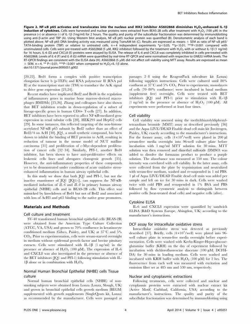
Figure 2. NF-kB p65 activates and translocates into the nucleus and IKK2 inhibitor AS602868 diminishes H2O2-enhanced IL-1binduction of cytokines. Cells were harvested and nuclear proteins were extracted from BEAS-2B cells after treatment with H2O2 (100 mM) in thepresence (+) or absence (-) of IL-1b (1ng/ml) for 2 hours. The quality and purity of the subcellular fractionation was determined by immunoblottingusing anti-b-actin and TBP (A). Using Western blot analysis, NF-kB p65 nuclear protein was quantified (B). Densitometric analysis of each band isplotted above. NF-kB p65 DNA binding activity was measured using TransAM kit (C). Results are expressed as means 6 SEM as ratio of NF-kB p65/TATA-binding protein (TBP) or relative to untreated cells. n = 4 independent experiments *p,0.05; **p,0.01; ***P,0.001 compared withunstimulated cells. Cells were pre-treated with AS602868 (5 mM, IKK2 inhibitor) followed by the treatment with H2O2 with or without IL-1b (1 ng/ml)for 16 hours. IL-6 (D) and CXCL8 (E) proteins were assayed by ELISA. The release of IL-6 and CXCL8 was completely inhibited in cells pre-treated withAS602868. Levels of IL-6 (F) and IL-8 (G) mRNA were quantified by real-time RT-QPCR and were normalised with respective to GNB2LI mRNA levels. TheRT-QPCR findings are consistent with the ELISA data (H). AS602868 (5 mM) did not affect cell viability using MTT assay. Results are expressed as mean6 SEM. n = 4. ** P,0.01; ***P,0.001 when compared to H2O2+IL-1b alone.doi:10.1371/journal.pone.0095051.g002
BET Inhibitors Reduce Inflammation
PLOS ONE | www.plosone.org 4 April 2014 | Volume 9 | Issue 4 | e95051

antibodies against cytoplasmic (b-actin) and nuclear (TATA
binding protein) proteins to demonstrate standardization of this
method.
NF-kB activation assayNF-kB activation after 2 hrs was measured in nuclear extracts
with TransAM NF-kB activation kit (Active Motif) according to
the manufacturer’s instructions.
Co-Immunoprecipitation (Co-IP)Whole cell lysates were prepared by incubating cells with IP
buffer (150 mM NaCl, 50 mM Tris-HCl pH 8, 0.5% Sodium
deoxycholate, 0.5% NP40, protease inhibitors cocktail [Roche
Applied Science, Burgess Hill, UK) for 30 mins on ice and
centrifuged at 14000 rpm for 10 mins. 500 mg of protein extract
was incubated with 5 mg of antibody Brd4 (Santa Cruz Biotech-
nology, Santa Cruz, California USA). The immune complex was
incubated overnight on rotator at 4uC. Protein A/G-plus agrose
beads (20 ml 50% slurry; Santa Cruz Biotechnology) were added to
the complex and left on the rotator at 4uC for 2 hours. The
Figure 3. NF-kB p65 acetylation and association with Brd4 protein. (A) BEAS-2B cells were stimulated with H2O2 in the presence (+) orabsence (-) of IL-1b (1 ng/ml) for 2 hours, nuclear (A) and cytoplasmic (B) extracts were fractioned by Western blot and membranes were probed withanti-acetylated NF-kB p65 antibody. The blots show that acetylated-310 (Ac310) NF-kB p65 is predominantly found in the nucleus when comparedwith the cytoplasm. (C) Brd4 protein was immunoprecipitated from whole cell extracts following treatments and separated by SDS-PAGE andsubsequently analysed by Western blotting using an anti-NF-kB p65 antibody. Each blot is representative of 3 independent experiments anddensitometric analysis of each band is plotted as bar graph above it. TBP: TATA-binding protein; *p,0.05; **p,0.01 compared with control(unstimulated).doi:10.1371/journal.pone.0095051.g003
BET Inhibitors Reduce Inflammation
PLOS ONE | www.plosone.org 5 April 2014 | Volume 9 | Issue 4 | e95051

BET Inhibitors Reduce Inflammation
PLOS ONE | www.plosone.org 6 April 2014 | Volume 9 | Issue 4 | e95051

complex was then washed using IP wash buffer (50 mM Tris-HCl
pH 7.4, 0.5% (v/v) NP40, 150 mM NaCl) three times. Proteins
were eluted from complex using 35 ml of elution buffer (50 mM
Glycine, HCl pH 2.4) followed by addition of 3.5 ml of neutralising
solution (2 M Tris pH 8, 1.5 M NaCl, 1 mM EDTA). Samples
were run on SDS-PAGE gel.
Western blottingNuclear and cytoplasmic extracts were fractionated by SDS-
PAGE gel as previously described [38]. Proteins were transferred
and immobilised on nitrocellulose membranes. Membranes were
incubated with specific antibodies directed against NF-kB p65, b-
actin, TATA-binding protein (All from Santa Cruz Biotechnolo-
gy), acetylated p65-lysine 310 (Abcam, Cambridge, UK), Brd2 and
Brd4 (Bethyl Lab, Montgomery, TX, USA). After washing,
membranes were incubated with horseradish peroxidase-linked
anti-rabbit immunoglobulin (DAKO Ltd, Ely, UK) and detected
by enhanced chemiluminescence (ECL) (GE healthcare, Amer-
sham, UK). The membranes were exposed to X-ray and the bands
density was quantified using the GelDoc Imaging System (UVP,
Upland, CA). This was standardised against the loading controls
and the data were represented in graphs.
Measurement of mRNA transcriptsCells were treated as above and total RNA was isolated using an
RNeasy mini kit (Qiagen, Crawley, UK) and the concentration of
RNA determined using the absorbance ratio of 260 nm/280 nm
by NanoDrop 2000c spectrophotometer (Thermo Fisher Scientif-
ic, Waltham, MA, USA). Single stranded cDNA was synthesized
using AMV-reverse transcriptase (Promega, Southampton, UK) as
described previously [39]. QPCR was performed in a Rotor-Gene
3000TM PCR machine (Corbett, Research, Cambridge, UK)
using a QuantiTect SYBR Green PCR Kit (Qiagen) according to
the manufacturer’s instructions. The RT-PCR primers used were
as follows: Brd2 (NCBI: NM_005104.3)-F, 59-GGGGTGGCAGT-
GCTGCTTTA-39; Brd2-R, 59-GCTCAGCTGCCGCTTCTC-
AT-39; Brd4 (NCBI: NM_014299.2)-F, 59-CCACACTGCGTGA-
GCTGGAG-39; Brd4-R, 59-ATCTTGGAGGAGCCGGCAAT-
39; GNB2L1 (NCBI: NM_006098.4)-F, 59-CTCCGCCTCTCGA-
GATAAGACC-39; GNB2L1-R, 59-GCAAACTGGCCATCTGA-
GGA-39; IL6 (NCBI: NM_000600.3)-F, 59-AGGAGACTTGCC-
TGGTGAAA-39; IL6-R, 59-GCTCTGGCTTGTTCCTCACT-
39; IL8 (NCBI: NM_000584.3)-F, 59-AGACAGCAGAGCACA-
CAAGC-39; IL8-R, 59-ATGGTTCCTTCCGGTGGT-39. The
PCR data for each gene of interest was normalized to
housekeeping gene (GNB2L1) and represented as fold change
using the delta-delta CT (2-DDCT) method.
Chromatin Immunoprecipitation (ChIP) assayChIP assay was carried out as previously described [38]. Briefly,
epithelial cells (BEAS-2B) were treated with H2O2 (100 mM) in the
presence or absence of IL-1b (1 ng/ml) or left untreated for 2 hrs.
In some experiments, cells were pre-treated with JQ1 (500 nM) for
4 hrs. After treatments, protein-DNA complex were fixed by
formaldehyde (1% final concentration) and immunoprecipitated
with either an anti-p65 (5 mg, Santa Cruz Biotechnology) or an
anti-Brd4 (5 mg, Bethyl Lab) antibody overnight. NF-kB p65 and
Brd4 binding to the IL-8 and the IL-6 promoter were quantified by
real-time QPCR using a QantiTech SYBR green PCR kit
(Qiagen) on Rotor gene 3000 (Corbett Research). Immunopre-
cipitated DNA was quantified by qPCR and compared to the level
of promoter specific DNA obtained at t = 0 which was given an
arbitrary value of 1. IL-8 [40,41] and IL-6 [42-44] primers have
been previously published. Primer pairs of IL-6 and IL-8 were as
follows: IL-6, forward, 59-AGCACTGGCAGCACAAGGCA-
AAC-39 and IL-6, reverse 59-CAAGCCTGGGATTATGAAG-
AAGG-39; and IL-8, forward, 59-GGGCCATCAGTTGCAA-
ATC-39 and IL-8, reverse, 59-TTCCTTCCGGTGGTTTCT-
TC-39.
siRNA transfectionBEAS-2Bs cells were seeded in 6-well plates at 0.36106 cells/
well and transfected with ON-Target plus SMART pool Brd2 (L-
004935-00-0005) or Brd4 (L-004937-00-0010) or control (D-
001810-10-05) small interfering RNAs (siRNA) at 20 nM concen-
tration (all from Dharmacon Thermo Scientific, Chicago, IL,
USA) using HiPerFect transfection reagent (Qiagen) as described
by the manufacturer. After 72 hrs, nuclear extracts were examined
for Brd2 and Brd4 protein expression using Western blotting.
Total mRNA was extracted and converted to cDNA and Brd2 and
Brd4 expression were assessed.
Confocal MicroscopyEpithelial cells were seeded on to 2-well chambered coverslips
(Sigma-Aldrich, Poole, UK) and allowed to attach overnight at
37uC and 5% CO2 in basal media. Cells were fixed and
permeabilized using 3% paraformaldehyde (PFA) with 0.5%
Triton-X-100 (Sigma-Aldrich) for 10 mins following stimulations.
Slides were incubated with blocking buffer (50% fetal calf serum
(FCS) 0.1%TX-100) for 30 min, followed by an hour incubation
with primary anti-acetyl-Histone 3 antibody (Milipore, Watford
UK) diluted to 1:300, followed by an hour incubation with
fluorescein isothiocyante-conjugated secondary antibody (Dako,
Ely, UK). Cells were also stained with Alexa Fluor 488 phalloidin
(Invitrogen) for cytoplasmic (Cytoskeleton actin) and DAPI (4’,6-
Diamidino-2-Phenylindole, Dihydrochloride; Invitrogen) for nu-
clear staining. Coverslips were mounted onto slides and allowed to
dry before quantifying fluorescence intensities with imaging
software (Leica Confocal Software Lite, Heidelberg, Germany).
Statistical analysisData are represented as mean 6 standard error mean (SEM).
Data were analysed by Student’s t test for two sets of data or by
one-way ANOVA/Dunn’s multiple comparison test for compar-
ing more than two sets of data. GraphPad Prism (La Jolla, CA,
US) was used to evaluate the data. Differences were considered
significant for P values of #0.05.
Figure 4. H3 acetylation, p65 and Brd4 binding to IL-6 and IL-8 kB promoter sites. Chromatin immunoprecipitation (ChIP) assays show thatIL-1b induces p65 DNA binding to both IL-6 (A) and IL-8 (B) promoters. H2O2 by itself does not affect p65 DNA binding activity; however, when co-treated with IL-1b, the affinity is enhanced by 7-fold at IL-6 promoter site and 20-fold at IL-8 promoters. Brd4 is also recruited to the same kBpromoter regions in the IL-6 (C) and IL-8 (D) promoters as p65. Histone 3 is acetylated at the IL-6 (E) and IL-8 (F) kB promoter sites followingtreatments. IgG is non-specific antibody used as a negative control. Furthermore, H3 acetylation was confirmed using confocal microscopy followingIL-1b stimulation whereas H2O2 had no effect on AcH3 alone or in combination with IL-1b (G). Results are representative of at least 4 independentexperiments.*p,0.05, **p,0.01 compared with control (unstimulated).doi:10.1371/journal.pone.0095051.g004
BET Inhibitors Reduce Inflammation
PLOS ONE | www.plosone.org 7 April 2014 | Volume 9 | Issue 4 | e95051

Figure 5. Concentration dependent reduction of IL-6 and CXCL8 by BET inhibitors. Cells were pre-treated with JQ1 and JQ1 (-)enantiomers (A, B) or PFI-1 (C, D) for 4 hours followed by IL-1b (1 ng/ml) for 16 hrs. IL-6 (A, C) and CXCL8 (B, D) proteins were assayed by ELISA. Theeffect of JQ1 and JQ1 (-) (E) and PFI-1 (F) on cell viability was assessed by MTT assay. n = 3 independent experiments. Points represent mean 6 SEM
BET Inhibitors Reduce Inflammation
PLOS ONE | www.plosone.org 8 April 2014 | Volume 9 | Issue 4 | e95051

Results
Hydrogen peroxide (H2O2) induces intracellular oxidativestress and enhances inflammatory mediator expression
Exogenous H2O2 (0–800 mM) enhanced intracellular ROS in
BEAS-2B cells a concentration-dependent manner after 2 hours
which reached significance at concentrations of 100 mM or above
(Fig. 1A). However, at concentrations .200 mM there was a
significant loss in cell viability (Fig. 1B) and H2O2 (100 mM) was
selected for all future experiments. H2O2 alone had no significant
effect on either IL-6 or CXCL8 release at 16 hrs; however, H2O2
significantly enhanced the production of IL-6 (Fig. 1C) and
CXCL8 (Fig. 1D) release from IL-1b (1 ng/ml) co-stimulated
cells. This effect was mirrored by changes in IL-6 and IL-8 mRNA
expression (Fig. 1E–F).
The expression of IL-6 and IL-8 are regulated by the NF-kB signalling pathway
The subcellular extraction process was demonstrated to be
highly reproducible with little or no cross-contamination of
cytoplasmic and nuclear extracts using Western blotting (Fig. 2A).
H2O2 alone had no significant effect on p65 nuclear translocation
after 2 hrs whereas it enhanced IL-1b-induced p65 nuclear import
(Fig. 2B). IL-1b significantly enhanced p65 DNA binding to a
consensus kB-response sequence (59-GGGACTTTCC-39) 7-fold
and this was further increased (up to 9-fold greater than baseline)
by pre-treatment with H2O2 (Fig. 2C). In contrast, H2O2 alone
had no significant effect on NF-kB p65 binding activity (Fig. 2C).
Furthermore, IL-1b+H2O2-induced release of both IL-6 and
CXCL8 proteins (Fig. 2D, E) and the expression of IL-6 and IL-8
mRNA (Fig. 2F, G) was completely suppressed by the selective
IKK2 inhibitor AS602868 [45,46]. Cell viability was not affected
by AS602868 (Fig. 2H).
Effect of IL-1b and H2O2 on p65 acetylation andassociation with Brd4
NF-kB p65 is subjected to post-translational modifications such
as acetylation that modulate its activity [47,48]. Western blot
analysis revealed that acetylated NF-kB p65 is found predomi-
nantly in the nuclear compartment in IL-1b stimulated cells after
2 hrs with little in the cytoplasmic compartment after any
treatment (Fig. 3A, B). Although, H2O2 alone did not induce
p65 K310 acetylation, IL-1b-induced p65 acetylation was
enhanced after IL-1b+H2O2 co-stimulation (Fig. 3A). This
suggests that acetylation of NF-kB at lysine-310 is associated with
maximal NF-kB activation and/or translocation into the nucleus
[49]. Brd4 can interact with acetylated p65 [24,28] as part of a
complex with p-TEFb and RNA polymerase II [28,30]. Using co-
immunoprecipitation experiments we found that there was an
association between acetylated p65 and Brd4 (Fig. 3C), however,
neither H2O2 nor IL-1b or in combination affected this
association. This suggests that acetylated p65-Brd4 association is
not directly linked to ROS-induced enhancement of NF-kB
function.
NF-kB p65 and Brd4 are recruited to the native IL-6 andIL-8 promoters
Following 2 hrs IL-1b treatment, p65 ChIP analysis (Fig. 4A,B) showed a marked enhancement in binding to kB elements
within the native IL-6 and IL-8 promoters which was enhanced by
co-stimulation with H2O2 (7- and 20-fold respectively). In contrast,
H2O2 alone had no effect on p65 binding to either the IL-6 or IL-8
promoters. IL-1b also significantly increased binding of Brd4 to
these kB sites in the IL-6 and IL-8 promoters (Fig. 4C, D). Again,
this recruitment was augmented by co-stimulation with H2O2 and
IL-1b at the IL-6 (6-fold) and IL-8 (8-fold) promoters compared to
unstimulated cells (Fig. 4C, D). Pan-histone H3 acetylation at the
IL-6 and IL-8 promoter kB sites was significantly elevated
following IL-1b stimulation and slightly further increased with
the addition of H2O2 although this did not reach significance
(Fig. 4E, F). H2O2 alone did not affect Brd4 nor pan-histone H3
acetylation at these sites (Fig. 4C-F). Confocal analysis also
confirmed an increased H3 acetylation in cells stimulated with IL-
1b in comparison with unstimulated cells whereas H2O2 alone had
little effect (Fig. 4D). We did not observe H4 acetylation at either
of the promoters at the 2 hr time point studied, suggesting that H4
acetylation might be time- and gene-dependent.
BET inhibitors reduce IL-1b induced inflammationBEAS-2Bs cells were treated with JQ1 and its inactive
enantiomer JQ1(-) at a range of concentrations (5610292
1026 M) for 4 hours followed by IL-1b (1 ng/ml) stimulation for
16 hours. JQ1 inhibited IL-1b-induced IL-6 (Fig. 5A) and
CXCL8 (Fig. 5B) release in a concentration-dependent manner
with a maximal suppression of 86.862.0% (IL-6) and 72.563.1%
(CXCL8) at 561027 M. In contrast, JQ1(-) (561027 M) had a
non-significant suppressive effect on IL-1b-stimulated IL-6
(35.165.9%) and CXCL8 (34.163.3%) release (Fig. 5A, B).
PFI-1, a structurally distinct BET inhibitor [50], also attenuated
the release of both IL-1b-induced IL-6 (Fig. 5C) and CXCL8
(Fig. 5D) in concentration-dependent manner. The IC50 for PFI-1
inhibition of IL-1b-induced IL-6 protein levels was 7.364.2610-
7 M and a similar effect was seen for the inhibition of IL-1b-
induced CXCL8 release (7.464.861027 M). PFI-1 inhibited IL-
1b-induced IL-6 and CXCL8 release with maximal suppression of
(80.761.9%) and (63.663.8%), respectively.
Cell viability was significantly affected at the highest concen-
trations of JQ1 and JQ1(-) tested (1026 M, Fig. 5E) and does not
therefore account for the reduction in IL-6 and CXCL8 release
seen at 561027 M. High concentrations of PFI-1 (461026 M) also
significantly reduced cell viability and were excluded from
subsequent experiments (Fig. 5F). These results indicated that
PFI-1 has a similar efficacy as JQ1. We used PFI-1 at 161026 M
in subsequent experiments. The lack of effect of JQ1 and PFI-1 on
cell viability as these concentrations was confirmed using FACS
analysis and Live/Dead Aqua blue staining (Fig. 5G). Cells
treated with DMSO (control), JQ1 (0.5 mM), JQ1(-) (0.5 mM) or
with PFI-1 (1 mM) resulted in only 5% overall cell death with 95%
cells being viable.
**p,0.01;***P,0.001 compared with IL-1b stimulation. #p,0.05 JQ1(-) versus JQ1. £££p,0.001when PFI-1 compared with IL-1b stimulation. $p,0.05;$$p,0.01when compared to control. (G) Cells were heat treated at 90uC or left untreated, mixed together and stained with LIVE/DEAD Fixable Aquastain then analysed by flow cytometry. Cells were checked with forward scatter detector (FSC) and side scatter detector (SSC) and analysed by densitygraph to check cell size and granularity. Fragmented cells were excluded from the study. Histogram shows separation of live cells (left) and dead cells(right). These parameters were used to assess cell viability following treatment with JQ1 (0.5 mM) and PFI-1 (1 mM) for 16 hours. DMSO/Control (,1%)alone, PFI-1, JQ1(-) or JQ1 resulted in only 5% decrease of overall cell viability. The data is representative of 3 independent experiments.doi:10.1371/journal.pone.0095051.g005
BET Inhibitors Reduce Inflammation
PLOS ONE | www.plosone.org 9 April 2014 | Volume 9 | Issue 4 | e95051

Figure 6. The BET inhibitors (JQ1 and PFI-1) reduce inflammatory mediator production. Cells were pre-treated with either JQ1 or JQ1(-)both at 500 nM for 4 hours followed by stimulation with H2O2 in the presence (+) or absence (-) of IL-1b (1 ng/ml) or both for 16 hours or leftunstimulated. IL-6 (A) and CXCL8 (B) proteins were assayed by ELISA. IL-6 (C) and IL-8 (D) transcripts were quantified by RT-PCR. n = 4 independentexperiments. Bar graph represents mean 6 SEM *p,0.05, **p,0.01, when compared JQ1(-) with JQ1 treated cells. Under similar experimentalconditions the effect of PFI-1 (1 mM) on IL-6 (E) and CXCL8 (F) proteins were assayed by ELISA. IL-6 (G) and IL-8 (H) mRNA levels were quantified byRT-QPCR. n = 4 independent experiments. Bar graph represent mean 6 SEM *p,0.05, **p,0.01, when compared cells treated with or without PFI-1.doi:10.1371/journal.pone.0095051.g006
BET Inhibitors Reduce Inflammation
PLOS ONE | www.plosone.org 10 April 2014 | Volume 9 | Issue 4 | e95051

BET Inhibitors Reduce Inflammation
PLOS ONE | www.plosone.org 11 April 2014 | Volume 9 | Issue 4 | e95051

BET inhibitors reduced oxidative stress-enhancedinflammation
We demonstrated above that H2O2 (100 mM) alone has
minimal effect on IL-6 and CXCL8 expression; however, it
enhanced the expression of both cytokines (IL-6 and CXCL8) in
IL-1b-induced cells. The release of IL-1b H2O2-induced IL-6
(Fig. 6A) and CXCL8 (Fig. 6B) proteins and IL-6 (Fig. 6C) and
IL-8 (Fig. 6D) mRNA was markedly suppressed in JQ1 but not
JQ1(-) treated cells. However, the levels of IL-6 and CXCL8 did
not return to baseline. A similar effect was seen with PFI-1 which
attenuated the release of IL-6 (Fig. 6E) and CXCL8 (Fig. 6F)
proteins and IL-6 (Fig. 6G) and IL-8 (Fig. 6H) mRNA when
compared with untreated cells. As with JQ1, mediator levels did
not return to baseline in PFI-1 treated cells.
The effects of JQ1 and PFI-1 are mimicked by Brd4, butnot Brd2, knockdown
To determine the specificity of the JQ1 and PF-1 effects, cells
were transfected with Brd4 or non-specific small interfering RNAs
for 72 hours resulting in significantly decreased expression of Brd4
mRNA (71.863.2%, p,0.05) (Fig. 7A) and protein 63.2667.5%,
p,0.05) expression (Fig. 7B). The release of IL-1b/H2O2-
stimulated IL-6 (Fig. 7C) and CXCL8 (Fig. 7D) proteins was
significantly suppressed in Brd4-knockdown cells in comparison
with control non-specific siRNA transfected cells. The degree of
suppression of IL-6 and CXCL8 was the similar to that seen in
JQ1- and PFI-1-treated cells (Fig. 6). In contrast, although there is
an 80% structural homology between Brd2 and Brd4 [51,52] we
were unable to show any effect of Brd2 knockdown on
inflammatory gene expression. Following treatment of cells with
Brd2 siRNA, there was a significant suppression of both Brd2
mRNA (Fig. 7E) and protein (Fig. 7F) expression, but no effect
on IL-1b/H2O2-stimulated IL-6 (Fig. 7G) or CXCL8 (Fig. 7H)
release. These data indicate that Brd4 is central to NF-kB-
mediated induction of IL-6 and IL-8 expression and that JQ1 and
PFI-1 preferentially target Brd4 and not Brd2 in airway epithelial
cells.
JQ1 inhibits p65 and Brd4 association at IL-6 and IL-8promoters
ChIP analysis was used to investigate the mechanism of JQ1 at
the p65 to the IL-6 and IL-8 promoters. Binding of Brd4 and p65
increased 5-fold at the IL-6 promoter in H2O2+IL-1b stimulated
cells when compared with unstimulated cells. This was not affected
by the presence of the inactive enantiomer JQ1(-). However, JQ1
significantly reduced Brd4 (Fig. 8A) and p65 (Fig. 8B) binding at
the IL-6 promoters. Similarly, Brd4 and p65 association with the
IL-8 promoter increased 4-fold and 10-fold respectively in
stimulated cells (H2O2+IL-1b). This association was abolished by
JQ1 but was unaffected by JQ1(-) (Fig. 8C, D). The results also
show that although the recruitment of p65 and Brd4 is completely
abrogated at both the IL-6 and IL-8 promoters the expression of
both IL-6 and IL-8 did not returned to baseline levels in JQ1 pre-
treated cells. This suggests that post-transcriptional factors may
also play a role in driving the expression of IL-6 and CXCL8
expression in these cells.
JQ1 reduced oxidative stressed enhanced inflammationin human primary epithelial cells
We repeated some key experiments in primary normal human
bronchial epithelial (NHBE) cells from 4 different donors. We
initially demonstrated that JQ1 had a significantly greater ability
to suppress IL-1b-stimulated IL-6 (Fig. 9A) and CXCL8 (Fig. 9B)
release than JQ1(-). JQ1 inhibited IL-1b-induced IL-6 and
CXCL8 release in concentration-dependant manner with maxi-
mal suppression of 72.461.8% (IL-6) and 64.063.8% (CXCL8) at
561027 M whereas the inactive enantiomer had much less effect
on IL-6 (49.962.9) and CXCL8 (35.165.3) release.
We then examined the effect of H2O2 on NHBE cells function.
H2O2 significantly induced intracellular ROS at concentrations .
100 mM when compared to untreated cells (Fig. 9C) although this
was associated with significantly reduced cell survival at concen-
trations .200 mM) (Fig. 9D). Therefore, NHBE cells were treated
with 100 mM of H2O2 in the presence or absence of IL-1b in
subsequent experiments. IL-1b-induced release of IL-6 (Fig. 9E)
and CXCL8 (Fig. 9F) was further enhanced when co-treated with
H2O2 which was mirrored by changes in IL-6 (Fig. 9G) and IL-8
(Fig. 9H) mRNA transcripts. There was no significant difference
between IL-6 and CXCL8 expression between JQ1(-) and JQ1
treated cells under control conditions. JQ1 markedly attenuated
the release of IL-1b/H2O2-induced IL-6 (Fig. 9E) and CXCL8
(Fig. 9F) proteins and IL-6 (Fig. 9G) and IL-8 (Fig. 9H) mRNA
expression when compared with JQ1(-) treated cells.
Discussion
In this study, we show that H2O2 induces intracellular ROS in
human airway epithelial cells (BEAS-2B and NHBE cells) and
enhances IL-1b-induced IL-6 and CXCL8 expression via
increased p65 and Brd4 promoter association at kB sites in the
native gene promoters. This is in conjunction with increases in
histone H3 acetylation at these kB sites. The BET inhibitors JQ1
and PFI-1 both suppressed IL-1b- and IL-1b/H2O2-induced IL-6
and CXCL8 protein and mRNA expression in a concentration-
dependent manner. Knockdown studies revealed that IL-6 and
CXCL8 release is significantly reduced in Brd4, but not Brd2,
depleted cells. Furthermore, H2O2-enhanced IL-1b-induced
recruitment of p65 and Brd4 to the IL-6 and IL-8 promoters is
abolished in JQ1-, but not in JQ1(-)-, pre-treated cells.
COPD primarily affects lungs; however, it now recognised a
disease with organ-specific characteristics and systemic manifesta-
tions such as chronic inflammation [53–55]. We have analysed the
expression of two of the most important pro-inflammatory
cytokines, IL-6 and CXCL8, that are elevated in plasma, BAL
fluids and sputum of COPD patients and whose expression
correlates with disease severity [56–58]. These cytokines are
regulated by the NF-kB signalling pathway which also orchestrates
many aspects of the cellular immune response, apoptosis,
differentiation and inflammation [59,60]. High concentrations of
H2O2 are found in breathe condensates of COPD patients and this
Figure 7. Knockdown of Brd4 reduces inflammation but not Brd2. Cells were transfected with either pooled Brd4 or Brd2 siRNAs (20 nM) ornon-specific siRNAs (20 nM). 72 hours post-transfection Brd4 mRNA (A) was quantified by RT-QPCR and nuclear extracts were analysed (B) byimmunoblotting using anti-Brd4 and -TATA-binding protein (TBP). IL-6 (C) and CXCL8 (D) proteins were measured by ELISA in post-transfected cellsfollowing stimulation with H2O2 and IL-1b together. n = 4 independent experiments. 72 hours post-transfection, Brd2 mRNA (E) was quantified by RT-QPCR and nuclear extracts were analysed (F) by immunoblotting using anti-Brd2 and TBP. Brd2 siRNA knockdown had no effect on either IL-6 (G) orCXCL8 (H) expression. n = 4 independent experiments. Results represent mean 6 SEM *p,0.05, **p,0.01, ***p,0.001, non-specific siRNA comparedto Brd2 or Brd4 siRNA.doi:10.1371/journal.pone.0095051.g007
BET Inhibitors Reduce Inflammation
PLOS ONE | www.plosone.org 12 April 2014 | Volume 9 | Issue 4 | e95051

has been linked to disease pathophysiology [61,62]. For example,
H2O2 can induce the release of potent chemotactic factors such as
CXCL8 from epithelial cells [63] by resulting in increased hyper-
acetylation at the IL-8 promoter via alteration in HDAC/HAT
balance [64].
NF-kB is a redox-sensitive transcription factor that converts
oxidative stress signals into changes in gene expression associated
with diverse cellular activities including inflammation [65]. NF-kB
activation requires translocation of the NF-kB p65/p50 dimer into
the nucleus where it binds to promoter regions of inflammatory
genes forming a transcriptional activator complex [22,66].
Optimal induction of NF-kB activity also requires post-translation
modification of p65 e.g. phosphorylation, acetylation and
ubiquitination, which modulates DNA binding and transcriptional
activity. For example, acetylation of p65/RelA at Lysine-310
(K310) is essential for optimal NF-kB transcriptional activity
[28,30,67]. Acetylated p65 provides a docking site for Brd4 and
may be an important target in the ability of BET inhibitors to
suppress inflammation [30]. In the present study, we demonstrated
that p65 is acetylated within the nucleus following stimulation of
cells with IL-1b and H2O2 although this did not affect the degree
of p65:Brd4 association in the nucleus under any of the conditions
tested. This data is in contrast to that observed in A549 lung
adenocarcinoma-like cells where TNFa stimulation was essential
for p65:Brd4 complex formation [30]. Our data suggests that in
bronchial airway epithelial cells that Brd4 and p65 may be part of
a pre-formed transcriptional complex.
An interesting observation made in this study is that the
recruitment of Brd4 is associated with enhanced pan-acetylation of
histone H3 but not of H4 at the IL-6 and IL-8 promoters
confirming previous data [68–70]. Although, H2O2 alone was
unable to induce H3 acetylation directly in BEAS-2B cells, it
slightly enhanced the histone acetylation seen with IL-1b alone.
This may reflect either a direct effect on localised HAT activity or
a reduction in HDAC activity as previously reported in BEAS-2B
cells [37]. This will result in an alteration in the local chromatin
environment/structure which will modulate transcription factor
association with promoter regions [71]. Future studies utilising
DNase1 hypersensitivity assays, for example, may reveal changes
in local chromatin structure following H2O2 treatment of cells
which will enable greater recruitment of pTEFb by Brd4 and
subsequent elongation of inflammatory gene expression by RNA
polymerase II [72].
The chemically distinct bromodomain inhibitors (JQ1 and PFI-
1) decreased recruitment of both Brd4 and p65 to the IL-6 and IL-
8 promoters and reduced IL-6 and CXCL8 protein release from
BEAS-2Bs and NHBE cells. This confirms data from Nicodeme
and colleagues who reported that I-BET disrupts the interaction of
bromodomain proteins and acetylated histones at the IL-6
promoter in LPS-stimulated macrophages [25]. Inhibition of
Brd4 either by JQ1 or knockdown reduced both IL-1b- and IL-
Figure 8. The effect of JQ1 on Brd4 and p65 binding to IL-6 and IL-8 promoters. Chromatin immunoprecipitation (ChIP) assay shows that IL-1b (1 ng/ml) and H2O2 (100 mM) induces Brd4 (A) and p65 (B) DNA binding to the IL-6 promoter by 5-fold which is abolished in cells pre-treated withJQ1 (500 nM). Similarly, Brd4 (C) and p65 (D) DNA binding at the IL-8 promoter is increased following H2O2 and IL-1b stimulation in JQ1(-) (500 nM)pre-treated cells by 10- and 4-fold. This binding is diminished in cells pre-treated with the active JQ1 (500 nM). Results are representative of at least 3independent experiments.*p,0.05, **p,0.01, ***p,0.001 when compared with unstimulated cells.doi:10.1371/journal.pone.0095051.g008
BET Inhibitors Reduce Inflammation
PLOS ONE | www.plosone.org 13 April 2014 | Volume 9 | Issue 4 | e95051

BET Inhibitors Reduce Inflammation
PLOS ONE | www.plosone.org 14 April 2014 | Volume 9 | Issue 4 | e95051

1b/H2O2-induced IL-6 and CXCL8 expression by ,80%
whereas JQ1 completely blocked p65 DNA binding and subse-
quent Brd4 recruitment at the kB sites studied. AS602868, an
IKK2 inhibitor, almost completely abrogated IL-1b- and IL-1b/
H2O2-induced IL-6 and CXCL8 protein and mRNA expression
confirming a key role for NF-kB in the transcriptional control of
these genes. This suggests that other kB sites within the IL-6 and
IL-8 promoters or enhancers may play a role in gene expression or
that other inflammatory signalling pathways may modify the effect
of JQ1 at the transcriptional and/or translational level. Future
studies using ChIP-seq may address this. The failure to completely
suppress CXCL8 gene expression may be important clinically as
total suppression of the innate immune response may lead to
opportunistic infections. The ability of JQ1 to dampen rather than
ablate the immune/inflammatory response may therefore be of
benefit in the treatment of patients with a chronic inflammatory
disease. It will be of interest to determine whether JQ1 protects
other innate immune genes in epithelial cells following stimulation
with IL-1b or IL-1b/H2O2.
This is the first report to our knowledge that demonstrates a
repressive effect of JQ1 and PFI-1 on IL-1b-induced CXCL8
expression. Previous studies [25,73] have showed that CXCL1
mRNA, the murine equivalent of CXCL8, was unaffected by I-
BET in mouse macrophages at baseline or after LPS-stimulation.
However, Huang et al. [30] showed that the Brd4 depletion in
LPS-stimulated THP-1 impaired CXCL8 expression. In our study
Brd2 depletion in BEAS-2B cells had no effect on inflammatory
cytokine expression. In contrast, Belkina et al. have shown that, in
addition to Brd4, LPS-induced inflammation in murine bone
marrow-derived macrophages (BMDMs) is mediated via Brd2
which binds to the IL-6 promoter [73]. This difference may be
species or cell type specific. ChIP-seq analysis indicates that Brd4
co-localizes with RNA pol II at both enhancers and promoters of
all active genes in primary human CD4+ T cells. This interaction
is disrupted upon JQ1 treatment leading to reduced lineage-
specific gene expression and acetylated histone-bromodomain
association [27]. The study implies that Brd4 could be used as a
tool to identify active promoters and enhancers in a genome-wide
manner in human epithelial cells. JQ1 has also been studied in
NUT (Nuclear protein in testis) midline carcinoma (NMC) mouse
model and various cancer cell lines, showing inhibition of cell
proliferation and differentiation [31,33]. Furthermore, Brd4 is
believed to be an inherited susceptibility gene for breast cancer
progression and metastasis. It has been shown that deletion of
Brd4 proline-rich domain at C-terminal results in a loss of contact
inhibition and changed in cell morphology from epithelial to a
mesenchymal-like phenotype [74].
There are some limitations to this study. Firstly, our data shows
a difference in IL-6 and CXCL8 protein release in response to IL-
1b and to IL-1b+H2O2 in the presence and absence of JQ1
suggesting that whatever effect IL-1b has on Brd4 function this is
modified at least to some extent by the presence of H2O2
particularly in primary cells. However, we are unable to rule out
the possibility that H2O2 stimulation modifies other aspects of p65
activity. Analysis of the effects of Brd4 knockdown on IL-1b- and
H2O2-stimulated cells on mediator protein, mRNA and promoter
ChIP analysis would help resolve this issue. Secondly, we show a
differential effect of BET mimics on IL-6 and CXCL8 protein
release compared with mRNA expression data. This indicates a
degree of disconnect in both BEAS-2B and primary epithelial cells.
Additional experiments, using combinations of BET protein
knockdowns and pharmacological inhibition, should be performed
to examine non-histone-based mechanisms of translational control
and secretion.
In summary, we have shown that IL-1b and IL-1b/H2O2 can
enhance recruitment of p65 and Brd4 to the native IL-6 and IL-8
promoters in epithelial cells. Inhibition of Brd4 by the structurally
distinct bromodomain inhibitors JQ1 and PFI-1 and by genetic
knockdown led to a reduction in IL-6/-8 mRNA and protein
expression. This was associated with an attenuation of p65 and
Brd4 binding to the native IL-6 and IL-8 promoters. The
specificity and long-term side effects of bromodomain inhibitors
needs to be evaluated if these agents are to be used in chronic
inflammatory diseases such as COPD and potentially in the
progression to lung cancer [75]. Despite these concerns, our
findings suggest that inhibition of Brd4 may have therapeutic
potential in inflammatory diseases where oxidative stress and NF-
kB activation is present.
Author Contributions
Conceived and designed the experiments: YMK IMA. Performed the
experiments: YMK. Analyzed the data: YMK. Wrote the paper: YMK
IMA. Manuscript corrections: PK PJB.
References
1. Barnes PJ (2008) Immunology of asthma and chronic obstructive pulmonary
disease. Nat Rev Immunol 8: 183–192.
2. Di Stefano A, Caramori G, Oates T, Capelli A, Lusuardi M, et al. (2002)
Increased expression of nuclear factor-kappaB in bronchial biopsies from
smokers and patients with COPD. Eur Respir J 20: 556–563.
3. Chung KF, Marwick JA Molecular mechanisms of oxidative stress in airways
and lungs with reference to asthma and chronic obstructive pulmonary disease.
Ann N Y Acad Sci 1203: 85–91.
4. Mortaz E, Rad MV, Johnson M, Raats D, Nijkamp FP, et al. (2008) Salmeterol
with fluticasone enhances the suppression of IL-8 release and increases the
translocation of glucocorticoid receptor by human neutrophils stimulated with
cigarette smoke. J Mol Med 86: 1045–1056.
5. Dekhuijzen PN, Aben KK, Dekker I, Aarts LP, Wielders PL, et al. (1996)
Increased exhalation of hydrogen peroxide in patients with stable and unstable
chronic obstructive pulmonary disease. Am J Respir Crit Care Med 154: 813–
816.
6. Kurien BT, Scofield RH (2008) Autoimmunity and oxidatively modified
autoantigens. Autoimmun Rev 7: 567–573.
7. Dalle-Donne I, Aldini G, Carini M, Colombo R, Rossi R, et al. (2006) Protein
carbonylation, cellular dysfunction, and disease progression. J Cell Mol Med 10:
389–406.
8. Adcock IM, Chou PC, Durham A, Ford P (2009) Overcoming steroid
unresponsiveness in airways disease. Biochem Soc Trans 37: 824–829.
9. Barnes PJ (2012) Development of New Drugs for COPD. Curr Med Chem.
Figure 9. JQ1 reduces oxidative stress-enhanced IL-6 and CXCL8 expression in NHBE cells. NHBE cells pre-treated with JQ1 but not JQ1(-)(both at 56102921026 M) reduced IL-1b induced release of IL-6 (A) and CXCL8 (B) in a concentration-dependent manner. Points represent mean 6SEM *p,0.05; **p,0.01;***P,0.001 when compared with IL-1b stimulation. #p,0.05 JQ1(-) versus JQ1. NHBE cells were treated with a range ofconcentrations of H2O2 and intracellular ROS (C) and cell viability (D) were measured using DCFH-DA and MTT assay, respectively. Results arepresented as mean 6 SEM. N = 4. *p,0.05; **p,0.01; ***p,0.0001; when compared to untreated cells (control). Cells were pre-treated with eitherJQ1 or JQ1(-) both at 5610-7 M for 4 hours followed by stimulation with IL-1b (1 ng/ml) in the presence (+) or absence (-) of H2O2 (100 mM) or bothfor 16 hours or left unstimulated. IL-6 (E) and CXCL8 (F) proteins were assayed by ELISA. IL-6 (G) and IL-8 (H) transcripts were quantified by RT-PCR.n = 5 independent experiments. Bar graph represents mean 6 SEM *p,0.05; **p,0.01; ***p,0.001 when compared with controls. #p,0.05; ##p,0.01; ###p,0.001 when comparing JQ1- with JQ1(-)-treated cells.doi:10.1371/journal.pone.0095051.g009
BET Inhibitors Reduce Inflammation
PLOS ONE | www.plosone.org 15 April 2014 | Volume 9 | Issue 4 | e95051

10. Liu F, Killian JK, Yang M, Walker RL, Hong JA, et al. (2010) Epigenomic
alterations and gene expression profiles in respiratory epithelia exposed tocigarette smoke condensate. Oncogene 29: 3650–3664.
11. Barnes PJ (2011) Glucocorticosteroids: current and future directions.
Br J Pharmacol 163: 29–43.
12. Barnes PJ, Adcock IM, Ito K (2005) Histone acetylation and deacetylation:importance in inflammatory lung diseases. Eur Respir J 25: 552–563.
13. Adcock IM, Cosio B, Tsaprouni L, Barnes PJ, Ito K (2005) Redox regulation of
histone deacetylases and glucocorticoid-mediated inhibition of the inflammatory
response. Antioxid Redox Signal 7: 144–152.
14. Adcock IM, Ford P, Ito K, Barnes PJ (2006) Epigenetics and airways disease.
Respir Res 7: 21.
15. Bannister AJ, Kouzarides T (2011) Regulation of chromatin by histone
modifications. Cell Res 21: 381–395.
16. Mujtaba S, Zeng L, Zhou MM (2007) Structure and acetyl-lysine recognition of
the bromodomain. Oncogene 26: 5521–5527.
17. Deckert J, Struhl K (2001) Histone acetylation at promoters is differentially
affected by specific activators and repressors. Mol Cell Biol 21: 2726–2735.
18. Barnes PJ (2009) Role of HDAC2 in the pathophysiology of COPD. Annu Rev
Physiol 71: 451–464.
19. Jakovcevski M, Akbarian S (2012) Epigenetic mechanisms in neurologicaldisease. Nat Med 18: 1194–1204.
20. Filippakopoulos P, Knapp S (2012) The bromodomain interaction module.
FEBS Lett 586: 2692–2704.
21. Picaud S, Wells C, Felletar I, Brotherton D, Martin S, et al. (2013) RVX-208, aninhibitor of BET transcriptional regulators with selectivity for the second
bromodomain. Proc Natl Acad Sci U S A.
22. Sanchez R, Zhou MM (2009) The role of human bromodomains in chromatin
biology and gene transcription. Curr Opin Drug Discov Devel 12: 659–665.
23. Rodriguez RM, Huidobro C, Urdinguio RG, Mangas C, Soldevilla B, et al.
(2012) Aberrant epigenetic regulation of bromodomain BRD4 in human colon
cancer. J Mol Med (Berl) 90: 587–595.
24. Chiang CM (2009) Brd4 engagement from chromatin targeting to transcrip-
tional regulation: selective contact with acetylated histone H3 and H4. F1000Biol Rep 1: 98.
25. Nicodeme E, Jeffrey KL, Schaefer U, Beinke S, Dewell S, et al. (2010)
Suppression of inflammation by a synthetic histone mimic. Nature 468: 1119–
1123.
26. Belkina AC, Nikolajczyk BS, Denis GV (2013) BET Protein Function Is
Required for Inflammation: Brd2 Genetic Disruption and BET Inhibitor JQ1
Impair Mouse Macrophage Inflammatory Responses. J Immunol, doi: 104049/
jimmunol1202838
27. Zhang W, Prakash C, Sum C, Gong Y, Li Y, et al. (2012) Bromodomain-
containing protein 4 (BRD4) regulates RNA polymerase II serine 2
phosphorylation in human CD4+ T cells. J Biol Chem 287: 43137–43155.
28. Zhang G, Liu R, Zhong Y, Plotnikov AN, Zhang W, et al. (2012) Down-
regulation of NF-kappaB transcriptional activity in HIV-associated kidneydisease by BRD4 inhibition. J Biol Chem 287: 28840–28851.
29. Kim JW, Jang SM, Kim CH, An JH, Kang EJ, et al. (2012) New molecular
bridge between RelA/p65 and NF-kappaB target genes via histone acetyltrans-
ferase TIP60 cofactor. J Biol Chem 287: 7780–7791.
30. Huang B, Yang XD, Zhou MM, Ozato K, Chen LF (2009) Brd4 coactivates
transcriptional activation of NF-kappaB via specific binding to acetylated RelA.
Mol Cell Biol 29: 1375–1387.
31. Filippakopoulos P, Qi J, Picaud S, Shen Y, Smith WB, et al. (2010) Selective
inhibition of BET bromodomains. Nature 468: 1067–1073.
32. Delmore JE, Issa GC, Lemieux ME, Rahl PB, Shi J, et al. (2011) BET
bromodomain inhibition as a therapeutic strategy to target c-Myc. Cell 146:
904–917.
33. Mertz JA, Conery AR, Bryant BM, Sandy P, Balasubramanian S, et al. (2011)Targeting MYC dependence in cancer by inhibiting BET bromodomains. Proc
Natl Acad Sci U S A 108: 16669–16674.
34. Ott CJ, Kopp N, Bird L, Paranal RM, Qi J, et al. (2012) BET bromodomain
inhibition targets both c-Myc and IL7R in high-risk acute lymphoblastic
leukemia. Blood 120: 2843–2852.
35. Picaud S, Da Costa D, Thanasopoulou A, Filippakopoulos P, Fish PV, et al.
(2013) PFI-1, a highly selective protein interaction inhibitor, targeting BET
Bromodomains. Cancer Res 73: 3336–3346.
36. Yellepeddi VK, Kumar A, Maher DM, Chauhan SC, Vangara KK, et al. (2011)
Biotinylated PAMAM dendrimers for intracellular delivery of cisplatin to
ovarian cancer: role of SMVT. Anticancer Res 31: 897–906.
37. Ito K, Hanazawa T, Tomita K, Barnes PJ, Adcock IM (2004) Oxidative stress
reduces histone deacetylase 2 activity and enhances IL-8 gene expression: role oftyrosine nitration. Biochem Biophys Res Commun 315: 240–245.
38. Ito K, Barnes PJ, Adcock IM (2000) Glucocorticoid receptor recruitment of
histone deacetylase 2 inhibits interleukin-1beta-induced histone H4 acetylation
on lysines 8 and 12. Mol Cell Biol 20: 6891–6903.
39. Shorter K, Farjo NP, Picksley SM, Randall VA (2008) Human hair follicles
contain two forms of ATP-sensitive potassium channels, only one of which is
sensitive to minoxidil. FASEB J 22: 1725–1736.
40. Ho SC, Lee KY, Chan YF, Kuo LW, Ito K, et al. (2009) Neutrophil elastase
represses IL-8/CXCL8 synthesis in human airway smooth muscle cells throughinduction of NF-kappa B repressing factor. J Immunol 183: 411–420.
41. Tsuchiya A, Imai K, Asamitsu K, Waguri-Nagaya Y, Otsuka T, et al. (2010)
Inhibition of inflammatory cytokine production from rheumatoid synovialfibroblasts by a novel IkappaB kinase inhibitor. J Pharmacol Exp Ther 333: 236–
243.
42. Hollingshead BD, Beischlag TV, Dinatale BC, Ramadoss P, Perdew GH (2008)
Inflammatory signaling and aryl hydrocarbon receptor mediate synergistic
induction of interleukin 6 in MCF-7 cells. Cancer Res 68: 3609–3617.
43. Goransson M, Elias E, Stahlberg A, Olofsson A, Andersson C, et al. (2005)
Myxoid liposarcoma FUS-DDIT3 fusion oncogene induces C/EBP beta-
mediated interleukin 6 expression. Int J Cancer 115: 556–560.
44. Nettles KW, Gil G, Nowak J, Metivier R, Sharma VB, et al. (2008) CBP Is adosage-dependent regulator of nuclear factor-kappaB suppression by the
estrogen receptor. Mol Endocrinol 22: 263–272.
45. Durham AL, McLaren A, Hayes BP, Caramori G, Clayton CL, et al. (2013)
Regulation of Wnt4 in chronic obstructive pulmonary disease. FASEB J.
46. Koch A, Giembycz M, Ito K, Lim S, Jazrawi E, et al. (2004) Mitogen-activated
protein kinase modulation of nuclear factor-kappaB-induced granulocyte
macrophage-colony-stimulating factor release from human alveolar macrophag-
es. Am J Respir Cell Mol Biol 30: 342–349.
47. Rothgiesser KM, Fey M, Hottiger MO (2010) Acetylation of p65 at lysine 314 isimportant for late NF-kappaB-dependent gene expression. BMC Genomics 11:
22.
48. Ito K, Charron CE, Adcock IM (2007) Impact of protein acetylation in
inflammatory lung diseases. Pharmacol Ther 116: 249–265.
49. Chen LF, Mu Y, Greene WC (2002) Acetylation of RelA at discrete sites
regulates distinct nuclear functions of NF-kappaB. EMBO J 21: 6539–6548.
50. Consortium SG (2012) PFI-1 - Selective chemical probe for BET Bromodo-
mains. pp. Structure of PFI-1 compound.
51. Belkina AC, Denis GV (2012) BET domain co-regulators in obesity,
inflammation and cancer. Nat Rev Cancer 12: 465–477.
52. Wu SY, Chiang CM (2007) The double bromodomain-containing chromatin
adaptor Brd4 and transcriptional regulation. J Biol Chem 282: 13141–13145.
53. Sinden NJ, Stockley RA (2010) Chronic obstructive pulmonary disease: an
update of treatment related to frequently associated comorbidities. Ther Adv
Chronic Dis 1: 43–57.
54. Joppa P, Petrasova D, Stancak B, Tkacova R (2006) Systemic inflammation in
patients with COPD and pulmonary hypertension. Chest 130: 326–333.
55. Sin DD, Man SF (2007) Systemic inflammation and mortality in chronic
obstructive pulmonary disease. Can J Physiol Pharmacol 85: 141–147.
56. Hacievliyagil SS, Mutlu LC, Temel I (2013) Airway inflammatory markers in
chronic obstructive pulmonary disease patients and healthy smokers. Niger J Clin
Pract 16: 76–81.
57. Chan KH, Yeung SC, Yao TJ, Ip MS, Cheung AH, et al. (2010) Elevated
plasma adiponectin levels in patients with chronic obstructive pulmonarydisease. Int J Tuberc Lung Dis 14: 1193–1200.
58. Sin DD, Man SF (2008) Interleukin-6: a red herring or a real catch in COPD?
Chest 133: 4–6.
59. Barnes PJ (2006) How corticosteroids control inflammation: Quintiles Prize
Lecture 2005. Br J Pharmacol 148: 245–254.
60. Watters TM, Kenny EF, O’Neill LA (2007) Structure, function and regulation of
the Toll/IL-1 receptor adaptor proteins. Immunol Cell Biol 85: 411–419.
61. Kostikas K, Papatheodorou G, Psathakis K, Panagou P, Loukides S (2003)
Oxidative stress in expired breath condensate of patients with COPD. Chest
124: 1373–1380.
62. Loukides S, Bouros D, Papatheodorou G, Panagou P, Siafakas NM (2002) The
relationships among hydrogen peroxide in expired breath condensate, airwayinflammation, and asthma severity. Chest 121: 338–346.
63. Gilmour PS, Rahman I, Donaldson K, MacNee W (2003) Histone acetylation
regulates epithelial IL-8 release mediated by oxidative stress from environmental
particles. Am J Physiol Lung Cell Mol Physiol 284: L533–540.
64. Bartling TR, Drumm ML (2009) Oxidative stress causes IL8 promoter
hyperacetylation in cystic fibrosis airway cell models. Am J Respir Cell Mol
Biol 40: 58–65.
65. Rahman I, Adcock IM (2006) Oxidative stress and redox regulation of lung
inflammation in COPD. Eur Respir J 28: 219–242.
66. Adcock IM (1997) Transcription factors as activators of gene transcription: AP-1
and NF-kappa B. Monaldi Arch Chest Dis 52: 178–186.
67. Huang B, Yang XD, Lamb A, Chen LF (2010) Posttranslational modifications ofNF-kappaB: another layer of regulation for NF-kappaB signaling pathway. Cell
Signal 22: 1282–1290.
68. Szulakowski P, Crowther AJ, Jimenez LA, Donaldson K, Mayer R, et al. (2006)
The effect of smoking on the transcriptional regulation of lung inflammation in
patients with chronic obstructive pulmonary disease. Am J Respir Crit CareMed 174: 41–50.
69. Yang Z, Yik JH, Chen R, He N, Jang MK, et al. (2005) Recruitment of P-TEFb
for stimulation of transcriptional elongation by the bromodomain protein Brd4.
Mol Cell 19: 535–545.
70. Tsaprouni LG, Ito K, Powell JJ, Adcock IM, Punchard N (2011) Differential
patterns of histone acetylation in inflammatory bowel diseases. J Inflamm (Lond)
8: 1.
71. Biddie SC, John S, Sabo PJ, Thurman RE, Johnson TA, et al. (2011)
Transcription factor AP1 potentiates chromatin accessibility and glucocorticoidreceptor binding. Mol Cell 43: 145–155.
BET Inhibitors Reduce Inflammation
PLOS ONE | www.plosone.org 16 April 2014 | Volume 9 | Issue 4 | e95051

72. Barboric M, Nissen RM, Kanazawa S, Jabrane-Ferrat N, Peterlin BM (2001)
NF-kappaB binds P-TEFb to stimulate transcriptional elongation by RNApolymerase II. Mol Cell 8: 327–337.
73. Belkina AC, Nikolajczyk BS, Denis GV (2013) BET protein function is required
for inflammation: Brd2 genetic disruption and BET inhibitor JQ1 impair mousemacrophage inflammatory responses. J Immunol 190: 3670–3678.
74. Alsarraj J, Walker RC, Webster JD, Geiger TR, Crawford NP, et al. (2011)
Deletion of the proline-rich region of the murine metastasis susceptibility geneBrd4 promotes epithelial-to-mesenchymal transition- and stem cell-like conver-
sion. Cancer Res 71: 3121–3131.
75. Barnes PJ, Adcock IM (2011) Chronic obstructive pulmonary disease and lungcancer: a lethal association. Am J Respir Crit Care Med 184: 866–867.
BET Inhibitors Reduce Inflammation
PLOS ONE | www.plosone.org 17 April 2014 | Volume 9 | Issue 4 | e95051