boundaries posteriorly by the thoracic part of the …...a rib has a head, neck, tubercle, shaft,...
TRANSCRIPT

THE THORACIC WALL
Posteriorly
by the thoracic part of the vertebral column
Boundaries
Anteriorly
by the sternum and costal cartilages
Laterally
by the ribs and intercostal spaces
Superiorly
by the suprapleural membrane
Inferiorly
by the diaphragm, which separates
the thoracic cavity from the abdominal cavity

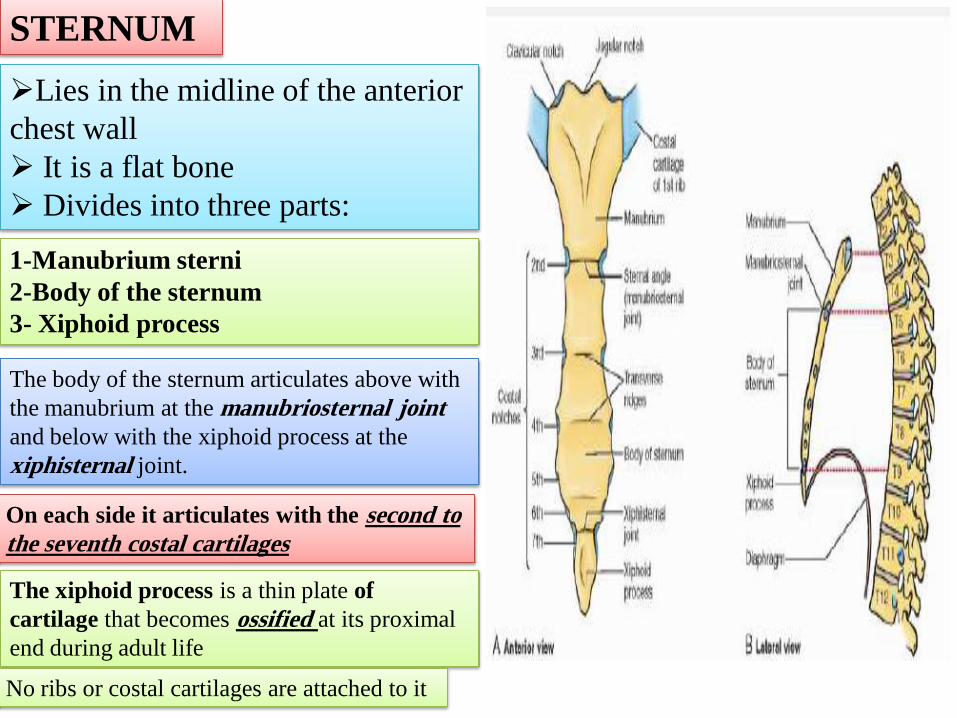
STERNUM
Lies in the midline of the anterior
chest wall
It is a flat bone
Divides into three parts:
1-Manubrium sterni
2-Body of the sternum
3- Xiphoid process
The body of the sternum articulates above with
the manubrium at the manubriosternal joint
and below with the xiphoid process at the
xiphisternal joint.
On each side it articulates with the second to
the seventh costal cartilages
The xiphoid process is a thin plate of
cartilage that becomes ossified at its proximal
end during adult life
No ribs or costal cartilages are attached to it
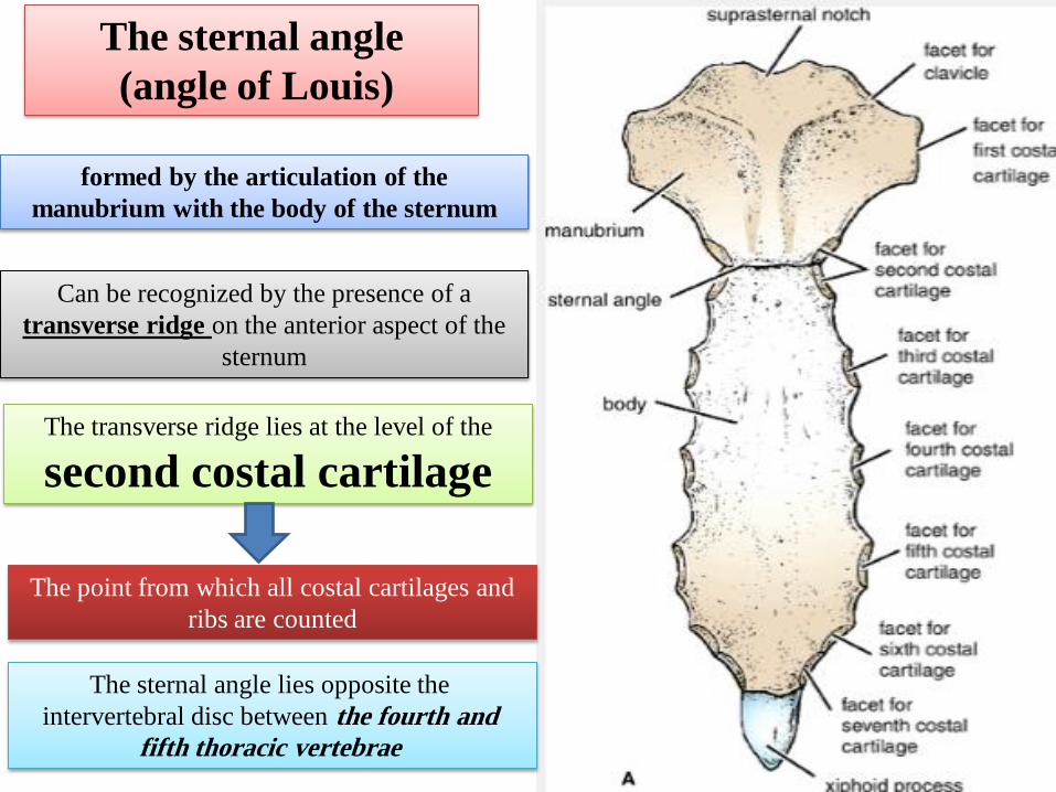
The sternal angle
(angle of Louis)
formed by the articulation of the
manubrium with the body of the sternum
Can be recognized by the presence of a
transverse ridge on the anterior aspect of the
sternum
The transverse ridge lies at the level of the
second costal cartilage
The point from which all costal cartilages and
ribs are counted
The sternal angle lies opposite the
intervertebral disc between the fourth and
fifth thoracic vertebrae

Sternum and Marrow Biopsy
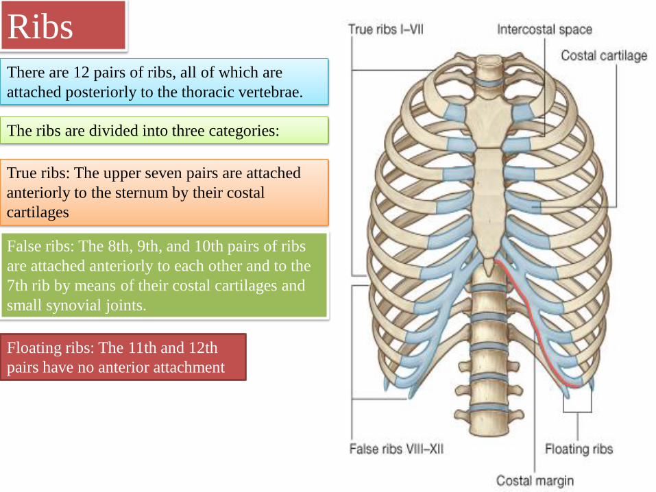
Ribs There are 12 pairs of ribs, all of which are
attached posteriorly to the thoracic vertebrae.
The ribs are divided into three categories:
False ribs: The 8th, 9th, and 10th pairs of ribs
are attached anteriorly to each other and to the
7th rib by means of their costal cartilages and
small synovial joints.
True ribs: The upper seven pairs are attached
anteriorly to the sternum by their costal
cartilages
Floating ribs: The 11th and 12th
pairs have no anterior attachment

Typical Rib
A typical rib is a long, twisted, flat bone
having a rounded, smooth superior border and
a sharp, thin inferior border
The inferior border overhangs and forms the
costal groove, which accommodates the
intercostal vessels and nerve.
The anterior end of each rib is attached to the
corresponding costal cartilage
A rib has a head, neck, tubercle, shaft, and
angle
The head has two facets for articulation with
the numerically corresponding vertebral body
and that of the vertebra immediately above
The neck is a constricted portion situated
between the head and the tubercle.
The tubercle is a prominence on the outer
surface of the rib at the junction of the neck
with the shaft.
It has a facet for articulation with the transverse
process of the numerically corresponding
vertebra
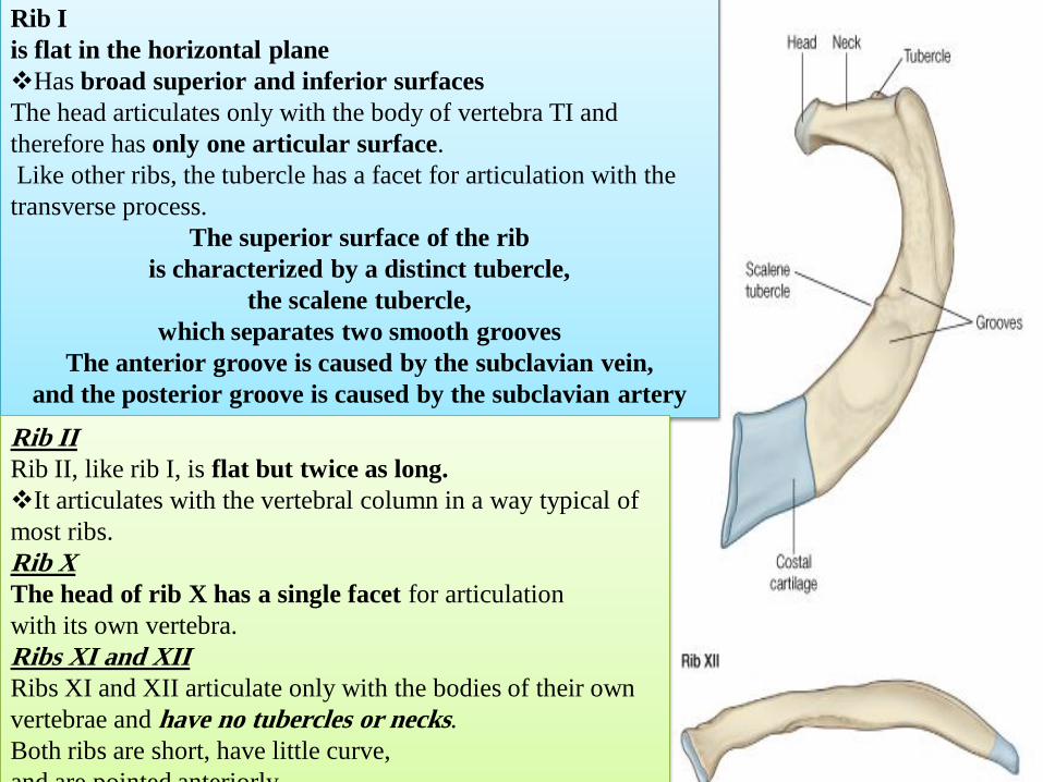
Rib I
is flat in the horizontal plane
Has broad superior and inferior surfaces
The head articulates only with the body of vertebra TI and
therefore has only one articular surface.
Like other ribs, the tubercle has a facet for articulation with the
transverse process.
The superior surface of the rib
is characterized by a distinct tubercle,
the scalene tubercle,
which separates two smooth grooves
The anterior groove is caused by the subclavian vein,
and the posterior groove is caused by the subclavian artery
Rib II
Rib II, like rib I, is flat but twice as long.
It articulates with the vertebral column in a way typical of
most ribs.
Rib X
The head of rib X has a single facet for articulation
with its own vertebra.
Ribs XI and XII
Ribs XI and XII articulate only with the bodies of their own
vertebrae and have no tubercles or necks.
Both ribs are short, have little curve,
and are pointed anteriorly.

The Vertebral Column
is composed of 33
vertebrae
7 cervical
12 thoracic
5 lumbar
5 sacral
(fused to form the sacrum)
4 coccygeal
(the lower 3 are commonly
fused)

A typical vertebra consists of: 1-a rounded body anteriorly
2-a vertebral arch posteriorly.
They enclose a space called The vertebral foramen
through which run the spinal cord and its coverings
The vertebral arch gives rise to seven
processes:
a-One spinous
b-Two transverse
c- Four articular
The spinous process is directed posteriorly
from the junction of the two laminae.
The transverse processes are directed laterally
from the junction of the laminae and the pedicles
The articular processes are vertically arranged and consist of:
Two superior & Two inferior processes
They arise from the junction of the laminae and the pedicles
.
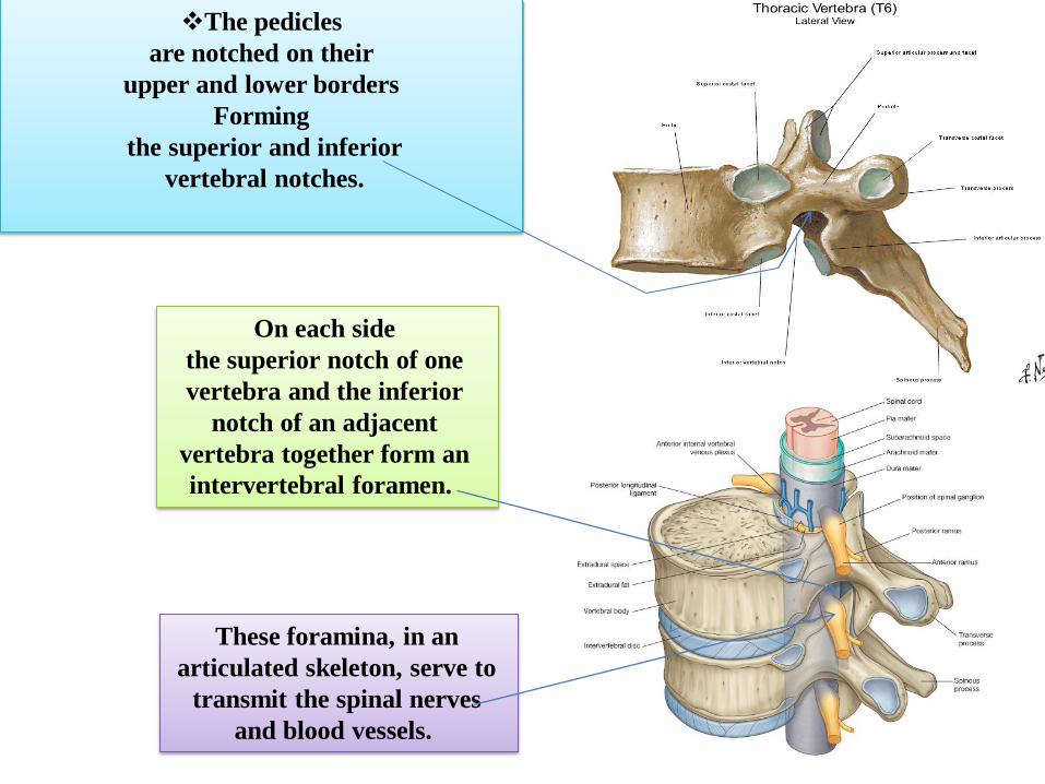
The pedicles
are notched on their
upper and lower borders
Forming
the superior and inferior
vertebral notches.
These foramina, in an
articulated skeleton, serve to
transmit the spinal nerves
and blood vessels.
On each side
the superior notch of one
vertebra and the inferior
notch of an adjacent
vertebra together form an
intervertebral foramen.
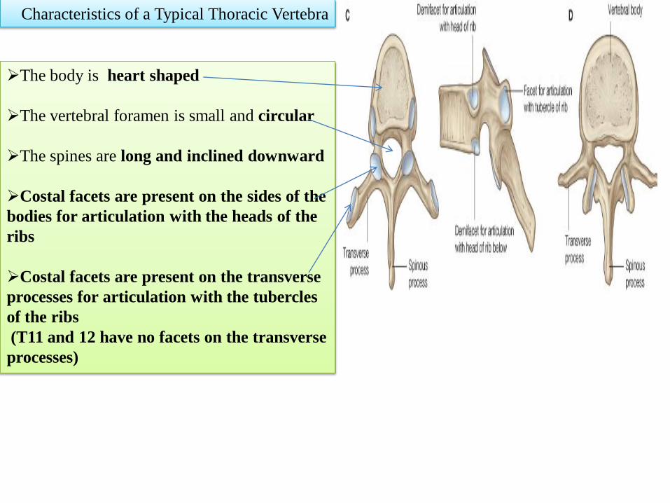
Characteristics of a Typical Thoracic Vertebra
The body is heart shaped
The vertebral foramen is small and circular
The spines are long and inclined downward
Costal facets are present on the sides of the
bodies for articulation with the heads of the
ribs
Costal facets are present on the transverse
processes for articulation with the tubercles
of the ribs
(T11 and 12 have no facets on the transverse
processes)
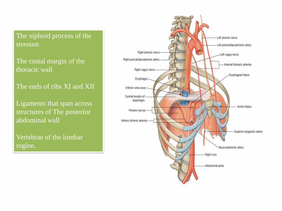
The xiphoid process of the
sternum
The costal margin of the
thoracic wall
The ends of ribs XI and XII
Ligaments that span across
structures of The posterior
abdominal wall
Vertebrae of the lumbar
region.


The inferior vena cava passes through
the central tendon at approximately
vertebral level T8
The esophagus passes through the muscular
part of the diaphragm, just to the left of
midline, approximately at vertebral level T10
The vagus nerves pass through the diaphragm
with the esophagus
The aorta passes behind the posterior
attachment of the diaphragm at vertebral
level T12
The thoracic duct passes behind the
diaphragm with the aorta
The azygos and hemiazygos veins may
also pass through the aortic hiatus or
through the crura of the diaphragm

the most superficial layer.
Its fibers are directed
downward and forward
ORIGIN:
FROM THE INFERIOR BORDER
OF THE RIB ABOVE
TO
INSERTION:
THE SUPERIOR BORDER OF THE
RIB BELOW
Intercostal Muscles
The muscle extends forward to the costal
cartilage where it is replaced by an
aponeurosis,
THE ANTERIOR (EXTERNAL)
INTERCOSTAL MEMBRANE
The external intercostal muscle
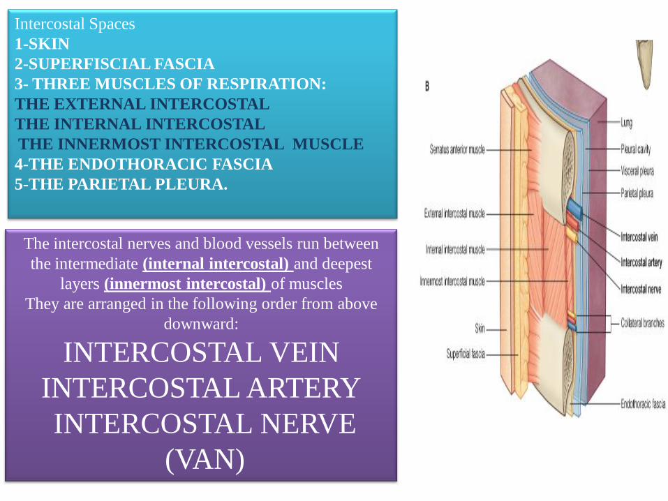
Intercostal Spaces
1-SKIN
2-SUPERFISCIAL FASCIA
3- THREE MUSCLES OF RESPIRATION:
THE EXTERNAL INTERCOSTAL
THE INTERNAL INTERCOSTAL
THE INNERMOST INTERCOSTAL MUSCLE
4-THE ENDOTHORACIC FASCIA
5-THE PARIETAL PLEURA.
The intercostal nerves and blood vessels run between
the intermediate (internal intercostal) and deepest
layers (innermost intercostal) of muscles
They are arranged in the following order from above
downward:
INTERCOSTAL VEIN
INTERCOSTAL ARTERY
INTERCOSTAL NERVE
(VAN)

THE INTERNAL INTERCOSTAL MUSCLE
forms the intermediate layer.
Its fibers are directed
downward and backward
from the subcostal groove of
the rib above to
the upper border of the rib below
The muscle extends
backward from the
sternum in front to the
angles of the ribs behind,
where the muscle is
replaced by an
aponeurosis, the posterior
(internal) intercostal
membrane
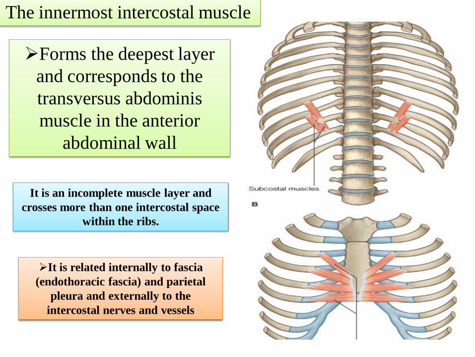
Forms the deepest layer
and corresponds to the
transversus abdominis
muscle in the anterior
abdominal wall
The innermost intercostal muscle
It is an incomplete muscle layer and
crosses more than one intercostal space
within the ribs.
It is related internally to fascia
(endothoracic fascia) and parietal
pleura and externally to the
intercostal nerves and vessels

Intercostal Arteries and Veins
Each intercostal space contains a large single
posterior intercostal artery and two small
anterior intercostal arteries.
The posterior intercostal arteries of the first two
spaces are branches from
the superior intercostal artery, a branch of
the costocervical trunk of the subclavian
artery
The posterior intercostal arteries of the lower nine
spaces are branches of
THE DESCENDING THORACIC AORTA
The anterior intercostal arteries of the first
six spaces are branches of
THE INTERNAL THORACIC ARTERY
which arises from the first part of the
subclavian artery.
The anterior intercostal arteries of the lower spaces are branches
of THE MUSCULOPHRENIC ARTERY, one of the terminal
branches of the internal thoracic artery.

The corresponding posterior
intercostal veins drain backward
into the azygos or hemiazygos
veins , and the anterior
intercostal veins drain forward
into the internal thoracic and
musculophrenic veins

Intercostal Nerves
The intercostal nerves are the
anterior rami of the first 11
thoracic spinal nerves
The anterior ramus of the 12th
thoracic nerve lies in the abdomen and
runs forward in the abdominal wall as
the subcostal nerve

Each intercostal nerve enters an
intercostal space between
the parietal pleura and the posterior
intercostal membrane
It then runs forward inferiorly to the
intercostal vessels in the subcostal
groove of the corresponding rib,
between the innermost intercostal and
internal intercostal muscle.

The first six nerves are
distributed within their
intercostal spaces.
The seventh to ninth intercostal nerves leave
the anterior ends of their intercostal spaces
by passing deep to the costal cartilages, to
enter the anterior abdominal wall.
The 10th and 11th nerves, since the
corresponding ribs are floating, pass directly
into the abdominal wall