borna disease virus p protein affects neural transmission...
TRANSCRIPT

JOURNAL OF VIROLOGY, Dec. 2008, p. 12487–12497 Vol. 82, No. 240022-538X/08/$08.00�0 doi:10.1128/JVI.00877-08Copyright © 2008, American Society for Microbiology. All Rights Reserved.
Borna Disease Virus P Protein Affects Neural Transmission throughInteractions with Gamma-Aminobutyric Acid
Receptor-Associated Protein�
Guiqing Peng,1 Yan Yan,1 Chengliang Zhu,1 Shiqun Wang,1 Xiaohong Yan,1 Lili Lu,1 Wei Li,1
Jing Hu,1 Wei Wei,1 Yongxin Mu,1 Yanni Chen,1 Yong Feng,1 Rui Gong,1 Kailang Wu,1Fengmin Zhang,2 Xiaolian Zhang,1 Ying Zhu,1 and Jianguo Wu1*
State Key Laboratory of Virology, College of Life Sciences, Wuhan University, Wuhan 430072, People’s Republic of China,1 andCollege of Basic Medical Science, Harbin Medical University, Harbin 150081, People’s Republic of China2
Received 24 April 2008/Accepted 18 September 2008
Borna disease virus (BDV) is one of the infectious agents that causes diseases of the central nervous systemin a wide range of vertebrate species and, perhaps, in humans. The phosphoprotein (P) of BDV, an essentialcofactor of virus RNA-dependent RNA polymerase, is required for virus replication. In this study, we identifiedthe gamma-aminobutyric acid receptor-associated protein (GABARAP) with functions in neurobiology as oneof the viral P protein-interacting cellular factors by using an approach of phage display-based protein-proteininteraction analysis. Direct binding between GABARAP and P protein was confirmed by coimmunoprecipita-tion, protein pull-down, and mammalian two-hybrid analyses. GABARAP originally was identified as a linkerbetween the gamma-aminobutyric acid receptor (GABAR) and the microtubule to regulate receptor traffickingand plays important roles in the regulation of the inhibitory neural transmitter gamma-aminobutyric acid(GABA). We showed that GABARAP colocalizes with P protein in the cells infected with BDV or transfectedwith the P gene, which resulted in shifting the localization of GABARAP from the cytosol to the nucleus. Wefurther demonstrated that P protein blocks the trafficking of GABAR, a principal GABA-gated ion channel thatplays important roles in neural transmission, to the surface of cells infected with BDV or transfected with theP gene. We proposed that during BDV infection, P protein binds to GABARAP, shifts the distribution ofGABARAP from the cytoplasm to the nucleus, and disrupts the trafficking of GABARs to the cell membranes,which may result in the inhibition of GABA-induced currents and in the enhancement of hyperactivity andanxiety.
Borna disease virus (BDV), a nonsegmental negative-strandRNA virus, belongs to the Bornaviridae family and is charac-terized by low productivity, neurotropism, and the nuclearlocalization of transcription and replication (12, 33). BDV wasreported to cause diseases of the central nervous system (CNS)in sheep and horses originally and then in a wide range of othervertebrate species (25). Epidemiological studies have shownthat a higher prevalence of BDV infection was found in psy-chiatric patients than in controls, indicating that BDV is apotential human pathogen related to psychiatric diseases (1,19). In contrast, some reports suggested that BDV does notplay significant roles in human health (9, 36).
One of the most prominent features of BDV infection is theheavy inflammatory reaction in the CNS and the rare degen-eration of neurons in naturally infected hosts (25). In experi-mentally infected rats, the histopathology of the CNS is de-pendent on the immune status of the host at the time ofinoculation, the genetic background, and the route of infection(33). The inoculation of immunocompetent adult rats withBDV results in marked immune-mediated meningoencephali-tis consistent with the classical Borna disease. In the process of
the persistent infection of BDV in adult rats, the degenerationof the neurons is observed. In contrast, immunoincompetentrats such as neonates show a tolerance to the infection of BDVwithout signs of Borna disease or encephalitis. Neonatal infec-tion with the virus causes significant alterations in the devel-opment of the CNS (23). These studies provided evidence fordirect effects of BDV infection on cellular functions in theabsence of immunopathological degeneration.
The phosphoprotein (P) of BDV, an essential cofactor ofvirus RNA-dependent RNA polymerase, is regulated by pro-tein kinase Cε and casein kinase II (26) and binds to the N, X,and L proteins of BDV to modulate the active viral polymerasecomplex (24, 27, 28). Our recent study showed that BDV andits P protein could inhibit the expression of inducible nitricoxide synthase (iNOS) in astrocytes, suggesting that BDV es-tablishes a persistent infection at least in part due to its inhi-bition of iNOS to impair immune responses (22). This viralprotein also can counteract the expression of TBK-1-depen-dent beta interferon, and thus it can establish an antiviral state(34). Moreover, P protein binds directly to a multifunctionalprotein (HMGB-1) and represses p53-mediated transcrip-tional activity (14, 38). Finally, the expression of BDV P pro-tein was demonstrated to induce behavioral and neurologicalabnormalities in transgenic mice (13).
Gamma-aminobutyric acid (GABA) exists predominantly inthe brain, with glycine as one of the two inhibitory amino acid
* Corresponding author. Mailing address: State Key Laboratory ofVirology, College of Life Sciences, Wuhan University, Wuhan 430072,People’s Republic of China. Phone: 86-27-68754979. Fax: 86-27-68754592. E-mail: [email protected].
� Published ahead of print on 24 September 2008.
12487
on June 12, 2018 by guesthttp://jvi.asm
.org/D
ownloaded from

neural transmitters (20). Its receptor, GABA type A receptor(GABAA-R), is the principal GABA-gated ion channel (17)and plays important roles in pharmacology. GABAA-R acts asthe target of benzodiazepine-related drugs, such as diazepam,triazolam, and alprazolam, which are widely used for the treat-ment of anxiety and insomnia. GABAA-R-associated protein(GABARAP) originally was identified as a linker betweenGABAA-Rs and microtubules. Recent study has demonstratedthat GABARAP can interact with the �2 subunit of GABAA-Rand has a functional effect on the regulation of GABA activ-ities by disrupting the trafficking of GABAA-Rs to the cellmembranes (4, 5).
In this study, we demonstrated that BDV P protein can binddirectly to GABARAP both in vitro and in vivo by using differentapproaches. Biological effects of the interaction of P protein withGABARAP and the molecular mechanisms underlying the effectsof P protein on the regulation of GABARAP and GABAA-Rsalso were investigated and discussed.
MATERIALS AND METHODS
Cell culture and virus propagation. 293T cells, HeLa cells, COS-7 cells, andOL cells were obtained from the ATCC and maintained in Dulbecco’s modifiedEagle’s medium (DMEM) containing 10% heat-inactivated fetal bovine serum(FBS). The BDV viral strain H1766 used for the infection of COS-7 and 293Tcells in this study and a BDV persistent infecting cell line (BDV/OL cells) weregifts from K. Ikuta of Osaka University. In general, adherent cells were infectedat a multiplicity of infection of 0.1 in DMEM containing 2% FBS and thencultured for 5 to 7 days (3). All cell cultures were maintained at 37°C in 5% CO2.
Plasmid construction. The plasmid pcDNA-P was kindly provided by ThorstenWolff and Peter Staeheli. Plasmids pCMV-�1, pCMV-�2, and pRK5-�2L-GFPN(15), expressing the �1, �2, and �2 subunits of the GABAA-R, respectively, werekindly provided by Bill Wisden and Erwin Sigel. The P gene of BDV wasamplified from pcDNA-P by PCR using primers 5�-TTTGGATCCATGGCAACGCGACCATCGAG-3� and 5�-TTTCTCGAGTGGTATGATGTCCCACTCATC-3�. The resulting PCR product then was subcloned into pET-28a to yieldplasmid pET-28a-P. To generate pVP16-P, the P gene was amplified frompcDNA-P by PCR using primers 5�-CGCGGATCCCCATGGCAACGCGACCATC-3� and 5�-CCCAAGCTTTTATGGTATGATGTCCCAC-3�, and the PCRproduct was subcloned into pVP16. To generate pEGFP-P, the P gene wasamplified from pcDNA-P using primers 5�-CCCAAGCTTATGGCAACGCGACCATCG-3� and 5�-CGCGGATCCTTATGGTATGATGTCCCAC-3�, and thePCR product then was subcloned into pEGFP-C3.
The GABARAP cDNA was amplified by PCR using primers 5�-CGCGGATCCGGATGAAGTTCGTGTACA-3� and 5�-CCCAAGCTTTCACAGACCGTAGACACT-3� to introduce BamHI and HindIII cloning sites and then wassubcloned into appropriate restriction sites of plasmids pM, pET-28a, andpCMV-Tag2C to generate the plasmids pM-GABARAP, pET-28a-GABARAP,and pCMV-Tag2C-GABARAP, respectively. The GABARAP cDNA was am-plified by PCR using primers 5�-CCCAAGCTTGGATGAAGTTCGTGTACA-3� and 5�-CGCGGATCCTCACAGACCGTAGACACT-3� and then sub-cloned into pAsRed2C1 to yield plasmid pAsRed2C1-GABARAP.
The mutant forms of the GABARAP and P genes in the expression plasmidswere generated from the wild-type plasmid by using PCR and recloning tech-niques. The primers used in PCR to create the mutant plasmids are available onrequest. The introduction of the correct mutations for each mutant was con-firmed by DNA sequencing.
Recombinant P protein purification and anti-P antibody generation. pET-28a-P, carrying the P gene fused in frame with an N-terminal hexahistidine tag,was transformed into Escherichia coli strain BL21, grown at 37°C to an opticaldensity at 600 nm of 0.5, induced with 1 mM isopropyl-ß-D-thiogalactopyranoside(IPTG), and cultured for another 2 to 4 h at 37°C. After harvest, the cells wereresuspended in buffer C (50 mM Tris-HCl, pH 8.0, 500 mM NaCl, 5 mM MgCl2,5% glycerol, 0.5% NP-40, 2 mM imidazole, and 7 mM �-mercaptoethanol), lysedby sonication, and centrifuged for 20 min at 8,500 � g and 4°C. Supernatants thenwere incubated at 4°C for 2 h with equilibrated Ni�-NTA agarose beads(Qiagen). The beads were washed with buffer C containing 20 mM imidazole,and recombinant proteins were eluted in buffer D (20 mM Tris-HCl, pH 8.0, 100mM NaCl, 5 mM MgCl2, 20% glycerol, 0.1% NP-40, 7 mM �-mercaptoethanol,
and 250 mM imidazole). Protein concentrations were determined using aBio-Rad protein assay. Purified recombinant protein was analyzed on a sodiumdodecyl sulfate-polyacrylamide gel electrophoresis (SDS-PAGE) gel by stainingwith Coomassie brilliant blue.
The recombinant P protein was used as an immunogen. Rabbits (4 weeks old)were immunized with the recombinant P protein with complete Freund’s adju-vant. The polyclonal anti-BDV-P antibody was prepared from immunized rab-bits’ hyperimmune sera by ammonium sulfate precipitation.
T7Select biopanning. A premade T7Select lung cDNA library (no. 70646-3;Novagen) was screened with the T7Select biopanning kit (Novagen) according tothe manufacturer’s instructions. The P protein expressed from bacteria andpurified by affinity chromatography was used as the bait. After five rounds ofselection, 96 randomly selected clones were picked up from each library, and thesequences of each of the clones were determined by DNA sequence analysis andsearched for homology to known sequences with the BLAST program (http://www.ncbi.nlm.nih.gov/BLAST).
Mammalian two-hybrid analysis. 293T cells or HeLa cells were transfectedwith luciferase reporter plasmid pG5luc (Promega) and test plasmids, respec-tively, using Lipofectamine 2000 transfection reagent in 24-well culture plates.Forty-eight hours after transfection, cells were lysed in 100 �l of lysis buffer for60 min with shaking at room temperature. After centrifugation at 12,000 � g for5 min at 4°C, the cell extracts were assayed for luciferase activity using thedual-luciferase reporter assay system (Promega) according to the manufacturer’srecommendations.
Immunoprecipitation assay. 293T cells were cotransfected with plasmidspCMV-GABARAP expressing Flag-tagged GABARAP and pCDNA-P express-ing P protein. Forty-eight hours posttransfection, transfected cells were lysed byfreeze-thaw cycling in a buffer containing 10 mM Tris, pH 7.6, 150 mM NaCl,0.5% NP-40, and 1.0 mM phenylmethylsulfonyl fluoride. After centrifugation,the soluble fraction was immunoprecipitated with anti-P antibody for 2 h at 4°C,and the precipitates then were recovered by incubation with protein A/G-agarosebeads (Santa Cruz Biotechnology, Inc., Santa Cruz, CA) for 2 h at 4°C. Afterbeing thoroughly washed, proteins bound to the agarose beads were separated bySDS-PAGE and electrotransferred onto a polyvinylidene difluoride membrane,which then was blocked with 5% skimmed milk in phosphate-buffered saline(PBS)–0.05% Tween 20 (PBS-T) overnight at 4°C. The membrane was reactedwith anti-Flag mouse monoclonal antibody in PBS-T containing 5% skimmedmilk for 1 h at room temperature. After being washed, the membranes wereincubated with horseradish peroxidase-conjugated goat anti-mouse immunoglob-ulin G (IgG) for 1 h at room temperature and visualized by the enhancedchemiluminescence system (Pierce).
To verify the interaction between BDV P protein and endogenousGABARAP, 293T cells were infected with BDV. Protein extracts prepared frominfected or noninfected cells were immunoprecipitated with anti-GABARAPantibody (sc-28938; Santa Cruz Biotechnology, Inc., Santa Cruz, CA) and de-tected by anti-P antibody.
Protein pull-down assays. Purified hexahistidine-tagged recombinant proteins(40 �g) were incubated with 300 �l Ni�-NTA agarose beads in 1 ml bindingbuffer (10 mM Tris-HCl, pH 7.5, 150 mM NaCl, 5 mM MgCl2, 0.5% NP-40, and1 mM dithiothreitol [DTT]) for 2 h at 4°C. After two washes with 1 ml bindingbuffer, some of the hybridized beads were analyzed by SDS-PAGE, and the restof the beads were incubated with extracts of BDV-infected (OL/BDV) or unin-fected (OL) cells in 1 ml of hybridization buffer (10 mM Tris-HCl, pH 8.0, 100mM NaCl, 0.5 mM EDTA, 0.1% NP-40, 1 mM DTT, and 10 mM imidazole) for1 h at 4°C. Beads then were washed four times with hybridization buffer, andbound proteins were eluted in Laemmli buffer and determined by Western blotanalysis.
Laser-scanning confocal microscopy analysis. Recombinant plasmids pAsRed2C1-GABARAP and/or pEGFP-P, pAsRed2C1-GABARAP, pEGFP-P134, and 1.2 �lLipofectamine 2000 were mixed in 30 �l DMEM for 20 min and then cotransfectedinto COS-7 cells. Transfected cells were cultured on coverslips in a 24-well plate.Twenty-four to 36 h posttransfection, cells were fixed with 4% paraformaldehyde for15 min, washed three times with 1� PBS, counterstained with Hoechst 33258 for 10min, and then washed three times with 1� PBS. Fluorescence was detected by usinga confocal laser-scanning microscope (LSM 510 META; Carl Zeiss). For the indirectimmunofluorescence assay, BDV-infected COS-7 cells were transfected with plas-mid pAsRed2C1-GABARAP and fixed with 4% paraformaldehyde prior to treat-ment with 0.4% Triton X-100. After a reaction with the optimal anti-P antibody(1:1,000) as the first antibody, cells were stained with secondary anti-rabbit IgGantibodies labeled with fluorescein isothiocyanate. AsRed2 fusion proteins werevisualized with red fluorescence. Fluorescence was detected by using a confocallaser-scanning microscope (LSM 510 META). The overlays were processed withPhotoshop 7.
12488 PENG ET AL. J. VIROL.
on June 12, 2018 by guesthttp://jvi.asm
.org/D
ownloaded from
Flow cytometry analysis. 293T cells and BDV-infected 293T cells were grownin 6-well plates until 50% confluence and then were transfected with pCMV-�1,pCMV-�2, and PRK5-�2L-GFPN using Lipofectamine reagent (Invitrogen) toexpress the GABAA-R. The ratio of the mixture of �1-�2-�2-P-GABARAP was1:1:2:2:2. �1-�2-�2, �1-�2-�2-GABARAP, and �1-�2-�2-P were used as controlsin the same ratio and were normalized with empty vector. For BDV-infected293T cells, the ratio of the mixture of �1-�2-�2-GABARAP was 1:1:2:2. �1-�2-�2 was used as the control in the same ratio. �1-�2-�2 and �1-�2-�2-GABARAP were used in 293T cells as additional controls in the same ratio andwere normalized with empty vector.
Forty-eight hours posttransfection, cells were dislodged from the plates withPBS and 50 mM EDTA, placed on ice, spun down at 4°C, resuspended inantibody incubation buffer (PBS, 0.2% bovine serum albumin, and 0.01% sodiumazide) containing rabbit anti-green fluorescent protein (GFP) antibody (1:500;Santa Cruz Biotechnology), and incubated on ice for 30 min. Treated cells thenwere washed three times with cold PBS, resuspended in antibody incubationbuffer containing R-PE-labeled goat anti-rabbit secondary antibody (1:100;PTGLAB), and incubated for 30 min, followed by three washes with PBS. Cells thenwere fixed in PBS and 4% paraformaldehyde for 5 min, washed three times withice-cold PBS, and held at 4°C until analysis by flow cytometry. Flow cytometryanalysis was performed using a flow cytometer (Cytomics Fc500; BeckmanCoulter). Fluorochromes excited from the argon laser were observed at 510 nmfor GFP and 570 nm for R-PE. A total of 10,000 cells were randomly selected foranalysis.
RESULTS
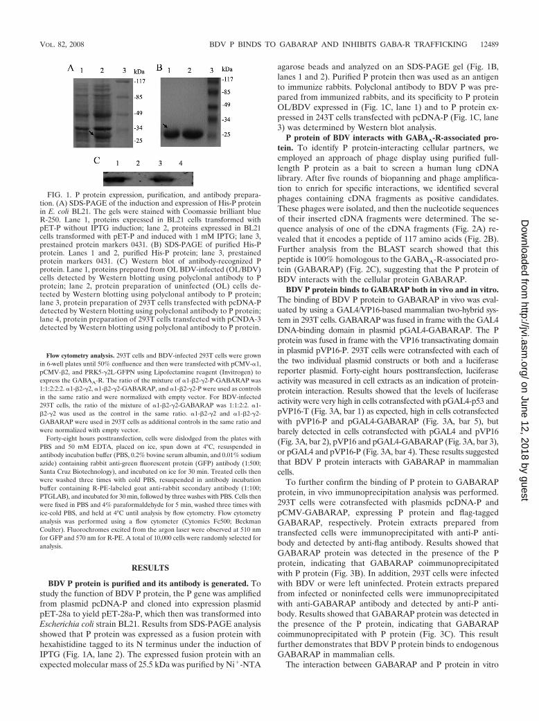
BDV P protein is purified and its antibody is generated. Tostudy the function of BDV P protein, the P gene was amplifiedfrom plasmid pcDNA-P and cloned into expression plasmidpET-28a to yield pET-28a-P, which then was transformed intoEscherichia coli strain BL21. Results from SDS-PAGE analysisshowed that P protein was expressed as a fusion protein withhexahistidine tagged to its N terminus under the induction ofIPTG (Fig. 1A, lane 2). The expressed fusion protein with anexpected molecular mass of 25.5 kDa was purified by Ni�-NTA
agarose beads and analyzed on an SDS-PAGE gel (Fig. 1B,lanes 1 and 2). Purified P protein then was used as an antigento immunize rabbits. Polyclonal antibody to BDV P was pre-pared from immunized rabbits, and its specificity to P proteinOL/BDV expressed in (Fig. 1C, lane 1) and to P protein ex-pressed in 243T cells transfected with pcDNA-P (Fig. 1C, lane3) was determined by Western blot analysis.
P protein of BDV interacts with GABAA-R-associated pro-tein. To identify P protein-interacting cellular partners, weemployed an approach of phage display using purified full-length P protein as a bait to screen a human lung cDNAlibrary. After five rounds of biopanning and phage amplifica-tion to enrich for specific interactions, we identified severalphages containing cDNA fragments as positive candidates.These phages were isolated, and then the nucleotide sequencesof their inserted cDNA fragments were determined. The se-quence analysis of one of the cDNA fragments (Fig. 2A) re-vealed that it encodes a peptide of 117 amino acids (Fig. 2B).Further analysis from the BLAST search showed that thispeptide is 100% homologous to the GABAA-R-associated pro-tein (GABARAP) (Fig. 2C), suggesting that the P protein ofBDV interacts with the cellular protein GABARAP.
BDV P protein binds to GABARAP both in vivo and in vitro.The binding of BDV P protein to GABARAP in vivo was eval-uated by using a GAL4/VP16-based mammalian two-hybrid sys-tem in 293T cells. GABARAP was fused in frame with the GAL4DNA-binding domain in plasmid pGAL4-GABARAP. The Pprotein was fused in frame with the VP16 transactivating domainin plasmid pVP16-P. 293T cells were cotransfected with each ofthe two individual plasmid constructs or both and a luciferasereporter plasmid. Forty-eight hours posttransfection, luciferaseactivity was measured in cell extracts as an indication of protein-protein interaction. Results showed that the levels of luciferaseactivity were very high in cells cotransfected with pGAL4-p53 andpVP16-T (Fig. 3A, bar 1) as expected, high in cells cotransfectedwith pVP16-P and pGAL4-GABARAP (Fig. 3A, bar 5), butbarely detected in cells cotransfected with pGAL4 and pVP16(Fig. 3A, bar 2), pVP16 and pGAL4-GABARAP (Fig. 3A, bar 3),or pGAL4 and pVP16-P (Fig. 3A, bar 4). These results suggestedthat BDV P protein interacts with GABARAP in mammaliancells.
To further confirm the binding of P protein to GABARAPprotein, in vivo immunoprecipitation analysis was performed.293T cells were cotransfected with plasmids pcDNA-P andpCMV-GABARAP, expressing P protein and flag-taggedGABARAP, respectively. Protein extracts prepared fromtransfected cells were immunoprecipitated with anti-P anti-body and detected by anti-flag antibody. Results showed thatGABARAP protein was detected in the presence of the Pprotein, indicating that GABARAP coimmunoprecipitatedwith P protein (Fig. 3B). In addition, 293T cells were infectedwith BDV or were left uninfected. Protein extracts preparedfrom infected or noninfected cells were immunoprecipitatedwith anti-GABARAP antibody and detected by anti-P anti-body. Results showed that GABARAP protein was detected inthe presence of the P protein, indicating that GABARAPcoimmunoprecipitated with P protein (Fig. 3C). This resultfurther demonstrates that BDV P protein binds to endogenousGABARAP in mammalian cells.
The interaction between GABARAP and P protein in vitro
FIG. 1. P protein expression, purification, and antibody prepara-tion. (A) SDS-PAGE of the induction and expression of His-P proteinin E. coli BL21. The gels were stained with Coomassie brilliant blueR-250. Lane 1, proteins expressed in BL21 cells transformed withpET-P without IPTG induction; lane 2, proteins expressed in BL21cells transformed with pET-P and induced with 1 mM IPTG; lane 3,prestained protein markers 0431. (B) SDS-PAGE of purified His-Pprotein. Lanes 1 and 2, purified His-P protein; lane 3, prestainedprotein markers 0431. (C) Western blot of antibody-recognized Pprotein. Lane 1, proteins prepared from OL BDV-infected (OL/BDV)cells detected by Western blotting using polyclonal antibody to Pprotein; lane 2, protein preparation of uninfected (OL) cells de-tected by Western blotting using polyclonal antibody to P protein;lane 3, protein preparation of 293T cells transfected with pcDNA-Pdetected by Western blotting using polyclonal antibody to P protein;lane 4, protein preparation of 293T cells transfected with pCNDA-3detected by Western blotting using polyclonal antibody to P protein.
VOL. 82, 2008 BDV P BINDS TO GABARAP AND INHIBITS GABA-R TRAFFICKING 12489
on June 12, 2018 by guesthttp://jvi.asm
.org/D
ownloaded from

also was determined by using protein pull-down analysis. His-tagged GABARAP was generated and immobilized on Ni�-NTAagarose beads. Cell extracts were prepared from OL cells infectedwith BDV or left uninfected and used in protein pull-down anal-ysis. Results indicated that His-tagged GABARAP protein couldspecifically pull down the viral P protein (Fig. 3D), as detected byWestern blot analysis probing with polyclonal antibodies to the Pprotein of BDV. Thus, these results demonstrate that BDV Pprotein interacts with GABARAP in vitro.
C-terminal 27 amino acids of the P protein are essential forits binding to GABARAP. To dissect the sequences of P proteinrequired for its binding to GABARAP, several P gene deletionsencoding the truncated proteins VP16-P�N1, VP16-P�N2,VP16-P�C1, and VP16-P�C2 were generated. Full-length andtruncated P genes were fused in frame with the VP16 transacti-vating domain to generate plasmids pVP16-P, pVP16-P�N1,pVP16-P�N2, pVP16-P�C1, and pVP16-P�C2, which expresscorresponding peptides (Fig. 4A). The GABARAP gene wasfused in frame with the GAL4 DNA-binding domain to createplasmid pGAL4-GABARAP, which expresses the GAL4-GABARAP fusion protein (Fig. 5A). HeLa cells were cotrans-fected with pGAL4-GABARAP and pVP16-P, pVP16-P�N1,pVP16-P�N2, pVP16-P�C1, or pVP16-P�C2 and tested for thebinding of GABARAP with P protein using a GAL4-VP16-basedmammalian two-hybrid system.
Results showed that the levels of luciferase activity werehigh in cells cotransfected with pGAL4-G and pVP16-P,pVP16-P�N1, or pVP16-P�N2, but they were very low in cellscotransfected with pGAL4-G and pVP6, pVP16-P�C1, orpVP16-P�C2 (Fig. 4B). These results indicated that GAL4-GABARAP could interact with VP16-P, VP16-P�N1, andVP16-P�N2 but failed to bind to VP16-P�C1 and VP16-P�C2.
Since both VP16-P�C1 and VP16-P�C2 lacked amino acids174 to 201 of the P protein, the C-terminal 27 amino acids areessential for the binding of P protein to GABARAP. On theother hand, since VP16-P�N2 contains the C-terminal se-quences from amino acids 87 to 201 of the P protein, theC-terminal 114 amino acids are sufficient for the binding of Pprotein to GABARAP.
Intact GABARAP protein is required for its binding to theviral P protein. We used a similar strategy to investigate thesequences of GABARAP that are required for their binding to Pprotein by constructing several deletions of the GABARAP geneencoding the truncated GABARAP proteins GAL4-G, GAL4-G�N1, GAL4-G�N2, GAL4-G�C1, and GAL4-G�C2 (Fig. 5A).Full-length and truncated GABARAP proteins were fused inframe with the GAL4 DNA-binding domain to generate plasmidspGAL4-G, pGAL4-G�N1, pGAL4-G�N2, pGAL4-G�C1, andpGAL4-G�C2. The P protein was fused in frame with the VP16transactivating domain to yield pVP16-P (Fig. 4A). HeLa cellswere cotransfected with pVP16-P and pGAL4-G, pGAL4-G�N1,pGAL4-G�N2, pGAL4-G�C1, and pGAL4-G�C2, respectively,and analyzed for the binding of the P protein with theGABARAP proteins using a GAL4-VP16-based mammaliantwo-hybrid system. Results indicated that the viral P protein couldinteract only with the wild-type protein GAL4-G and failed tobind to the four GABARAP mutants GAL4-G�N1, GAL4-G�N2, GAL4-G�C1, and GAL4-G�C2 (Fig. 5B). These resultssuggested that GABARAP with an intact structure is essential forits binding to BDV P protein.
BDV P protein stimulates the translocation of GABARAPfrom the cytosol to nuclei. Subcellular localizations of P proteinand GABARAP were investigated by examining COS-7 cellstransfected with plasmids expressing enhanced GFP-P (EGFP-P)
FIG. 2. Identification and sequencing of protein interactions with P protein. (A) The nucleotide sequences of a cDNA fragment isolated froma P protein-reactive phage in a phage display-based protein interaction screen. GAATTC and AAGCTT are sequences of restriction sites.Underlined are the nucleotide sequences with homology to the GABARAP gene. (B) Sequence of 117 amino acids translated from the underlinednucleotide sequences of panel A. (C) Results from a BLAST search of the GenBank databases at the NCBI website, which indicated that thesequences of protein expressed from the phage display that interacted with the P protein were 100% homologous to the cellular proteinGABARAP.
12490 PENG ET AL. J. VIROL.
on June 12, 2018 by guesthttp://jvi.asm
.org/D
ownloaded from

or AsRed2-GABARAP fusion proteins, respectively. The cellularlocalization of the fusion proteins was visualized by laser-scanningconfocal microscopy. Results showed that EGFP-P was localizedmainly in nuclei (Fig. 6A) and that AsRed2-GABARAP wasuniformly distributed in the cytosol (Fig. 6B). To show the loca-tions of the nuclei, cells were stained with Hoechst.
The biological relevance of the interaction between BDV Pprotein and GABARAP was determined by the examination ofthe colocalization of the two proteins. COS-7 cells were co-transfected with plasmids expressing EGFP-P and AsRed2-GABARAP fusion proteins. The distributions of the fusionproteins were examined by laser-scanning confocal micros-copy. Results showed that the majority of the EGFP-P andAsRed2-GABARAP fusion proteins were localized in nuclei(Fig. 6C). These results demonstrated that BDV P proteincolocalized with GABARAP resulted in the translocation ofGABARAP from the cytosol to the nuclei of the cells.
To evaluate the effects of P protein on the localization ofGABARAP, one of the P protein mutants, P�C1 (or P134),was included in the study. In the presence of P�C1, the ma-jority of GABARAP remained in the cytosol of the cells (Fig.6D). This result was consistent with previous finding that theP�C1 protein failed to interact with GABARAP (Fig. 4). This
result also suggested that the translocation of GABARAPfrom the cytosol to the nuclei in the presence of P protein wasdue to its interaction with a functional P protein.
To further investigate the effects of P protein on the cellulardistribution of GABARAP in infected cells, COS-7 cells wereinfected with BDV and then transfected with plasmid express-ing AsRed2-GABARAP fusion protein. The subcellular distri-bution of the fusion protein was examined by laser-scanningconfocal microscopy. Results showed that the viral P proteinalso could interact with the fusion protein by shifting the sub-cellular distributions of AsRed2-GABARAP from the cytosolto the nuclei in BDV-infected COS-7 cells (Fig. 6E).
BDV P protein inhibits the translocation of GABAA-Rs to thecell surface. The fact that GABARAP interacts with GABAA-Rsand enhances the translocation receptors to the cell membraneprompted us to determine the effect of the P protein on thetranslocation of GABAA-Rs to the cell surface regulated byGABARAP. 293T cells were cotransfected with plasmids express-ing the three subunits of GABAA-Rs (�, �, and GFP-�),GABARAP, and P protein. The numbers of cells with expressedGFP-� and cells with expressed GFP-� on the membrane weremeasured by flow cytometry analyses. Results showed that thepercentage of cells with GABAA-Rs translocated to the cell sur-
FIG. 3. Determination of the interaction between BDV P protein and GABARAP in vivo. (A) Determination of the protein-protein interactionin 293T cells by mammalian two-hybrid analysis. 293T cells were cotransfected with pG5luc (a luciferase reporter plasmid), pVP16-P, andpGAL4-GABARAP or with control plasmids. Binding between proteins was monitored by luciferase activity, which was measured 48 h aftertransfection using the dual-luciferase reporter assay system. Values are expressed as the means plus standard errors of the means (three values persample). (B) Determination of the protein-protein interaction in 293T cells by in vivo immunoprecipitations. 293T cells were cotransfected withpcDNA-P (expressing P protein) and pCMV-GABARAP (expressing Flag-tagged GABARAP). The immunoprecipitation was performed withanti-P antibody, and the Western blotting was performed with anti-Flag antibody. Lane 1, P protein alone; lane 2, P protein plus GABARAP; lane3, extracts of untransfected 293T cells; lane 4, GABARAP alone. (C) Determination of the interaction between BDV P protein and endogenousGABARAP in 293T cells by in vivo immunoprecipitations. 293T cells were infected with BDV, and the interaction between proteins was analyzedby immunoprecipitation. In vivo immunoprecipitation was performed with anti-GABARAP antibody or anti-P antibody, and Western blotting wasperformed with anti-P antibody. Lane 1, 293T cells infected with BDV; lane 2, uninfected cells. (D) Determination of the interaction between BDVP protein and GABARAP in vitro. His-tagged GABARAP was bound to His-bound Sepharose beads. Some of the beads were detected bySDS-PAGE, and the rest were incubated with the extracts of BDV-infected (OL/BDV) or uninfected (OL) cells. After being washed, boundproteins were eluted with elution buffer, separated by SDS-PAGE, and probed with antibodies to P protein or anti-His antibody. Western blottingwas performed with anti-P antibody. Lane 1, extracts of BDV-infected (OL/BDV) cells treated with His-GABARAP-bound agarose beads; lane2, extracts of BDV-infected (OL/BDV) cells treated with unbound agarose beads; lane 3, extracts of uninfected (OL) cells treated withHis-GABARAP-bound agarose beads; lane 4, extracts of uninfected (OL) cells treated with unbound agarose beads.
VOL. 82, 2008 BDV P BINDS TO GABARAP AND INHIBITS GABA-R TRAFFICKING 12491
on June 12, 2018 by guesthttp://jvi.asm
.org/D
ownloaded from

face was 35.2% (in the presence of vector), 32.0% (in the pres-ence of P protein), 66.9% (in the presence of GABARAP), and43.2% (in the presence of GABARAP and P protein) (Fig. 7Aand B). These results demonstrated that GABARAP stimulatesthe translocation of GABAA-Rs to the cell surface as expected,while the BDV P protein inhibits the translocation of GABAA-Rsto the cell surface by blocking the function of GABARAP.
To further evaluate the effects of the P protein of BDV onthe trafficking of GABAA-Rs in infected cells, 293T cells wereinfected with BDV and then cotransfected with plasmids ex-pressing the three subunits of GABAA-Rs (�, �, and GFP-�)and GABARAP. Cells with expressed GFP-� and cells withexpressed GFP-� on the membrane were measured by flowcytometry analyses. Results showed that the percentage of cellswith GABAA-Rs translocated to the cell surface was 43.1% (inthe presence of vector), 38.1% (infected with BDV), 70.7% (inthe presence of GABARAP), and 41.8% (in the presence ofBDV and GABARAP), respectively (Fig. 7C and D). Theseresults further demonstrated that GABARAP stimulates thetranslocation of GABAA-Rs to the cell surface as expected,while BDV inhibits the translocation of GABAA-Rs to the cellsurface through the viral P protein blocking the function ofGABARAP.
DISCUSSION
BDV is an infectious agent that causes CNS diseases in awide range of vertebrate species and perhaps in humans (1, 19,25). The P protein of BDV, which is abundantly expressed ininfected cells and animal brains, plays important roles in theregulation of the polymerase complex of BDV by binding tothe viral proteins N, X, and L (16, 24, 27, 28). In addition to itsrole in viral replication, P protein plays a role in the inductionof behavioral and neurological abnormalities in transgenicmice (13). This viral protein can counteract TBK-1-dependentbeta interferon expression in cells to form an antiviral state(34). Our recent study showed that P protein can inhibit iNOSexpression in astrocytes, which perhaps results in the establish-ment of a persistent infection (22).
To explore the neurological functions of P protein, we usedpurified P protein as the bait to identify its cellular targets byscreening a human cDNA library using a phage display ap-proach. Using this technique, we identified several cellularfactors, including HMGB-1 (data not shown) and GABARAP.Previous results from far-Western blotting assays showed thatP protein interacts directly with HMGB-1 and inhibits its func-tions in cultured neural cells (14, 38). GABARAP, a 14-kDapolypeptide, originally was identified as an intracellular protein
FIG. 4. Functional analysis of P protein in its binding to GABARAPby the mammalian two-hybrid system. (A) Schematic representations of Pprotein deletions and its fusion proteins. The numbers indicate the aminoacid positions of BDV P protein. VP16, transactivating domain of VP16.(B) Functional analysis of P protein in its binding to GABARAP by themammalian two-hybrid system. HeLa cells were cotransfected with theluciferase reporter plasmid pG5luc along with pGAL4-GABARAP,pVP16, pVP16-P, pVP16-P�N1, pVP16-P�N2, pVP16-P�C1, andpVP16-P�C2 as indicated. Forty-eight hours posttransfection, cell ex-tracts were prepared and assayed for luciferase activity. The values areexpressed as means plus standard errors of the means.
FIG. 5. Functional analysis of GABARAP in its binding to P pro-tein by the mammalian two-hybrid system. (A) Schematic representa-tion of GABARAP deletions and GAL4-GABARAP fusion proteins.The numbers indicate amino acid positions in GABARAP. GAL4, theDNA-binding domain of GAL4. (B) Functional analysis ofGABARAP in the binding of P protein by the mammalian two-hybridsystem. HeLa cells were cotransfected with luciferase reporter plasmidpG5luc along with pVP16-P, pGAL4-G, pGAL4-G�N1, pGAL4-G�N2, pGAL4-G�C1, and pGAL4-G�C2 as indicated. Forty-eighthours posttransfection, cell extracts were prepared and assayed forluciferase activity. The values are expressed as means plus standarderrors of the means.
12492 PENG ET AL. J. VIROL.
on June 12, 2018 by guesthttp://jvi.asm
.org/D
ownloaded from

with sequence similarity to light chain 3 of microtubule-asso-ciated proteins 1A and 1B (18). GABARAP can interact withthe �2 subunits of GABAA-Rs (35) and facilitate the surfacepresentation of GABAA-Rs (2, 4).
The direct binding of P protein to GABARAP was confirmedby several independent approaches, such as mammalian two-
hybrid assay, coimmunoprecipitation, and protein pull-downanalyses. All results were consistent and demonstrated that Pprotein interacts directly with GABARAP both in vivo and invitro, indicating that P protein is involved in the regulation of thefunction of GABAA-Rs.
The C terminus of P protein has been reported to have
FIG. 6. Determination of the subcellular distribution and colocalization of GABARAP and BDV P protein. (A and B) pEGFP-P orpAsRed2C1-GABARAP was transfected individually into COS-7 cells. The expression and distribution of fusion proteins GFP-P (A) andAsRed2-GABARAP (B) in transfected cells were examined by laser-scanning confocal microscopy. (C and D) pAsRed2C1-GABARAP andpEGFP-P or pEGFP-P�C1 were cotransfected into COS-7 cells. The colocalization of fusion proteins GFP-P with AsRed2-GABARAP(C) and GFP-P�C1 with AsRed2-GABARAP (D) in transfected cells was examined by laser-scanning confocal microscopy with appropriate filters.(E) For investigating the colocalization of P and GABARAP in BDV-infected COS-7 cells, cells were infected with BDV and then transfected withpAsRed2C1-GABARAP. Anti-P antibody was used as the first antibody, and anti-rabbit IgG antibody labeled with fluorescein isothiocyanate(FITC) was used as the second antibody. Proteins were examined by laser-scanning confocal microscopy. To show the location of the nuclei, cellswere stained with Hoechst 33258. Scale bar, 20 �m.
VOL. 82, 2008 BDV P BINDS TO GABARAP AND INHIBITS GABA-R TRAFFICKING 12493
on June 12, 2018 by guesthttp://jvi.asm
.org/D
ownloaded from

FIG. 7. Determination of the effect of P protein on GABARAP in the trafficking of GABAA-Rs. (A) 293T cells were cotransfected with plasmids pCMV-�1(expressing receptor subunit �1), pCMV-�2 (expressing receptor subunit �2), pRK5-�2L-GFPN (expressing receptor subunit �2 and GFP fusion protein�2-GFP), pCMV-GABARAP, and pCMV-P. Forty-eight hours posttransfection, cells were harvested and the receptors translocated on the cell surface weremeasured as described in Materials and Methods. Data are expressed as percentages of GFP-positive and R-PE-positive cells and as percentages of GFP-positiveand R-PE-negative cells. (B) Flow cytometry data were analyzed, and the percentages of GFP, R-PE double-positive cells in GFP single-positive cells wereillustrated. A total of 10,000 cells were randomly selected for analysis. (C) BDV-infected 293T cells or uninfected 293T cells were cotransfected with pCMV-�1,pCMV-�2, pRK5-�2L-GFPN, and pCMV-GABARAP. Forty-eight hours posttreatment, cells were harvested and the receptors translocated on the cell surfacewere measured according to the method described in Materials and Methods. Data are expressed as percentages of GFP-positive and R-PE-positive cells.(D) Flow cytometry data were analyzed, and the percentages of R-PE-positive cells and GFP-positive cells were illustrated. A total of 10,000 cells were randomlyselected for analysis.
12494
on June 12, 2018 by guesthttp://jvi.asm
.org/D
ownloaded from

several functions. Residues 135 to 182 are responsible for itsinteracting with the viral polymerase (28), and residues 194 to201 are responsible for its interacting with BDV N protein(30). All the findings demonstrated that the C terminus of Pprotein is critical for protein-protein interaction. Our data inthis study were consistent with these results, which indicatedthat amino acids 175 to 201 of the P protein are important, andthe deletion of this region results in the loss of P proteinfunction in its interaction with GABARAP.
In the process of dissecting functional domains that areresponsible for the binding of GABARAP to P protein, wefound that the interaction of the viral protein requires an intact
full-length GABARAP. It was reported that GABARAP con-tains two domains, a larger C-terminal domain (residues 27 to117) that mediates GABAA-R binding (4, 35) and a smallerN-terminal one (residues 1 to 26) that contains a noveltubulin-binding motif (7, 32, 35). Glycine 116 is required forthe C-terminal processing of GABARAP, the localizationof the protein, and its functions as a trafficking protein (5).
The cellular colocalization of GABARAP and P protein andthe status of their interaction were explored in mammalian cells inthis study. When the two proteins were expressed separately,BDV P protein was rich in the nucleus, while GABARAP wasdistributed predominantly in the cytosol, which was consistent
FIG. 8. Model for the effect of BDV P protein on GABARAP in the trafficking of GABAA-Rs. (A) In the absence of BDV P protein,GABAA-R is the principal GABA-gated ion channel and plays important roles in neural transmission (17). GABARAP distributes in the cytosolof the cells, interacts with the �2 subunit of GABAA-Rs, links GABAA-Rs to microtubules, facilitates the trafficking of GABAA-Rs to the plasmamembrane (4, 5), and thus enhances GABA-induced currents (10). ER, endoplasmic reticulum. (B) In the presence of BDV P protein (P) or inBDV-infected cells, P protein binds to GABARAP, shifts the distribution of GABARAP from the cytoplasm to the nucleus, and blocks thetrafficking of GABAA-Rs to the cell surface by inhibiting GABARAP translocation, which may result in neural disorder.
VOL. 82, 2008 BDV P BINDS TO GABARAP AND INHIBITS GABA-R TRAFFICKING 12495
on June 12, 2018 by guesthttp://jvi.asm
.org/D
ownloaded from

with previous reports (29, 31, 37). However, when both proteinswere present in the same cells, P protein could bind toGABARAP and recruit GABARAP to P protein-rich loci (in thenucleus). In contrast, the coexpression of the GABARAP proteinand the truncated P protein (P�N1) resulted in the failure ofP�N1 to bind to GABARAP and in an inhibitory effect on therecruitment of GABARAP from the cytosol to the nucleus. Inaddition, in BDV-infected COS-7 cells, GABARAP also was dis-tributed in the nuclei of the cells, which is predominantly wherethe viral P protein was found. These results demonstrated that theviral P protein may play a role in the regulation of the neurolog-ical functions of GABARAP.
Previous studies have demonstrated that GABARAP is impor-tant for interactions with several cellular transcription factors,including the �2 subunit, tubulin, gephyrin, NSF, ULK1, p130,and transferin GABAA-Rs, resulting in shipping GABAA-Rs tothe cell surface, organizing them into postsynaptic clusters, andregulating the steady-state receptor density (4, 6). Our findingsraise the possibility that the viral P protein and its cellular part-ners competitively interfere with each other in binding toGABARAP. Among these cellular factors, GABAA-R subunit �2is a particularly interesting one, due to the facts that it plays adirect role in neurological functions and that BDV infects a lot ofbig neurons, including GABA-ergic neurons (11).
Interestingly, in this study we demonstrated that the Pprotein of BDV inhibits the function of GABARAP in pro-moting the translocation of GABAA-Rs to the cell surfaceby using heterologous expression systems. Thus, we predictthat BDV P protein inhibits GABA-induced currents basedon the previous literature (4, 10), although further studieshave to be done in order to confirm this hypothesis. Never-theless, our results suggested that the BDV P protein playsan important role in the regulation of the neurological func-tions of the cells, since it has been reported that decreasedGABAA-R translocation results in enhanced anxiety and abias for threat cues (8).
Although the clinical course and histopathology of Bornadisease vary with the age of the animal at the time of infection,our results may explain the neurological effects (hyperactivityand enhanced anxiety) in some cases. For example, many ex-perimental data on BDV infection have been obtained in ratmodels in which the infection of adult immunocompetent ratswith BDV causes acute disease characterized by hyperactivityand aggressiveness, followed by tremors, paralysis, and oftendeath (21). Neonatal BDV brain infection results in selectivedevelopmental damage to the hippocampal dentate gyrus andthe cerebellum. When mature, neonatally BDV-infected ratsshow extreme locomotor hyperactivity (23). The possible in-terpretation of these behavioral abnormalities suggests de-creased GABAA-R translocation in infected rats compared tothat in normal animals.
In summary, we demonstrated that BDV P protein interactsdirectly with the cellular protein GABARAP, shifts the distri-bution of GABARAP from the cytoplasm to the nucleus, andinhibits the GABARAP trafficking of GABAA-Rs to the cellmembrane (Fig. 8), which may lead to the inhibition of GABA-induced currents and the pathogenesis of neural abnormalitycaused by BDV infection.
ACKNOWLEDGMENTS
We thank Bill Wisden and Erwin Sigel for kindly providing plasmidspCMV-�1, pCMV-�2, and pRK5-�2L-GFPN; Thorsten Wolff and Pe-ter Staeheli for kindly providing plasmid pcDNA-P; and K. Ikuta forthe gift of BDV strain H1766.
This work was supported by research grants from the Major StateBasic Research Development Program of China (973 project nos.2005CB522901 and 2005CB522905), the National Natural ScienceFoundation of China (nos. 30570070 and 30730001), the Program forChangjiang Scholars and Innovative Research Team in Universities(no. IRT0745), and the Department of Science Technology of HubeiProvince (no. 2005ABC003) to J. Wu.
REFERENCES
1. Bode, L., and H. Ludwig. 2003. Borna disease virus infection, a humanmental-health risk. Clin. Microbiol. Rev. 16:534–545.
2. Boileau, A. J., R. A. Pearce, and C. Czajkowski. 2005. Tandem subunitseffectively constrain GABAA receptor stoichiometry and recapitulate recep-tor kinetics but are insensitive to GABAA receptor-associated protein.J. Neurosci. 25:11219–11230.
3. Bourteele, S., K. Oesterle, S. Pleschka, G. Unterstab, C. Ehrhardt, T. Wolff,S. Ludwig, and O. Planz. 2005. Constitutive activation of the transcriptionfactor NF-B results in impaired Borna disease virus replication. J. Virol.79:6043–6051.
4. Chen, L., H. Wang, S. Vicini, and R. W. Olsen. 2000. The gamma-aminobu-tyric acid type A (GABAA) receptor-associated protein (GABARAP) pro-motes GABAA receptor clustering and modulates the channel kinetics.Proc. Natl. Acad. Sci. USA 97:11557–11562.
5. Chen, Z. W., C. S. Chang, T. A. Leil, and R. W. Olsen. 2007. C-terminalmodification is required for GABARAP-mediated GABA(A) receptor traf-ficking. J. Neurosci. 27:6655–6663.
6. Coyle, J. E., and D. B. Nikolov. 2003. GABARAP: lessons for synaptogen-esis. Neuroscientist 9:205–216.
7. Coyle, J. E., S. Qamar, K. R. Rajashankar, and D. B. Nikolov. 2002. Struc-ture of GABARAP in two conformations: implications for GABA(A)receptor localization and tubulin binding. Neuron 33:63–74.
8. Crestani, F., M. Lorez, K. Baer, C. Essrich, D. Benke, J. P. Laurent, C.Belzung, J. M. Fritschy, B. Luscher, and H. Mohler. 1999. DecreasedGABAA-receptor clustering results in enhanced anxiety and a bias for threatcues. Nat. Neurosci. 2:833–839.
9. Durrwald, R., J. Kolodziejek, S. Herzog, and N. Nowotny. 2007. Meta-analysis of putative human bornavirus sequences fails to provide evidenceimplicating Borna disease virus in mental illness. Rev. Med. Virol. 17:181–203.
10. Everitt, A. B., T. Luu, B. Cromer, M. L. Tierney, B. Birnir, R. W. Olsen, andP. W. Gage. 2004. Conductance of recombinant GABA (A) channels isincreased in cells coexpressing GABA(A) receptor-associated protein.J. Biol. Chem. 279:21701–21706.
11. Gonzalez-Dunia, D., M. Watanabe, S. Syan, M. Mallory, E. Masliah, andJ. C. De La Torre. 2000. Synaptic pathology in Borna disease virus persistentinfection. J. Virol. 74:3441–3448.
12. Jordan, I., and W. I. Lipkin. 2001. Borna disease virus. Rev. Med. Virol.11:37–57.
13. Kamitani, W., E. Ono, S. Yoshino, T. Kobayashi, S. Taharaguchi, B. J. Lee,M. Yamashita, M. Okamoto, H. Taniyama, K. Tomonaga, and K. Ikuta.2003. Glial expression of Borna disease virus phosphoprotein induces be-havioral and neurological abnormalities in transgenic mice. Proc. Natl. Acad.Sci. USA 100:8969–8974.
14. Kamitani, W., Y. Shoya, T. Kobayashi, M. Watanabe, B. J. Lee, G. Zhang, K.Tomonaga, and K. Ikuta. 2001. Borna disease virus phosphoprotein binds aneurite outgrowth factor, amphoterin/HMG-1. J. Virol. 75:8742–8751.
15. Kittler, J. T., J. Wang, C. N. Connolly, S. Vicini, T. G. Smart, and S. J. Moss.2000. Analysis of GABAA receptor assembly in mammalian cell lines andhippocampal neurons using gamma 2 subunit green fluorescent protein chi-meras. Mol. Cell Neurosci. 16:440–452.
16. Kobayashi, T., G. Zhang, B. J. Lee, S. Baba, M. Yamashita, W. Kamitani, H.Yanai, K. Tomonaga, and K. Ikuta. 2003. Modulation of Borna disease virusphosphoprotein nuclear localization by the viral protein X encoded in theoverlapping open reading frame. J. Virol. 77:8099–8107.
17. Macdonald, R. L., and R. W. Olsen. 1994. GABAA receptor channels. Annu.Rev. Neurosci. 17:569–602.
18. Mann, S. S., and J. A. Hammarback. 1994. Molecular characterization oflight chain 3. A microtubule binding subunit of MAP1A and MAP1B. J. Biol.Chem. 269:11492–11497.
19. Miranda, H. C., S. O. Nunes, E. S. Calvo, S. Suzart, E. N. Itano, and M. A.Watanabe. 2006. Detection of Borna disease virus p24 RNA in peripheralblood cells from Brazilian mood and psychotic disorder patients. J. AffectDisord. 90:43–47.
20. Moss, S. J., and T. G. Smart. 2001. Constructing inhibitory synapses. Nat.Rev. Neurosci. 2:240–250.
12496 PENG ET AL. J. VIROL.
on June 12, 2018 by guesthttp://jvi.asm
.org/D
ownloaded from

21. Narayan, O., S. Herzog, K. Frese, H. Scheffers, and R. Rott. 1983. Patho-genesis of Borna disease in rats: immune-mediated viral ophthalmoencepha-lopathy causing blindness and behavioral abnormalities. J. Infect. Dis. 148:305–315.
22. Peng, G., F. Zhang, Q. Zhang, K. Wu, F. Zhu, and J. Wu. 2007. Bornadisease virus P protein inhibits nitric oxide synthase gene expression inastrocytes. Virology 366:446–452.
23. Pletnikov, M. V., S. A. Rubin, G. J. Schwartz, T. H. Moran, T. J. Sobotka,and K. M. Carbone. 1999. Persistent neonatal Borna disease virus (BDV)infection of the brain causes chronic emotional abnormalities in adult rats.Physiol. Behav. 66:823–831.
24. Poenisch, M., G. Unterstab, T. Wolff, P. Staeheli, and U. Schneider. 2004.The X protein of Borna disease virus regulates viral polymerase activitythrough interaction with the P protein. J. Gen. Virol. 85:1895–1898.
25. Rott, R., and H. Becht. 1995. Natural and experimental Borna disease inanimals. Curr. Top. Microbiol. Immunol. 190:17–30.
26. Schmid, S., D. Mayer, U. Schneider, and M. Schwemmle. 2007. Functionalcharacterization of the major and minor phosphorylation sites of the Pprotein of Borna disease virus. J. Virol. 81:5497–5507.
27. Schneider, U. 2005. Novel insights into the regulation of the viral polymerasecomplex of neurotropic Borna disease virus. Virus Res. 111:148–160.
28. Schneider, U., K. Blechschmidt, M. Schwemmle, and P. Staeheli. 2004.Overlap of interaction domains indicates a central role of the P protein inassembly and regulation of the Borna disease virus polymerase complex.J. Biol. Chem. 279:55290–55296.
29. Schwemmle, M., C. Jehle, T. Shoemaker, and W. I. Lipkin. 1999. Charac-terization of the major nuclear localization signal of the Borna disease virusphosphoprotein. J. Gen. Virol. 80:97–100.
30. Schwemmle, M., M. Salvatore, L. Shi, J. Richt, C. H. Lee, and W. I. Lipkin.
1998. Interactions of the borna disease virus P, N, and X proteins and theirfunctional implications. J. Biol. Chem. 273:9007–9012.
31. Shoya, Y., T. Kobayashi, T. Koda, K. Ikuta, M. Kakinuma, and M. Kishi.1998. Two proline-rich nuclear localization signals in the amino- and car-boxyl-terminal regions of the Borna disease virus phosphoprotein. J. Virol.72:9755–9762.
32. Stangler, T., L. M. Mayr, and D. Willbold. 2002. Solution structure of humanGABA(A) receptor-associated protein GABARAP: implications for biolgoi-cal funcrion and its regulation. J. Biol. Chem. 277:13363–13366.
33. Tomonaga, K., T. Kobayashi, and K. Ikuta. 2002. Molecular and cellularbiology of Borna disease virus infection. Microbes Infect. 4:491–500.
34. Unterstab, G., S. Ludwig, A. Anton, O. Planz, B. Dauber, D. Krappmann, G.Heins, C. Ehrhardt, and T. Wolff. 2005. Viral targeting of the interferon-�-inducing Traf family member-associated NF-B activator (TANK)-bindingkinase-1. Proc. Natl. Acad. Sci. USA 102:13640–13645.
35. Wang, H., F. K. Bedford, N. J. Brandon, S. J. Moss, and R. W. Olsen. 1999.GABA(A)-receptor-associated protein links GABA(A) receptors and thecytoskeleton. Nature 397:69–72.
36. Wolff, T., G. Heins, G. Pauli, R. Burger, and R. Kurth. 2006. Failure todetect Borna disease virus antigen and RNA in human blood. J. Clin. Virol.36:309–311.
37. Wu, M., G. Yin, X. Zhao, C. Ji, S. Gu, R. Tang, H. Dong, Y. Xie, and Y. Mao.2006. Human RAB24, interestingly and predominantly distributed in thenuclei of COS-7 cells, is colocalized with cyclophilin A and GABARAP. Int.J. Mol. Med. 17:749–754.
38. Zhang, G., T. Kobayashi, W. Kamitani, S. Komoto, M. Yamashita, S. Baba,H. Yanai, K. Ikuta, and K. Tomonaga. 2003. Borna disease virus phospho-protein represses p53-mediated transcriptional activity by interference withHMGB1. J. Virol. 77:12243–12251.
VOL. 82, 2008 BDV P BINDS TO GABARAP AND INHIBITS GABA-R TRAFFICKING 12497
on June 12, 2018 by guesthttp://jvi.asm
.org/D
ownloaded from