specific collagen peptides improve bone mineral density
TRANSCRIPT

nutrients
Article
Specific Collagen Peptides Improve Bone MineralDensity and Bone Markers in PostmenopausalWomen—A Randomized Controlled Study
Daniel König 1,* ID , Steffen Oesser 2, Stephan Scharla 3, Denise Zdzieblik 1 andAlbert Gollhofer 1
1 Department for Nutrition, Institute for Sports and Sports Science, University of Freiburg,Schwarzwaldstr. 175, 79117 Freiburg, Germany; [email protected] (D.Z.);[email protected] (A.G.)
2 CRI, Collagen Research Institute GmbH, Schauenburgerstr. 116, 24118 Kiel, Germany;[email protected]
3 Independent Reasercher, Salinenstr. 8, 83435 Bad Reichenhall, Germany; [email protected]* Correspondence: [email protected]; Tel.: +49-761-203-54085
Received: 4 December 2017; Accepted: 9 January 2018; Published: 16 January 2018
Abstract: Introduction: Investigations in rodents as well as in vitro experiments have suggested ananabolic influence of specific collagen peptides (SCP) on bone formation and bone mineral density(BMD). The goal of the study was to investigate the effect of 12-month daily oral administration of 5 gSCP vs. placebo (CG: control group) on BMD in postmenopausal women with primary, age-relatedreduction in BMD. Methods: 131 women were enrolled in this randomized, placebo-controlleddouble-blinded investigation. The primary endpoint was the change in BMD of the femoral neck andthe spine after 12 months. In addition, plasma levels of bone markers—amino-terminal propeptideof type I collagen (P1NP) and C-telopeptide of type I collagen (CTX 1)—were analysed. Results: Atotal of 102 women completed the study, but all subjects were included in the intention-to-treat (ITT)analysis (age 64.3 ± 7.2 years; Body Mass Index, BMI 23.6 ± 3.6 kg/m2; T-score spine −2.4 ± 0.6;T-score femoral neck −1.4 ± 0.5). In the SCP group (n = 66), BMD of the spine and of the femoralneck increased significantly compared to the control group (n = 65) (T-score spine: SCP +0.1 ± 0.26;CG −0.03 ± 0.18; ANCOVA p = 0.030; T-score femoral neck: SCP +0.09 ± 0.24; CG −0.01 ± 0.19;ANCOVA p = 0.003). P1NP increased significantly in the SCP group (p = 0.007), whereas CTX 1increased significantly in the control group (p = 0.011). Conclusions: These data demonstrate thatthe intake of SCP increased BMD in postmenopausal women with primary, age-related reductionof BMD. In addition, SCP supplementation was associated with a favorable shift in bone markers,indicating increased bone formation and reduced bone degradation.
Keywords: osteoporosis; collagen hydrolysate; SCP; bone marker; protein supplementation
1. Introduction
The etiology of osteoporosis includes a lack of physical activity, malnutrition, underlying diseases,drug ingestion and non-modifiable factors, such as ageing, gender, and familiar predisposition.Adequate prevention or therapy of osteoporosis is a very important goal for individual and publichealth, because osteoporotic bone fractures are responsible for chronic pain, inactivity and invalidityin the elderly. It is estimated that, worldwide, every third women, and one in five men over theage of 50, will sustain an osteoporotic-induced bone fracture [1]. At present, there are a numberof therapeutic approaches for the prevention and treatment of osteoporosis. Non-pharmacologicalapproaches, such as daily physical activity, smoking cessation and reduction of alcohol consumption,
Nutrients 2018, 10, 97; doi:10.3390/nu10010097 www.mdpi.com/journal/nutrients

Nutrients 2018, 10, 97 2 of 11
are very important cofactors in maintaining bone health. In addition, supplementation with calciumand vitamin D is recommended in osteoporosis management, but has not been shown to significantlyreduce bone fracture risk. Pharmacological treatment includes substances such as bisphosphonates,human monoclonal antibody therapy and selective estrogen receptor modulators. Bisphosphonates arethe most widely-used medication and are designated as the “gold standard” anti-catabolic therapy infracture prophylaxis. However, although the risk-benefit ratio favors treatment with bisphosphonates,some side effects, such as bisphosphonate-related osteonecrosis of the jaw (BRONJ), gastrointestinaldisorders, ulcer of the mucosa, hypocalcaemia or predisposed renal failure have been described in theliterature [2–4]. Other therapy forms focus on recombinant synthesized hormones or the manipulationof hormone receptors; however, these therapies, may also induce a number of side effects [5–7].Compared to other chronic medications, compliance problems are relatively high in patients takinganti-osteoporotic medication. Although a relationship with acute or chronic side effects has not beenclearly established, about 40% of patients with oral bisphosphonates discontinue their medicationduring the first year of therapy and approximately 75% of patients do so by 5 years [8].
Once osteopenia or osteoporosis has been diagnosed, basic therapies, with increased physicalactivity, a balanced, calcium-rich diet and the reduction of alcohol and nicotine, may protect against afurther loss of bone mineral density (BMD). Nevertheless, these approaches are not likely to inducean improvement in BMD. Hence, effective and compliant therapeutic approaches currently rank veryhigh, particularly in a period of worldwide demographic change.
Preclinical in vitro studies or investigations with rodents have shown that administration ofcollagen peptides increased the organic component of bones [9], improved bone metabolism as well asbone microarchitecture [10–12] and enhanced the biomechanical resistance of vertebrae [13]. Moreover,supplementation with collagen peptides in combination with calcitonin has shown positive effectsin postmenopausal women [14]. In the latter study, supplementation with collagen peptides led toa statistically significant decreased excretion of bone collagen breakdown products, in comparisonto placebo treatment. Moreover, the effect of therapy with collagen peptides was persistent over aperiod of at least three months after the last administration, suggesting an anabolic effect of collagenpeptide treatment.
Therefore, in the present study, the longer-term effects of a specific bioactive collagen peptidesupplementation on BMD of the lower spine and the femoral neck, determined by DXA (dual energyX-ray absorptiometry), were tested. For this purpose, postmenopausal women, aged 46–80 years(mean age 63 years) received 5 g specific collagen peptide (SCP)/day or 5 g maltodextrin as a placebo(control group: CG), in a randomized, placebo-controlled design, for 12 months.
2. Subjects and Methods
2.1. Subjects and Consort Flow Diagram
A total of 131 postmenopausal women with a reduced bone mineral density (DXA T-score of −1or lower on either the femoral neck or the lumbar spine) were included in this study. Specific dataregarding patient recruitment, allocation and follow-up are presented in Figure 1.
2.2. Inclusion Criteria
The inclusion criteria were as follows: female subjects with reduced BMD of the lower spine orthe femoral neck, determined by DXA; menopause (amenorrhea for at least 1 year); no diagnosis of anysevere chronical disease or co-morbidity; steady state body weight and nutrition; no contraindicationsfor nutraceuticals or protein-rich supplements.
2.3. Exclusion Criteria
The exclusion criteria were as follows: medical treatment for osteoporosis within the last year;osteoporosis with high risk for bone fractures and indication for medical treatment; allergy to

Nutrients 2018, 10, 97 3 of 11
collagen; medical or endocrinological induced osteoporosis; malignant diseases within the previous5 years; diabetes mellitus type I or II; renal or liver diseases induced by a high protein load; recentimmobilization for several weeks.Nutrients 2018, 10, 97 3 of 11
Figure 1. Flow diagram of patient recruitment, randomization and follow up.
2.4. Study Design
The investigation was designed as a single-center, prospective, randomized, double-blind, placebo-controlled study. The clinical trial was a phase III study and was carried out according to GCP (good clinical practice) requirements. The study protocol was approved by the ethical committee of the University of Freiburg. All subjects gave written informed consent. The trial was registered with DRKS-ID: DRKS0009708.
The participants of the study were randomly assigned to the treatment group (supplementation with collagen peptides, SCP) or to the placebo group (maltodextrin). Randomization was performed using a random number generator [15]. Blinding of investigators and participants was not lifted until all data were entered, the dataset was secured, and the statistical analyses were performed. The participants had to dissolve the content of one sachet of the investigational product (SCP or maltodextrin) and drink it in a glass of water, before breakfast. Subjects in both groups were instructed by a physician on the beneficial effects of regular physical activity and balanced nutrition on osteopenia or osteoporosis [16,17]. In addition, subjects were encouraged to take calcium and vitamin D supplements in a daily dose of approximately 0.5–0.8 g (depending on weight) and 400–800 IU, respectively. However, the supplements were not prescribed, and the intake was not controlled. Compliance, regarding the intake of the investigational product, was checked by the collection of unused supplements. In addition, subjects were asked to keep daily records about side effects or other problems related to the supplements. In addition, blood samples were taken at the beginning and the end of the study for analysis of bone markers and to evaluate the safety of the product and to verify adverse reactions.
The study was conducted over a total timeframe of 12 months. All phases of the study are summarized in Figure 2.
Figure 1. Flow diagram of patient recruitment, randomization and follow up.
2.4. Study Design
The investigation was designed as a single-center, prospective, randomized, double-blind,placebo-controlled study. The clinical trial was a phase III study and was carried out according to GCP(good clinical practice) requirements. The study protocol was approved by the ethical committee ofthe University of Freiburg. All subjects gave written informed consent. The trial was registered withDRKS-ID: DRKS0009708.
The participants of the study were randomly assigned to the treatment group (supplementationwith collagen peptides, SCP) or to the placebo group (maltodextrin). Randomization was performedusing a random number generator [15]. Blinding of investigators and participants was not lifteduntil all data were entered, the dataset was secured, and the statistical analyses were performed.The participants had to dissolve the content of one sachet of the investigational product (SCP ormaltodextrin) and drink it in a glass of water, before breakfast. Subjects in both groups wereinstructed by a physician on the beneficial effects of regular physical activity and balanced nutrition onosteopenia or osteoporosis [16,17]. In addition, subjects were encouraged to take calcium and vitaminD supplements in a daily dose of approximately 0.5–0.8 g (depending on weight) and 400–800 IU,respectively. However, the supplements were not prescribed, and the intake was not controlled.Compliance, regarding the intake of the investigational product, was checked by the collection of

Nutrients 2018, 10, 97 4 of 11
unused supplements. In addition, subjects were asked to keep daily records about side effects or otherproblems related to the supplements. In addition, blood samples were taken at the beginning and theend of the study for analysis of bone markers and to evaluate the safety of the product and to verifyadverse reactions.
The study was conducted over a total timeframe of 12 months. All phases of the study aresummarized in Figure 2.Nutrients 2018, 10, 97 4 of 11
Figure 2. Procedure of the clinical trial and points of examinations. BMD, bone mineral density; DXA, dual energy X-ray absorptiometry.
2.5. DXA
BMD of the lower lumbar spine (L1–L4) and the femoral neck was measured before and after the 12-month study period using DXA (Stratos DR 2D Fan Beam, Degen Medizintechnik, Heppenheim, Germany).
2.6. Dietary Behavior and Physical Activity
Dietary behavior, with special reference to macronutrients (fat, carbohydrate, protein), calcium and vitamin D, was evaluated before and at the end of the study, by using a 4-day nutritional protocol. Subjects were asked to fill out the protocol using household measurements on 3 working days and one day off work. The protocols were analyzed using PRODI 6.0 (Prodi, Stuttgart, Germany). Physical activity was assessed with an evaluated questionnaire, in German language, in which the amount and intensity of physical activity was queried, and the total amount of calories burned by these activities was calculated [18].
2.7. Protein Supplementation
For this study, a mixture of specific bioactive collagen peptides (SCP) with a mean molecular weight of approx. 5 kDa, derived from a complex multi-step hydrolysis of collagen, was used (FORTIBONE®, GELITA AG, Eberbach, Germany). The sachets containing 5 g SCP or placebo (maltodextrin, CARGILL, Paris, France) were identical in appearance and the products were equal in flavor and texture.
2.8. Sample Size
The sample size for the study was calculated on the basis of the recent statistical publication from the Center of Disease Control (CDC) of March 2012 [19]. A power analysis was performed, based on the assumption that the bone mineral density of postmenopausal women between 50–59 years was 0.99 ± 0.1 g/cm2, as described by CDC. Considering the fact that a therapy with bisphosphonates increased bone density by about 5%, similar to findings by Adam et al. [14], for SCP a hypothetical growth to 1.04 g/cm2 was expected. With the standard deviation of 0.1 g/cm2 and an intended test power of 80% with a significance level of α = 0.05 and a calculated drop-out rate of 10%, the number of 64 participants per study group was calculated and considered sufficient.
2.9. Endpoints
The primary endpoint of this study was defined as comparing differences in bone mineral density (BMD) of the spine (L1–L4) between both study groups (SCP versus placebo). The second
Figure 2. Procedure of the clinical trial and points of examinations. BMD, bone mineral density; DXA,dual energy X-ray absorptiometry.
2.5. DXA
BMD of the lower lumbar spine (L1–L4) and the femoral neck was measured before andafter the 12-month study period using DXA (Stratos DR 2D Fan Beam, Degen Medizintechnik,Heppenheim, Germany).
2.6. Dietary Behavior and Physical Activity
Dietary behavior, with special reference to macronutrients (fat, carbohydrate, protein), calciumand vitamin D, was evaluated before and at the end of the study, by using a 4-day nutritional protocol.Subjects were asked to fill out the protocol using household measurements on 3 working days and oneday off work. The protocols were analyzed using PRODI 6.0 (Prodi, Stuttgart, Germany). Physicalactivity was assessed with an evaluated questionnaire, in German language, in which the amount andintensity of physical activity was queried, and the total amount of calories burned by these activitieswas calculated [18].
2.7. Protein Supplementation
For this study, a mixture of specific bioactive collagen peptides (SCP) with a mean molecularweight of approx. 5 kDa, derived from a complex multi-step hydrolysis of collagen, was used(FORTIBONE®, GELITA AG, Eberbach, Germany). The sachets containing 5 g SCP or placebo(maltodextrin, CARGILL, Paris, France) were identical in appearance and the products were equal inflavor and texture.
2.8. Sample Size
The sample size for the study was calculated on the basis of the recent statistical publication fromthe Center of Disease Control (CDC) of March 2012 [19]. A power analysis was performed, based onthe assumption that the bone mineral density of postmenopausal women between 50–59 years was0.99 ± 0.1 g/cm2, as described by CDC. Considering the fact that a therapy with bisphosphonates

Nutrients 2018, 10, 97 5 of 11
increased bone density by about 5%, similar to findings by Adam et al. [14], for SCP a hypotheticalgrowth to 1.04 g/cm2 was expected. With the standard deviation of 0.1 g/cm2 and an intended testpower of 80% with a significance level of α = 0.05 and a calculated drop-out rate of 10%, the number of64 participants per study group was calculated and considered sufficient.
2.9. Endpoints
The primary endpoint of this study was defined as comparing differences in bone mineral density(BMD) of the spine (L1–L4) between both study groups (SCP versus placebo). The second primaryoutcome was defined as changes in BMD of the femoral neck. Changes in bone metabolism wereevaluated using the bone biomarkers, amino-terminal propeptide of type I collagen (P1NP) andC-telopeptide of type I collagen (CTX 1). Amino-terminal propeptide of type I collagen (P1NP) wasassessed as an indicator of bone formation, whereas C-telopeptide of type I collagen (CTX 1) wasmeasured as marker for bone resorption. Bone turnover was calculated by comparing the number ofbiomarkers (in ng/mL) in the plasma samples at the end of the study (t12), to the value recorded at thebeginning of the study (t0). An in vitro enzyme immunoassay was used for the quantitative analysesof CTX 1 (BlueGene Biotech., Shanghai, China) and P1NP (Cloud-Clone Corp., Houston, TX, USA).The ELISA tests were performed according to the respective instruction manuals. The sensitivities ofthe tests were indicated as 12.5 ng/mL and 0.91 ng/mL, respectively.
2.10. Statistical Methods
All quantitative parameters are presented as mean ± SD. Statistical analyses were performedusing the Statistical Package for the Social Sciences Software (IBM SPSS Statistics 23, IBM, Armonk,NY, USA).
The whole statistical evaluation was based on the intention-to-treat population (ITT). Missingvalues of all test parameters after the 12-month intervention were completed by a linear trend at point(LTAP) examination. Missing values were estimated by SPSS, based on the whole study population.The baseline values of all parameters were compared between the study groups, using the unpairedStudent’s T-test. Testing for changes of the primary study objectives on bone mineral density betweenthe study groups was performed by using an Analysis of the Variances, with the baseline values asCovariate (ANCOVA). Differences between the examination at the baseline level and after the 12-monthintervention within the study groups were carried out by the paired Student’s T-test. Alterations inthe bone blood biomarkers, P1NP and CTX 1, within the study groups, were tested using the Student’sT-test for paired samples. All the tests in the descriptive analysis were performed as two-sided tests;the levels of significance were assessed to α = 0.05 at any one time. A p-value of < 0.05 was consideredto indicate statistical significance. As no hierarchy for the two primary end-points had been defined inthe protocol, an analysis, according to Bonferroni–Holm, was performed.
3. Results
3.1. Subjects
A total of 131 women (age 64.3 ± 7.2 years; body mass index, BMI 23.6 ± 3.6 kg/m2; BMD spineT-score: −2.4 ± 0.6; BMD femoral neck T-score: −1.4 ± 0.5) were enrolled in the study and wereincluded in the statistical analysis (ITT). Twenty-nine women dropped out during the study; therewere no significant differences in the number of drop-outs between the groups. None of the drop-outswere related to any side effects caused by the intake of the specific collagen peptides or the placebo.No adverse events were noted, and, in particular, no pathological findings were observed due to theintake of the test substances.
To evaluate the homogeneity of the data, at baseline, between the SCP and placebo group, allparameters were compared between both study groups. At the baseline visit, the participants in theSCP group had statistically significant lower bone densities in the spine, compared to the placebo

Nutrients 2018, 10, 97 6 of 11
group (p = 0.005; Table 1). Therefore, an ANCOVA model was chosen for the statistical analysis,thereby considering the imbalance in baseline data. Also, BMI, despite being in the normal range inboth groups, was significantly different at baseline. For all the other data, no statistically significantdifferences between the study groups were determined at baseline (Table 1).
Table 1. Baseline data [t0].
SCP (n = 66) Placebo (n = 65) p-Value
Age [years] 63.8 ± 7.4 64.9 ± 7.1 0.378Height [m] 1.64 ± 6.8 1.62 ± 7.0 0.222
BMI 24.4 ± 3.7 22.9 ± 3.4 0.016BMD spine [T] −2.5 ± 0.6 −2.3 ± 0.6 0.005
BMD femoral neck [T] −1.4 ± 0.5 −1.4 ± 0.5 0.903
The data represent mean ± SD. BMI, body mass index; SCP, specific collagen peptides.
3.2. Changes in Bone Mineral Density
During the course of the study, the bone mineral density increased significantly in the spine(p = 0.021) and in the femoral neck (p = 0.002) after the SCP treatment (Table 2). In contrast, nosignificant changes for these parameters were determined in the placebo group (p = 0.185 and p = 0.552respectively; Table 2).
Table 2. Overview of the T-scores at t0, t12 for each treatment group.
Group n X0 ± SD0 X12 ± SD12 p-Value * ANCOVA p-Value
BMD spine [T] SCP 66 −2.54 ± 0.6 −2.47 ± 0.6 0.0210.030Placebo 65 −2.25 ± 0.6 −2.28 ± 0.6 0.185
BMD femoral neck [T]SCP 66 −1.41 ± 0.5 −1.32 ± 0.5 0.002
0.003Placebo 65 −1.42 ± 0.5 −1.44 ± 0.5 0.552
The data represent mean ± SD, * Paired Student’s test. X0 = initial examination (t0); X12 = examination after12 months (t12).
The differences observed between the SCP group and the placebo group were verified by analysisof covariance (ANCOVA), considering the unbalanced baseline values. The analysis showed thatbone density significantly (p = 0.030) increased in the spine and the femoral neck after SCP treatmentcompared to placebo (p = 0.003; Table 3). In the SCP group, BMD increased by almost 3.0% in the spineand 6.7% in the femoral neck, whereas, in the same period, bone density decreased in the placebogroup (−1.3% for spine and −1.0% in the femoral neck) (Figure 3).
Table 3. Changes in bone biomarkers, between t0 and t12, in the confirmatory study groups.
Group n X0 ± SD0 X12 ± SD12 p-Value *
P1NP [ng/mL] SCP 66 33.34 ± 24.70 37.22 ± 27.70 0.007Placebo 65 38.74 ± 27.00 40.6 ± 28.35 0.248
CTX 1 [ng/mL] SCP 66 0.81 ± 0.40 0.80 ± 0.35 0.747Placebo 65 0.68 ± 0.31 0.80 ± 0.58 0.011
* Paired Student’s T-test. CTX 1, C-telopeptide of type I collagen; P1NP, amino-terminal propeptide of type I collagen.
Nutrients 2018, 10, 97 7 of 11
Nutrients 2018, 10, 97 6 of 11
Table 1. Baseline data [t0].
SCP (n = 66) Placebo (n = 65) p-Value Age [years] 63.8 ± 7.4 64.9 ± 7.1 0.378 Height [m] 1.64 ± 6.8 1.62 ± 7.0 0.222
BMI 24.4 ± 3.7 22.9 ± 3.4 0.016 BMD spine [T] −2.5 ± 0.6 −2.3 ± 0.6 0.005
BMD femoral neck [T] −1.4 ± 0.5 −1.4 ± 0.5 0.903 The data represent mean ± SD. BMI, body mass index; SCP, specific collagen peptides.
3.2. Changes in Bone Mineral Density
During the course of the study, the bone mineral density increased significantly in the spine (p = 0.021) and in the femoral neck (p = 0.002) after the SCP treatment (Table 2). In contrast, no significant changes for these parameters were determined in the placebo group (p = 0.185 and p = 0.552 respectively; Table 2).
Table 2. Overview of the T-scores at t0, t12 for each treatment group.
Group n X0 ± SD0 X12 ± SD12 p-Value * ANCOVA
p-Value
BMD spine [T] SCP 66 −2.54 ± 0.6 −2.47 ± 0.6 0.021
0.030 Placebo 65 −2.25 ± 0.6 −2.28 ± 0.6 0.185
BMD femoral neck [T] SCP 66 −1.41 ± 0.5 −1.32 ± 0.5 0.002
0.003 Placebo 65 −1.42 ± 0.5 −1.44 ± 0.5 0.552
The data represent mean ± SD, * Paired Student’s test. X0 = initial examination (t0); X12 = examination after 12 month (t12).
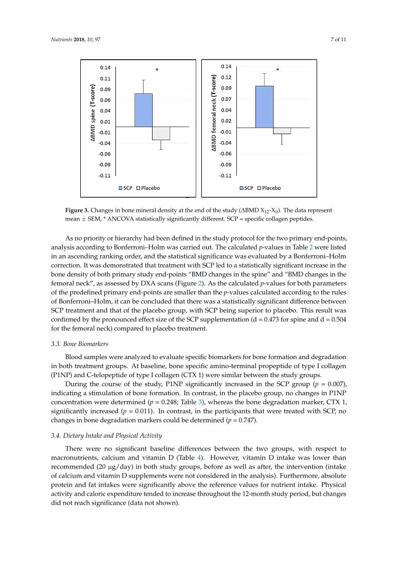
The differences observed between the SCP group and the placebo group were verified by analysis of covariance (ANCOVA), considering the unbalanced baseline values. The analysis showed that bone density significantly (p = 0.030) increased in the spine and the femoral neck after SCP treatment compared to placebo (p = 0.003; Table 3). In the SCP group, BMD increased by almost 3.0% in the spine and 6.7% in the femoral neck, whereas, in the same period, bone density decreased in the placebo group (−1.3% for spine and −1.0% in the femoral neck) (Figure 3).
Figure 3. Changes in bone mineral density at the end of the study (ΔBMD X12-X0). The data represent mean ± SEM, * ANCOVA statistically significantly different. SCP = specific collagen peptides. Figure 3. Changes in bone mineral density at the end of the study (∆BMD X12-X0). The data representmean ± SEM, * ANCOVA statistically significantly different. SCP = specific collagen peptides.
As no priority or hierarchy had been defined in the study protocol for the two primary end-points,analysis according to Bonferroni–Holm was carried out. The calculated p-values in Table 2 were listedin an ascending ranking order, and the statistical significance was evaluated by a Bonferroni–Holmcorrection. It was demonstrated that treatment with SCP led to a statistically significant increase in thebone density of both primary study end-points “BMD changes in the spine” and “BMD changes in thefemoral neck”, as assessed by DXA scans (Figure 2). As the calculated p-values for both parametersof the predefined primary end-points are smaller than the p-values calculated according to the rulesof Bonferroni–Holm, it can be concluded that there was a statistically significant difference betweenSCP treatment and that of the placebo group, with SCP being superior to placebo. This result wasconfirmed by the pronounced effect size of the SCP supplementation (d = 0.473 for spine and d = 0.504for the femoral neck) compared to placebo treatment.
3.3. Bone Biomarkers
Blood samples were analyzed to evaluate specific biomarkers for bone formation and degradationin both treatment groups. At baseline, bone specific amino-terminal propeptide of type I collagen(P1NP) and C-telopeptide of type I collagen (CTX 1) were similar between the study groups.
During the course of the study, P1NP significantly increased in the SCP group (p = 0.007),indicating a stimulation of bone formation. In contrast, in the placebo group, no changes in P1NPconcentration were determined (p = 0.248; Table 3), whereas the bone degradation marker, CTX 1,significantly increased (p = 0.011). In contrast, in the participants that were treated with SCP, nochanges in bone degradation markers could be determined (p = 0.747).
3.4. Dietary Intake and Physical Activity
There were no significant baseline differences between the two groups, with respect tomacronutrients, calcium and vitamin D (Table 4). However, vitamin D intake was lower thanrecommended (20 µg/day) in both study groups, before as well as after, the intervention (intakeof calcium and vitamin D supplements were not considered in the analysis). Furthermore, absoluteprotein and fat intakes were significantly above the reference values for nutrient intake. Physicalactivity and caloric expenditure tended to increase throughout the 12-month study period, but changesdid not reach significance (data not shown).

Nutrients 2018, 10, 97 8 of 11
Table 4. Intake of energy and nutrients compared to recommended dietary allowance (RDA)(US) [20,21].
Group X0 ± SD0 X12 ± SD12RDA US
(Women 51–70 Years)
Energy [kcal] SCP 1978 ± 387 2010 ± 4852403 (19 years) 1
Placebo 2029 ± 524 2030 ± 576
Protein [g] SCP 75 ± 18 a 73 ± 19 a46 (=̂0.8 g/kg BW)
Placebo 85 ± 39 a 87 ± 29 a
Fat [%]SCP 35 ± 7 34 ± 7
NDPlacebo 38 ± 7 34 ± 7
Calcium [mg] SCP 1086 ± 262 1064 ± 3481200Placebo 1383 ± 590 1220 ± 466
Vitamin D [µg] SCP 1.94 ± 2.9 b 2.13 ± 2.4 b15Placebo 2.92 ± 2.8 b 4.00 ± 3.1 b
a = Significant higher intake than recommended, b = significant lower intake than recommended, ND = notdetermined, 1 = For healthy active Americans and Canadians; subtract 7 kcal/day for females for each year of ageabove 19 years, BW, bodyweight.
3.5. Blood and Safety Parameters
Blood safety parameters (i.e., hemogram, kidney, liver and inflammatory parameters) showedno clinically relevant changes during the course of the 12-month treatment. However, systolic bloodpressure (134.5 ± 16.8 vs. 128.1 ± 12.9 mmHg; p < 0.001) and also diastolic blood pressure (81.2 ± 9.6vs. 78.6 ± 7.0 mmHg; p = 0.015) were significantly lower in the SCP group after the intervention, ascalculated by paired Student’s t-tests. In the placebo group, blood pressure remained rather constant.
4. Discussion
The main outcome of this randomized, double-blinded and placebo-controlled study inpostmenopausal women was that specific collagen peptides significantly increased bone mineraldensity (BMD) in both the lumbar spine and femoral neck. In contrast, no significant changes for theseparameters were determined in the placebo group. Considering the decrease in BMD in the controlgroup, subjects in the SCP group showed a 4.2% higher BMD in the spine and a 7.7% higher BMDin the femoral neck, suggesting a clinically relevant effect of the 12-month treatment with SCP [22].The anabolic effect of SCP intake was also confirmed by a significant increase in the bone formationbiomarker, amino-terminal propeptide of type I collagen (P1NP). In contrast, only in the control group,a significant increase in the concentration of the bone degradation marker C-telopeptide of type Icollagen (CTX 1) could be detected after the 12-month study period.
To the author’s knowledge, only two studies have investigated the effect of collagen peptides onbone markers and bone mineral density in humans. In one study, the effects of collagen peptides wereinvestigated, with and without calcitonin [14]. The authors measured urinary pyridinoline cross-linksand suggested, from their results, that calcitonin plus collagen peptides had a greater effect on theinhibition of bone collagen breakdown. Another investigation, using a supplement with a combinationof collagen + calcium + vitamin D, found that the loss in BMD was substantially lower in the collagensupplemented group than in the group with calcium + vitamin D alone [23].
Direct scientific evidence to explain the positive effects of collagen supplementation in humansis still lacking. However, some findings from cell experiments and in vivo studies in rodentshave enhanced our knowledge of how collagen peptides could enhance bone formation andincrease BMD. First of all, it has been shown that collagen peptides are rapidly absorbed fromthe gastrointestinal tract [24,25]. In addition, collagen peptides are absorbed in the small intestineto a considerable amount in peptide form and may act as signaling molecules, thereby positivelyinfluencing anabolic processes [25,26]. Especially for connective tissue, this stimulating effect has

Nutrients 2018, 10, 97 9 of 11
previously been demonstrated [27–29]. With respect to myoblast differentiation and myotubehypertrophy, Kitakaze et al. identified the dipeptide (Hyp-Gly) from collagen as a signaling peptide thatactivates the PI3K/Akt/mTOR pathway [30]. Therefore, it may be assumed that also the stimulationof collagen formation in the bone could be mediated via signaling proteins derived from collagenpeptides. Collagen peptides have been shown to increase gene expression of collagen type 1, alpha 1(COLIA1). In addition, the authors found that the ERK/MAPK signaling pathway was involved inthe collagen-induced increase in COLIA1 expression [31]. Collagen is by far the major constituentof bone mass. Results from rodent studies have demonstrated that collagen peptides significantlyincrease the organic substance of the bone [9]. Therefore, an increase in this organic fraction andthe following mineralization of the bone may result in an increased BMD. In the present study, ananabolic effect, with respect to collagen synthesis and bone anabolism, was reflected by an increasein the bone marker P1NP in the SCP supplemented group. In contrast, the bone degradation marker,CTX1, was significantly increased in the control group only. Comparable results were also found inovariectomized rats, following oral administration of bovine collagen peptides [32].
Other animal studies have shown that collagen peptides or gelatin hydrolysates increase thelongitudinal bone growth in rats [33], increase the bone mass in both growing rats following treadmilltraining [34] as well as mature rats [35], inhibit bone loss in ovariectomized rats [12,36] and preventbone loss in estrogen-deficient rats, probably by reducing the levels of proinflammatory cytokines [11].
Together, the increasing knowledge about the signaling characteristics of collagenpeptides [30,31,37], as well as the aforementioned encouraging findings in animal studies, makethe results of the current investigations comprehensible, although considerably more data are neededfrom human studies.
Nevertheless, there is still insufficient knowledge about which type of collagen peptides (marine,porcine, bovine etc.) exerts the most favorable effect. Moreover, not all collagen peptides mayhave the same effects in different kind of diseases, and, finally, the manufacturing process couldalso have an influence on the biological and physiological properties of collagen peptides and thus,their effectiveness.
Apart from the fact that we need more and larger human studies, we also need additionaldata regarding the optimal timing and dosage as well as findings related to longer-term effects ofsupplementation with collagen peptides.
5. Conclusions
In conclusion, the findings of this randomized, placebo-controlled trial demonstrate, thatsupplementation with 5 g of specific collagen peptides significantly increases bone mineral densityof the lumbar spine and the femoral neck as well as blood levels of the bone marker, P1NP, inpostmenopausal women with age-related decline in BMD.
Acknowledgments: Part of the study was financially supported by GELITA AG, Germany. The planning,organization of the study as well as data analyses was performed solely by the investigators.
Author Contributions: Daniel König was the principal investigator of the study. Stephan Scharla and SteffenOesser were involved in the design and/or execution of the study and manuscript preparation. All authors readand approved the final manuscript.
Conflicts of Interest: Daniel König, Steffen Oesser, Stephan Scharla, Denise Zdzieblik and Albert Gollhoferdeclare that they have no conflict of interest.
References
1. Hernlund, E.; Svedbom, A.; Ivergard, M.; Compston, J.; Cooper, C.; Stenmark, J.; McCloskey, E.V.; Jönsson, B.;Kanis, J.A. Osteoporosis in the European Union: Medical management, epidemiology and economic burden.A report prepared in collaboration with the International Osteoporosis Foundation (IOF) and the EuropeanFederation of Pharmaceutical Industry Associations (EFPIA). Arch. Osteoporos. 2013, 8, 136. [CrossRef][PubMed]

Nutrients 2018, 10, 97 10 of 11
2. Kreutle, V.; Blum, C.; Meier, C.; Past, M.; Müller, B.; Schütz, P.; Borm, K. Bisphosphonate inducedhypocalcaemia - report of six cases and review of the literature. Swiss Med. Wkly. 2014, 144, w13979.[CrossRef] [PubMed]
3. Kaehling, C.; Streckbein, P.; Schmermund, D.; Henrich, M.; Burchert, D.; Gattenloehner, S.; Howaldt, H.P.;Wilbrand, J.F. Lethal cervical abscess following bisphosphonate related osteonecrosis of the jaw.J. Cranio-Maxillofac. Surg. 2014, 42, 1203–1206. [CrossRef] [PubMed]
4. Rachner, T.D.; Platzbecker, U.; Felsenberg, D.; Hofbauer, L.C. Osteonecrosis of the jaw after osteoporosistherapy with denosumab following long-term bisphosphonate therapy. Mayo Clin. Proc. 2013, 88, 418–419.[CrossRef] [PubMed]
5. Montagnani, A. Bone anabolics in osteoporosis: Actuality and perspectives. World J. Orthop. 2014, 5, 247–254.[CrossRef] [PubMed]
6. Dore, R.K. Long-term safety, efficacy, and patient acceptability of teriparatide in the management ofglucocorticoid-induced osteoporosis. Patient Prefer. Adherence 2013, 7, 435–446. [CrossRef] [PubMed]
7. Hadji, P. The evolution of selective estrogen receptor modulators in osteoporosis therapy. Climacteric 2012,15, 513–523. [CrossRef] [PubMed]
8. Gallagher, A.M.; Rietbrock, S.; Olson, M.; van Staa, T.P. Fracture outcomes related to persistence andcompliance with oral bisphosphonates. J. Bone Miner. Res. 2008, 23, 1569–1575. [CrossRef] [PubMed]
9. Watanabe-Kamiyama, M.; Shimizu, M.; Kamiyama, S.; Taguchi, Y.; Sone, H.; Morimatsu, F.; Shirakawa, H.;Furukawa, Y.; Komai, M. Absorption and effectiveness of orally administered low molecular weight collagenhydrolysate in rats. J. Agric. Food Chem. 2010, 58, 835–841. [CrossRef] [PubMed]
10. Guillerminet, F.; Fabien-Soule, V.; Even, P.C.; Tomé, D.; Benhamou, C.L.; Roux, C.; Blais, A. Hydrolyzedcollagen improves bone status and prevents bone loss in ovariectomized C3H/HeN mice. Osteoporos. Int.2012, 23, 1909–1919. [CrossRef] [PubMed]
11. Han, X.; Xu, Y.; Wang, J.; Pei, X.; Yang, R.; Li, N.; Li, Y. Effects of cod bone gelatin on bone metabolism andbone microarchitecture in ovariectomized rats. Bone 2009, 44, 942–947. [CrossRef] [PubMed]
12. Guillerminet, F.; Beaupied, H.; Fabien-Soule, V.; Tomé, D.; Benhamou, C.L.; Roux, C.; Blais, A. Hydrolyzedcollagen improves bone metabolism and biomechanical parameters in ovariectomized mice: An in vitro andin vivo study. Bone 2010, 46, 827–834. [CrossRef] [PubMed]
13. De Almeida, J.E.; Cuneo, F.; Amaya-Farfan, J.; de Assuncao, J.V.; Quintaes, K.D. A food supplement ofhydrolyzed collagen improves compositional and biodynamic characteristics of vertebrae in ovariectomizedrats. J. Med. Food 2010, 13, 1385–1390. [CrossRef] [PubMed]
14. Adam, M.; Spacek, P.; Hulejova, H.; Galianova, A.; Blahos, J. Postmenopausal osteoporosis. Treatment withcalcitonin and a diet rich in collagen proteins. Cas. Lek. Cesk. 1996, 135, 74–78. [PubMed]
15. Urbaniak, G.C.; Plous, S. Research Randomizer, Version 4.0; 2011. Available online: http://www.randomizer.org/(accessed on 16 January 2018).
16. Management of osteoporosis in postmenopausal women: 2010 position statement of The North AmericanMenopause Society. Menopause 2010, 17, 25–54.
17. Scharla, S.H. Nutritional medicine for the prevention and treatment of osteoporosis. Dtsch. Med. Wochenschr.2003, 128, 946–950. [CrossRef] [PubMed]
18. Frey, I.; Berg, A. Erfassung der körperlichen Aktivität in Klinik und Praxis. In Körperliche Aktivität inPrävention und Therapie; Samitz, G.M.G., Ed.; Hans Marseille Verlag GmhH: München, Germany, 2002;pp. 81–86.
19. Looker, A.C.; Borrud, L.G.; Hughes, J.P.; Fan, B.; Shepherd, J.A.; Melton, L.J., III. Lumbar spine and proximalfemur bone mineral density, bone mineral content, and bone area: United States, 2005–2008. Vital Health Stat.2012, 11, 1–132.
20. Institute of Medicine (US). Committee to Review Dietary Reference Intakes for Vitamin D and Calcium; NationalAcademies Press: Washington, DC, USA, 2011.
21. Institute of Medicine. Dietary Reference Intakes for Energy, Carbohydrate, Fiber, Fat, Fatty Acids, Cholesterol,Protein, and Amino Acids; The National Academies Press: Washington, DC, USA, 2005.
22. Liberman, U.A.; Weiss, S.R.; Broll, J.; Minne, H.W.; Quan, H.; Bell, N.H.; Rodriguez-Portales, J.;Downs, R.W., Jr.; Dequeker, J.; Favus, M.; et al. Effect of oral alendronate on bone mineral density and theincidence of fractures in postmenopausal osteoporosis. The Alendronate Phase III Osteoporosis TreatmentStudy Group. N. Engl. J. Med. 1995, 333, 1437–1443. [CrossRef] [PubMed]

Nutrients 2018, 10, 97 11 of 11
23. Elam, M.L.; Johnson, S.A.; Hooshmand, S.; Feresin, R.G.; Payton, M.E.; Gu, J.; Arjmandi, B.H.A calcium-collagen chelate dietary supplement attenuates bone loss in postmenopausal women withosteopenia: A randomized controlled trial. J. Med. Food 2015, 18, 324–331. [CrossRef] [PubMed]
24. Walrand, S.; Chiotelli, E.; Noirt, F.; Mwewa, S.; Lassel, T. Consumption of a functional fermented milkcontaining collagen hydrolysate improves the concentration of collagen-specific amino acids in plasma.J. Agric. Food Chem. 2008, 56, 7790–7795. [CrossRef] [PubMed]
25. Ohara, H.; Matsumoto, H.; Ito, K.; Iwai, K.; Sato, K. Comparison of quantity and structures ofhydroxyproline-containing peptides in human blood after oral ingestion of gelatin hydrolysates fromdifferent sources. J. Agric. Food Chem. 2007, 55, 1532–1535. [CrossRef] [PubMed]
26. Shimizu, K.; Sato, M.; Zhang, Y.; Kouguchi, T.; Takahata, Y.; Morimatsu, F.; Shimizu, M. The bioavailableoctapeptide Gly-Ala-Hyp-Gly-Leu-Hyp-Gly-Pro stimulates nitric oxide synthesis in vascular endothelialcells. J. Agric. Food Chem. 2010, 58, 6960–6965. [CrossRef] [PubMed]
27. Oesser, S.; Seifert, J. Stimulation of type II collagen biosynthesis and secretion in bovine chondrocytescultured with degraded collagen. Cell Tissue Res. 2003, 311, 393–399. [PubMed]
28. Bello, A.E.; Oesser, S. Collagen hydrolysate for the treatment of osteoarthritis and other joint disorders:A review of the literature. Curr. Med. Res. Opin. 2006, 22, 2221–2232. [CrossRef] [PubMed]
29. Ng, K.W.; Saliman, J.D.; Lin, E.Y.; Statman, L.Y.; Kugler, L.E.; Lo, S.B.; Ateshian, G.A.; Hung, C.T.Culture duration modulates collagen hydrolysate-induced tissue remodeling in chondrocyte-seeded agarosehydrogels. Ann. Biomed. Eng. 2007, 35, 1914–1923. [CrossRef] [PubMed]
30. Kitakaze, T.; Sakamoto, T.; Kitano, T.; Inoue, N.; Sugihara, F.; Harada, N.; Yamaji, R. The collagen deriveddipeptide hydroxyprolyl-glycine promotes C2C12 myoblast differentiation and myotube hypertrophy.Biochem. Biophys. Res. Commun. 2016, 478, 1292–1297. [CrossRef] [PubMed]
31. Kim, H.K.; Kim, M.G.; Leem, K.H. Osteogenic activity of collagen peptide via ERK/MAPK pathwaymediated boosting of collagen synthesis and its therapeutic efficacy in osteoporotic bone by back-scatteredelectron imaging and microarchitecture analysis. Molecules 2013, 18, 15474–15489. [CrossRef] [PubMed]
32. Liu, J.; Wang, Y.; Song, S.; Wang, X.; Qin, Y.; Si, S.; Guo, Y. Combined oral administration of bovine collagenpeptides with calcium citrate inhibits bone loss in ovariectomized rats. PLoS ONE 2015, 10, e0135019.[CrossRef] [PubMed]
33. Bortolin, R.H.; da Graca Azevedo Abreu, B.J.; Ururahy, M.A.; de Souza, K.S.; Bezerra, J.F.; Loureiro, M.B.; daSilva, F.S.; da Silva Marques, D.E.; de Sousa Batista, A.A.; Oliveira, G.; et al. Protection against T1DM-InducedBone Loss by Zinc Supplementation: Biomechanical, Histomorphometric, and Molecular Analyses inSTZ-Induced Diabetic Rats. PLoS ONE 2015, 10, e0125349. [CrossRef] [PubMed]
34. Takeda, S.; Park, J.H.; Kawashima, E.; Ezawa, I.; Omi, N. Hydrolyzed collagen intake increases bone mass ofgrowing rats trained with running exercise. J. Int. Soc. Sports Nutr. 2013, 10, 35. [CrossRef] [PubMed]
35. Wu, J.; Fujioka, M.; Sugimoto, K.; Mu, G.; Ishimi, Y. Assessment of effectiveness of oral administration ofcollagen peptide on bone metabolism in growing and mature rats. J. Bone Miner. Metab. 2004, 22, 547–553.[CrossRef] [PubMed]
36. Liu, J.; Zhang, B.; Song, S.; Ma, M.; Si, S.; Wang, Y.; Xu, B.; Feng, K.; Wu, J.; Guo, Y. Bovine collagen peptidescompounds promote the proliferation and differentiation of MC3T3-E1 pre-osteoblasts. PLoS ONE 2014,9, e99920. [CrossRef] [PubMed]
37. Iba, Y.; Yokoi, K.; Eitoku, I.; Goto, M.; Koizumi, S.; Sugihara, F.; Oyama, H.; Yoshimoto, T. Oral Administrationof Collagen Hydrolysates Improves Glucose Tolerance in Normal Mice Through GLP-1-Dependent andGLP-1-Independent Mechanisms. J. Med. Food 2016, 19, 836–843. [CrossRef] [PubMed]
© 2018 by the authors. Licensee MDPI, Basel, Switzerland. This article is an open accessarticle distributed under the terms and conditions of the Creative Commons Attribution(CC BY) license (http://creativecommons.org/licenses/by/4.0/).