bone and muscle: structure, force, and motion€¦ · · 2010-03-31physiology of bone 61 calcium...
TRANSCRIPT
CONTENTS20
41
Introduction 12
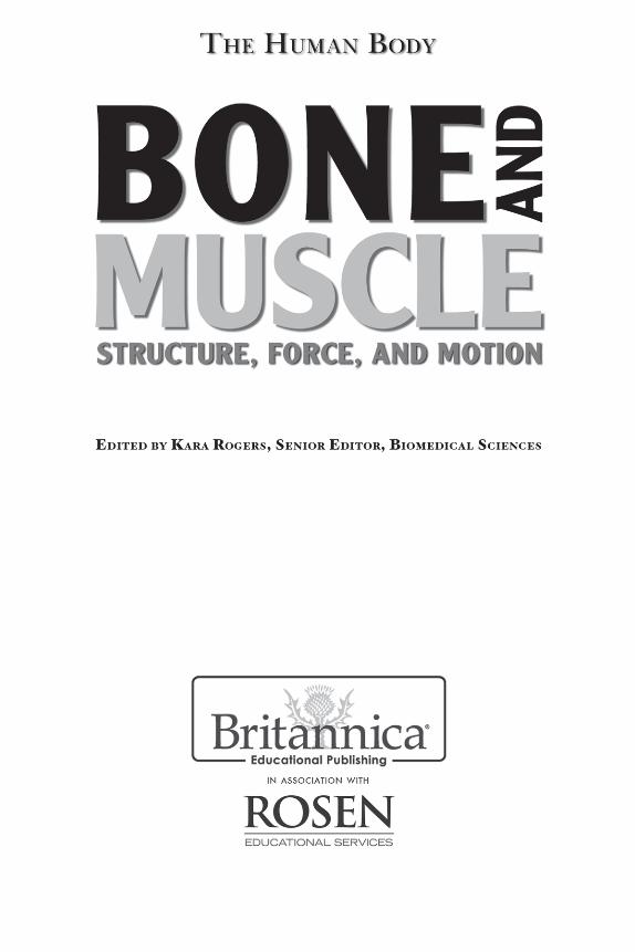
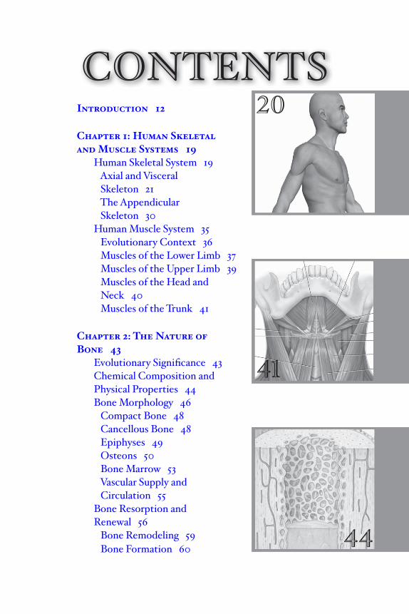
Chapter 1: Human Skeletaland Muscle Systems 19 Human Skeletal System 19 Axial and Visceral Skeleton 21 The Appendicular Skeleton 30 Human Muscle System 35 Evolutionary Context 36 Muscles of the Lower Limb 37 Muscles of the Upper Limb 39 Muscles of the Head and Neck 40 Muscles of the Trunk 41
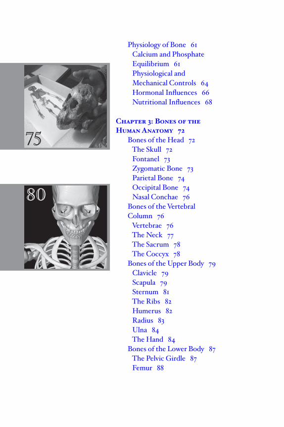
Chapter 2: The Nature of Bone 43 Evolutionary Signifi cance 43 Chemical Composition and Physical Properties 44 Bone Morphology 46 Compact Bone 48 Cancellous Bone 48 Epiphyses 49 Osteons 50 Bone Marrow 53 Vascular Supply and Circulation 55 Bone Resorption and Renewal 56 Bone Remodeling 59 Bone Formation 60 44
Physiology of Bone 61 Calcium and Phosphate Equilibrium 61 Physiological and Mechanical Controls 64 Hormonal Influences 66 Nutritional Influences 68
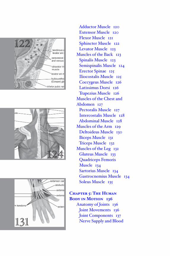
Chapter 3: Bones of the Human Anatomy 72 Bones of the Head 72 The Skull 72 Fontanel 73 Zygomatic Bone 73 Parietal Bone 74 Occipital Bone 74 Nasal Conchae 76 Bones of the Vertebral Column 76 Vertebrae 76 The Neck 77 The Sacrum 78 The Coccyx 78 Bones of the Upper Body 79 Clavicle 79 Scapula 79 Sternum 81 The Ribs 82 Humerus 82 Radius 83 Ulna 84 The Hand 84 Bones of the Lower Body 87 The Pelvic Girdle 87 Femur 88
80
75
93
Tibia 89 Fibula 89 The Foot 90
Chapter 4: The Nature ofMuscle 93
Striated Muscle 94 Muscle Fibres 94 Myofi brils 96 Myofi laments 98 Proteins of the Myofi laments 100 Actin-Myosin Interaction and Its Regulation 103 Energy Stores 104 Molecular Mechanisms of Muscle Contraction 105 Smooth Muscle 106 Structure and Organization 107 Initiation of Contraction 109 Cross-Bridge Cycle and ATP Breakdown 110 Mechanical Properties 111 Cardiac Muscle 112 Structure and Organization 113 The Frequency of Contraction 114 Excitation/Contraction Coupling 116 Force and Velocity of Contraction 117 Response of the Heart to Stress 117 Muscles of Movement 119 Abductor Muscle 119
91
122
124
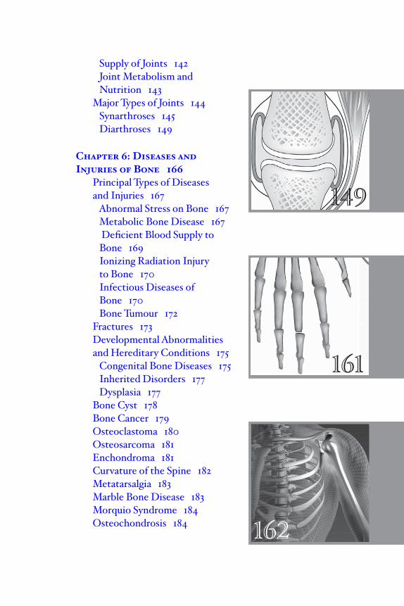
Adductor Muscle 120 Extensor Muscle 120 Flexor Muscle 121 Sphincter Muscle 122 Levator Muscle 123 Muscles of the Back 123 Spinalis Muscle 123 Semispinalis Muscle 124 Erector Spinae 125 Iliocostalis Muscle 125 Coccygeus Muscle 126 Latissimus Dorsi 126 Trapezius Muscle 126 Muscles of the Chest and
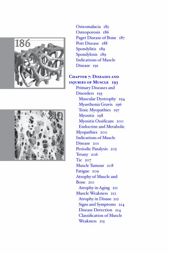
Abdomen 127 Pectoralis Muscle 127 Intercostalis Muscle 128 Abdominal Muscle 128 Muscles of the Arm 129 Deltoideus Muscle 130 Biceps Muscle 131 Triceps Muscle 132 Muscles of the Leg 132 Gluteus Muscle 133 Quadriceps Femoris Muscle 134 Sartorius Muscle 134 Gastrocnemius Muscle 134 Soleus Muscle 135
Chapter 5: The Human Body in Motion 136 Anatomy of Joints 136 Joint Movements 136 Joint Components 137 Nerve Supply and Blood 131
149
161
162
Supply of Joints 142 Joint Metabolism and Nutrition 143 Major Types of Joints 144 Synarthroses 145 Diarthroses 149
Chapter 6: Diseases andInjuries of Bone 166 Principal Types of Diseases and Injuries 167 Abnormal Stress on Bone 167 Metabolic Bone Disease 167 Deficient Blood Supply to Bone 169 Ionizing Radiation Injury to Bone 170 Infectious Diseases of Bone 170 Bone Tumour 172 Fractures 173 Developmental Abnormalities and Hereditary Conditions 175 Congenital Bone Diseases 175 Inherited Disorders 177 Dysplasia 177 Bone Cyst 178 Bone Cancer 179 Osteoclastoma 180 Osteosarcoma 181 Enchondroma 181 Curvature of the Spine 182 Metatarsalgia 183 Marble Bone Disease 183 Morquio Syndrome 184 Osteochondrosis 184
Osteomalacia 185 Osteoporosis 186 Paget Disease of Bone 187 Pott Disease 188 Spondylitis 189 Spondylosis 189 Indications of Muscle Disease 191
Chapter 7: Diseases and injuries of Muscle 193 Primary Diseases and Disorders 193 Muscular Dystrophy 194 Myasthenia Gravis 196 Toxic Myopathies 197 Myositis 198 Myositis Ossificans 200 Endocrine and Metabolic
Myopathies 200 Indications of Muscle Disease 201 Periodic Paralysis 205 Tetany 206 Tic 207 Muscle Tumour 208 Fatigue 209 Atrophy of Muscle and Bone 210 Atrophy in Aging 211 Muscle Weakness 212 Atrophy in Disuse 212 Signs and Symptoms 214 Disease Detection 214 Classification of Muscle Weakness 215
186
210
219
Chapter 8: Diseases andInjuries of Joints 218 Types of Arthritis 218 Bursitis 220 Infectious Arthritis 221 Rheumatoid Arthritis and Allied Disorders 223 Gout 226 Collagen Diseases 228 Miscellaneous Types of Arthritis 229 Traumatic Joint Diseases 230 Dislocation 231 Fracture–Dislocation 232 Sprain 233 Elbow Injuries 233 Knee Injuries 234 Degenerative Joint Disease 235 Congenital and Hereditary Joint Abnormalities 237 Secondary Joint Diseases 240 Hemorrhagic Joint Diseases 240 Aseptic Necrosis 241 Endocrine Factors 242 Neurogenic Arthropathy 242 Hypertrophic Osteoarthropathy 243 Reflex Sympathetic Dystrophy 243 Tumours of Joints 244
Conclusion 246Glossary 247Bibliography 250Index 252
231
INTRODUCTION
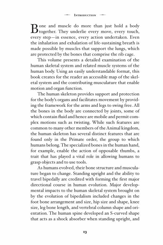
Bone and muscle do more than just hold a body together. They underlie every move, every touch,
every step—in essence, every action undertaken. Even the inhalation and exhalation of life-sustaining breath is made possible by muscles that support the lungs, which are protected by the bones that comprise the rib cage.
This volume presents a detailed examination of the human skeletal system and related muscle systems of the human body. Using an easily understandable format, this book creates for the reader an accessible map of the skel-etal system and the contributing musculature that enable motion and organ function.
The human skeleton provides support and protection for the body’s organs and facilitates movement by provid-ing the framework for the arms and legs to swing free. All the bones in the body are connected by joints, some of which contain fl uid and hence are mobile and permit com-plex motions such as twisting. While such features are common to many other members of the Animal kingdom, the human skeleton has several distinct features that are found only in the Primate order, the group to which humans belong. The specialized bones in the human hand, for example, enable the action of opposable thumbs, a trait that has played a vital role in allowing humans to grasp objects and to use tools.
As humans evolved, their bone structure and muscula-ture began to change. Standing upright and the ability to travel bipedally are credited with forming the fi rst major directional course in human evolution. Major develop-mental impacts to the human skeletal system brought on by the evolution of bipedalism included changes in the foot bone arrangement and size, hip size and shape, knee size, leg bone length, and vertebral column shape and ori-entation. The human spine developed an S-curved shape that acts as a shock absorber when standing upright, and
13
7 Introduction 7
helps to hold up the heavy skull. Walking on two feet also demanded monumental changes to the musculature of the upper and lower limbs, the head and neck, and the muscles of the trunk or torso. Such changes were required to sup-port and maintain this new, upright posture.
This posture has provided humans with some distinct evolutionary advantages. Unlike chimpanzees, humans do not spend much time hanging from trees or otherwise using their hands for locomotion. Instead, bipedal move-ment freed the hands, allowing humans to use their hands to grasp tools and weapons, to build shelters, and to skin animals for clothes. As civilizations evolved, humans began to use their hands to create great works of art and to play instruments.This allowed humans to create a hand-mind connection that made their brains grow larger as well.
There are two primary types of bone material. Compact bone, which makes up 80 percent of all bone, is the dense, rigid outer layer of that provides strength and structural integrity. Cancellous bone, of which the remain-ing 20 percent is comprised, is honeycombed bone in structure and makes up much of the enlarged ends of the long bones and ribs. The structure of this type of bone enables it to absorb large amounts of stress.
The bones of the human skeleton are not static. The skeletal system is vibrant and continuously active, con-tributing a steady stream of new blood cells through bone marrow. Bone tissue is also subject to loss or damage through wear and tear, disease, or injury. Through the pro-cess of bone remodeling, bone tissue is constantly being dissolved, removed, and replaced by new building blocks of remodeled bone. Bone formation is affected by factors such as diet and hormonal influences and by a lack of nutrients, such as calcium and vitamins C and D. The absence of these nutrients from the diet can harm bone development.
7 Bone and Muscle 7
14
Muscles aid in movement and protect and support the body. They also have an important role in automatic func-tions, such as breathing and digestion, and they provide a source of heat to keep the body warm. There are three major kinds of muscles: striated, smooth, and cardiac. Striated muscles are muscles that enable humans to move in various ways. They make up a large fraction of total body weight. Most of these muscles are attached to the skeleton at both ends by tendons, and they serve primar-ily to move the limbs and to maintain posture. Smooth muscle is found in the walls of many hollow organs, such as those of the gastrointestinal tract and the reproductive system. It does not have the striped appearance of stri-ated muscle. Cardiac muscle, which is found only in the heart, is a special kind of muscle that contracts rhythmi-cally. This rhythmic contraction allows the heart to pump blood through the body. Muscles perform a number of different functions in terms of allowing the human body to move freely. Extensor muscles, for instance, allow the limbs to straighten, whereas the flexor muscles allow the limbs to bend.
Joints permit humans to move in a variety of ways, from nodding their heads to flexing their knees. Joints, which consist of a variety of components, including carti-lage, collagen, and ligaments, are specifically designed to be flexible. Flexibility gives joints such as the shoulder a wide range of motion. There are two basic structural types of joints—diarthroses (fluid-containing joints) and synarthroses (characterized by the absence of fluid). Synarthroses are located in the skull, the jaw, the spinal column, and the pelvic bone, and they perform highly spe-cialized functions. They allow for infant skull compression during childbirth, which eases the infant’s passage through the birth canal, and they allow the hip bones to swing upward and outward during childbirth. They also allow for
15
7 Introduction 7
vertebrae to compress during physical activities, and they act as virtual hinges in the skull, allowing for the growth of adjacent bones. Diarthroses are much more varied in their structure and assigned tasks, and they are primarily responsible for movement and locomotion. The joints of the elbows, knees, and ankles are all examples of diarthroses.
It is important to note the relationship of injury and disease when examining the skeletal system, muscles, and joints. Injury to bone, such as a fracture, can lead to the onset of disease within that bone. On the other hand, dis-ease within a bone, such as osteoporosis, can contribute to bone fractures. The leading contributor to bone injury and fracture is abnormal stress on the bone tissue. This can occur as a result of physical exertion, a fall, or pres-sure upon the bone in a nonsupported direction. Inactivity can also cause fractures. For instance, in a patient who is bedridden, bone formation is reduced, weakening bone tissues. If a patient receives radiation to fight cancer, that same radiation can have a negative impact on bone strength and integrity. Bone tissue is also subject to infec-tion, such as when microorganisms are introduced into the tissue through the bloodstream or when a fractured bone breaks the surface of the skin. The skeletal system can also exhibit developmental abnormalities, such as scoliosis, or curvature of the spine. Several cancers can affect bone tissue as well.
The most common indications of muscle disease or injury are pain weakness, and atrophy (a noticeable decrease in size of the affected muscle tissue). By defini-tion, muscle weakness is the failure of the muscle to develop an expected rate of force. Muscle weakness can be caused by disease of the brain, spinal cord, or peripheral nerves, since these conditions may interfere with the proper electrical stimulation of the muscle tissue. In
7 Bone and Muscle 7
16
addition, a defect within the muscle tissue itself can give rise to weakness. There are several classifications of mus-cle weakness. Motor neuron disease, also known as Lou Gehrig disease, is characterized by the degeneration of neurons that regulate muscle movements. The neurons eventually atrophy, causing the affected muscle tissue to waste away. Some of the most well-known muscle diseases include muscular dystrophy, which causes degeneration of the skeletal muscles; myasthenia gravis, a chronic autoim-mune disorder caused by failure of nerve impulses; and myositis, inflammation of muscle tissue.
Joint diseases fall into one of two categories; they are either inflammatory or noninflammatory. Arthritis is a generic term for inflammatory joint disease. Inflammation causes swelling, stiffness, and pain in the joint, and often fluid will accumulate in the joint as well. Examination of any such fluid can be a critical clue in establishing the nature and cause of the inflammation. Bacteria, fungi, or viruses may infect joints by direct contamination through a penetrating wound, by migration through the blood-stream as a result of systemic infection, or by migration from infected adjacent bone tissues. These joint inflam-mations are classified as infectious arthritis. Rheumatoid arthritis, which is similar to infectious arthritis except that no causative agent is known, typically affects the same joints on both sides of the body. The fingers, wrists, and knees are particularly susceptible to this form of arthritis. Symptoms include joint stiffness upon waking, fatigue and anemia, an occasional slight fever, and skin lesions outside the joints. In roughly one-third of patients, the condition progresses to the point where joint func-tionality is totally compromised.
Noninflammatory joint diseases include degenerative joint disorders and blunt force injuries varying in intensity from mild sprains to fractures and dislocations. A sprain
17
7 Introduction 7
1818
involves damage to a ligament, tendon, or muscle follow-ing a sudden wrench and partial dislocation of a joint. Traumatic dislocations must be treated by prolonged immobilization to allow supportive tissue tears in muscles and ligaments to heal. In the case of fractures in the vicin-ity of joints or fractures that extend into the joint space, it is imperative that the normal contour of the joint be restored. If the joint is not repaired correctly, long-term arthritic complications may develop.
There are many different types and causes of degen-erative joint disease. From the simple, ubiquitous disorder osteoarthritis that affects all adults to a greater or lesser degree by the time they reach old age, to joint abnormali-ties such as hemarthrosis (bleeding into the joints), all are defi ned, explained, and made understandable within this body of text.
This volume presents an exceptional tool and is a meaningful addition to the library of anyone who is mak-ing a study of anatomy and medicine.
7 Bone and Muscle 7