body area network: implications for rehabilitation dr. lori maria walton, phd, dpt, mph(s) professor...
TRANSCRIPT

Body Area Network: Implications for Rehabilitation
Dr. Lori Maria Walton, PhD, DPT, MPH(s)Professor & Director of Research
Andrews UniversityMichigan, USA

IntroductionBody Area Networks have numerous applications in medicine and rehabilitation:
Cardiopulmonary
Vascular
Endocrine
Neurological
Physical Medicine
Perioperative monitoring
Clinical Application of
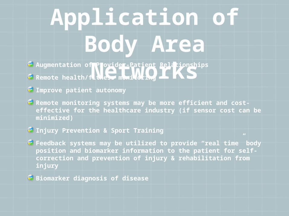
Body Area NetworksAugmentation of Provider-Patient Relationships
Remote health/fitness monitoring
Improve patient autonomy
Remote monitoring systems may be more efficient and cost-effective for the healthcare industry (if sensor cost can be minimized)
Injury Prevention & Sport Training
Feedback systems may be utilized to provide “real time” body position and biomarker information to the patient for self-correction and prevention of injury & rehabilitation from injury
Biomarker diagnosis of disease

Health Care Expenditures
Canada spends 68 billion dollars per year and USA spends 75% of $2 trillion dollar budget on chronic diseases such as diabetes, cardiovascular disease, pulmonary disease, etc.
One third of Canadians have at least one chronic health condition (Brief to House of Commons, 2011)
Patients who are well informed about their medical condition are able to take action to correct it and make behavioral changes accordingly.

Type of Body Data Monitored
Blood pressure, Heart Rate, Respiratory Rate
Body Movements according to specified anatomical landmarks
Biomarkers for acute and chronic disease process

Placement of Body Area SensorsSubcutaneous (biomarkers)
Clothing (external temperature, vital signs, etc.)
Body Part Accessory (Heart monitors, pace makers)
Includes: accelerometers, gyroscopes, smart fabrics and actuators, wireless communication networks, and data capture technology

BAS for

AccelerometersElectrochemical sensors
Measure acceleration of objects in motion
Monitors posture, walking, running, etc
Reference axis landmarks
Provide information on basic steps and activity counts
Quantitative measurement
Velocity and displacement measurement
Triaxial accelerometers (3-D)Provide information on movement 3-dimensions
Posture
Gait analysis
GyroscopesBased on angular momentum (3-D)
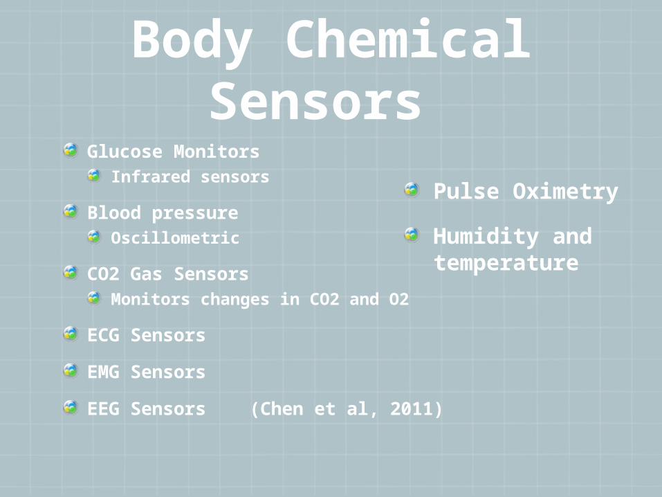
Body Chemical Sensors
Glucose MonitorsInfrared sensors
Blood pressureOscillometric
CO2 Gas SensorsMonitors changes in CO2 and O2
ECG Sensors
EMG Sensors
EEG Sensors (Chen et al, 2011)
Pulse Oximetry
Humidity and temperature

Placement of the Body Area SensorsWrist
Ankle
Waist
Chest
Arm
Legs
Subcutaneous and superficial placement depending upon the utilization
*speed, distance, steps taken, floors climbed, calories burned, ambulation, and posture, SpO2, HR, body temps, , ECG, RR, gait, biomarkers such as lactate, glucose, etc..

WBAN

Monitoring Patient reported outcomes (PRO)
Telemonitoring
Quantifying self-hybrid model (QSHM)
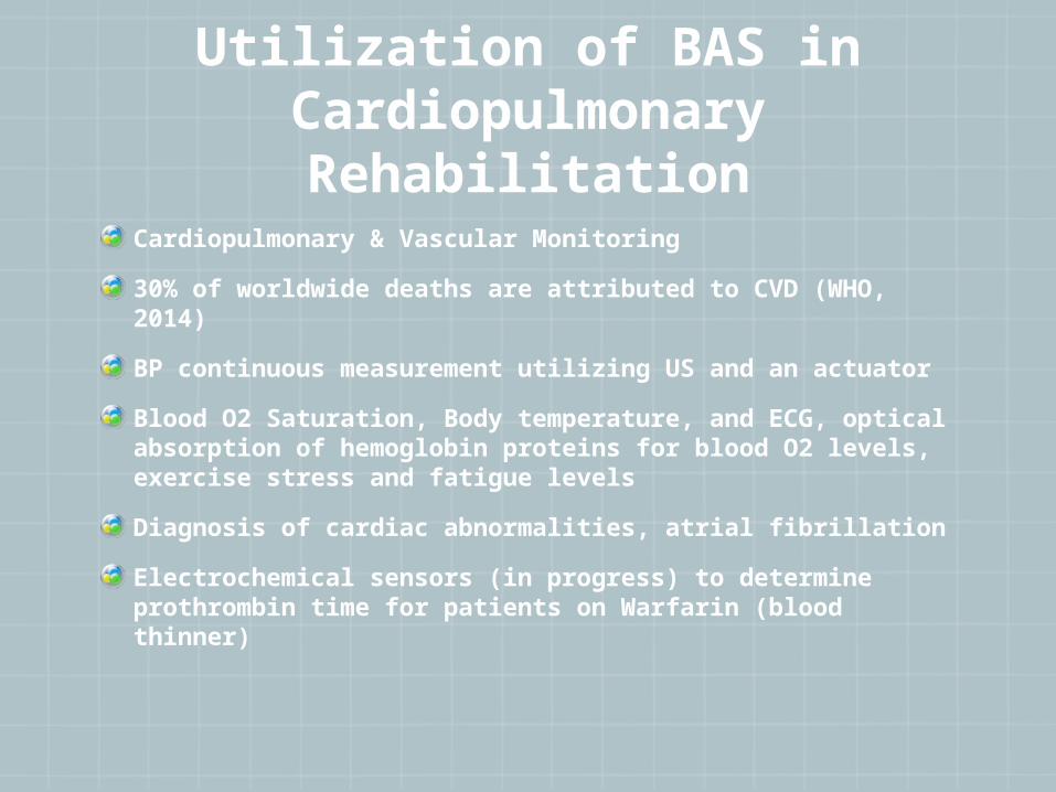
Utilization of BAS in Cardiopulmonary
RehabilitationCardiopulmonary & Vascular Monitoring
30% of worldwide deaths are attributed to CVD (WHO, 2014)
BP continuous measurement utilizing US and an actuator
Blood O2 Saturation, Body temperature, and ECG, optical absorption of hemoglobin proteins for blood O2 levels, exercise stress and fatigue levels
Diagnosis of cardiac abnormalities, atrial fibrillation
Electrochemical sensors (in progress) to determine prothrombin time for patients on Warfarin (blood thinner)
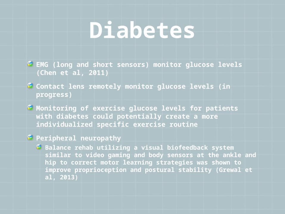
DiabetesEMG (long and short sensors) monitor glucose levels (Chen et al, 2011)
Contact lens remotely monitor glucose levels (in progress)
Monitoring of exercise glucose levels for patients with diabetes could potentially create a more individualized specific exercise routine
Peripheral neuropathyBalance rehab utilizing a visual biofeedback system similar to video gaming and body sensors at the ankle and hip to correct motor learning strategies was shown to improve proprioception and postural stability (Grewal et al, 2013)

Neurologic Diagnosis
Gait & Posture Analysis Analysis Parkinson’s Disease (Conceptual model by Cassimassima et al, 2014)
Limb ParalysisMeulen et al, 2015 Optimal guidance of rehab for 13 subjects with stroke utilized 17 sensors in full body ambulatory system to track measurements for maximal reaching distance, vertical reaching range, hand movement relative to sternum & pelvis
Cerebral PalsyTwo sensors placed on low back and R ankle to monitor gait in children with CP (reliability and validity was more predictable in the minimod vs AMP sensors) (Kuo et al, 2009)
Another study (Baram et al, 2011), showed a 21% residual improvement in walking speed & 8% improvement in stride length for children w/ CP and sensor feedback.

Other Measurement
Exercise progression to maximize therapeutic recovery
Pulmonary rehabilitationGraded exercises
Self-management education
Strength & flexibility training
Physical activity
Monitoring of home exercise program

Subcutaneous Biomarker DetectionElectrochemical biochips
Bajj-Rossi et al (2014) proposed utilization of multi-walled carbon nanotubes w/enzyme catalyst to assure sensitivity and specificity of biosensing
Lactate (SN: .77)
glucose (SN: .64)
pH (SN: .75)
temperature (SN=1.08)
Inflammation (C-Reactive Protein) (Fakanya et al, 2014)

Glucose SensorsAmperometric sensor
utilizing enzyme-electrochemical sensors & thick film technology
Fibre Optic fluorometric glucose sensor
based on O2 measurement
Spectroscopic glucose sensor
utilizing mid-infrared spectroscopy

Implications for Women’s Health Rehabilitation
Prenatal/Postpartum DiagnosisMagnetoencephalogram for fetal and maternal monitoring (during activity) (Vairavan et al, 2010)
Brain growth in the fetus
Cardiac anomolies in mother
ObstetricsEarly detection of preeclampsia biomarker predictors including Corticotropin Releasing Hormone and Vitronectin (Song et al, 2015)
Cancer (Hunter et al, 2014)Subcutaneous temperature sensors in mice sample utilized to detect lymph tumor progression (EMu Mic Lymphoma) (r=.68, p<.001)
Common Prenatal Problems
Placenta Previa
Preeclampsia to Eclampsia
Placenta Abruptio
Subchorionic Hemorrhage
Gestational Diabetes

Placenta previa
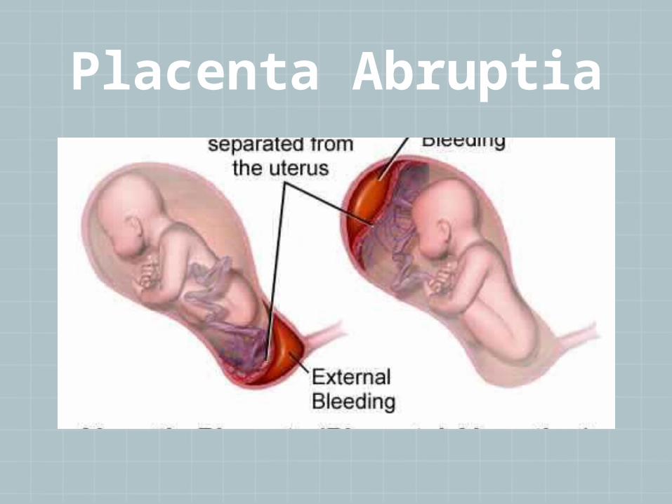
Placenta Abruptia
Preeclampsia3rd Most common cause of maternal mortality world wide (12% all deaths)
May be reduced by up to 55% in women who begin 20 min of exercise 5 X per week in first trimester
Symptoms: High blood pressure and protein in the urine (due to kidney failure) after 20th week in pregnancy
Caused by autoimmune disorders, diet, lack of exercise, blood vessel problems
Risk factors: first pregnancy, twins, obesity, > 35 y/o, diabetes

Testing for Preeclampsia
Protein lab tests (urinalysis)
Weight gain greater than 2 lbs/week
BP > 140/90
Elevated liver enzymes
Swelling hands/face/feet
Decreased platelet count (less than 100,000)


Preeclampsia Monitoring
2010 Study of 50 women by Callaway and Colditz showed improvements in fasting glucose at 28 weeks and insulin at 36 weeks for those who exercised greater than 900 cal/week
2003 Study showed Magnesium sulfate to reduce risk of preeclampsia more than 50% for women and is “drug of choice” above any hypertensives or other treatment
2003 Sorensen et al found a 54% reduction in pre-eclampsia diagnosis for women who exercised vigorously in the year preceeding pregnancy and in early pregnancy.. 34% reduction for those with ANY form of physical activity that was regular, and 24% reduction for women who reported light to moderate (less than 6 METS) compared with non-exercise group
Saftlass et al (2004) suggested that women who engaged in any Leisure time physical activity regardless of caloric intake were significantly decreased their chance of getting preeclampsia

Caesarean-Section
Urinary incontinence
Bowel/bladder scar tissue symptoms
Endometriomas
Placenta previa and abruptio in subsequent pregnancies
Pain
Hypotonia & Hypertonia
Postpartum Infections
Pelvic Floor Spasticity
Pelvic and Abdominal floor Flaccidity,Levator Abnormalities

Normal DeliveryPerineal Trauma
Coccygeal Fracture
Pubic Diastasis
Neuropathy
Post Epidural Pain
Low Back Pain & Pelvic Girdle Pain
Urinary Incontinence

Postpartum Evaluation
Uterus changes in size, location, and volume
Pelvic Floor changes
Urogenital and GI Changes
Wound Healing
Superficial Nerve Entrapment at site of C-Section
Pain
Low Back & Pelvic Girdle Instability

Postpartum Cardiomyopathy1 out of every 1,300 deliveries
Mortality Rate is 25-50%
Weak heart diagnosed within fifth month post delivery
Risk Factors: obesity, alcohol, cardiac diagnosis prior to pregnancy, smoking, multiple pregnancies, undernourished
Symptoms: fatigue, increased nocturia, racing or skipping beats, SOB lying flat, swollen ankles
Complications: CHF, Embolism (Pulmonary), Arhythmias
Treatment: Hospital/ER, immunosuppressive treatment and aortic balloon, heart transplant, medications, fluid restrictions,activity limitations

Venous Air Embolism
More commonly associated with C-section
Incidence 50-95%
1% of all maternal deaths
Occurs more often with Steep Trendelenburg positions
Symptoms: Hypotension, hypoxemia, chest pain
Treatment: Change position, 100% forced O2, IV fluids, encourage fast delivery without CS
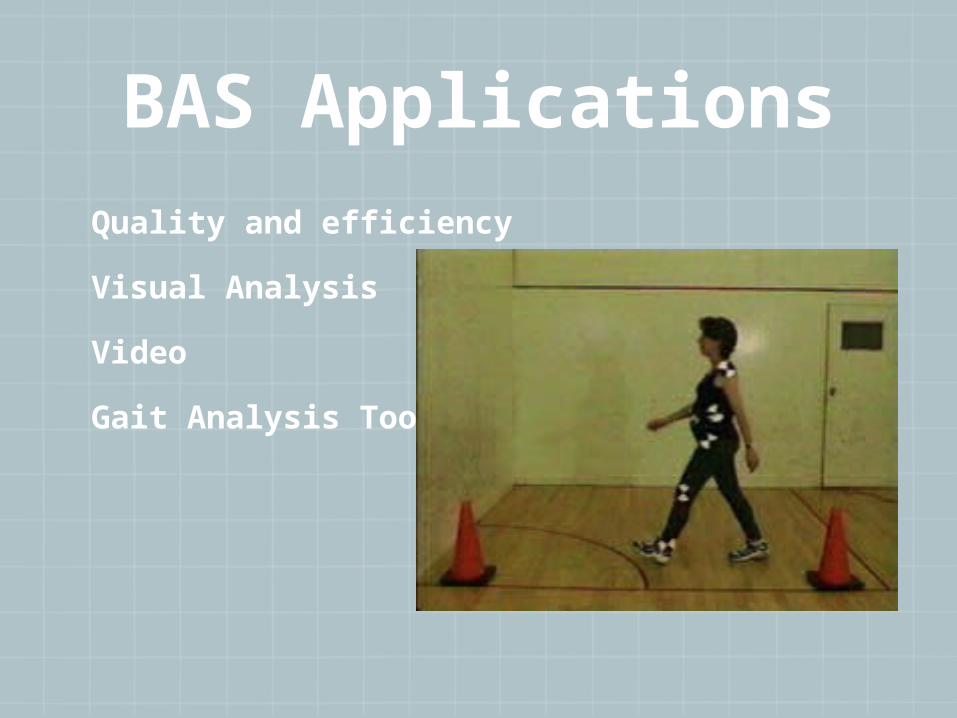
BAS ApplicationsQuality and efficiency
Visual Analysis
Video
Gait Analysis Tools

BAN Applications Special Diagnostic
TestsSLR (SN=.98; SP= .61)
ASLR (SN= .25; SP=.86)
Gaenslen’s (SN=.71; SP= .26)
FABER (SN> .82, SP>.60)
SIJ ant distraction (SN=.60;SP=.81): post compression (SN=.69; SP=.69)
Hip Scour
Anterior labral

Barriers to BAN implementation
Reliability and efficiency of sensor systems
Many sensor systems had same authors for system developers (bias)
Expensive
Accelerometers should be utilized with more than one sensor…
One accelerometer (65% accuracy)
Two accelerometers (87% accuracy)

Legal & Ethical Issues
Personal Data safety
Technology for elders may be seen as a limit to their independence (constant monitoring of symptoms that may impact social and community movement)

Future ResearchEvidence for reliability, validity, and responsiveness of wireless network body sensors must be established for each protocol
Collaborations between health care provider experts in diagnostics and rehabilitation, patients, computer and bioengineers, and wireless industry
Examination of this type of BAN system to be utilized in countries of challenged socio-economic needs and where access to health care is limited.

Thank You!“The doctor of the future will give no medicine, but will interest his patients in the care of the human frame, in diet, and in the cause and prevention of disease.” -Thomas Edison, Inventor

ReferencesFrancois KE, Foley MR. Antepartum and postpartum hemorrhage. In: Gabbe SG, Niebyl JR, Simpson JL, eds. Obstetrics - Normal and Problem Pregnancies. 5th ed. Philadelphia, Pa: Elsevier Churchill Livingstone; 2007:chap 18
Houry DE, Salhi BA. Acute complications of pregnancy. In: Marx J, Hockberger RS, Walls RM, et al, eds. Rosen’s Emergency Medicine: Concepts and Clinical Practice. 7th ed. Philadelphia, Pa: Mosby Elsevier; 2009:chap 176
Cunningham FG, Leveno KL, Bloom SL, et al. Obstetrical hemorrhage. In: Cunningham FG, Leveno KL, Bloom SL, et al., eds. Williams Obstetrics. 23rd ed. New York, NY: McGraw-Hill: 2010:chap 35.
Rosmans C, Holtz S, & Stanton C. Socioeconomic differentials in caesarean rates in developing countries: a retrospective analysis. The Lancet. 2006;368: 1516–1523.
Dumont A, de Bernis L, Bouvier-Colle MH, Breart G (2001). Caesarean section rate for maternal indication in sub-Saharan Africa: a systematic review, Lancet, 358: 1328-1333.
De Brouwere V, Dubourg D, Richard F, Van Lerberghe W (2002). Need for caesarean sections in west Africa. Lancet, 359: 974–75.
Rosmans C, Holtz S, & Stanton C (2006). Socioeconomic differentials in caesarean rates in developing countries: a retrospective analysis. The Lancet, 368: 1516–1523.