bml-111 alleviated the inflammatory response and apoptosis
TRANSCRIPT

3439
Abstract. – OBJECTIVE: The effect of lipoxin receptor agonist BML-111 on acute kidney inju-ry and AKI in rats with hemorrhagic shock (HS) and its mechanism were investigated.
MATERIALS AND METHODS: A model of hemorrhagic shock in Sprague-Dawley (SD) rats was established, and recovered after 30 min of shock. 1 mg/kg BML-111 was intraperitone-ally injected at the beginning of resuscitation in group BML-111. The concentration of serum creatinine, serum KIM-1 content, NGAL and in-flammatory factors were detected. Renal tissue injury was examined by HE staining; TUNEL staining was used to detect the apoptosis of rat kidney cells. Western blot was performed for the detection of the expression level of MAPK (mito-gen-activated protein kinase), Bax, cytochrome C and caspase-3,9 in rat renal tissue.
RESULTS: HE staining showed pathological changes in groups group comparing to sham group. BML-111 group had a significant decrease in renal tissue injury. The scores of renal injury in each group were in accordance with the histo-logical changes. The expression level of inflam-matory factors in HS group was significantly higher than that in sham group (p<0.05). After BML-111 intervention, the levels of inflammatory factors in renal tissue were significantly lower than those in HS group (p<0.05). Meanwhile, NGAL and KIM-1 also showed the same trend. TUNEL staining showed that compared with sham group, the number of apoptotic cells in renal tissue of HS group increased significantly, and the apoptosis rate of renal tissue cells in group BML-111 decreased significantly. Western blot showed that the expression level of JNK, p38MAPK, and apoptosis-related protein in HS group was significantly increased, whereas the expression level of p38MAPK and JNK in group BML-111 was significantly decreased.
CONCLUSIONS: BML-111 can reduce the in-flammatory response and apoptosis of renal tissue by inhibiting the activation of MAPK sig-naling pathway in acute renal injury induced by hemorrhagic shock.
Key WordsBML-111, Inflammatory response, Apoptosis, Hem-
orrhagic shock, MAPK.
Introduction
Hemorrhagic shock (HS) is a critical disea-se and one of the main reasons for the disability and death of traumatic patients1. In patients with hemorrhagic shock, the effective circulating blo-od volume is significantly reduced, which causes insufficient blood perfusion in important tissues and organs and leads to the disorder of metabolic function of normal tissues and cells. Pathological damage caused by hemorrhagic shock is a conti-nuous development and deterioration process, whi-ch eventually leads to the occurrence of multiple organ dysfunction syndrome (MODS). Related studies2 indicated that hemorrhagic shock activates cell stress signals and leads to the occurrence of systemic inflammation response syndrome (SIRS). Acute kidney injury (AKI) caused by hemorrhagic shock often occurs in critically ill patients3. Recent works showed that pro-inflammatory factors like IL-1, IL-1β, and TNF-α play an important role in the occurrence and development of AKI induced by HS3-5. Hemorrhagic shock can activate mito-gen-activated protein kinase (MAPK) pathway, which functions as sensor mediating the expres-sion of related pro-inflammatory factors6,7. ERK, JNK, and P38MAPK are important members of the MAPK pathway. Recent investigations8-11 have shown that MAPK signal transduction pathway is important to mediate MODS of HS.
At present, P38 and JNK are considered to be “stress-induced” MAPK12. After double pho-sphorylation of serine-63 and serine-73 site in c-jun activated region, JNK plays an important
European Review for Medical and Pharmacological Sciences 2018; 22: 3439-3447
S.-Y. CHANG1, R.-Q. SUN1, M. FENG1, Y.-X. LI1, H.-L. WANG1, Y.-M. XU2
1Department of Critical Care Medicine, The First Affiliated Hospital of Zhengzhou University, Zhengzhou, China2Department of Neurology, The First Affiliated Hospital of Zhengzhou University, Zhengzhou, China
Corresponding Author: Yuming Xu, MD; e-mail: [email protected]
BML-111 inhibits the inflammatory response and apoptosis of renal tissue in rats with hemorrhagic shock by inhibiting the MAPK pathway

S.-Y. Chang, R.-Q. Sun, M. Feng, Y.-X. Li, H.-L. Wang, Y.-M. Xu
3440
role in cell proliferation, differentiation and tumor transformation. Activated JNK can activate Bcl-2 and Bcl-XI by endogenous pathways. It participa-tes in the release of apoptotic molecules (such as the release of cytochrome C from mitochondria), which leads to activation of caspase and apopto-sis. Furthermore, JNK is not limited to the sub-strates of MAPK transcription factors and nucle-ar proteins. In U937 cells, once activated by UV irradiation, JNK MAPK moves to the mitochon-dria, and phosphorylates anti-apoptosis protein cl-XI, suggesting that JNK MAPK also plays a role in apoptosis induced by genotoxic stress13. The-refore, to control and block the MAPK signaling pathway may be an effective clinical method for the treatment of HS induced by AKI and MODS.
Lipoxin (LXs) is a newly discovered metabo-lite of arachidonic acid oxygenase, which serves as an endogenous anti-inflammatory lipid media-tor. It is, therefore, regarded as the brake signal of inflammatory reaction14. By binding to lipoxin A4 receptor (ALX), lipoxin exerts its anti-inflam-matory effect. ALX is highly conservative in hu-man, rat, and mouse. It is a G protein coupled with 7 transmembrane protein receptors with a wide expression on the surface of neutrophils, mo-nocytes and other tissue cells15.
After synthesis, natural lipoxin is quickly inacti-vated by enzymes in vivo. Therefore, a more stable and efficient lipoxin has currently been under de-velopment. Lipoxin and its analogues have shown strong anti-inflammatory effects in many kinds of inflammatory-related diseases such as asthma, arthri-tis, glomerulonephritis16. Researches17 have shown that aspirin induced lipoxin (ATL) can effectively reduce the inflammatory response induced by lipo-polysaccharide (LPS) in BV-2 microglia by inhibiting the activation of MAPK signaling pathway. BML-111 is a lipoxin A4 receptor agonist, which has similarly strong anti-inflammatory and promotion effect as li-poxin A4 and higher biological stability18. This study will detect whether BML-111 protects against acute kidney injury caused by hemorrhagic shock, and will explore whether its mechanism is related to the inhi-bition of MAPK signaling pathway.
Materials and Methods
Animal Grouping and Model Establishment
A total of 30 specific pathogen free (SPF) heal-thy male Sprague-Dawley (SD) rats weighing 200-260 g were randomly divided into 3 groups: control
group (Sham group), hemorrhagic shock/resuscita-tion group (HS group), and BML-111 intervention group (BML-111 group). The rats were anesthetized with intraperitoneal injection of 2% pentobarbital so-dium at a dose of 80 mg/kg. The left common carotid artery catheter was used for mean arterial pressure (MAP) detection and bloodletting. The right jugu-lar vein catheter was used for resuscitation. Hemor-rhagic shock model was established by bloodletting (35% of animal blood volume) and would last for 30 min. Next, the blood and twice the amount of Rin-ger’s fluid were resuscitated by venous transfusion for recovery. The rats in the Sham group were only anesthetized without bloodletting and resuscitation. Group BML-111 was given BML-111 (1 mg/kg) by intraperitoneal injection at the beginning of the re-suscitation. The rats were sacrificed 2 hours after the resuscitation, and the blood was collected for the de-tection of creatinine, NGAL, and KIM-1. The renal tissue was isolated and paraformaldehyde was used for pathological sections. The remaining kidney tis-sue was cryopreserved at-80°C after cryopreserva-tion. This study was approved by the Animal Ethics Committee of Zhengzhou University Animal Center.
Histologic Examination of KidneyThe right kidney of rat was taken for parafor-
maldehyde fixation, and embedded in paraffin. He-matoxylin eosin (HE) staining was conducted, and the pathological changes of the renal tissues of rats were observed with a microscope to record Paller score. The scoring criteria included tubular dilata-tion, cell flat scores (1), brush border injury scores (1), abscission as score (2), tubular type scores (2), and detached and necrotic cells (untubular or cell debris) in renal tubule lumen scores (1).
Detection of Inflammatory Factors and Creatinine, NGAL and KIM-1 in Rat Serum
The collected blood of rats was centrifuged for 10 min at 3000 r/min, and the supernatant serum was taken. The content of creatinine in the serum of rats was detected by the automatic biochemical analyzer. The content of serum inflammatory factors (IL-1β, IL-6, TNF-α) and NGAL and KIM-1 in serum was detected by ELISA kit according to the instructions.
Detection of Apoptosis in Renal Tissue by TUNEL Staining
The renal tissue was made into paraffin section, then dewaxed by xylene, and immersed in gradient ethanol for dehydration. 100 μL proteinase K were pipetted to cover the sample area and react for 30
BML-111 alleviated the inflammatory response and apoptosis of renal tissue cells in rats
3441
min at 37°C. The slices were immersed in blocking solution, incubated at room temperature for 10 min, then washed by PBS solution for 3 times. 50 μL TUNEL reaction mixture were added to the sam-ples, and incubated in a wet box for 60 min at 37°C. After incubation the samples were washed by pho-sphate-buffered saline (PBS) solution for 3 times. 50 μL conversion agent-POD were added and the samples were incubated in wet box for 30 min at 37°C. Pre-prepared diaminobenzidine (DAB) so-lution was added to cover samples. The reaction was terminated until the color reached expected depth. Next, samples were slightly stained with hematoxylin. The optical microscope was used for observation, and apoptosis rate was calculated.
Western Blotting0.1 g (+ 0.05 g) of the kidney tissue was wei-
ghed by the analytical balance and put in the liquid nitrogen in the centrifuge tube, grinded to pow-der and treated with 1 mL protein lysis buffer for 2 hours on the ice. The lysate was centrifuged at 12000 r/min for 10 min, and the supernatants were mixed with the same volume of 2 x loading buffer. The sample was boiled at 100°C for 8 min. The electrophoresis was performed. The protein was transferred and incubated with specific antibody. Lastly gel exposure was performed and the images were taken, and gray scale analysis was carried out.
Statistical AnalysisThe experimental data were analyzed with Sta-
tistical Product and Service Solutions (SPSS 17.0 Inc., Chicago, IL, USA) statistical software, and the measurement data were expressed as (̀ x±s). Single factor analysis of variance was used in the group data analysis. The test of homogeneity of variance was performed by Levene’s method. When the variance was homogeneous, LSD post-hoc test was used for average comparison betwe-en groups. The Tamhane’s test was used when the variance was not homogeneous, and p<0.05 was considered statistically significant.
Result
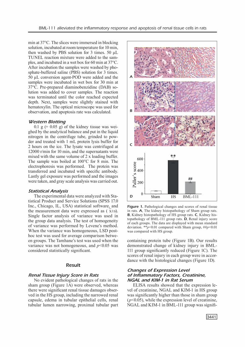
Renal Tissue Injury Score in RatsNo evident pathological changes of rats in the
sham group (Figure 1A) were observed, whereas there were significant renal tissue damages obser-ved in the HS group, including the narrowed renal capsule, edema in tubular epithelial cells, renal tubular lumen narrowing, proximal tubular part
containing protein tube (Figure 1B). Our results demonstrated change of kidney injury in BML-111 group significantly reduced (Figure 1C). The scores of renal injury in each group were in accor-dance with the histological changes (Figure 1D).
Changes of Expression Level of Inflammatory Factors, Creatinine,NGAL and KIM-1 in Rat Serum
ELISA results showed that the expression le-vel of creatinine, NGAL and KIM-1 in HS group was significantly higher than those in sham group (p<0.05), while the expression level of creatinine, NGAL and KIM-1 in BML-111 group was signifi-
Figure 1. Pathological changes and scores of renal tissue in rats. A, The kidney histopathology of Sham group rats. B, Kidney histopathology of HS group rats. C, Kidney his-topathology of BML-111 group rats. D, Renal injury score of each groups. The data are displayed with mean standard deviation. **p<0.01 compared with Sham group, ##p<0.01 was compared with HS group.
A
B
C
D

S.-Y. Chang, R.-Q. Sun, M. Feng, Y.-X. Li, H.-L. Wang, Y.-M. Xu
3442
cantly lower than those in HS group (p<0.05) (Fi-gure 2 A-C). The expression level of IL-1β, IL-6, and TNF-α in HS group was significantly higher than that in sham group (p<0.05). Furthermore, levels of IL-1 β, IL-6, and TNF-α in renal tissues of BML-111 group were significantly lower than those in HS group (Figure 2 D, E&F).
Change of Apoptosis Rate of Renal Tissue Cells
Paraffin sections of renal tissues of rats were stained with TUNEL. There were a few apoptotic cells in renal tissue of sham group rats (apopto-tic factor 0.55 + 0.071%) (Figure 3A). Apoptosis of renal tubular cells in HS rats was significant, and the number of apoptotic cells increased si-gnificantly (p<0.05) (apoptotic coefficient 5.5 + 0.48%) (Figure 3B). Of note, the apoptosis rate of renal cells decreased significantly after BML-111 intervention (p<0.05) (Figure 3C-D).
Expression of Apoptosis-RelatedProteins in Renal Tissue
Compared with Sham group, the expression le-vel of JNK and p38MAPK in HS group was signi-ficantly increased (p<0.05) (Figure 4A, B). Com-
pared with HS group, the expression level of JNK and p38MAPK in BML-111 group was significantly decreased (p<0.05). The expression level of apopto-sis-related protein including Bax, Cyto C and caspa-se3,9 of HS group was significantly higher than that of Sham group (p<0.05), while the expression levels were significantly decreased after the intervention of BML-111 (p<0.05) (Figure 4C, D).
Discussion
Hemorrhagic shock is an acute critical disease, which is one of the main causes of death among ICU patients20. Liquid resuscitation is a routine method for the treatment of hemorrhagic shock, which aims to restore the body’s circulating blood volume and the effective perfusion of the tissues21. However, the re-sulting ischemia-reperfusion in the body may cause a systemic inflammatory response and further deterio-rates to multiple organ failure22. The kidney is most vulnerable to hemorrhagic shock. In this study, we demonstrated that there were significant pathological changes in the renal tissue of hemorrhagic shock rats. Specifically, early renal damage index such as KIM-1 and NGAL in serum increased significantly.
A
D
B
E
C
F
Figure 2. Changes of expression level of inflammatory factors, creatinine, NGAL and KIM-1 in the serum of rats. A, The changes of serum creatinine in rats. B, The changes of serum KIM-1 in rats. C, The change of NGAL in serum of rats. D, The change of IL-1β in serum of rats, E, The change of IL-6 in serum of rats. F, The change of TNF-α in serum of rats. **p<0.01 compared with Sham group, ##p<0.01 was compared with HS group.

BML-111 alleviated the inflammatory response and apoptosis of renal tissue cells in rats
3443
Hemorrhagic shock induces a large number of pro-inflammatory factors. It promotes neu-trophil infiltration into renal tissue and activates pro-inflammatory genes, thereby leading to the occurrence of acute kidney injury. It is difficult to completely cure hemorrhagic shock only by liquid resuscitation. Therefore, only by control-ling and preventing the development of systemic inflammatory reaction, we can effectively treat acute renal injury. Inflammatory factors and neu-trophil activation are important characteristics of acute inflammation. The timely subsiding of the inflammatory response can reconstruct the body homeostasis environment and limit the tissue da-mage caused by the inflammatory reaction. If the inflammatory reaction is over-activated or subsi-ded, it will lead to chronic inflammatory reaction, further aggravation of tissue as well as organ da-mage and failure23. Lipoxin is anti-inflammatory
and subsiding medium, which was first known as the “brake signal” of the inflammatory reaction. Lipoxin can significantly reduce the production of pro-inflammatory factors, inhibit the activation and migration of neutrophils, promote macropha-ges for phagocytosis of apoptotic neutrophils, and prevent the activation and transcription of pro-inflammatory genes. BML-111 is a lipoxin receptor agonist, which has a higher biological stability and a more powerful biological efficacy than lipoxygenin A424. Studies25,26 have shown that BML-111 has significant anti-inflammatory effects on diseases like carbon tetrachloride-indu-ced liver injury and yeast polysaccharide induced arthritis. However, the specific biological effects of BML-111 on hemorrhagic shock remain to be studied. Previous experiments27 have shown that BML-111 could reduce renal injury induced by ischemia-reperfusion.
C D
A B
Figure 3. Changes in apoptosis rate of renal tissue cells in rats. A, In Sham group, TUNEL staining in rat kidney tissue. B, Renal tissue TUNEL staining of HS group. C, Rats kidney tissue TUNEL staining of BML-111 group. D, Changes of apoptosis coefficient in kidneys of rats were observed. **p<0.01 compared with Sham group, ##p<0.01 is compared to HS group.

S.-Y. Chang, R.-Q. Sun, M. Feng, Y.-X. Li, H.-L. Wang, Y.-M. Xu
3444
Hemorrhagic shock can lead to upregulation of a large number of various pro-inflammatory factors in vivo28. IL-1β and IL-6 play an important role in the inflammatory reaction of acute kidney injury. Moreover, they also play an important role in the development and progression of renal injury caused by hemorrhagic shock. Related studies29 have shown that in a large number of hemorrhagic rat models, inhibition of JNK activation before hemorrhagic re-covery can reduce the death of hepatocytes and re-duce the degree of inflammation of liver cells. Our results also showed that hemorrhagic shock incre-ased expression of JNK and p38MAPK in the re-nal tissue, while this phenotype was partly reversed after BML-111 was given to rats. At the same time, our results also showed that BML-111 could reduce the expression of IL-1β, IL-6 and TNF-α in renal tis-sue after hemorrhagic shock.
In a subsequent study, our data showed that BML-111 could significantly inhibit MAPK signa-ling pathway. The mitogen activated protein kinase (MAPK)30 family belongs to serine/threonine pro-tein kinase, and is a group of signal transduction molecules widely present in diverse cell types. The-re are four MAPK signal transduction pathways in-cluding P38MAPK. P38MAPK plays an important role in cell proliferation, differentiation and apop-tosis, and has become a research hotspot in signal transduction field in recent years. P38MAPK is mainly distributed in the cytoplasm during resting state, and activated after the stimulation of phy-siological stress (such as hypoxia), osmotic stress (such as hypertonic environment), lipopolysaccha-ride (LPS) and ultraviolet radiation31. P38 activates nuclear translocation, which further phosphoryla-tes and activates many protein kinases and tran-
Figure 4. Expression of MAPK and apoptosis-related proteins in renal tissue of rats. A, The expression of MAPK protein in renal tissue of rats. B, The expression level of MAPK protein in rat kidney tissue was changed. C, The expression of apopto-sis-related protein in renal tissue of rats was observed. D, The expression level of apoptosis-related protein in renal tissue of rats was changed. **p<0.01 compared with Sham group, ##p<0.01 was compared with HS group.
A
C
B
D

BML-111 alleviated the inflammatory response and apoptosis of renal tissue cells in rats
3445
scription factors; therefore, it plays a key role in the regulation of inflammatory response by regulating the activity of transcription factors and the synthe-sis of cytokines32-34. The activation of p38MAPK can not only promote the production of monocyte macrophages, such as TNF α, IL 1, IL 4, IL 6, IL 8, and IL 12, but also increase the production of an-ti-inflammatory factor IL-10. However, the inflam-mation stimulation, such as LPS, TNF α, IL-1, PAF and ischemia reperfusion can induce p38MAPK activation in monocytes, neutrophils and endothe-lial cells and leads to the release of a large number of inflammatory mediators and acute inflamma-tion evoked response Consequently, it results in the imbalance of SIRS/CARS and the damage of cells in the body tissue35.
At the same time, MAPK is thought to be the most important signal molecule to transfer the apoptosis signal to the mitochondria. JNK and p38 are located upstream of the typical apopto-tic pathway, which can reduce the expression and activity of anti-apoptotic Bcl-2 protein family. MAPKs can also increase the expression and activity of apoptotic protein and induce apopto-sis through intrinsic apoptosis pathway36-39. Mito-chondria are the key units in the process of cell apoptosis, which can be activated by a variety of death signals. Mitochondrial dysfunction will si-gnificantly affect cell apoptosis, which can lead to cytochrome C release from mitochondrial membrane gap to cytoplasm. The release of cyto-chrome C from mitochondria into the cytopla-sm and APAF1 complexes can further activate caspase-9 and caspase-3, eventually leading to apoptosis40-42. In this research, we also found that the renal tissue apoptosis coefficiency increased significantly in hemorrhagic shock rats, and the apoptosis rate of renal tissue in BML-111 inter-vention group was lower than that in hemorrha-gic shock group. However, compared with the hemorrhagic shock group, the expression level of apoptosis-related protein Bax, Cyto C and caspa-se-3,9 in BML-111 intervention group decreased significantly, implying that BML-111 may inhibit MAPK pathway, downregulate the expression of apoptosis-related protein, inhibit the mitochon-drial apoptotic pathway, thereby reducing renal cell apoptosis of rats with traumatic shock.
In conclusion, our research showed that BML-111 can significantly reduce the pathological da-mage of renal tissue in hemorrhagic shock and the expression of inflammatory factors in kidney tissue. BML-111 can also inhibit the activation of MAPK signaling pathway, and decrease the
expression of cytochrome C as well as other apop-tosis-related proteins. In recent years, promoting the inflammation subsiding in kidney tissue has become a new strategy for the treatment of acute kidney injury. Lipoxin as an endogenous inflam-matory regression factor provides a new way for drug treatment of acute kidney injury. However, the prototypes and analogues of these substances have short half-life and poor biological stability in vivo, so the drugs development that are based on these mediums, such as BML-111, will have high clinical significance and therapeutic value. The pathogenesis of acute kidney injury and the specific mechanism of lipoxin still needs further investigation in the future.
Conclusions
BML-111 can inhibit the activation of MAPK signaling pathway to reduce the inflammatory re-sponse and apoptosis of renal tissue in rats with hemorrhagic shock.
Conflict of interestThe authors declared no conflict of interest.
References
1) Megevand B, Celi J, niquille M. [Hemorrhagic shock]. Rev Med Suisse 2014; 10: 1501-1505.
2) Fan J, li Y, levY RM, Fan JJ, HaCkaM dJ, vodovotz Y, Yang H, tRaCeY kJ, BilliaR tR, Wilson Ma. Hemor-rhagic shock induces NAD(P)H oxidase activation in neutrophils: role of HMGB1-TLR4 signaling. J Immunol 2007; 178: 6573-6580.
3) CHen s, sHi Js, YiBulaYin X, Wu ts, Yang XW, zHang J, BaiHeti P. Cystatin C is a moderate predictor of acute kidney injury in the early stage of traumatic hemorrhagic shock. Exp Ther Med 2015; 10: 237-240.
4) RaBB H, gRiFFin Md, MCkaY dB, sWaMinatHan s, PiCk-keRs P, RosneR MH, kelluM Ja, RonCo C. Inflamma-tion in AKI: current understanding, key questions, and knowledge gaps. J Am Soc Nephrol 2016; 27: 371-379.
5) CHen g, song X, Yin Y, Xia s, liu q, You g, zHao l, zHou H. C-type natriuretic peptide prevents kidney injury and attenuates oxidative and inflam-matory responses in hemorrhagic shock. Amino Acids 2017; 49: 347-354.
6) koCHanek aR, FukudoMe eY, li Y, sMitH eJ, liu B, velMaHos gC, deMoYa M, king d, alaM HB. His-tone deacetylase inhibitor treatment attenuates MAP kinase pathway activation and pulmonary inflammation following hemorrhagic shock in a rodent model. J Surg Res 2012; 176: 185-194.

S.-Y. Chang, R.-Q. Sun, M. Feng, Y.-X. Li, H.-L. Wang, Y.-M. Xu
3446
7) Fan J, MaRsHall JC, JiMenez M, sHek Pn, zagoRski J, Rotstein od. Hemorrhagic shock primes for increased expression of cytokine-induced neu-trophil chemoattractant in the lung: role in pulmo-nary inflammation following lipopolysaccharide. J Immunol 1998; 161: 440.
8) JaRRaR d, kueBleR JF, Matalon s, Wang P, Bland ki, CHaudRY iH. Alveolar macrophage activation after trauma-hemorrhage and sepsis is dependent on NF-kappaB and MAPK/ERK mechanisms. Am J Physiol Lung Cell Mol Physiol 2002; 283: L799.
9) liu FC, liu FW, Yu HP. Ondansetron attenuates hepatic injury via p38 MAPK-dependent pathway in a rat haemorrhagic shock model. Resuscitation 2011; 82: 335-340.
10) sato H, tanaka t, kasai k, tanaka n. A quantitative study of p38 mitogen-activated protein kinase on renal dysfunction after hemorrhagic shock in rats. J Trauma 2011; 71: 973-981.
11) lv kY, Yu XY, Bai Ys, zHu sH, tang Ht, Ben dF, Xiao sC, Wang gY, Ma B, Xia zF. Role of inhibition of p38 mitogen-activated protein kinase in liver dysfunction after hemorrhagic shock and resus-citation. J Surg Res 2012; 178: 827-832.
12) FioRe s, MaddoX JF, PeRez Hd, seRHan Cn. Identi-fication of a human cDNA encoding a functional high affinity lipoxin A4 receptor. J Exp Med 1994; 180: 253-260.
13) takano t, FioRe s, MaddoX JF, BRadY HR, Petasis na, seRHan Cn. Aspirin-triggered 15-Epi-Lipoxin a4 (LXA4) and LXA4 stable analogues are potent inhibitors of acute inflammation: evidence for an-ti-inflammatory receptors. J Exp Med 1997; 185: 1693.
14) sHi Y, Pan H, zHang Hz, zHao XY, Jin J, Wang HY. Lipoxin A4 mitigates experimental autoimmune myocarditis by regulating inflammatory response, NF-kappaB and PI3K/Akt signaling pathway in mice. Eur Rev Med Pharmacol Sci 2017; 21: 1850-1859.
15) FioRe s, MaddoX JF, PeRez Hd, seRHan Cn. Identi-fication of a human cDNA encoding a functional high affinity lipoxin A4 receptor. J Exp Med 1994; 180: 253-260.
16) sun YP, tJonaHen e, keledJian R, zHu M, Yang R, ReCCHiuti a, Pillai Ps, Petasis na, seRHan Cn. An-ti-inflammatory and pro-resolving properties of benzo-lipoxin A(4) analogs. Prostaglandins Leu-kot Essent Fatty Acids 2009; 81: 357-366.
17) Wang YP, Wu Y, li lY, zHeng J, liu Rg, zHou JP, Yuan sY, sHang Y, Yao sl. Aspirin-triggered lipox-in A4 attenuates LPS-induced pro-inflammatory responses by inhibiting activation of NF-kappaB and MAPKs in BV-2 microglial cells. J Neuroin-flammation 2011; 8: 95.
18) HaWkins ke, deMaRs kM, singH J, Yang C, CHo Hs, FRankoWski JC, doRe s, CandelaRio-Jalil e. Neuro-vascular protection by post-ischemic intravenous injections of the lipoxin A4 receptor agonist, BML-111, in a rat model of ischemic stroke. J Neurochem 2014; 129: 130-142.
19) PalleR Ms, Hoidal JR, FeRRis tF. Oxygen free rad-icals in ischemic acute renal failure in the rat. J Clin Invest 1984; 74: 1156-1164.
20) gutieRRez g, Reines Hd, WulF-gutieRRez Me. Clinical review: hemorrhagic shock. Crit Care 2004; 8: 373-381.
21) dong W, Cai B, Pena g, PisaRenko v, vida g, douCet d, lee M, sHaRPe s, lu q, Xu dz, RaMos l, deitCH ea, ulloa l. Ethyl pyruvate prevents inflammatory responses and organ damage during resusci-tation in porcine hemorrhage. Shock 2010; 34: 205-213.
22) kauvaR ds, leFeRing R, Wade Ce. Impact of hem-orrhage on trauma outcome: an overview of epi-demiology, clinical presentations, and therapeutic considerations. J Trauma 2006; 60: S3-S11.
23) Maderna P, Godson C. Lipoxins: resolutionary road. Br J Pharmacol 2009; 158: 947-959.
24) FoRsMan H, daHlgRen C. Lipoxin A(4) metabolites/analogues from two commercial sources have no effects on TNF-alpha-mediated priming or activation through the neutrophil formyl peptide receptors. Scand J Immunol 2009; 70: 396-402.
25) zHang l, Wan J, li H, Wu P, Jin s, zHou X, Yuan P, Xiong W, li Y, Ye d. Protective effects of BML-111, a lipoxin A(4) receptor agonist, on carbon tetrachloride-induced liver injury in mice. Hepatol Res 2007; 37: 948-956.
26) Conte FP, Menezes-de-liMa oJ, veRRi WJ, CunHa Fq, Penido C, HenRiques Mg. Lipoxin A(4) attenuates zy-mosan-induced arthritis by modulating endothelin-1 and its effects. Br J Pharmacol 2010; 161: 911-924.
27) Wu sH, CHen Xq, lu J, Wang MJ. BML-111 attenu-ates renal ischemia/reperfusion injury via peroxi-some proliferator-activated receptor-alpha-regu-lated heme oxygenase-1. Inflammation 2016; 39: 611-624.
28) al-aMRan Fg, Hadi nR, HasHiM aM. Leukotriene biosynthesis inhibition ameliorates acute lung in-jury following hemorrhagic shock in rats. J Car-diothorac Surg 2011; 6: 81.
29) RelJa B, sCHWestka B, lee vs, HenRiCH d, CzeRnY C, BoRsello t, MaRzi i, leHneRt M. Inhibition of c-Jun N-terminal kinase after hemorrhage but before resuscitation mitigates hepatic damage and in-flammatory response in male rats. Shock 2009; 32: 509-516.
30) gaestel M. MAPK-Activated protein kinases (MKs): novel insights and challenges. Front Cell Dev Biol 2015; 3: 88.
31) CaRgnello M, RouX PP. Activation and function of the MAPKs and their substrates, the MAPK-ac-tivated protein kinases. Microbiol Mol Biol Rev 2011; 75: 50-83.
32) kiM ek, CHoi eJ. Pathological roles of MAPK signaling pathways in human diseases. Biochim Biophys Acta 2010; 1802: 396-405.
33) takekaWa M, kuBota Y, nakaMuRa t, iCHikaWa k. Reg-ulation of stress-activated MAP kinase pathways during cell fate decisions. Nagoya J Med Sci 2011; 73: 1-14.
34) klein aM, zaganJoR e, CoBB MH. Chromatin-teth-ered MAPKs. Curr Opin Cell Biol 2013; 25: 272-277.
35) Jiang Y, gong XW. [Regulation of inflammatory re-sponses by MAPK signal transduction pathways]. Acta Physiologica Sinica 2000; 52: 267.
BML-111 alleviated the inflammatory response and apoptosis of renal tissue cells in rats
3447
36) CHen YJ, liu WH, kao PH, Wang JJ, CHang ls. Involvement of p38 MAPK- and JNK-modulated expression of Bcl-2 and Bax in Naja nigricollis CMS-9-induced apoptosis of human leukemia K562 cells. Toxicon 2010; 55: 1306-1316.
37) deng Yt, Huang HC, lin Jk. Rotenone induc-es apoptosis in MCF-7 human breast cancer cell-mediated ROS through JNK and p38 signal-ing. Mol Carcinog 2010; 49: 141-151.
38) kang YH, lee sJ. The role of p38 MAPK and JNK in Arsenic trioxide-induced mitochondrial cell death in human cervical cancer cells. J Cell Physiol 2008; 217: 23-33.
39) su JC, lin kl, CHien CM, lu CM, CHen Yl, CHang ls, lin sR. Novel indoloquinoline derivative, IQD-MA, induces G(2)/M phase arrest and apoptosis
in A549 cells through JNK/p38 MAPK signaling activation. Life Sci 2009; 85: 505-516.
40) santRa s, kaittanis C, PeRez JM. Cytochrome C encapsulating theranostic nanoparticles: a novel bifunctional system for targeted delivery of ther-apeutic membrane-impermeable proteins to tu-mors and imaging of cancer therapy. Mol Pharm 2010; 7: 1209-1222.
41) YaMada Y, HaRasHiMa H. Mitochondrial drug deliv-ery systems for macromolecule and their thera-peutic application to mitochondrial diseases. Adv Drug Deliv Rev 2008; 60: 1439-1462.
42) CHang z, Xing J, Yu X. Curcumin induces osteo-sarcoma MG63 cells apoptosis via ROS/Cyto-C/Caspase-3 pathway. Tumour Biol 2014; 35: 753-758.