bmc urology biomed central - pdfs.semanticscholar.org · lee q robinson - [email protected] *...
TRANSCRIPT

BioMed CentralBMC Urology
ss
Open AcceCase reportCase report – ancient schwannoma of the scrotumPeter T Chan*1, Sankalp Tripathi2, Su E Low3 and Lee Q Robinson1Address: 1Surgical Directorate, Warrington Hospital, Lovely Lane, Warrington, UK, 2Department of Radiology, Warrington Hospital, Lovely Lane, Warrington, UK and 3Department of Pathology, Warrington Hospital, Lovely Lane, Warrington, UK
Email: Peter T Chan* - [email protected]; Sankalp Tripathi - [email protected]; Su E Low - [email protected]; Lee Q Robinson - [email protected]
* Corresponding author
AbstractBackground: Scrotal schwannoma is a rare neoplasm and poses a diagnostic challenge tourologists. This article describes a rare case of ancient scrotal schwannoma and reviews the currentmodality of investigation and treatment of this tumour.
Case report: A 28 year old man presented with a 3-month history of an asymptomatic scrotalswelling. Ultrasonography and computer topography revealed an intra-scrotal and extra-testicularmass without local invasion. Surgical excision was undertaken and histology was an ancientschwannoma of the scrotum.
Conclusion: Schwannoma is a benign encapsulating neoplasm with an overall low incidence,occurring mostly in the head and neck region and seldom in the scrotum. Histology shows twodistinctive patterns, Antoni type A and B areas. Variations of schwannoma such as cellular, ancient,glandular and epithelioid are observed based on the appearances. Ancient schwannoma exhibitspleomorphism without mitosis as the result of cellular degeneration, which can lead to anerroneous diagnosis of malignancy. Imaging modalities are non-specific for schwannomas, but candefine tumour size, site and extension. The mainstay treatment is complete excision, although localrecurrence may occur in large and incompletely excised lesions. Malignant change is exceedinglyrare.
BackgroundThe "ancient" variant of schwannomas is a rare subtype ofa benign encapsulated neoplasm of the nerve sheath [1].A review of current literature has revealed several reportedsites but not in the scrotum. Here we report such a scrotaltumour arising in a 28-year old man, paying particularattention to the potential diagnostic difficulties prior tosurgical intervention.
Case reportA 28-year old man presented with a three month historyof an asymptomatic scrotal swelling. He had previouslyundergone orchidopexy in childhood for a mal-descended right testis. There is otherwise no other signifi-cant medical or family history. The mid-scrotally locatedmass measured 4 × 7 cm and was nodular, hard and clin-ically separate from the testes. It was not attached to thescrotal skin or other underlying structures. Lymphaden-opathy was not detected.
Published: 23 January 2007
BMC Urology 2007, 7:1 doi:10.1186/1471-2490-7-1
Received: 22 September 2006Accepted: 23 January 2007
This article is available from: http://www.biomedcentral.com/1471-2490/7/1
© 2007 Chan et al; licensee BioMed Central Ltd. This is an Open Access article distributed under the terms of the Creative Commons Attribution License (http://creativecommons.org/licenses/by/2.0), which permits unrestricted use, distribution, and reproduction in any medium, provided the original work is properly cited.
Page 1 of 4(page number not for citation purposes)

BMC Urology 2007, 7:1 http://www.biomedcentral.com/1471-2490/7/1
Initial ultrasonography (Fig 1a) showed a large extra-tes-ticular and multinodular mass in the midline of the scro-tum. The largest nodule, located behind the right testiswas cystic. Both testes appeared normal. Subsequent stag-ing Computer Tomography (CT) of the scrotum, abdo-men and chest was performed, which identified asimilarly localised heterogeneous mass of 6.9 × 3.7 cm,separate from the testes (Fig 1b) and did not extend intothe tunica vaginalis. There was no evidence of distant ornodal metastases.
An initial diagnostic wedge biopsy indicated the lesioncould be an ancient schwannoma. Curative surgical exci-sion with partial scrotectomy was undertaken for removalof the mass and for definitive diagnosis. At resection, thetumour appeared to be superficial to the tunica vaginalis,testes and corpus spongiosum. Inferiorly, the tumour wasattached to the bulbospongiosus requiring partial resec-tion of the superficial muscle fibres.
The resected specimen (Fig 2) was white and multinodu-lar, with areas of cystic change and haemorrhage. Micro-scopically (Fig 3), proliferation of spindled shaped cellswith fibrillary cytoplasm was seen, with dense fibrousbands arranging the cells into nodules. In some cells,marked nuclear hyperchromaticism and atypia were seen.Mitoses were not present. Within the lesion, cellular areaswere interspersed with looser myxoid and cystic areas.Blood vessels with thickened hyalinised walls were noted.Staining for S100 protein was positive in the tumour cells.The histological features are that of an ancient schwan-noma. The margins of this specimen were ragged and thecompleteness of excision was difficult to assess.
ConclusionA schwannoma is a benign encapsulated neoplasmderived from schwann cells of the nerve sheath. The exactincidence of schwannomas is unknown, but they are rare.Schwannomas are found in all age groups but are morecommon in the first four decades and affect both sexesequally. They may occur in association with neurofi-bromatosis or arise sporadically. The microscopic appear-ance of schwannoma is distinctive [1], with tworecognisable patterns. Antoni A areas are composed ofcompacted spindle cells often arranged in palisades or inan organoid arrangement (Verocay bodies). Antoni Bareas consist of tumour cells suspended in a myxomatousmatrix that may appear microcystic. Several variants ofschwannomas based on appearances have been observed,including cellular, glandular, epithelioid and ancienttypes, and all exhibit benign progression. Cellularschwannomas are almost exclusively composed of AntoniA areas but lack Verocay bodies. The glandular and epith-eloid variants compose of epitheloid areas and glandularcomponent, respectively, to acquire their descriptivenames. Ancient schwannomas show bizarre hyperchro-matic nuclei without mitoses. To the unwary, these fea-tures can lead to an erroneous diagnosis of malignancy,although the very low mitotic activity should allow thesetumours to be distinguished from malignant nerve sheathtumours.
A literature review showed that whilst ancient schwanno-mas are rare, most cases occur in the head and neck region[2] (trigeminal nerve, facial nerve, vestibular nerve, vagusnerve, parotid, thyroid, vocal cord, floor of mouth, orbitand infra-temporal fossa). Other less common sitesinclude the extremities, mediastinum, thorax [3], retro-
Imaging of scrotal ancient schwannomaFigure 1Imaging of scrotal ancient schwannoma. a) Ultrasound image and b) CT image showing an intra-scrotal and extra-testic-ular mass in the mid-scrotal region [Capital red T – tumour, red t – testes].
Page 2 of 4(page number not for citation purposes)
BMC Urology 2007, 7:1 http://www.biomedcentral.com/1471-2490/7/1
peritoneum [4], pancreas [5] and pelvis [6]. Scrotalschwannomas have been infrequently described in themedical literature [7,8,10], however, we could not find
references pertaining to the ancient variant in the scrotalregion.
Schwannomas pose a difficult diagnostic challenge tourologists. Radiological findings are often non-specific[9]. Ultrasonography can differentiate between solid andcystic tumours. CT can be helpful in determining the size,location, local involvement and distant spread. Magneticresonance imaging (MRI) provides similarly useful infor-mation as CT, but yields better visualisation of thetumour. Fine needle aspirate (FNA) cytology is not oftenhelpful because the tissue architectural informationrequired is not obtainable from cytological specimen [4].The only gold standard diagnostic investigation is histol-ogy of either biopsy or excised specimen.
Surgical excision has remained the mainstay of treatment.Although benign, large and incompletely excised lesionsare capable of recurrence; malignant change is exceedinglyrare [3,10]. This patient will require a prolonged period ofsurveillance due to the large size of the tumour and theuncertainty of complete excision.
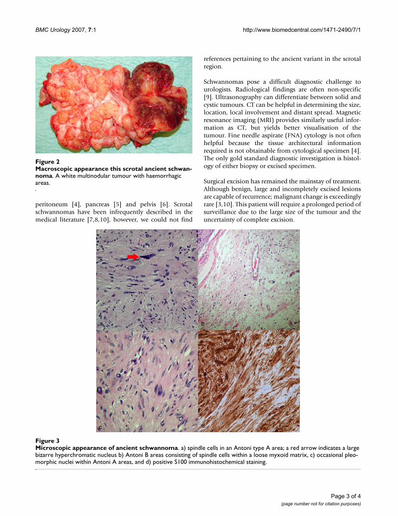
Microscopic appearance of ancient schwannomaFigure 3Microscopic appearance of ancient schwannoma. a) spindle cells in an Antoni type A area; a red arrow indicates a large bizarre hyperchromatic nucleus b) Antoni B areas consisting of spindle cells within a loose myxoid matrix, c) occasional pleo-morphic nuclei within Antoni A areas, and d) positive S100 immunohistochemical staining.
Macroscopic appearance this scrotal ancient schwannomaFigure 2Macroscopic appearance this scrotal ancient schwan-noma. A white multinodular tumour with haemorrhagic areas.
Page 3 of 4(page number not for citation purposes)

BMC Urology 2007, 7:1 http://www.biomedcentral.com/1471-2490/7/1
Publish with BioMed Central and every scientist can read your work free of charge
"BioMed Central will be the most significant development for disseminating the results of biomedical research in our lifetime."
Sir Paul Nurse, Cancer Research UK
Your research papers will be:
available free of charge to the entire biomedical community
peer reviewed and published immediately upon acceptance
cited in PubMed and archived on PubMed Central
yours — you keep the copyright
Submit your manuscript here:http://www.biomedcentral.com/info/publishing_adv.asp
BioMedcentral
Competing interestsThe author(s) declare that they have no competing inter-ests.
Authors' contributionsPTC drafted the manuscript, prepared illustrations andperformed the literature search. ST helped to draft themanuscript and helped to acquire ultrasound and CTimages. SEL helped to draft the manuscript, paying partic-ular attention to the pathological aspect and kindlyacquired histological images for illustration. LQR con-ceived this study and supervised the drafting and overallstructure of the manuscript. All authors read andapproved the final manuscript.
References1. Rosai J: Ackerman's surgical pathology. Mosby 8th edition. 1995,
2:2264-66.2. Bayindir T, Kalcioglu T, Kizilay A, Karadag N, Akarcay M: Ancient
schwannoma of the parotid gland: a case report and reviewof the literature. J Craniomaxillofac Surg 2006, 34:38-42.
3. Chu YC, Yoon YH, Han HS, Han JY, Kim JM, Park IS: Malignanttransformation of intrathoracic ancient neurilemmoma in apatient without Von Recklinghausen's disease. J Korean MedSci 2003, 18:295-8.
4. Daneshmand S, Youssefzadeh D, Chamie K, Boswell W, Wu N, SteinJP, Boyd S, Skinner DG: Benign retroperitoneal schwannoma: acase series and review of the literature. Urology 2003, 62:993-7.
5. Von Dobschuetz E, Walch A, Werner M, Hopt UT, Adam U: Giantancient schwannoma of pancreatic head treated byextended pancreatoduodenectomy. Pancreatology 2004,4:505-8.
6. Hennigan TW, Branfoot AC, Theodorou NA: Ancient neurilem-oma of the pelvis. J R Soc Med 1992, 85:416-7.
7. Matsui F, Kobori Y, Takashima H, Amano T, Takemae K: A case ofintrascrotal schwannoma. Acta Urol Jap 2002, 48:749-51.
8. Zarate RE, Fernandez GI, Lujan GM, Ortega MP, Berenguer SA:Schwannoma of the scrotum: report of case and review ofthe literature. Actas Urol Esp 1997, 21:1012-3.
9. Isobe K, Shimizu T, Akahane T, Kato H: Imaging of ancientschwannoma. AJR Am J Roentgenol 2004, 183:331-6.
10. Safak M, Baltaci S, Ozer G, Turkolmez K, Uluoglu O: Long-termoutcome of a patient with intrascrotal extratesticular malig-nant schwannoma. Urol Int 1998, 60:202-4.
Pre-publication historyThe pre-publication history for this paper can be accessedhere:
http://www.biomedcentral.com/1471-2490/7/1/prepub
Page 4 of 4(page number not for citation purposes)