blood - welcome to welsh! · blood and place in tube. centrifuge 2 the blood sample. ......
TRANSCRIPT
PowerPoint® Lecture Slides prepared by Janice Meeking, Mount Royal College
C H A P T E R
Copyright © 2010 Pearson Education, Inc.
17 Blood

Copyright © 2010 Pearson Education, Inc.
Warm Up 4/21/17
•What are the functions of the following components of blood:
1. Plasma2. White Blood Cells (WBCs)3. Platelets 4. Red Blood Cells (RBCs)
Copyright © 2010 Pearson Education, Inc.
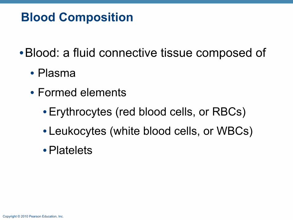
Blood Composition
•Blood: a fluid connective tissue composed of• Plasma
• Formed elements
•Erythrocytes (red blood cells, or RBCs)
• Leukocytes (white blood cells, or WBCs)
•Platelets
Copyright © 2010 Pearson Education, Inc.
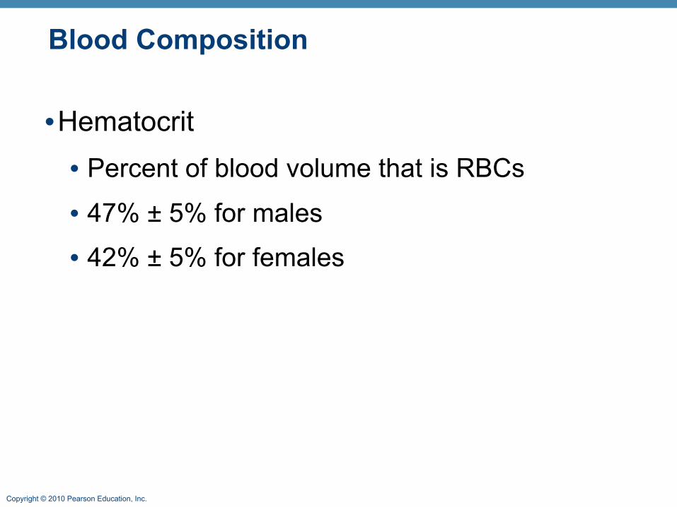
Blood Composition
•Hematocrit• Percent of blood volume that is RBCs
• 47% ± 5% for males
• 42% ± 5% for females
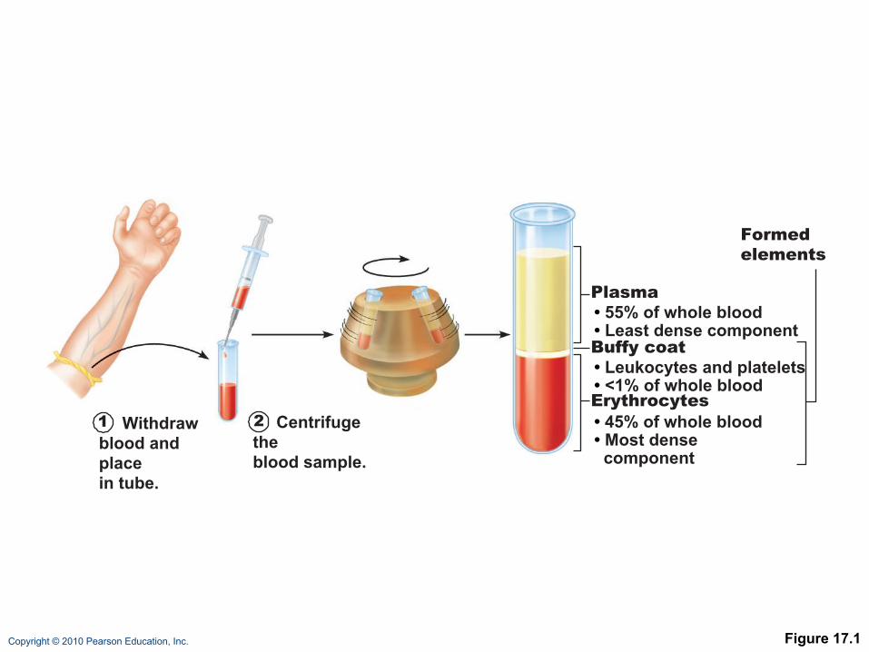
Copyright © 2010 Pearson Education, Inc. Figure 17.1
1 Withdrawblood and placein tube.
2 Centrifuge theblood sample.
Plasma• 55% of whole blood• Least dense componentBuffy coat• Leukocytes and platelets• <1% of whole bloodErythrocytes• 45% of whole blood• Most dense component
Formedelements
Copyright © 2010 Pearson Education, Inc.
Physical Characteristics and Volume
•Sticky, opaque fluid
•Color scarlet to dark red
•pH 7.35–7.45
•38°C
•~8% of body weight
•Average volume: 5–6 L for males, and 4–5 L for females
Copyright © 2010 Pearson Education, Inc.
Functions of Blood
1. Distribution of• O2 and nutrients to body cells
• Metabolic wastes to the lungs and kidneys for elimination
• Hormones from endocrine organs to target organs
Copyright © 2010 Pearson Education, Inc.
Functions of Blood
2. Regulation of• Body temperature by absorbing and
distributing heat
• Normal pH using buffers
• Adequate fluid volume in the circulatory system
Copyright © 2010 Pearson Education, Inc.
Functions of Blood
3. Protection against• Blood loss• Plasma proteins and platelets initiate clot
formation• Infection • Antibodies• Complement proteins• WBCs defend against foreign invaders

Copyright © 2010 Pearson Education, Inc.
Blood Plasma
•90% water
•Proteins are mostly produced by the liver• 60% albumin
• 36% globulins
• 4% fibrinogen
Copyright © 2010 Pearson Education, Inc.
Blood Plasma
•Nitrogenous by-products of metabolism—lactic acid, urea, creatinine
•Nutrients—glucose, carbohydrates, amino acids
•Electrolytes—Na+, K+, Ca2+, Cl–, HCO3–
•Respiratory gases—O2 and CO2
•Hormones
Copyright © 2010 Pearson Education, Inc.
Formed Elements
•Only WBCs are complete cells
•RBCs have no nuclei or organelles
•Platelets are cell fragments
•Most formed elements survive in the bloodstream for only a few days
•Most blood cells originate in bone marrow and do not divide
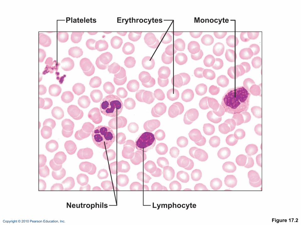
Copyright © 2010 Pearson Education, Inc. Figure 17.2
Platelets
Neutrophils Lymphocyte
Erythrocytes Monocyte
Copyright © 2010 Pearson Education, Inc.
Erythrocytes
•Biconcave discs, anucleate, essentially no organelles•Filled with hemoglobin (Hb) for gas transport•Contain the plasma membrane protein spectrin and other proteins• Provide flexibility to change shape as
necessary•Are the major factor contributing to blood viscosity
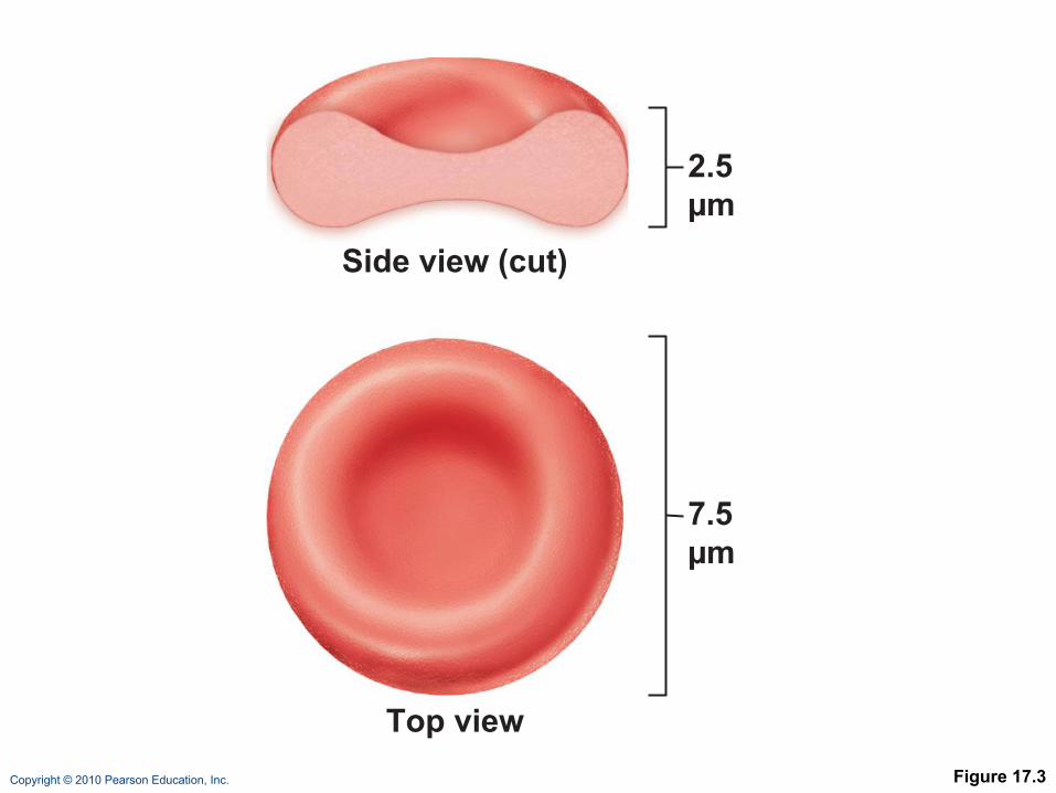
Copyright © 2010 Pearson Education, Inc. Figure 17.3
2.5 µm
7.5 µm
Side view (cut)
Top view
Copyright © 2010 Pearson Education, Inc.
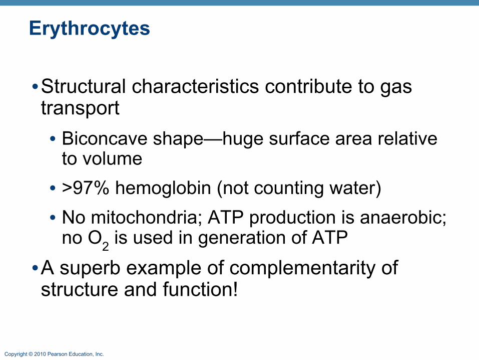
Erythrocytes
•Structural characteristics contribute to gas transport • Biconcave shape—huge surface area relative
to volume• >97% hemoglobin (not counting water)• No mitochondria; ATP production is anaerobic;
no O2 is used in generation of ATP•A superb example of complementarity of structure and function!
Copyright © 2010 Pearson Education, Inc.
Erythrocyte Function
•RBCs are dedicated to respiratory gas transport
•Hemoglobin binds reversibly with oxygen

Copyright © 2010 Pearson Education, Inc.
Erythrocyte Function
•Hemoglobin structure• Protein globin: two alpha and two beta chains
• Heme pigment bonded to each globin chain
• Iron atom in each heme can bind to one O2 molecule
•Each Hb molecule can transport four O2

Copyright © 2010 Pearson Education, Inc. Figure 17.4
Hemegroup
(a) Hemoglobin consists of globin (two alpha and two beta polypeptide chains) and four heme groups.
(b) Iron-containing heme pigment.α Globin chains
β Globin chains
Copyright © 2010 Pearson Education, Inc.
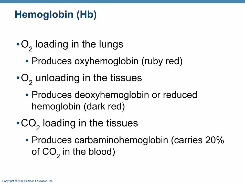
Hemoglobin (Hb)
•O2 loading in the lungs• Produces oxyhemoglobin (ruby red)
•O2 unloading in the tissues• Produces deoxyhemoglobin or reduced
hemoglobin (dark red)
•CO2 loading in the tissues• Produces carbaminohemoglobin (carries 20%
of CO2 in the blood)
Copyright © 2010 Pearson Education, Inc.
Hematopoiesis
•Hematopoiesis (hemopoiesis): blood cell formation • Occurs in red bone marrow of axial skeleton,
girdles and proximal epiphyses of humerus and femur
Copyright © 2010 Pearson Education, Inc.
Hematopoiesis
•Hemocytoblasts (hematopoietic stem cells)• Give rise to all formed elements
• Hormones and growth factors push the cell toward a specific pathway of blood cell development
•New blood cells enter blood sinusoids
Copyright © 2010 Pearson Education, Inc.
Erythropoiesis
• Erythropoiesis: red blood cell production• A hemocytoblast is transformed into a
proerythroblast
• Proerythroblasts develop into early erythroblasts
Copyright © 2010 Pearson Education, Inc.
Erythropoiesis
• Phases in development
1. Ribosome synthesis
2. Hemoglobin accumulation
3. Ejection of the nucleus and formation of reticulocytes
• Reticulocytes then become mature erythrocytes

Copyright © 2010 Pearson Education, Inc. Figure 17.5
Stem cell
Hemocytoblast
Proerythro-blast
Earlyerythroblast
Lateerythroblast
Normoblast
Phase 1Ribosomesynthesis
Phase 2Hemoglobinaccumulation
Phase 3Ejection ofnucleus
Reticulo-cyte
Erythro-cyte
Committedcell
Developmental pathway

Copyright © 2010 Pearson Education, Inc.
Regulation of Erythropoiesis
•Too few RBCs leads to tissue hypoxia
•Too many RBCs increases blood viscosity
•Balance between RBC production and destruction depends on• Hormonal controls
• Adequate supplies of iron, amino acids, and B vitamins
Copyright © 2010 Pearson Education, Inc.
Hormonal Control of Erythropoiesis
•Erythropoietin (EPO)• Direct stimulus for erythropoiesis
• Released by the kidneys in response to hypoxia
Copyright © 2010 Pearson Education, Inc.
Hormonal Control of Erythropoiesis
•Causes of hypoxia• Hemorrhage or increased RBC destruction
reduces RBC numbers
• Insufficient hemoglobin (e.g., iron deficiency)
• Reduced availability of O2 (e.g., high altitudes)
Copyright © 2010 Pearson Education, Inc.
Hormonal Control of Erythropoiesis
•Effects of EPO (Erythropoietin)• More rapid maturation of committed bone
marrow cells
• Increased circulating reticulocyte count in 1–2 days
•Testosterone also enhances EPO production, resulting in higher RBC counts in males
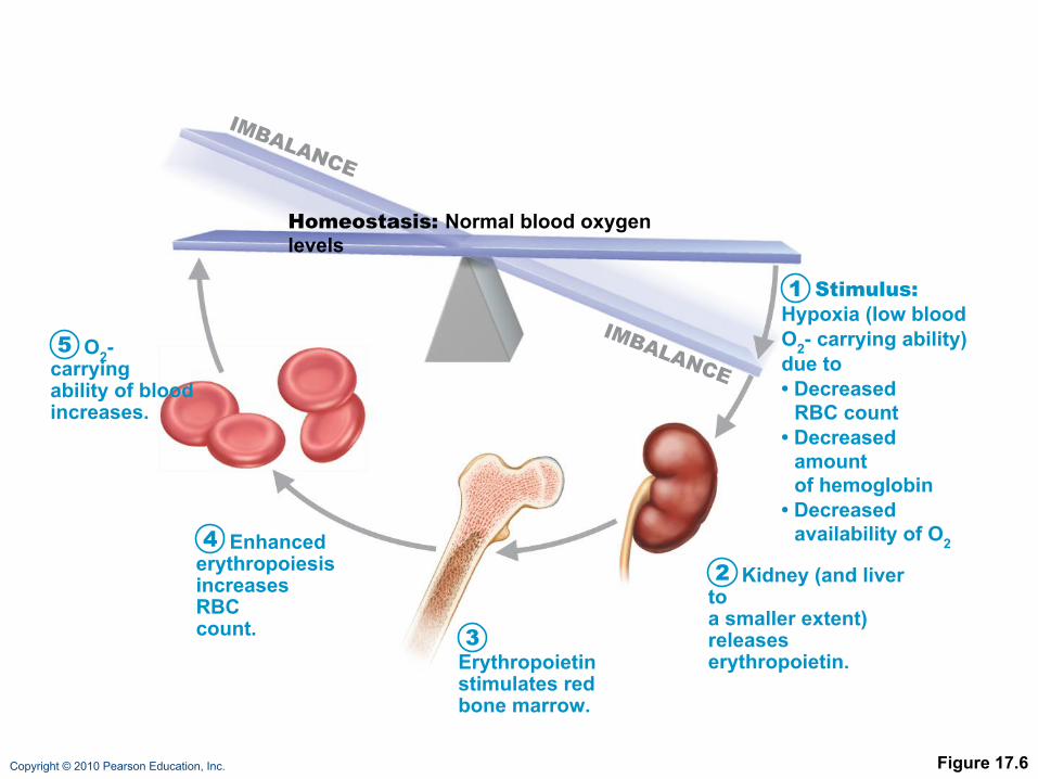
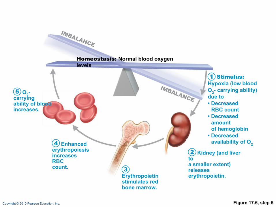
Copyright © 2010 Pearson Education, Inc. Figure 17.6
Kidney (and liver toa smaller extent)releaseserythropoietin.
Erythropoietinstimulates redbone marrow.
Enhancederythropoiesisincreases RBCcount.
O2- carryingability of bloodincreases.
Homeostasis: Normal blood oxygen levels
Stimulus:Hypoxia (low bloodO2- carrying ability)due to• Decreased
RBC count• Decreased
amountof hemoglobin
• Decreasedavailability of O2
1
2
3
4
5
IMBALANCE
IMBALANCE
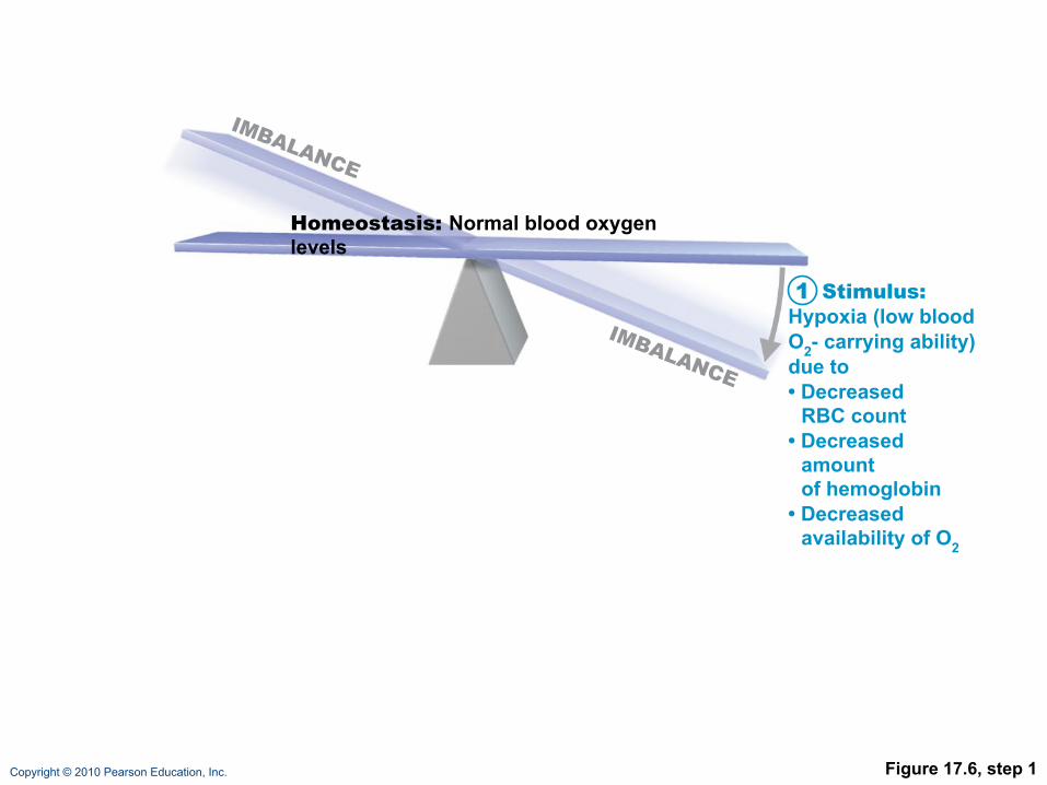
Copyright © 2010 Pearson Education, Inc. Figure 17.6, step 1
Homeostasis: Normal blood oxygen levels
Stimulus:Hypoxia (low bloodO2- carrying ability)due to• Decreased
RBC count• Decreased
amountof hemoglobin
• Decreasedavailability of O2
1
IMBALANCE
IMBALANCE

Copyright © 2010 Pearson Education, Inc. Figure 17.6, step 2
Kidney (and liver toa smaller extent)releaseserythropoietin.
Homeostasis: Normal blood oxygen levels
Stimulus:Hypoxia (low bloodO2- carrying ability)due to• Decreased
RBC count• Decreased
amountof hemoglobin
• Decreasedavailability of O2
1
2
IMBALANCE
IMBALANCE
Copyright © 2010 Pearson Education, Inc. Figure 17.6, step 3
Kidney (and liver toa smaller extent)releaseserythropoietin.
Erythropoietinstimulates redbone marrow.
Homeostasis: Normal blood oxygen levels
Stimulus:Hypoxia (low bloodO2- carrying ability)due to• Decreased
RBC count• Decreased
amountof hemoglobin
• Decreasedavailability of O2
1
2
3
IMBALANCE
IMBALANCE
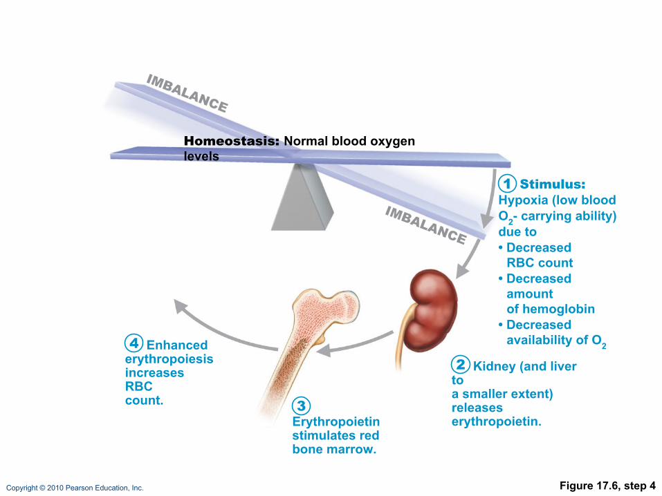
Copyright © 2010 Pearson Education, Inc. Figure 17.6, step 4
Kidney (and liver toa smaller extent)releaseserythropoietin.
Erythropoietinstimulates redbone marrow.
Enhancederythropoiesisincreases RBCcount.
Homeostasis: Normal blood oxygen levels
Stimulus:Hypoxia (low bloodO2- carrying ability)due to• Decreased
RBC count• Decreased
amountof hemoglobin
• Decreasedavailability of O2
1
2
3
4
IMBALANCE
IMBALANCE
Copyright © 2010 Pearson Education, Inc. Figure 17.6, step 5
Kidney (and liver toa smaller extent)releaseserythropoietin.
Erythropoietinstimulates redbone marrow.
Enhancederythropoiesisincreases RBCcount.
O2- carryingability of bloodincreases.
Homeostasis: Normal blood oxygen levels
Stimulus:Hypoxia (low bloodO2- carrying ability)due to• Decreased
RBC count• Decreased
amountof hemoglobin
• Decreasedavailability of O2
1
2
3
4
5
IMBALANCE
IMBALANCE
Copyright © 2010 Pearson Education, Inc.
Dietary Requirements for Erythropoiesis
•Nutrients—amino acids, lipids, and carbohydrates
• Iron• Stored in Hb (65%), the liver, spleen, and bone
marrow
• Stored in cells as ferritin and hemosiderin
• Transported loosely bound to the protein transferrin
• Vitamin B12 and folic acid—necessary for DNA synthesis for cell division
Copyright © 2010 Pearson Education, Inc.
Fate and Destruction of Erythrocytes
•Life span: 100–120 days
•Old RBCs become fragile, and Hb begins to degenerate
•Macrophages engulf dying RBCs in the spleen
Copyright © 2010 Pearson Education, Inc.
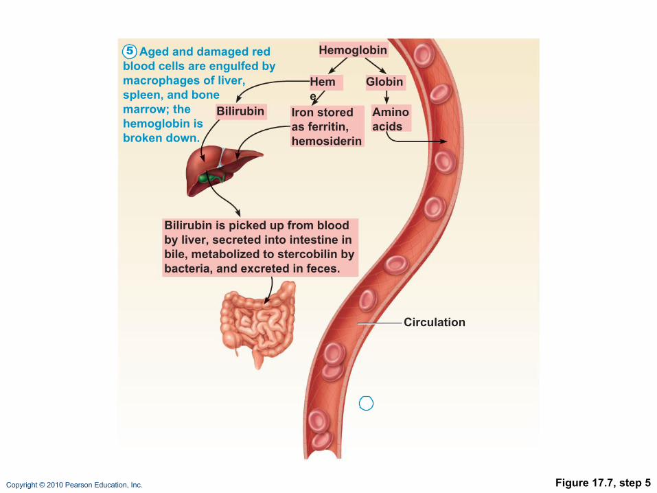
Fate and Destruction of Erythrocytes
•Heme and globin are separated• Iron is salvaged for reuse• Heme is degraded to yellow the pigment
bilirubin• Liver secretes bilirubin (in bile)) into the
intestines • Degraded pigment leaves the body in feces as
stercobilin • Globin is metabolized into amino acids
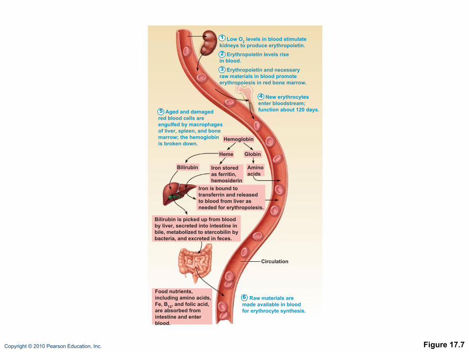
Copyright © 2010 Pearson Education, Inc. Figure 17.7
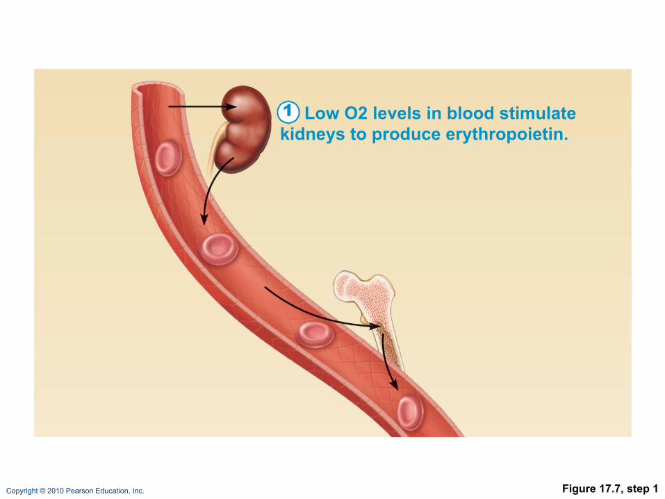
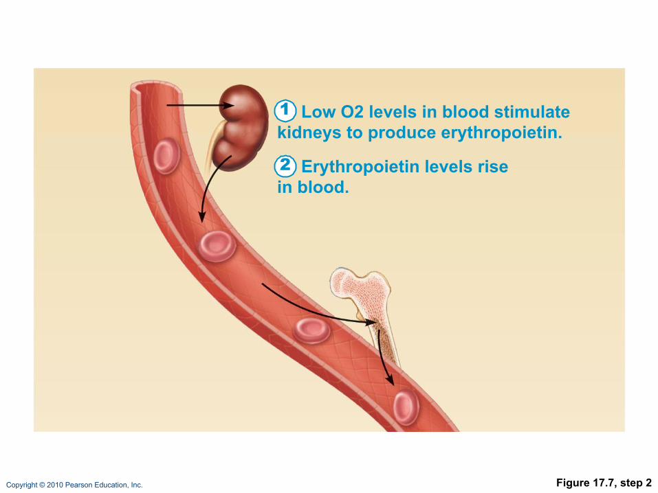
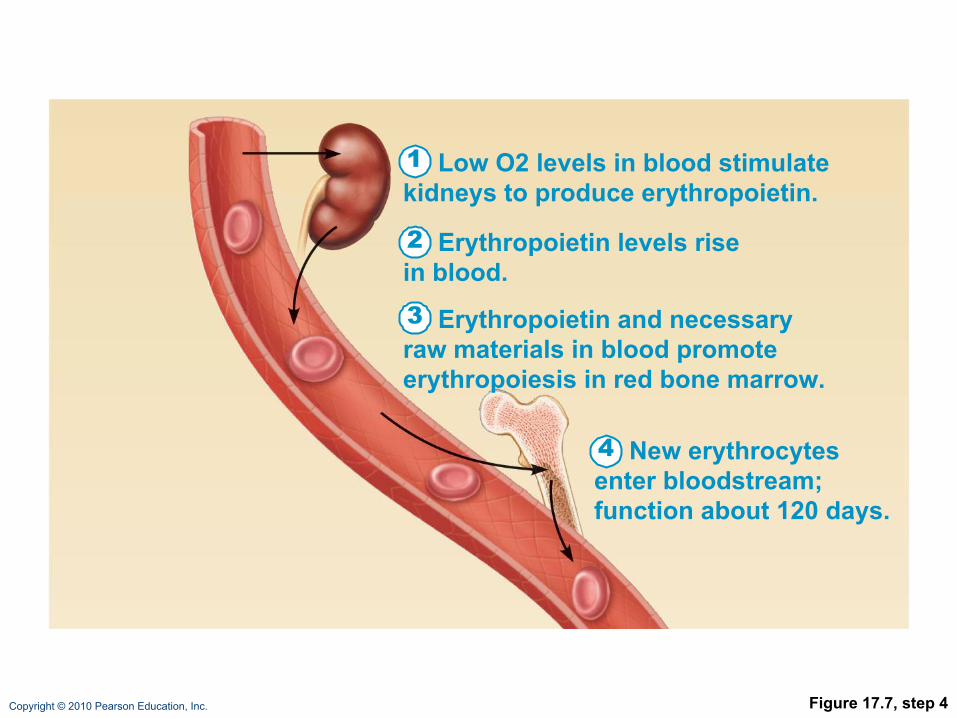
Low O2 levels in blood stimulate kidneys to produce erythropoietin.1
Erythropoietin levels risein blood.2
Erythropoietin and necessaryraw materials in blood promoteerythropoiesis in red bone marrow.
3
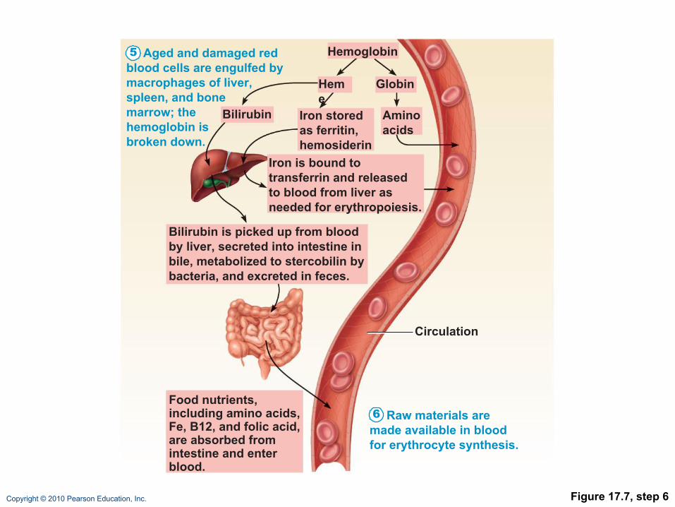
Aged and damagedred blood cells areengulfed by macrophagesof liver, spleen, and bonemarrow; the hemoglobinis broken down.
5
New erythrocytesenter bloodstream;function about 120 days.
4
Raw materials aremade available in bloodfor erythrocyte synthesis.
6
Hemoglobin
Aminoacids
Globin
Iron is bound totransferrin and releasedto blood from liver asneeded for erythropoiesis.
Food nutrients,including amino acids,Fe, B12, and folic acid,are absorbed fromintestine and enterblood.
Heme
Circulation
Iron storedas ferritin,hemosiderin
Bilirubin
Bilirubin is picked up from bloodby liver, secreted into intestine inbile, metabolized to stercobilin bybacteria, and excreted in feces.
Copyright © 2010 Pearson Education, Inc. Figure 17.7, step 1
Low O2 levels in blood stimulatekidneys to produce erythropoietin.1
Copyright © 2010 Pearson Education, Inc. Figure 17.7, step 2
Low O2 levels in blood stimulatekidneys to produce erythropoietin.1
Erythropoietin levels risein blood.2

Copyright © 2010 Pearson Education, Inc. Figure 17.7, step 3
Low O2 levels in blood stimulatekidneys to produce erythropoietin.1
Erythropoietin levels risein blood.2
Erythropoietin and necessaryraw materials in blood promoteerythropoiesis in red bone marrow.
3
Copyright © 2010 Pearson Education, Inc. Figure 17.7, step 4
Low O2 levels in blood stimulatekidneys to produce erythropoietin.1
Erythropoietin levels risein blood.2
Erythropoietin and necessaryraw materials in blood promoteerythropoiesis in red bone marrow.
3
New erythrocytesenter bloodstream;function about 120 days.
4
Copyright © 2010 Pearson Education, Inc. Figure 17.7, step 5
Aged and damaged redblood cells are engulfed bymacrophages of liver,spleen, and bonemarrow; thehemoglobin isbroken down.
5 Hemoglobin
Aminoacids
GlobinHeme
Circulation
Iron storedas ferritin,hemosiderin
Bilirubin
Bilirubin is picked up from bloodby liver, secreted into intestine inbile, metabolized to stercobilin bybacteria, and excreted in feces.
Copyright © 2010 Pearson Education, Inc. Figure 17.7, step 6
Aged and damaged redblood cells are engulfed bymacrophages of liver,spleen, and bonemarrow; thehemoglobin isbroken down.
5
Raw materials aremade available in bloodfor erythrocyte synthesis.
6
Hemoglobin
Aminoacids
Globin
Iron is bound totransferrin and releasedto blood from liver asneeded for erythropoiesis.
Food nutrients,including amino acids,Fe, B12, and folic acid,are absorbed fromintestine and enterblood.
Heme
Circulation
Iron storedas ferritin,hemosiderin
Bilirubin
Bilirubin is picked up from bloodby liver, secreted into intestine inbile, metabolized to stercobilin bybacteria, and excreted in feces.

Copyright © 2010 Pearson Education, Inc. Figure 17.7
Low O2 levels in blood stimulate kidneys to produce erythropoietin.1
Erythropoietin levels risein blood.2
Erythropoietin and necessaryraw materials in blood promoteerythropoiesis in red bone marrow.
3
Aged and damagedred blood cells areengulfed by macrophagesof liver, spleen, and bonemarrow; the hemoglobinis broken down.
5
New erythrocytesenter bloodstream;function about 120 days.
4
Raw materials aremade available in bloodfor erythrocyte synthesis.
6
Hemoglobin
Aminoacids
Globin
Iron is bound totransferrin and releasedto blood from liver asneeded for erythropoiesis.
Food nutrients,including amino acids,Fe, B12, and folic acid,are absorbed fromintestine and enterblood.
Heme
Circulation
Iron storedas ferritin,hemosiderin
Bilirubin
Bilirubin is picked up from bloodby liver, secreted into intestine inbile, metabolized to stercobilin bybacteria, and excreted in feces.

Copyright © 2010 Pearson Education, Inc.
Erythrocyte Disorders
•Anemia: blood has abnormally low O2-carrying capacity• A sign rather than a disease itself
• Blood O2 levels cannot support normal metabolism
• Accompanied by fatigue, paleness, shortness of breath, and chills
Copyright © 2010 Pearson Education, Inc.
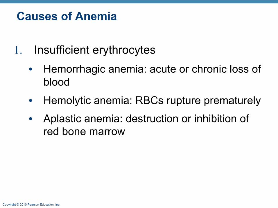
Causes of Anemia
1. Insufficient erythrocytes• Hemorrhagic anemia: acute or chronic loss of
blood
• Hemolytic anemia: RBCs rupture prematurely
• Aplastic anemia: destruction or inhibition of red bone marrow
Copyright © 2010 Pearson Education, Inc.
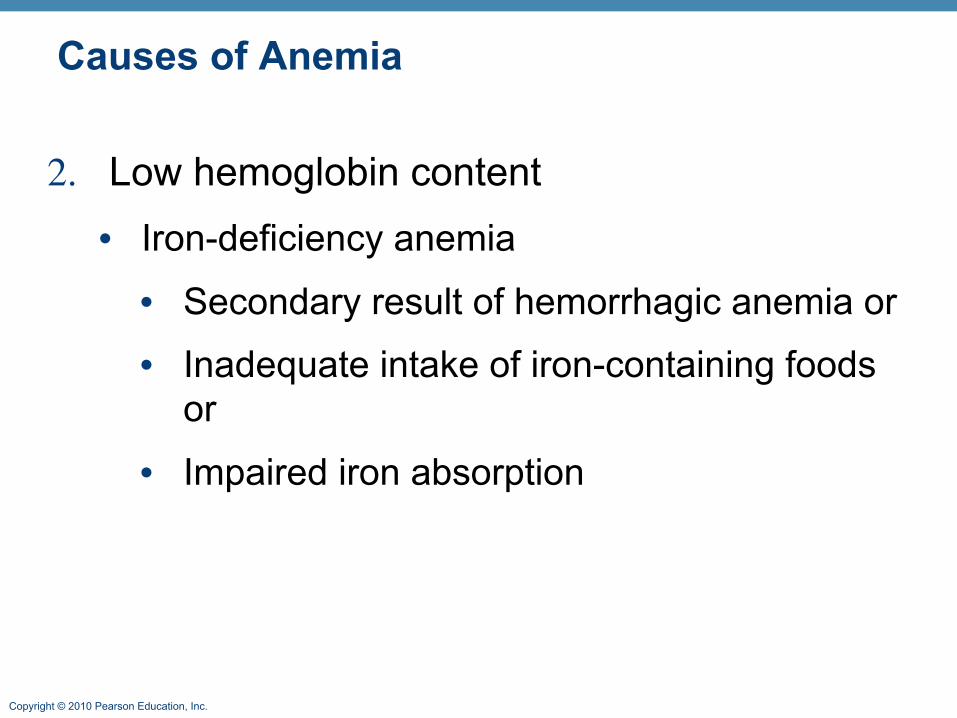
Causes of Anemia
2. Low hemoglobin content• Iron-deficiency anemia
• Secondary result of hemorrhagic anemia or
• Inadequate intake of iron-containing foods or
• Impaired iron absorption
Copyright © 2010 Pearson Education, Inc.
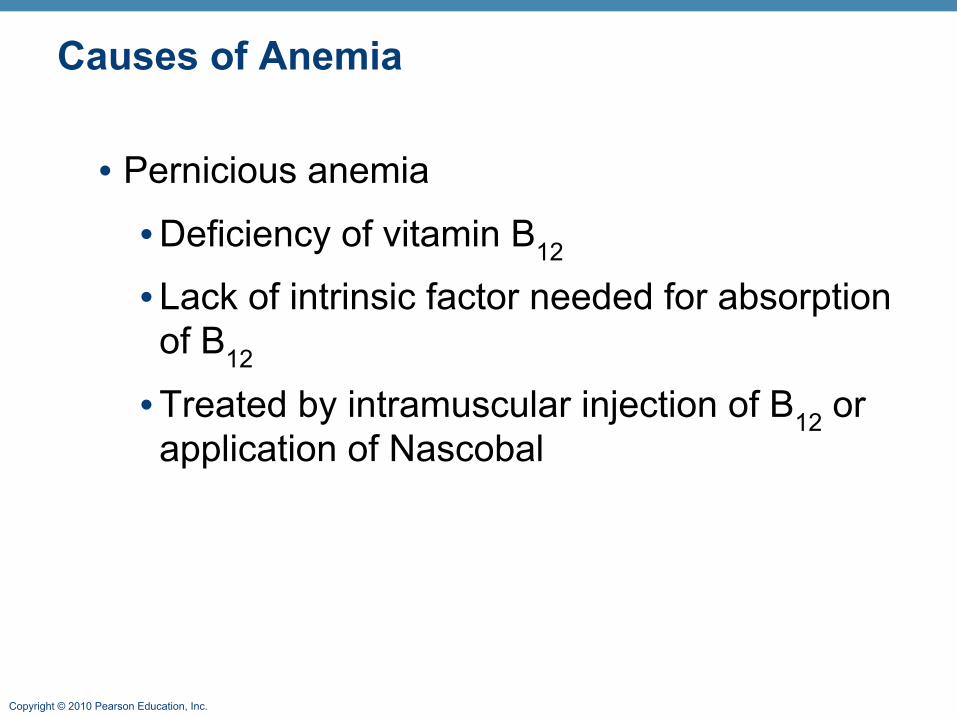
Causes of Anemia
• Pernicious anemia
•Deficiency of vitamin B12
• Lack of intrinsic factor needed for absorption of B12
•Treated by intramuscular injection of B12 or application of Nascobal
Copyright © 2010 Pearson Education, Inc.
Causes of Anemia
3. Abnormal hemoglobin• Thalassemias
• Absent or faulty globin chain
• RBCs are thin, delicate, and deficient in hemoglobin
Copyright © 2010 Pearson Education, Inc.
Causes of Anemia
• Sickle-cell anemia
•Defective gene codes for abnormal hemoglobin (HbS)
•Causes RBCs to become sickle shaped in low-oxygen situations

Copyright © 2010 Pearson Education, Inc. Figure 17.8
1 2 3 4 5 6 7 146
1 2 3 4 5 6 7 146
(a) Normal erythrocyte has normal hemoglobin amino acid sequence in the beta chain.
(b) Sickled erythrocyte results from a single amino acid change in the beta chain of hemoglobin.
Copyright © 2010 Pearson Education, Inc.
Erythrocyte Disorders
•Polycythemia: excess of RBCs that increase blood viscosity
•Results from:• Polycythemia vera—bone marrow cancer
• Secondary polycythemia—when less O2 is available (high altitude) or when EPO production increases
• Blood doping

Copyright © 2010 Pearson Education, Inc.
17B
Copyright © 2010 Pearson Education, Inc.
Leukocytes
•Make up <1% of total blood volume
•Can leave capillaries via diapedesis
•Move through tissue spaces by ameboid motion and positive chemotaxis
•Leukocytosis: WBC count over 11,000/mm3
• Normal response to bacterial or viral invasion
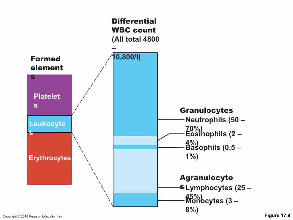
Copyright © 2010 Pearson Education, Inc. Figure 17.9
Formedelements
Platelets
Leukocytes
Erythrocytes
DifferentialWBC count(All total 4800 –10,800/l)
Neutrophils (50 – 70%)
Lymphocytes (25 – 45%)
Eosinophils (2 – 4%)Basophils (0.5 – 1%)
Monocytes (3 – 8%)
Agranulocytes
Granulocytes

Copyright © 2010 Pearson Education, Inc.
Granulocytes
•Granulocytes: neutrophils, eosinophils, and basophils• Cytoplasmic granules stain specifically with
Wright’s stain
• Larger and shorter-lived than RBCs
• Lobed nuclei
• Phagocytic

Copyright © 2010 Pearson Education, Inc.
Neutrophils
•Most numerous WBCs•Polymorphonuclear leukocytes (PMNs)•Fine granules take up both acidic and basic dyes•Give the cytoplasm a lilac color•Granules contain hydrolytic enzymes or defensins •Very phagocytic—“bacteria slayers”
Copyright © 2010 Pearson Education, Inc.
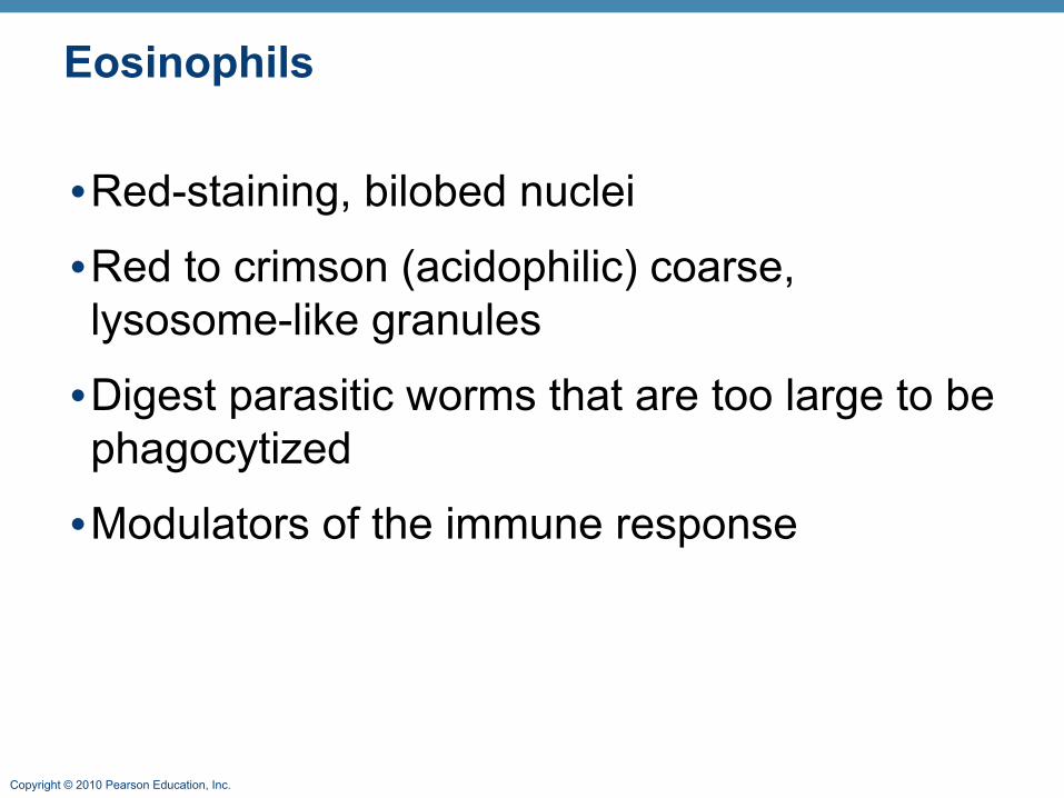
Eosinophils
•Red-staining, bilobed nuclei
•Red to crimson (acidophilic) coarse, lysosome-like granules
•Digest parasitic worms that are too large to be phagocytized
•Modulators of the immune response
Copyright © 2010 Pearson Education, Inc.
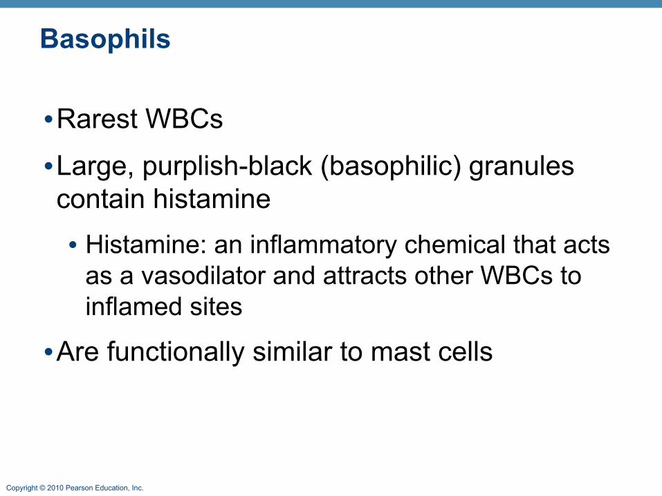
Basophils
•Rarest WBCs
•Large, purplish-black (basophilic) granules contain histamine• Histamine: an inflammatory chemical that acts
as a vasodilator and attracts other WBCs to inflamed sites
•Are functionally similar to mast cells

Copyright © 2010 Pearson Education, Inc. Figure 17.10 (a-c)
(a) Neutrophil; multilobed nucleus
(b) Eosinophil; bilobed nucleus, red cytoplasmic granules
(c) Basophil; bilobed nucleus, purplish-black cytoplasmic granules

Copyright © 2010 Pearson Education, Inc.
Agranulocytes
•Agranulocytes: lymphocytes and monocytes• Lack visible cytoplasmic granules
• Have spherical or kidney-shaped nuclei

Copyright © 2010 Pearson Education, Inc.
Lymphocytes
•Large, dark-purple, circular nuclei with a thin rim of blue cytoplasm
•Mostly in lymphoid tissue; few circulate in the blood
•Crucial to immunity

Copyright © 2010 Pearson Education, Inc.
Lymphocytes
•Two types • T cells act against virus-infected cells and
tumor cells
• B cells give rise to plasma cells, which produce antibodies
Copyright © 2010 Pearson Education, Inc.
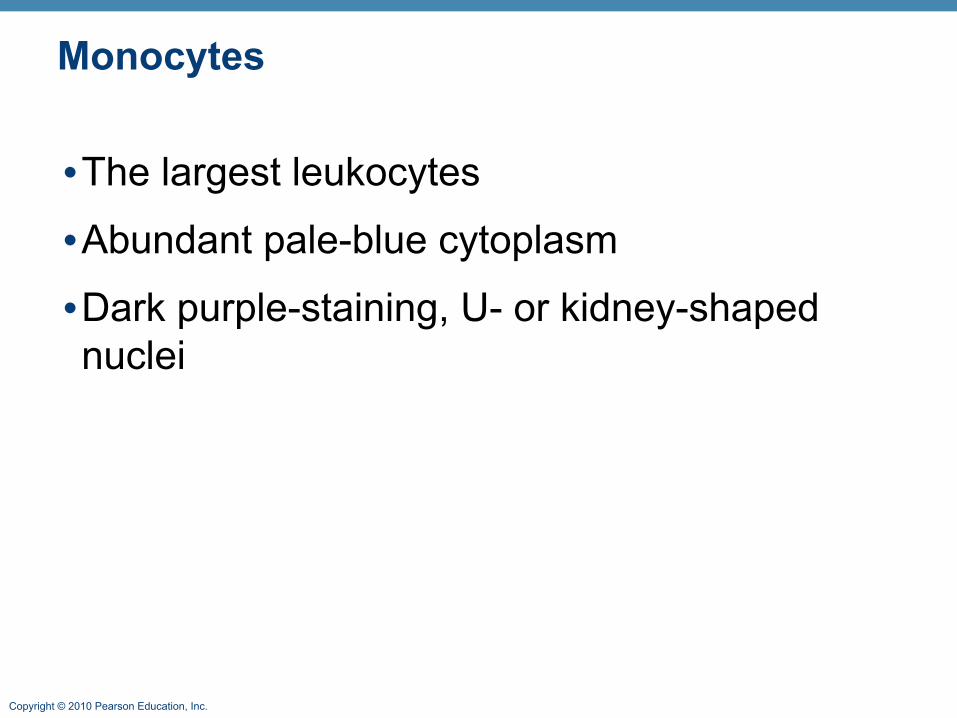
Monocytes
•The largest leukocytes
•Abundant pale-blue cytoplasm
•Dark purple-staining, U- or kidney-shaped nuclei
Copyright © 2010 Pearson Education, Inc.
Monocytes
•Leave circulation, enter tissues, and differentiate into macrophages• Actively phagocytic cells; crucial against
viruses, intracellular bacterial parasites, and chronic infections
•Activate lymphocytes to mount an immune response

Copyright © 2010 Pearson Education, Inc. Figure 17.10d, e
(d) Small lymphocyte; large spherical nucleus
(e) Monocyte; kidney-shaped nucleus
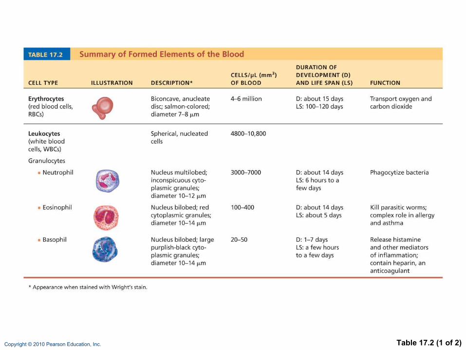
Copyright © 2010 Pearson Education, Inc. Table 17.2 (1 of 2)

Copyright © 2010 Pearson Education, Inc. Table 17.2 (2 of 2)
Copyright © 2010 Pearson Education, Inc.
Warm Up 4/14
Describe the function and composition of blood. Name all of the major cell types!
Copyright © 2010 Pearson Education, Inc.
Leukopoiesis
•Production of WBCs
•Stimulated by chemical messengers from bone marrow and mature WBCs• Interleukins (e.g., IL-1, IL-2)
• Colony-stimulating factors (CSFs) named for the WBC type they stimulate (e.g., granulocyte-CSF stimulates granulocytes)
•All leukocytes originate from hemocytoblasts
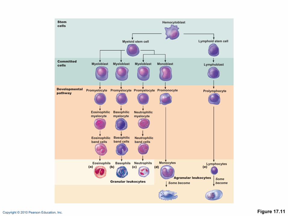
Copyright © 2010 Pearson Education, Inc. Figure 17.11
Hemocytoblast
Myeloid stem cell Lymphoid stem cell
Myeloblast Myeloblast MonoblastMyeloblast Lymphoblast
Stem cells
Committedcells
Promyelocyte PromyelocytePromyelocyte Promonocyte Prolymphocyte
Eosinophilicmyelocyte
Neutrophilicmyelocyte
Basophilicmyelocyte
Eosinophilicband cells
Neutrophilicband cells
Basophilicband cells
Developmentalpathway
Eosinophils NeutrophilsBasophils
Granular leukocytes
(a) (b) (c) (d) (e)Monocytes Lymphocytes
Agranular leukocytes
Some becomeSomebecome
Copyright © 2010 Pearson Education, Inc.
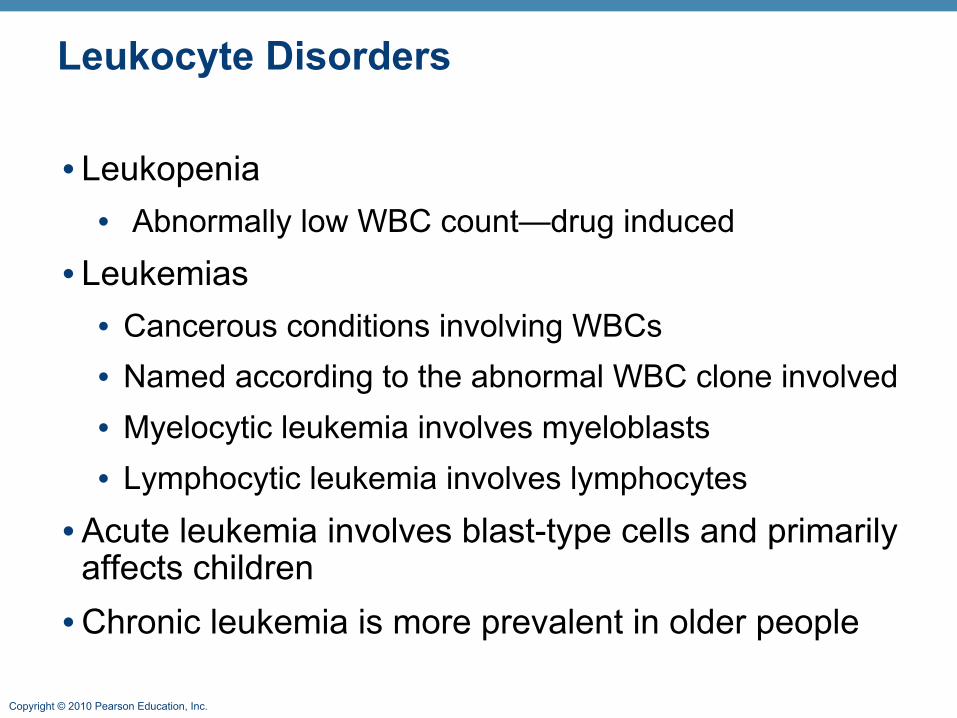
Leukocyte Disorders
• Leukopenia• Abnormally low WBC count—drug induced
• Leukemias• Cancerous conditions involving WBCs• Named according to the abnormal WBC clone involved• Myelocytic leukemia involves myeloblasts• Lymphocytic leukemia involves lymphocytes
• Acute leukemia involves blast-type cells and primarily affects children•Chronic leukemia is more prevalent in older people
Copyright © 2010 Pearson Education, Inc.
Leukemia
•Bone marrow totally occupied with cancerous leukocytes
• Immature nonfunctional WBCs in the bloodstream
•Death caused by internal hemorrhage and overwhelming infections
•Treatments include irradiation, antileukemic drugs, and stem cell transplants
Copyright © 2010 Pearson Education, Inc.
Platelets
•Small fragments of megakaryocytes
•Formation is regulated by thrombopoietin
•Blue-staining outer region, purple granules
•Form a temporary platelet plug that helps seal breaks in blood vessels
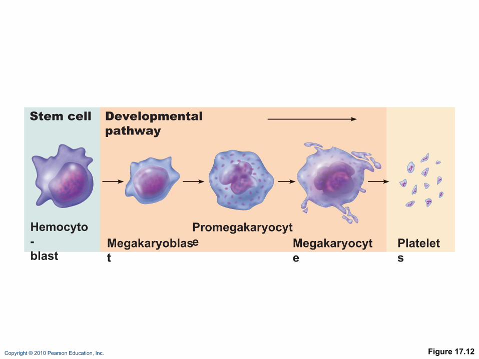
Copyright © 2010 Pearson Education, Inc. Figure 17.12
Stem cell Developmental pathway
Hemocyto-blast
Megakaryoblast
Promegakaryocyte Megakaryocyt
ePlatelets
Copyright © 2010 Pearson Education, Inc.
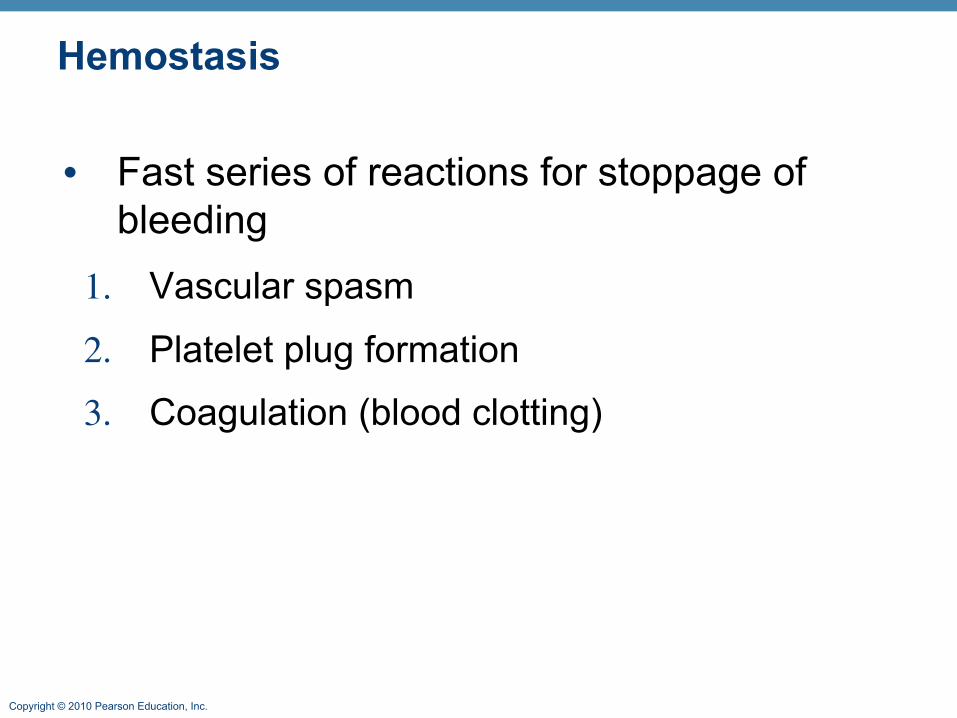
Hemostasis
• Fast series of reactions for stoppage of bleeding
1. Vascular spasm
2. Platelet plug formation
3. Coagulation (blood clotting)
Copyright © 2010 Pearson Education, Inc.
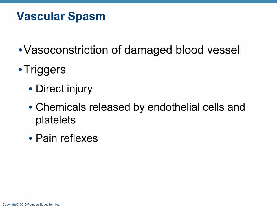
Vascular Spasm
•Vasoconstriction of damaged blood vessel
•Triggers• Direct injury
• Chemicals released by endothelial cells and platelets
• Pain reflexes

Copyright © 2010 Pearson Education, Inc.
Platelet Plug Formation
• Positive feedback cycle• At site of blood vessel injury, platelets
• Stick to exposed collagen fibers with the help of von Willebrand factor, a plasma protein
• Swell, become spiked and sticky, and release chemical messengers
• ADP causes more platelets to stick and release their contents
• Serotonin and thromboxane A2 enhance vascular spasm and more platelet aggregation
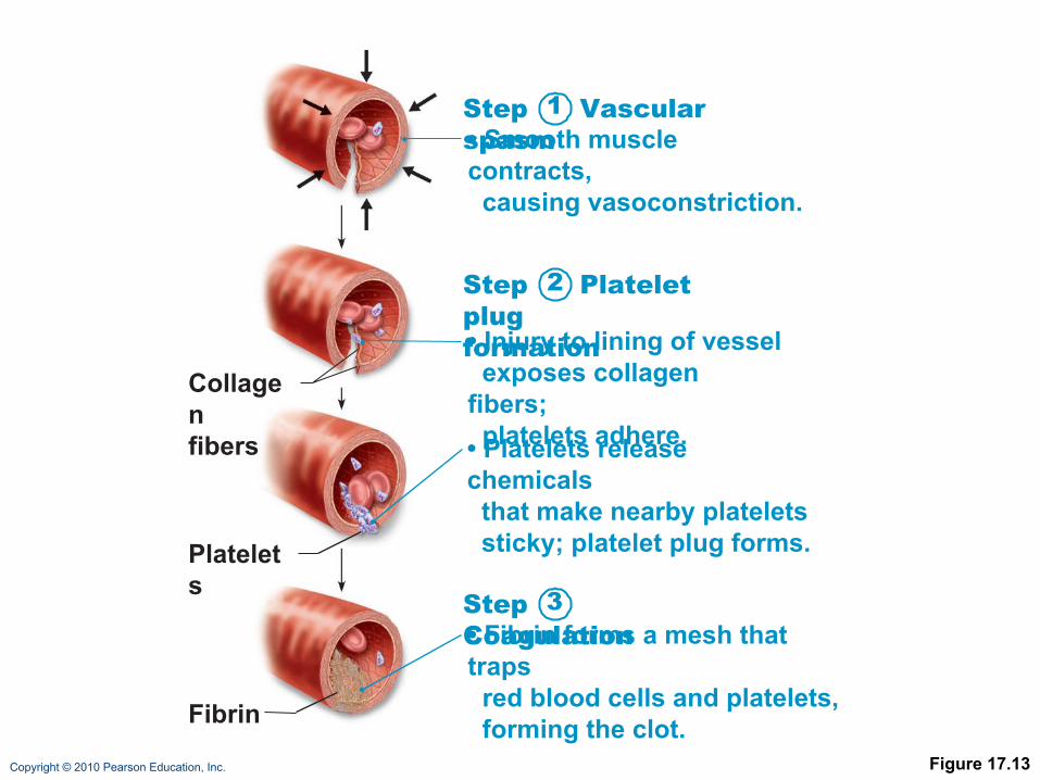
Copyright © 2010 Pearson Education, Inc. Figure 17.13
Collagenfibers
Platelets
Fibrin
Step Vascular spasm• Smooth muscle contracts, causing vasoconstriction.
Step Platelet plugformation• Injury to lining of vessel exposes collagen fibers; platelets adhere.
• Platelets release chemicals that make nearby platelets sticky; platelet plug forms.
Step Coagulation• Fibrin forms a mesh that traps red blood cells and platelets, forming the clot.
1
2
3

Copyright © 2010 Pearson Education, Inc.
Coagulation
• A set of reactions in which blood is transformed from a liquid to a gel
• Reinforces the platelet plug with fibrin threads

Copyright © 2010 Pearson Education, Inc.
Transfusions
•Whole-blood transfusions are used when blood loss is substantial
•Packed red cells (plasma removed) are used to restore oxygen-carrying capacity
•Transfusion of incompatible blood can be fatal

Copyright © 2010 Pearson Education, Inc.
Human Blood Groups
•RBC membranes bear 30 types glycoprotein antigens that are• Perceived as foreign if transfused blood is
mismatched• Unique to each individual • Promoters of agglutination and are called
agglutinogens •Presence or absence of each antigen is used to classify blood cells into different groups
Copyright © 2010 Pearson Education, Inc.
Blood Groups
•Humans have 30 varieties of naturally occurring RBC antigens
•Antigens of the ABO and Rh blood groups cause vigorous transfusion reactions
•Other blood groups (MNS, Duffy, Kell, and Lewis) are usually weak agglutinogens

Copyright © 2010 Pearson Education, Inc.
ABO Blood Groups
•Types A, B, AB, and O•Based on the presence or absence of two agglutinogens (A and B) on the surface of the RBCs •Blood may contain anti-A or anti-B antibodies (agglutinins) that act against transfused RBCs with ABO antigens not normally present •Anti-A or anti-B form in the blood at about 2 months of age

Copyright © 2010 Pearson Education, Inc. Table 17.4
Copyright © 2010 Pearson Education, Inc.
Rh Blood Groups
•There are 45 different Rh agglutinogens (Rh factors)
•C, D, and E are most common
•Rh+ indicates presence of D
Copyright © 2010 Pearson Education, Inc.
Rh Blood Groups
•Anti-Rh antibodies are not spontaneously formed in Rh– individuals
•Anti-Rh antibodies form if an Rh– individual receives Rh+ blood
•A second exposure to Rh+ blood will result in a typical transfusion reaction
Copyright © 2010 Pearson Education, Inc.
Transfusion Reactions
•Occur if mismatched blood is infused•Donor’s cells• Are attacked by the recipient’s plasma agglutinins• Agglutinate and clog small vessels• Rupture and release free hemoglobin into the
bloodstream
•Result in• Diminished oxygen-carrying capacity• Hemoglobin in kidney tubules and renal failure
Copyright © 2010 Pearson Education, Inc.
Blood Typing
•When serum containing anti-A or anti-B agglutinins is added to blood, agglutination will occur between the agglutinin and the corresponding agglutinogens
•Positive reactions indicate agglutination

Copyright © 2010 Pearson Education, Inc.
ABO Blood Typing

Copyright © 2010 Pearson Education, Inc. Figure 17.16
SerumAnti-A
RBCs
Anti-B
Type AB (containsagglutinogens A and B;agglutinates with bothsera)
Blood being tested
Type A (containsagglutinogen A;agglutinates with anti-A)
Type B (containsagglutinogen B;agglutinates with anti-B)
Type O (contains noagglutinogens; does notagglutinate with eitherserum)
Copyright © 2010 Pearson Education, Inc.
Restoring Blood Volume
•Death from shock may result from low blood volume
• Volume must be replaced immediately with• Normal saline or multiple-electrolyte solution that
mimics plasma electrolyte composition
• Plasma expanders (e.g., purified human serum albumin, hetastarch, and dextran)
• Mimic osmotic properties of albumin
• More expensive and may cause significant complications

Copyright © 2010 Pearson Education, Inc.
Diagnostic Blood Tests
•Hematocrit
•Blood glucose tests
•Microscopic examination reveals variations in size and shape of RBCs, indications of anemias
Copyright © 2010 Pearson Education, Inc.
Diagnostic Blood Tests
•Differential WBC count
•Prothrombin time and platelet counts assess hemostasis
•SMAC, a blood chemistry profile
•Complete blood count (CBC)

Copyright © 2010 Pearson Education, Inc.
Heart Anatomy / Physiology Whiteboard Review
-Label the interior and exterior anatomy of the heart-Describe and show the pathway of blood through the heart-Describe and show the 5 steps of excitation (challenge: correlate each step with the 3 waves of an ECG)-Describe and show the 3 phases of the cardiac cycle-What is the difference in function of the atrium and ventricles?-What is the difference between pulmonary, systemic, and coronary circulation?-Label and describe the function of the 4 valves in our heart.