blood coagulation by low energy plasma jet
TRANSCRIPT

Blood Coagulation by Low Energy Plasma Jet E. Janani1, M. Ghoranneviss1, M. Al-Ebrahim2, P. Mortazavi3, A .H. Sari1, Sh. Mirpour1,
M. Hadipour Jahromy4, S. M. Borghei1, and Z. Ghoannevis1
1Plasma Physics Research Center, Science and Research Branch, Islamic Azad University, Tehran, Iran 2Department of Biology, Science and Research Branch, Islamic Azad University, Tehran, Iran
3Department of Pathology, Faculty of specialized Veterinary Science and Research Branch, Islamic Azad University, Tehran, Iran
4Tehran Medical Branch, Islamic Azad University, Tehran, Iran
Abstract: In this paper we demonstrate blood coagulation by atmospheric plasma jet operating at room temperature. The non-thermal characteristic of our device confirms the possibility of using this device for biomedical applications. The results on the ability of non-thermal plasma to promote blood coagulation are presented in this paper. Plasma treatment performed on skin and liver of living animal tissues in our experiments and our results suggest that there is no any visible or microscopic damage on the skin and tissue using plasma treatment at few seconds plasma irradiation. Keywords: non-thermal argon plasma, blood coagulation, tissue processing.
1. Introduction
Recently, a new area for the application of plasma source operating at atmospheric pressures has emerged: biomedical applications. During the last decade, the basics of sterilizing effects of plasmas were well studied. Recently, therapeutic applications are of great concern. Existing plasma surgical technologies such as coagulation or ablation are mainly based on lethal plasma effects on living systems [1-5]. The effect of the Plasma on different tissues remains relatively constant across the tissue types tested. A total depth of necrosis of about 2.0 mm was seen in the dense and vascular tissue of the liver, spleen and kidney. Thermal damage leads to necrosis, or accidental cell death. Necrosis is defined as the consequence of a catastrophic injury to the mechanisms that maintain the integrity of the cell. A well-established technique based on plasma heat is argon plasma coagulation (APC) [6-8]. In this article we demonstrate atmospheric plasma jet operating at room temperature. The non-thermal characteristic of our device confirms the possibility of this device for biomedical applications. Here we describe the ability of non-thermal plasma to promote blood coagulation, where plasma treatment was performed on the skin and liver of living animal tissues. The results confirmed that there was not any visible or microscopic damage on skin and tissue using plasma treatment at few seconds. [1, 5, 6].
2. Experimental
Argon plasma was produced inside a glass tube with 5cm in length and diameter of 8mm, (Fig (1.a)). Two electrodes were used. The first electrode was placed inside the tube, which is made of Ni-Cr with the length of 11cm and the diameter of 1mm (Fig (1.b)) The first electrode is movable and it is possible to adjust the length inside the tube in order to have stable plasma, which in our setup the optimum length was found to be 7 cm inside the tube as it is shown in Fig. 2.c. The second electrode was also made from aluminum, which was 2 cm in length and was placed around the tube. The tube was made of pyrex and surrounded by insulator of 4mm in diameter (see Fig. 2.c and 2.d). The argon flow rate is set at between 1.5 and 2 lit/min and the process time is 21 min while 1.1 kv voltage and AC power of 60 HZ was applied.
Figure 1.a. glass tube, b. first electrode, c. and d. plasma irradiation.
3. Results and Discussion
Blood coagulation initially in vitro performed on a certain volume of blood from cadaver organs. The results consistently show faster coagulation when exposed to APC: for example, blood treated for 10 seconds completely coagulates while untreated sample coagulates in 14 minutes.
Figure 2. two drops of blood at the same time were dropping. Plasma-assisted blood coagulation with 10-second treatment (left) and control drop (right). Right drop after 10 minutes still does not coagulate.
Figure 3.a. and b. blood clot formation under APC treatment in 10 seconds. Fig. 3.b shows control drop after 10 minutes without coagulation.
Blood coagulation on Living animal during liver surgery was also tested in another experiment on the 12 living rat’s liver. Initially 4 rats were operated and in each of them slices of 1 cm on rat liver were created. Bleeding time to complete blood coagulation in rat liver was about 4 to 5 minutes. Then the remained eight rats Surgery was performed immediately after cutting 1cm on liver and plasma treatment begun and in 12 seconds that is less than a minute Coagulation was completed. On tissues after exposure of plasma, treated samples of liver in 1cm3
were examined with blood coagulation histology tests on cadaver organs with subsequent gross and microscopic evaluation of tissue to test for damage. Our analysis demonstrates blood clotting within 12 seconds without gross or microscopic evidence of tissue damage. A total depth of necrosis of about 12 micro meter was seen in the dense and vascular tissue of the liver.
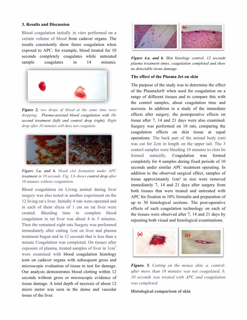
Figure 4.a. and b. Skin histology control, 12 seconds plasma treatment times, coagulation completed and show no detectable tissue damage.
The effect of the Plasma Jet on skin
The purpose of the study was to determine the effect of the PlasmaJet® when used for coagulation on a range of different tissues and to compare this with the control samples, about coagulation time and necrosis. In addition to a study of the immediate effects after surgery, the postoperative effects on tissue after 7, 14 and 21 days were also examined. Surgery was performed on 10 rats, comparing the coagulation effects on skin tissue at equal operations. The back part of the animal body (rat) was cut for 2cm in length on the upper tail. The 3 control samples were bleeding 10 minutes to clots be formed naturally. Coagulation was formed completely for 4 samples during fixed periods of 10 seconds under similar APC treatment operating. In addition to the observed surgical effect, samples of tissue approximately 1cm³ in size were removed immediately 7, 14 and 21 days after surgery from both tissues that were treated and untreated with APC for fixation in 10% formalin and preparation of up to 30 histological sections. The post-operative effects of each coagulation technology on each of the tissues were observed after 7, 14 and 21 days by repeating both visual and histological examinations.
Figure. 5. Cutting on the mouse skin, a. control: after more than 10 minutes was not coagulated, b. 10 seconds was treated with APC and coagulation was completed.
Histological comparison of skin

Figure 6.a. and b. Skin histology: control samples, 0, 21 days after surgery
Figure 7.a., b., and c. Skin histology (left to right): APC treated samples with 10 seconds treatment times, 0, 14, 21 days after surgery show no detectable tissue damage
Figure. 8. Skin histology: APC treated samples after 5 minutes treatment and 7days after surgery show no detectable tissue damage
Therefore we observed that 21 days after APC treatment wound size is decreased so dramatically and results and images suggest that the method of using APC designed in this stage reduced the clotting time from 10 minutes in normal samples to 10 seconds on APC treated samples with no damage.
4. Conclusion
The scope of the presented research included only initial studies of plasma influence on blood coagulation. In our experiment using the APC to blood coagulation in vitro and also was used during surgery have less time for blood coagulation compared with other methods as well as some other APC and the DBD in the absence of tissue damage and also reduce the coagulation time in 10 seconds which has been reported for more than a minute by other researchers at the DBD technique previously [6-8]. The device competitive with other samples made in the world and less than 15 seconds was Completed blood coagulation with no damage.
References [1] M. Laroussi. IEEE Trans. Plasma Sci. 37 714 (2009). [2] G. Fridman, G. Friedman, A. Gutsol, A. B. Shekhter, V. N. Vasilets, A. Fridman. Plasma Process. Polym. 5 503 (2008). [3] E. Stoffels, I. E. Kieft, R. E. J. Sladek, E. P. van der Laan, D. W. Slaaf. Crit. Rev. Biomed. Eng. 34 53 (2004). [4] C. Q. Jiang, M. T. Chen, C. Schaudinn, A. Gorur, P. T. Vernier, J. W. Costerton, D. E. Jaramillo, P. P. Sedghizadeh, M. A. Gundersen. IEEE Trans. Plasma Sci. 37 1190 (2009). [5]K. D. Weltmann, Pure Appl. Chem., 82 1223 (2010).
[6] J. J. Vargo, Gastrointestinal Endoscopy. 3 81 (2004).
[7] S. Kalghatgi Ninth Annual Research Innovation Scholarship and Creativity (RISC) Day, Philadelphia, USA (2007).
[8] M. Thiyagarajan. IEEE Transactions on Plasma Science (2005).