biomolecular archaeology (an introduction) || sources of ancient biomolecules
TRANSCRIPT
PART II
PRESERVATION AND DECAY OF BIOMOLECULES
IN ARCHAEOLOGICAL SPECIMENS
p02.indd 89p02.indd 89 12/6/2010 6:43:23 AM12/6/2010 6:43:23 AM
Biomolecular Archaeology: An Introduction, by Terry Brown and Keri Brown © 2011 Terry Brown & Keri Brown

7
Sources of Ancient Biomolecules
Now that we are familiar with the different types of ancient biomolecule we must examine the materials from which those biomolecules are obtained. The most important of these are the bones and teeth of humans and animals, these being the types of biological material most frequently recovered by archaeological excavation. But we must also consider specimens such as mummies and bog bodies, which contain preserved soft tissue as well as skeletal remains, and hair and coprolites (preserved excrement), both of which are important sources of human and animal biomolecules. Our survey must also include plants, which are preserved in the archaeological record mainly as charred, desiccated, or waterlogged remains, often but not always of seeds. Invertebrates are also important in archaeology, discarded shells being good indicators of a marine or freshwater diet and the identities of land snails present at a site providing information on climate, but invertebrate remains have not yet been exploited as sources of ancient biomolecules, and we can therefore exclude them from our study.
When we consider each type of remain, we must remember that the material from which ancient biomolecules are extracted is not the original biological structure, but the preserved version of that structure, which almost certainly has undergone transformation since it was alive. We must therefore understand not just the nature of the living material but the changes that might have occurred to that material since its death. These changes can be grouped into three categories:● Changes occurring soon after death due to human activity, including cremation or mum-mification of human and animal remains.● Changes due to diagenesis, the general breakdown and decay of dead biological material over time.● Changes occurring after excavation, due to continued decay during storage or resulting from treatments applied by curators in attempts to preserve the material. Post-excavational
Biomolecular Archaeology: An Introduction, by Terry Brown and Keri Brown © 2011 Terry Brown & Keri Brown
c07.indd 91c07.indd 91 12/6/2010 6:44:21 AM12/6/2010 6:44:21 AM

92 Preservation and Decay of Biomolecules in Archaeological Specimens
change is particularly important, as biomolecular archaeologists often work with material that has been stored in museums or elsewhere for years or decades.
Clearly, we must deal with a range of materials and a number of different issues. To sim-plify our task we will focus in this chapter specifically on the biological materials. The related question of how the biomolecules themselves change and decay over time will be the subject of the next chapter.
7.1 Bones and Teeth
The preserved bones and teeth of animals and humans are by far the commonest types of biological material recovered by archaeological excavation. Partly for this reason, bone and teeth are also the types of material most frequently studied by biomolecular archaeologists. Bone and teeth are the most important sources of ancient DNA, the demonstration in 1989 that DNA is preserved in at least some archaeological skeletons being the major stimulus in development of ancient DNA research from a backwater into the mainstream of archaeol-ogy, zoology, and paleontology. Bone also contains proteins, such as collagen and osteocal-cin, which can be studied in their own right or, along with the mineral component, used as sources of stable isotopes for dietary studies. Stable isotopes are also obtained from teeth, and teeth contain the strontium isotopes that are used to provide information on human and animal mobility.
7.1.1 The structure of living bone
Bone is a living tissue that constantly undergoes remodelling during the lifetime of the organism. The cells responsible for the synthesis and resorption of bone are called osteob-lasts and osteoclasts, respectively. A third type of cell, osteocytes, are osteoblasts that have become embedded in the bone that they have formed, but which remain alive and continue to contribute to turnover of the bone matrix.
Osteoblasts secrete proteins including collagen, along with calcium, magnesium, and phosphate ions, which combine to form the inorganic component of bone. This inorganic component is, in chemical terms, a crystalline hydroxyapatite carbonate mineral called dahllite, more commonly referred to simply as bioapatite. The organic component, which makes up about 20% of the dry weight of a bone, largely comprises collagen molecules that have combined into triple helical structures called tropocollagen, which in turn combine to form collagen fibrils. These fibrils are embedded within the bioapatite matrix, which also permeates between the individual tropocollagen units of a fibril, giving an overall structure that combines flexibility with rigidity.
Collagen fibrils can be organized in different ways to give three distinct types of bone microstructure:● Woven bone, in which the fibrils takes up a random arrangement. Woven bone is relatively weak but can be synthesized rapidly and so is often the first type of bone to be made at points of bone growth or repair.● Lamellar bone contains aligned collagen fibrils and is much harder than woven bone, which it usually replaces as the bone structure matures. Lamellar bone is usually organized into
c07.indd 92c07.indd 92 12/6/2010 6:44:21 AM12/6/2010 6:44:21 AM

Sources of Ancient Biomolecules 93
cylindrical osteons, each one a few mm in length and made up of concentric layers of bone surrounding a central Haversian canal containing blood vessels and nerves (Figure 7.1).● Parallel-fibered bone has an intermediate arrangement between woven and lamellar. It is the least frequent type and is found mainly on new bone surfaces.
At the macroscopic level, two types of bone are recognized. The first is compact or corti-cal bone, as shown in Figure 7.1, which is made up of parallel bundles of osteons and has great strength. Compact bone makes up the shafts of the long bones in the arms and legs, as well as the outer parts of ribs and wrist and ankle bones. Layers of compact bone also form the various components of the skull. The second macroscopic type is cancellous or trabec-ular bone, often called “spongy,” which has a much less rigid organization, being less dense and permeated with channels. Spongy bone is found in the central parts of long bones,
Haversian canalOsteon
Osteocytes
Figure 7.1 The typical appearance of
well- preserved lamellar bone. The micrograph
shows a cross-section with individual osteons
clearly visible, each approximately 0.2 mm in
diameter. The histology would be graded as “5” in
the index described in Table 7.1. Reprinted from
Robert Hedges, Andrew Millard, and Alistair Pike,
“Measurements and relationships of diagenetic
alteration of bone from three archaeological sites,”
Journal of Archaeological Science, 22, 201–9,
copyright 1995, with permission from Elsevier
and the authors.
Table 7.1 The Oxford Histological Index of archaeological bone decay.
Index % intact bone Description
0 < 5 No original features identifiable, other than Haversian canals
1 <15 Small areas of well-preserved bone present, or some lamellar
structure preserved by pattern of destructive foci
2 <50 Clear lamellate structure preserved between destructive foci
3 >50 Clear preservation of some osteocyte lacunae
4 >85 Only minor amounts of destructive foci, otherwise generally
well preserved
5 >95 Very well preserved, virtually indistinguishable from fresh bone
c07.indd 93c07.indd 93 12/6/2010 6:44:21 AM12/6/2010 6:44:21 AM

94 Preservation and Decay of Biomolecules in Archaeological Specimens
forming the bone marrow in which blood cells are made, and is also present within the bones of the skull. Vertebrae and the hip bones have a larger spongy component, though this component is denser than that found in bone marrow. The compact part of these “irregular” bones is confined to a thin outer surface.
7.1.2 The decay processes for bone after death are complex
The diagenetic changes occurring in bones after death are complex and dependent on the burial environment, in particular the presence of water. Even the water aspect is complex, modelling studies showing that the movement of water through a bone leads to different decay processes than those occurring when a bone is simply soaked in water. Neither is there any reason to suppose that the environment will remain static during the entire period between burial and excavation, adding another layer of complexity. This multifaceted nature of diagenesis applies not just to bones but to all biological materials, but we tend to recognize it more clearly with bones because the most detailed attempts to understand diagenesis have been made with this material.
Arguably the most important cause of bone decay in the period immediately following death is attack by microorganisms, both bacteria and fungi. Microbial activity leads to changes in bone histology that are clearly visible when a transverse section is examined with a light microscope, the first signs of attack being the presence of small holes in the bone structure. These holes or “destructive foci” are not all uniform in appearance and different types of foci are associated with different burial environments. Whether or not they are caused by different species of microorganism is not known. Continued micro-bial attack leads to an increasing amount of structural damage, culminating in badly decayed bones lacking almost all of their original histological features (Figure 7.2). Microbial attack has been observed in some bones after just a few months of burial, but in other cases bones buried for over 30 years have displayed very little of this kind of decay. These differences might not be entirely due to the burial environment but instead might reflect the nature of the burial. Burial of an entire animal or human body, as opposed to butchered segments, exposes the skeleton to microorganisms released from the guts, which are thought to promote bone decay. There might also be differences between butchered bones buried with attached flesh and those from which the flesh has been removed. Experiments with animal bones suggest that ones that are partially cooked before burial undergo more rapid microbial attack than those that are buried without this treatment.
These considerations prompt the question of what exactly is being broken down by the microorganisms present in a bone. The destructive foci indicate that the inorganic compo-nent is being broken down in some way, but this is almost certainly not the only effect that microorganisms have on a bone. To survive, bacteria and fungi must obtain nutrients and it will be the organic fraction of the bone that satisfies this requirement. Collagen, however, is a difficult compound to degrade, so some type of pretreatment such as partial cooking might promote microbial attack by partially breaking down the bone collagen, or making it more accessible by freeing parts of the collagen fibrils from the inorganic component of
c07.indd 94c07.indd 94 12/6/2010 6:44:22 AM12/6/2010 6:44:22 AM

Sources of Ancient Biomolecules 95
the bone. Microbial attack can certainly result in considerable depletion of the organic content of a bone, and the process is sufficiently rapid to reach completion after a few hundred years.
Non-biological processes can also contribute to loss of collagen, and will act on any that remains after microbial decay, and can also result in rapid decay of collagen in bones that are not subject to attack by microorganisms. These chemical decay reactions are water driven and, like all chemical reactions, act more quickly at higher temperatures. Other proc-esses are thought to prevent or at least delay the chemical breakdown of collagen. These include the formation of a complex between collagen and humic acids, which can move from the soil into buried bones.
The inorganic matrix of bone also undergoes chemical change, additional to the focal destruction caused by microbial attack. These changes include dissolution of the bone matrix in water, which is more rapid in acidic environments and when the bone is constantly exposed to fresh sources of water. If the water is not recharged, then its content of calcium and phosphate ions eventually reaches equilibrium with that of bone and no further dissolu-tion occurs. There can also be uptake of ions from the groundwater, possibly resulting in substantial changes in the inorganic content of the bone. One important consequence of uptake is that certain ions naturally present in the bone might become exchanged with ions of the same element present in the burial environment. This has been shown to happen with strontium, which means that the characteristic strontium isotope ratio present in a bone at the time of death will change over time due to the diagenetic exchange between bone and environmental strontium ions. This change must be accounted for when strontium isotope ratios from bones are used in studies of human and animal mobility (Section 6.2).
Figure 7.2 The typical appearance of badly degraded lamellar bone, histology index “0.” The
micrograph is at the same magnification as Figure 7.1. Reprinted from Robert Hedges, Andrew
Millard, and Alistair Pike, “Measurements and relationships of diagenetic alteration of bone
from three archaeological sites,” Journal of Archaeological Science, 22, 201–9, copyright 1995,
with permission from Elsevier and the authors.
c07.indd 95c07.indd 95 12/6/2010 6:44:22 AM12/6/2010 6:44:22 AM

96 Preservation and Decay of Biomolecules in Archaeological Specimens
7.1.3 Methods that enable the extent of bone diagenesis to be measured are being sought
Faced with such a complex process, is it possible to devise any methods for estimating the amount of diagenesis that has occurred within a bone? Various methods have been devised to address this question, but with most of these it is debatable if diagenesis as a whole is being measured, or just some component of it.
The simplest way of measuring bone diagenesis is to examine thin transverse sections with the light microscope in order to assess the amount of structural damage that has occurred. To aid this type of analysis, a histological index has been devised (Table 7.1) to grade the extent of destruction between the two extremes illustrated in Figures 7.1 and 7.2. This index is largely based on the nature and extent of the destructive foci that are present, and so is essen-tially a measure of microbiological attack rather than the overall degree of diagenesis. The amount of organic decay that has occurred can be estimated by assaying the collagen content of the bone, by measuring the weight loss that occurs when the mineral component is dis-solved away by soaking in 0.6M hydrochloric acid. Collagen makes up approximately 23% of the dry weight of a living bone, so values less than this indicate increasing organic destruc-tion. Diagenesis is also associated with an increase in the crystallinity of the bone, probably due to loss of the smallest bioapatite crystals along with recrystallization of the less small crystals to produce even larger structures. Both factors lead to a greater crystalline content that can be measured by X-ray diffraction analysis or infrared spectroscopy.
The methods that we have considered so far give good indications of the extent to which particular components of the diagenetic process have proceeded to completion, but do not give an overall measure of diagenesis as a whole. One parameter that might be used to obtain a more complete picture is bone porosity. About 12% of living compact bone is made up of pore spaces, but this value increases during diagenesis as the bone is gradually destroyed. An important observation is that different kinds of diagenetic process produce pores of different sizes. Degradation of the collagen fibrils, for example, results in pores between 0.01 and 0.1 μm in diameter, microbiological attack produces slightly larger pores up to 8.5 μm, and destruction of the mineral component gives pores up to 70 μm in diam-eter. By measuring porosity it is therefore possible to assess both the overall degree of dia-genesis and the relative contributions of organic, microbial, and mineral decay.
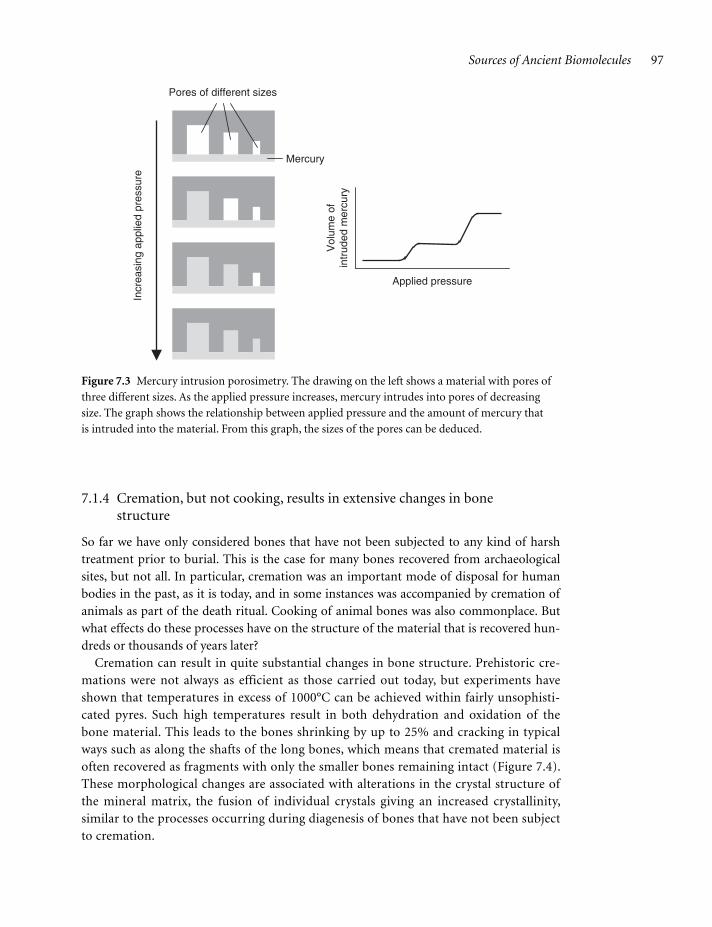
How is bone porosity measured? The technique called mercury intrusion porosimetry ena-bles the total volume of pores of different sizes to be measured in materials ranging from con-crete to fine pharmaceutical powders and has also been applied with success to archaeological bones. The principle of the method is the inability of a non-wetting liquid such as mercury to penetrate into the pores of a material unless it is forced in by physical pressure. A porosimeter comprises a bath of mercury, into which the test material is placed, linked to a device that gradu-ally increases the hydraulic pressure acting on the mercury. As the pressure increases the mer-cury is squeezed into ever smaller pores, resulting in a gradual decrease in its volume. By measuring the volume change at different pressures the volume of pores in different size ranges can be estimated (Figure 7.3). Mercury intrusion porosimetry can therefore provide a measure-ment of the overall porosity of a bone as well as individual estimates of the relative contributions made by pores of the different diameters associated with organic, microbial, and mineral decay.
c07.indd 96c07.indd 96 12/6/2010 6:44:23 AM12/6/2010 6:44:23 AM
Sources of Ancient Biomolecules 97
7.1.4 Cremation, but not cooking, results in extensive changes in bone structure
So far we have only considered bones that have not been subjected to any kind of harsh treatment prior to burial. This is the case for many bones recovered from archaeological sites, but not all. In particular, cremation was an important mode of disposal for human bodies in the past, as it is today, and in some instances was accompanied by cremation of animals as part of the death ritual. Cooking of animal bones was also commonplace. But what effects do these processes have on the structure of the material that is recovered hun-dreds or thousands of years later?
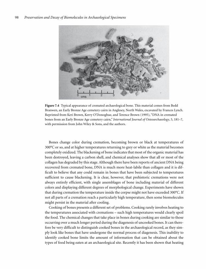
Cremation can result in quite substantial changes in bone structure. Prehistoric cre-mations were not always as efficient as those carried out today, but experiments have shown that temperatures in excess of 1000°C can be achieved within fairly unsophisti-cated pyres. Such high temperatures result in both dehydration and oxidation of the bone material. This leads to the bones shrinking by up to 25% and cracking in typical ways such as along the shafts of the long bones, which means that cremated material is often recovered as fragments with only the smaller bones remaining intact (Figure 7.4). These morphological changes are associated with alterations in the crystal structure of the mineral matrix, the fusion of individual crystals giving an increased crystallinity, similar to the processes occurring during diagenesis of bones that have not been subject to cremation.
Applied pressure
Vol
ume
ofin
trud
ed m
ercu
ry
Pores of different sizes
Mercury
Incr
easi
ng a
pplie
d pr
essu
re
Figure 7.3 Mercury intrusion porosimetry. The drawing on the left shows a material with pores of
three different sizes. As the applied pressure increases, mercury intrudes into pores of decreasing
size. The graph shows the relationship between applied pressure and the amount of mercury that
is intruded into the material. From this graph, the sizes of the pores can be deduced.
c07.indd 97c07.indd 97 12/6/2010 6:44:23 AM12/6/2010 6:44:23 AM
98 Preservation and Decay of Biomolecules in Archaeological Specimens
Bones change color during cremation, becoming brown or black at temperatures of 300°C or so, and at higher temperatures returning to grey or white as the material becomes completely oxidized. The blackening of bone indicates that most of the organic material has been destroyed, leaving a carbon shell, and chemical analyses show that all or most of the collagen has degraded by this stage. Although there have been reports of ancient DNA being recovered from cremated bone, DNA is much more heat-labile than collagen and it is dif-ficult to believe that any could remain in bones that have been subjected to temperatures sufficient to cause blackening. It is clear, however, that prehistoric cremations were not always entirely efficient, with single assemblages of bone including material of different colors and displaying different degrees of morphological change. Experiments have shown that during cremation the temperature inside the corpse might not have exceeded 300°C. If not all parts of a cremation reach a particularly high temperature, then some biomolecules might persist in the material after cooling.
Cooking of bones presents a different set of problems. Cooking rarely involves heating to the temperatures associated with cremations – such high temperatures would clearly spoil the food. The chemical changes that take place in bones during cooking are similar to those occurring over a much longer period during the diagenesis of uncooked bones. It can there-fore be very difficult to distinguish cooked bones in the archaeological record, as they sim-ply look like bones that have undergone the normal process of diagenesis. This inability to identify cooked bone limits the amount of information that can be obtained about the types of food being eaten at an archaeological site. Recently it has been shown that heating
Figure 7.4 Typical appearance of cremated archaeological bone. This material comes from Bedd
Branwen, an Early Bronze Age cemetery cairn in Anglesey, North Wales, excavated by Frances Lynch.
Reprinted from Keri Brown, Kerry O’Donoghue, and Terence Brown (1995), “DNA in cremated
bones from an Early Bronze Age cemetery cairn,” International Journal of Osteoarchaeology, 5, 181–7,
with permission from John Wiley & Sons, and the authors.
0 1cms 2 3
c07.indd 98c07.indd 98 12/6/2010 6:44:23 AM12/6/2010 6:44:23 AM

Sources of Ancient Biomolecules 99
bone to temperatures used in cooking results in gradual demineralization of collagen fibrils, which take on an unpacked appearance that is observable by electron microscopy. Demineralization and unpacking of collagen fibrils also occurs during diagenesis, but it is suggested that at a single site, where all the buried bones are subject to similar environmen-tal conditions and hence equivalent diagenetic regimes, any bones that have been cooked can be distinguished, as these display a greater degree of collagen unpacking.
7.1.5 Bone may continue to deteriorate after excavation
Finally, we must consider the changes that might occur within an archaeological bone after excavation. A few decades ago, archaeologists and museum curators tended to place greater importance on cultural remains – pottery, metalwork, and suchlike – rather than skeletons, and bones collected prior to the 1960s were not always conserved as well as they might have been. Biomolecular deterioration in material excavated in the past has been demonstrated in convincing fashion by comparisons between the ancient DNA contents of two parts of a 3200-year-old aurochs bone, one part excavated in 1947 and the other in 2004. When exam-ined in 2005, no DNA could be detected in the 1947 segment, whereas the 2004 fragment yielded positive results with a range of PCRs. It was estimated that DNA degradation within the 1947 fragment had increased seventyfold after its excavation.
In recent years, the curation and conservation of archaeological bones has become a highly professional discipline, but the effectiveness of the strategies that have been adopted has been measured largely by their ability to preserve the overall morphological appearance of a bone, rather than by their effects on the biomolecular content. It is common practice to wash bones immediately after excavation, to remove dirt, which can potentially lead to some biomolecules and ions being lost if these are not bound within the bone matrix in an insoluble form. There is also a possibility that washing increases the chances that a bone becomes contaminated with modern DNA. Fortunately, more and more excavators are becoming aware of the requirements of biomolecular analysis and are setting aside some bones for this purpose prior to washing. Additionally, many biomolecular archaeolo-gists now direct their research specifically at freshly excavated material, so that potential problems associated with curation are circumvented.
Although not a major problem for archaeological material, bones that have adhering soft tissue or grease are usually treated in some way to clean them and hence prevent subsequent attack by microorganisms and other pests. The procedures that are used are necessarily harsh and can include boiling the bones for short periods, treating with enzymes designed to break down the protein content of adhering flesh, or soaking in organic solvents for periods of days to remove grease. The combination of these procedures would be expected to have a detrimental, one might say disastrous, consequence for the biomolecular content of a bone. If a bone is fragile then a consolidant might be used to stabilize it. Most consolidants are polymeric compounds that soak into the bone and then harden, improv-ing its structural integrity. A wide range of chemicals are used, including acrylic, polyvinyl, and cellulose resins, none of which would be expected to have any damaging effect on the biomolecules within a bone. Consolidants can, however, hinder recovery of those biomol-ecules by making it less easy to break the bone up and to make a soluble extract.
c07.indd 99c07.indd 99 12/6/2010 6:44:25 AM12/6/2010 6:44:25 AM

100 Preservation and Decay of Biomolecules in Archaeological Specimens
Although conservation practices can have a detrimental effect on the biomolecular con-tent of a bone, these problems pale into insignificance compared with the post-excavation procedure that is most damaging to biomolecular, and indeed all archaeological, research. This is reburial. Few archaeologists dispute the imperative for reburial of skeletal material that can be clearly identified as ancestral to a group of living people. Less easy to accept is the reburial of British and European skeletons due to nebulous concepts of “ownership” promoted by groups whose cultural identities lie in New Ageism or, at best, 19th century romanticism.
7.1.6 Teeth are more stable than bones
Teeth are the second major hard tissue present in vertebrates and commonly recovered at archaeological sites. Strictly speaking, teeth are not part of the skeleton, but form a separate component of the body called the dentition.
Like bones, teeth are made up primarily of collagen and bioapatite, but structurally they are quite different to bones. Each tooth comprises a central pulp cavity surrounded by dentine which in turn is coated with enamel and cementum. The enamel covers that part of the tooth that is exposed within the mouth, and the cementum covers the part that is attached to jaw. The pulp cavity contains living cells, blood vessels, and nerves and connects directly with the soft tissue of the jaw via a canal present at the tip of each root. Being soft tissue, the pulp usually degrades soon after death and is not present in archae-ological remains. The other three components have significant collagen and mineral contents and are much more resistant to deterioration. Dentine is made up of collagen fibrils mineralized in bioapatite and arranged as tubular structures, between 1.0 and 2.5 μm in diameter, radiating out from the center of the tooth. The organic collagen component makes up about 20% of living dentine, with the mineral part contributing some 70%, and the rest water. Cementum has a slightly higher organic content, approxi-mately 35%, again mainly collagen, with 45% bioapatite and 20% water. Enamel is virtu-ally entirely made up of bioapatite, with less than 5% organic content. The latter does not include collagen, but instead is made up of special enamel proteins such as amelogenin, which we will meet again in Section 10.3, as its gene is used in a DNA test for identifying the sex of a skeleton.
Compared with bone, there have been relatively few studies of the diagenesis of teeth. Most of the detailed studies that have been carried out have focussed on uptake and exchange of ions between the mineral fraction of teeth and the burial environment, the isotopic signature of preserved teeth being important not only in archaeological research but in studies of past climates. This type of work has shown that there is minimal exchange between tooth and environmental strontium during diagenesis, lending support to the use of dental strontium in studies of human and animal mobility. It has generally been assumed that the enamel and cementum layers are so solid that microorganisms are unable to penetrate into a tooth, although microbial invasion does occur through the openings at the tips of the roots. Despite the relative lack of work on tooth diagenesis it is clear that teeth degrade more slowly than bone, although burial conditions that result in complete destruc-tion of bone, such as acidic environments, will destroy teeth also.
c07.indd 100c07.indd 100 12/6/2010 6:44:25 AM12/6/2010 6:44:25 AM
Sources of Ancient Biomolecules 101
As well as their use in isotope studies, teeth are attractive sources of ancient DNA, partly because of their relatively good preservation but also because their outer surfaces can be rigorously cleaned to remove contaminating modern DNA, which is less easy to do with bone because the surface is porous. Samples for DNA analysis can be taken from the interior of a tooth by drilling through the root canal which, if carried out carefully, results in minimal damage to the overall morphology of the tooth. The use of laser ablation (Section 6.3) simi-larly enables samples for isotopic analysis to be taken without damaging the tooth.
These non-destructive methods are important because morphological examination of teeth can provide information relevant to studies of kinship and diet. Small inherited struc-tural variations can be used to infer if groups of skeletons might be related (Section 11.1), and microwear patterns can indicate the types of food that were eaten (Section 12.1). This information would be lost if teeth were damaged when used in biomolecular research.
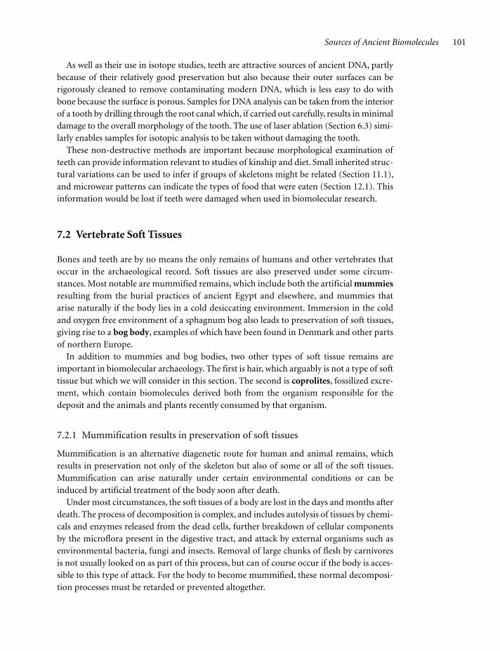
7.2 Vertebrate Soft Tissues
Bones and teeth are by no means the only remains of humans and other vertebrates that occur in the archaeological record. Soft tissues are also preserved under some circum-stances. Most notable are mummified remains, which include both the artificial mummies resulting from the burial practices of ancient Egypt and elsewhere, and mummies that arise naturally if the body lies in a cold desiccating environment. Immersion in the cold and oxygen free environment of a sphagnum bog also leads to preservation of soft tissues, giving rise to a bog body, examples of which have been found in Denmark and other parts of northern Europe.
In addition to mummies and bog bodies, two other types of soft tissue remains are important in biomolecular archaeology. The first is hair, which arguably is not a type of soft tissue but which we will consider in this section. The second is coprolites, fossilized excre-ment, which contain biomolecules derived both from the organism responsible for the deposit and the animals and plants recently consumed by that organism.
7.2.1 Mummification results in preservation of soft tissues
Mummification is an alternative diagenetic route for human and animal remains, which results in preservation not only of the skeleton but also of some or all of the soft tissues. Mummification can arise naturally under certain environmental conditions or can be induced by artificial treatment of the body soon after death.
Under most circumstances, the soft tissues of a body are lost in the days and months after death. The process of decomposition is complex, and includes autolysis of tissues by chemi-cals and enzymes released from the dead cells, further breakdown of cellular components by the microflora present in the digestive tract, and attack by external organisms such as environmental bacteria, fungi and insects. Removal of large chunks of flesh by carnivores is not usually looked on as part of this process, but can of course occur if the body is acces-sible to this type of attack. For the body to become mummified, these normal decomposi-tion processes must be retarded or prevented altogether.
c07.indd 101c07.indd 101 12/6/2010 6:44:25 AM12/6/2010 6:44:25 AM

102 Preservation and Decay of Biomolecules in Archaeological Specimens
In the natural environment, mummification is usually associated with dry and/or cold conditions. If the body dries out then autolysis is prevented and the action of microbial and insect decomposers is greatly retarded. The best known of these natural mummies are the “frozen” mammoths of Siberia and the Tyrolean iceman. The mammoths are thought to have become frozen shortly after death, possibly after drowning by falling through the ice on the surface of a lake. Subsequently their bodies became encased in permafrost until ero-sion eventually exposed their remains on the tundra surface. Almost 50 of these, between 20,000–60,000 years in age, are known to science; many others have probably been discov-ered in the past but only their ivory collected.
The iceman, sometimes called Ötzi as he was found in the Ötstal region of the Alps on the border between Austria and Italy, is one of the most famous of all archaeological remains. He dates to about 3300 bc and was preserved along with many items of clothing and other arti-facts, which have revealed a great deal about life in Copper Age Europe. Originally it was thought that Ötzi died from exposure on the high mountains, even though his clothing should have been sufficient to protect him from the cold. More recently, the discovery of an arrow head in his left shoulder has suggested a more sinister end. His body became mummi-fied in the cold conditions on the mountain, probably under a thin layer of snow that pro-tected him from animal predation, and possibly aided by a dry, desiccating wind during the days after his death. His body then became trapped in a glacier until its discovery in 1991. He is one of the most complete and best preserved of all natural mummies that have been found, anatomical changes being restricted to shrinkage and displacement of some of his organs, and the stomach contents sufficiently well preserved to enable identification of the food that he ate prior to his death. A greater amount of decomposition is evident at the microscopic level, the cellular structure of individual tissues being indistinct with no cell nuclei visible, though with well-preserved collagen fibrils and fat deposits.
The iceman is the most famous, oldest, and arguably best preserved of the natural human mummies that have been discovered (Table 7.2). Equally evocative, however, is the Siberian ice maiden, a female frozen mummy from the 5th century bc, found in one of a number of tombs belonging to the Pazyryk culture. Her tomb probably became flooded soon after her burial, the water freezing and remaining as permafrost until excavation in 1993. The ice maiden was heavily tatooed and buried with rich trappings, suggesting that she held high status in her community. Other, much younger frozen mummies, from 500 to 600 years ago, have been discovered in Greenland and British Columbia in North America. Mummies of Inca children, again dating to 500–600 years ago have been found on mountaintops in the Andes, thought to be sacrificial victims.
7.2.2 Artificial mummification was not restricted to ancient Egypt
The distinction between natural and artificial mummification becomes blurred at the edges. Mummification in ancient Egypt began with simple burial in the sand, probably as a convenient means of disposing of the dead rather than with any aim of preserving the body. Natural mummification is promoted by the dry, desiccating conditions in the hot desert and it is possible that the ancient Egyptians “discovered” mummification by recog-nizing that bodies buried in this way do not decompose. During the 4th millennium bc,
c07.indd 102c07.indd 102 12/6/2010 6:44:25 AM12/6/2010 6:44:25 AM
Sources of Ancient Biomolecules 103
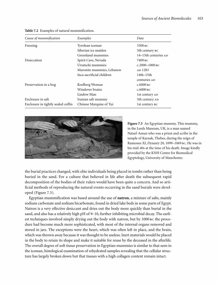
the burial practices changed, with elite individuals being placed in tombs rather than being buried in the sand. For a culture that believed in life after death the subsequent rapid decomposition of the bodies of their rulers would have been quite a concern. And so arti-ficial methods of reproducing the natural events occurring in the sand burials were devel-oped (Figure 7.5).
Egyptian mummification was based around the use of natron, a mixture of salts, mainly sodium carbonate and sodium bicarbonate, found in dried lake beds in some parts of Egypt. Natron is a very effective desiccant and dries out the body more quickly than burial in the sand, and also has a relatively high pH of 9–10, further inhibiting microbial decay. The earli-est techniques involved simply drying out the body with natron, but by 1000 bc the proce-dure had become much more sophisticated, with most of the internal organs removed and stored in jars. The exceptions were the heart, which was often left in place, and the brain, which was thrown away because it was thought to be useless. Inert materials would be placed in the body to retain its shape and make it suitable for reuse by the deceased in the afterlife. The overall degree of soft tissue preservation in Egyptian mummies is similar to that seen in the iceman, histological examination of rehydrated samples revealing that the cellular struc-ture has largely broken down but that tissues with a high collagen content remain intact.
Figure 7.5 An Egyptian mummy. This mummy,
in the Leeds Museum, UK, is a man named
Natsef-Amun who was a priest and scribe in the
temple of Karnak, Thebes, during the reign of
Ramesses XI, Dynasty 20, 1099–1069 bc. He was in
his mid-40s at the time of his death. Image kindly
provided by the KNH Centre for Biomedical
Egyptology, University of Manchester.
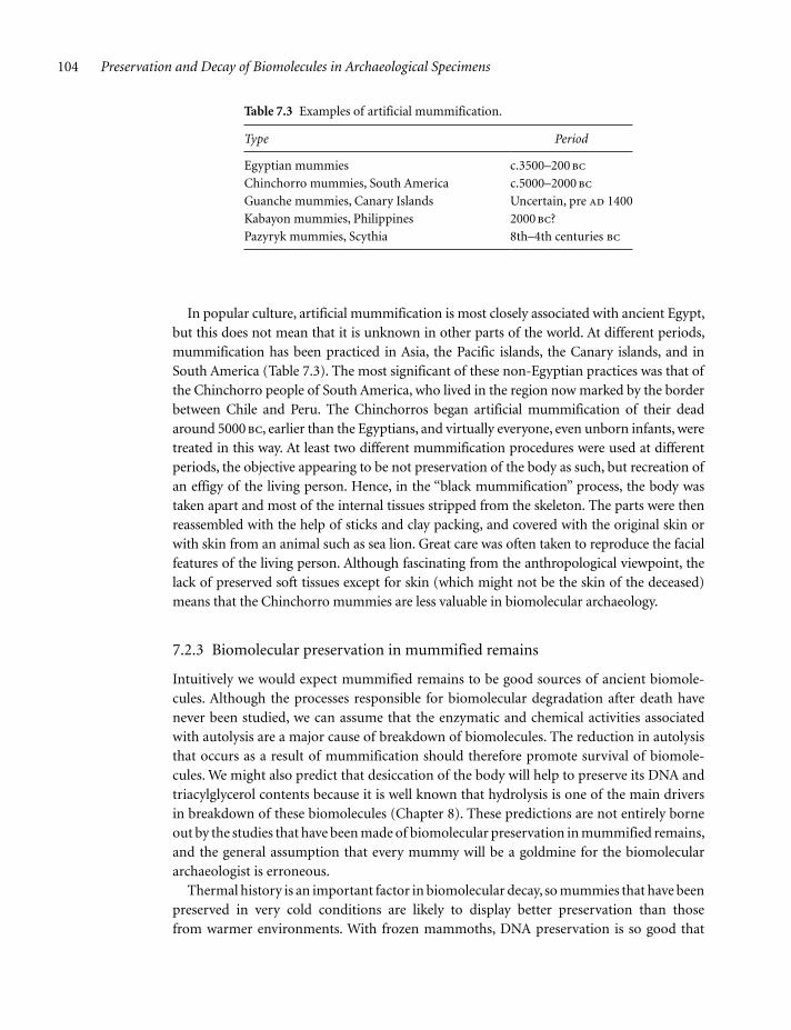
Table 7.2 Examples of natural mummification.
Cause of mummification Examples Date
Freezing Tyrolean iceman
Siberian ice maiden
3300 bc
5th century bc
Greenland mummies 14–15th centuries ad
Desiccation Spirit Cave, Nevada
Urumchi mummies
Maronite mummies, Lebanon
7400 bc
c.2000–1000 bc
ad 1283
Inca sacrificial children 14th–15th
centuries ad
Preservation in a bog Koelberg Woman
Windover brains
Lindow Man
c.6000 bc
c.6000 bc
1st century ad
Enclosure in salt Iranian salt mummy 5th century ad
Enclosure in tightly sealed coffin Chinese Marquise of Tui 1st century bc
c07.indd 103c07.indd 103 12/6/2010 6:44:25 AM12/6/2010 6:44:25 AM
104 Preservation and Decay of Biomolecules in Archaeological Specimens
In popular culture, artificial mummification is most closely associated with ancient Egypt, but this does not mean that it is unknown in other parts of the world. At different periods, mummification has been practiced in Asia, the Pacific islands, the Canary islands, and in South America (Table 7.3). The most significant of these non-Egyptian practices was that of the Chinchorro people of South America, who lived in the region now marked by the border between Chile and Peru. The Chinchorros began artificial mummification of their dead around 5000 bc, earlier than the Egyptians, and virtually everyone, even unborn infants, were treated in this way. At least two different mummification procedures were used at different periods, the objective appearing to be not preservation of the body as such, but recreation of an effigy of the living person. Hence, in the “black mummification” process, the body was taken apart and most of the internal tissues stripped from the skeleton. The parts were then reassembled with the help of sticks and clay packing, and covered with the original skin or with skin from an animal such as sea lion. Great care was often taken to reproduce the facial features of the living person. Although fascinating from the anthropological viewpoint, the lack of preserved soft tissues except for skin (which might not be the skin of the deceased) means that the Chinchorro mummies are less valuable in biomolecular archaeology.
7.2.3 Biomolecular preservation in mummified remains
Intuitively we would expect mummified remains to be good sources of ancient biomole-cules. Although the processes responsible for biomolecular degradation after death have never been studied, we can assume that the enzymatic and chemical activities associated with autolysis are a major cause of breakdown of biomolecules. The reduction in autolysis that occurs as a result of mummification should therefore promote survival of biomole-cules. We might also predict that desiccation of the body will help to preserve its DNA and triacylglycerol contents because it is well known that hydrolysis is one of the main drivers in breakdown of these biomolecules (Chapter 8). These predictions are not entirely borne out by the studies that have been made of biomolecular preservation in mummified remains, and the general assumption that every mummy will be a goldmine for the biomolecular archaeologist is erroneous.
Thermal history is an important factor in biomolecular decay, so mummies that have been preserved in very cold conditions are likely to display better preservation than those from warmer environments. With frozen mammoths, DNA preservation is so good that
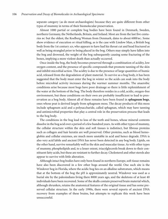
Table 7.3 Examples of artificial mummification.
Type Period
Egyptian mummies c.3500–200 bc
Chinchorro mummies, South America c.5000–2000 bc
Guanche mummies, Canary Islands Uncertain, pre ad 1400
Kabayon mummies, Philippines 2000 bc?
Pazyryk mummies, Scythia 8th–4th centuries bc
c07.indd 104c07.indd 104 12/6/2010 6:44:25 AM12/6/2010 6:44:25 AM

Sources of Ancient Biomolecules 105
substantial parts of the nuclear genome have been sequenced, but the DNA for this work was extracted not from the preserved tissues but from the bones within the specimens and from hairs. The mitochondrial genome of the iceman, on the other hand, has been obtained from soft tissue extracts. Quantitative PCR of these extracts suggested that the DNA con-tent of the iceman’s tissues was similar to that of the mammoth bones. As the iceman is substantially younger than the mammoths, the implication from this observation might be that DNA preservation in mummified soft tissue is no better than that in the bones of the same specimen. The iceman’s skin has also been examined, showing that although its gross structure is well preserved, proteins other than collagen have broken down, and the tria-cylglycerols and phospholipids have been hydrolyzed to fatty acids. We will look in more detail at the breakdown pathways for different types of biomolecule in Chapter 8: here it is sufficient to note that the degree of preservation in mummified tissues is not as great as we might have assumed.
With Egyptian mummies, there have been numerous reports of the successful extraction of ancient DNA and detection of proteins via immunological and other methods, but very few rigorous studies aimed at understanding the general biomolecular content. The simi-larities between the histological appearance of tissue from Egyptian mummies and that of the iceman suggest that the protein and lipid contents might be degraded to an equivalent extent. As well as its own biomolecules, most Egyptian mummies also contain compounds of biological origin from the embalming fluids and other preparations applied during the mummification process. Identification of the resins, oils, waxes, and spices used in these preparations is an important part of biomolecular archaeology in its own right, but their presence as contaminants in extracts also has the potential to interfere with studies directed at a mummy’s endogenous biomolecules.
Additional practical problems have to be taken into account when mummies are studied. Mummified remains include many of the most valuable bioarchaeological treasures, and destructive sampling, as is needed for most types of biomolecular study, has to be carefully justified. In the early days of scientific Egyptology, researchers had to make do with pieces that had literally dropped off the mummy they wished to study. Scientific study is nowadays valued more highly, but still the samples taken for biomolecular research are often not the ones that would be chosen if there were no other considerations. It is also clear that some mummies have deteriorated since their collection. Natural mummification is not an irre-versible process, and exposure of the body to moisture or heat will cause partial rehydration and the possibility of gradual decomposition. For some mummies, notably the Siberian ice maiden, substantial deterioration is thought to have occurred since excavation, and even for the most carefully curated specimens such as the iceman concerns have been raised about the onset of decay. This is less of a problem for Egyptian mummies as the embalming process continues to preserve the specimen even today, but some have still suffered as a result of deficiencies in the curation practices adopted during the earlier decades of the last century.
7.2.4 Bog bodies are special types of mummy
The strict definition of a mummy is a specimen in which some of the soft tissues are pre-served. This means that bog bodies are a type of mummy, although we will treat them as a
c07.indd 105c07.indd 105 12/6/2010 6:44:25 AM12/6/2010 6:44:25 AM
106 Preservation and Decay of Biomolecules in Archaeological Specimens
separate category (as do most archaeologists) because they are quite different from other types of mummy in terms of their biomolecular preservation.
Almost 1000 partial or complete bog bodies have been found in Denmark, Sweden, northern Germany, the Netherlands, Britain, and Ireland. Most are from the last five centu-ries bc but the oldest, the Koelberg Woman from Denmark, dates to about 6000 bc. Some show evidence of execution or ritual killing, as is the case with Lindow Man, a British bog body from the 1st century ad, who appears to have had his throat cut and head fractured as well as being strangled prior to being placed in the bog. Others may simply have fallen into the bog and drowned, the weight of the bog subsequently causing some fracturing of the bones, implying a more violent death than actually occurred.
Once inside the bog, the body becomes preserved through a combination of acidity, low oxygen content, and the presence of specific compounds that promote tanning of the skin and inhibit microbial action. The acidity is due to the presence of large quantities of humic acid, released from the degradation of plant material. To survive as a bog body, it has been suggested that the body must enter the bog in winter so the acids can soak into the body before microbial activity increases during the warmer summer months. The anaerobic conditions arise because most bogs have poor drainage so there is little replenishment of the water at the bottom of the bog. The body therefore resides in a cold, acidic, oxygen-free environment, but these conditions on their own are probably insufficient to lead to pres-ervation as a bog body. Almost all of these remains have been found in sphagnum bogs, ones whose peat is derived largely from sphagnum moss. The decay products of this moss include sphagnum acid and a polysaccharide, called sphagnan, which may have tanning and antimicrobial properties that play a central role in the preservation process that leads to the bog body.
The conditions in the bog lead to loss of the teeth and bones, whose mineral contents dissolve in the bog acid over a period of a few hundred years. As with other types of mummy, the cellular structure within the skin and soft tissues is indistinct, but fibrous proteins such as collagen and hair keratin are well preserved. Other proteins, such as blood hemo-globin and cellular enzymes, are much more unstable in acid and hence degrade. DNA is also very acid labile and ancient DNA has never been detected in any bog body. Lipids, on the other hand, survive remarkably well in the skin and muscular tissue. As with other types of mummy, phospholipids and, to a lesser extent, triacylglycerols break down to their con-stituent fatty acids, but these are resistant to further decay. Cholesterol and other sterols also appear to survive with little alteration.
Although intact bog bodies have only been found in northern Europe, soft tissue remains have also been discovered in a few other bogs around the world. One such site is the Windover bog in Florida, where the acidic bog lies on an alkaline substratum, which means that at the bottom of the bog the pH is approximately neutral. Windover was used as a burial site by the paleoindians living there 8000 years ago, and the skeletons of at least 40 individuals have been recovered. Some of the skulls contain preserved brain material which, although shrunken, retains the anatomical features of the original tissue and has some pre-served cellular structure. In the early 1990s, there were several reports of ancient DNA recovery from examples of these brains, but attempts to replicate this work have been unsuccessful.
c07.indd 106c07.indd 106 12/6/2010 6:44:25 AM12/6/2010 6:44:25 AM
Sources of Ancient Biomolecules 107
7.2.5 Hair is important in DNA and stable isotope studies
Hair is a characteristic feature of all mammals and is one of the parts of the body that is most resistant to decay. Attached hair is frequently found on natural mummies, and also on artificial ones, if it was not removed when the body was prepared for burial. Hair is also sometimes found attached to the skull of a body that is otherwise completely skeletonized. It is quite possible that detached human hair is present at many archaeological sites, not only associated with burials, but is simply overlooked during excavation. In those cases where hair has been searched for, for example in cave sites, it has often been found.
Hair has a more complex structure than is often appreciated. Most people are familiar with the lengthwise division into the shaft or hair fiber, which protrudes from the skin and is the visible component, and the root or follicle which is embedded in the dermis. At the base of the root are a small number of living cells, comprising the dermal papilla, whose division results in elongation of the fiber and growth of the hair. The new cells that bud off from the dermal papilla form the structure of the hair shaft. These cells become filled with the fibrous protein keratin, and with pigments that give the hair its natural color. In cross-section, the hair fiber is made up of three concentric layers, referred to as the medulla (in the middle of the hair), the cortex, and the outer cuticle. The cortex contains densely packed keratinized cells and usually makes up the greatest part of the bulk of the hair. The medulla also contains keratinized cells, but these are less densely packed and possibly separated by air spaces. This part of the shaft helps provide the hair with its insulating properties. The cuticle is just a single layer of cells which controls the water content of the hair and gives it most of its mechanical strength. The hairs of different mammals all have the same structure, the major differences between species being the relative thicknesses of the hairs on different parts of the body. Humans have a similar density of hair follicles on all parts of their body as other mammals, but most of the follicles produce very fine downy hair. On the other hand, the hair on our heads and on masculine faces is thicker than the hair on the heads and faces of other mammals.
The textile and cosmetics industries have carried out extensive research into the changes that occur during processing and aging of animal hair and as a result of cosmetic treatment of human hair. It is not clear to what extent this work is relevant to the changes that hair under-goes in an archaeological setting, and very few studies have addressed this particular issue. Hair from mummies retains much of its cross-sectional structure, though usually the outlines of individual cells are no longer visible. In more extreme cases, the keratin fibers become unpacked and the melanin pigment granules aggregate into larger structures. Although looked on as relatively resistant to decay, hair can decompose over a period of months under some conditions, probably when the burial environment contains fungi or bacteria able to degrade keratin. Such microorganisms are not ubiquitous in the biosphere but a number of different species are known and these have a wide geographical distribution.
As the hair fiber has a cellular structure, it is no surprise that living hair contains DNA. Both nuclear and mitochondrial DNA can be obtained from the root of the hair, and both are also easily recoverable from the shaft. The presence of DNA in shed and cut hair, even after a few years of storage, has been one of the pillars on which modern forensic biology has been built since the development of genetic profiling. It has also been known since the 1980s that hair from archaeological specimens often contains DNA, but hair has only
c07.indd 107c07.indd 107 12/6/2010 6:44:25 AM12/6/2010 6:44:25 AM

108 Preservation and Decay of Biomolecules in Archaeological Specimens
infrequently been exploited in this way because usually other parts of the body, such as soft tissues, bones and teeth, are also available from the same specimen. The alternative sources are usually present in larger amounts than the hair, and can be sampled with less visi-ble damage to the specimen. It has been suggested that the ancient DNA in hairs of frozen mammoths has undergone less chemical degradation than that in bones, and so gives more accurate nucle-otide sequences, but this report has been disputed and remains unproven.
The second application of hair in biomolecular archaeology is as a source of stable isotopes. The δ13C and δ15N ratios in hair protein are influenced by diet in the same way as the ratios in bones and teeth, which means that hair can be used along with the hard tissues in dietary studies. Hair has the additional advantage that it provides a time course for the period immediately before the death of the individual, or the time when the hair was shed. The isotope ratios in segments closest to the root reflect the very recent diet, and those further along the shaft relate to earlier periods when those parts of the hair were being formed. Seasonal variations and sudden changes in diet can therefore be discerned by segmental analysis along a hair (Figure 7.6).
7.2.6 Biomolecular archaeologists are becoming increasing interested in coprolites
Coprolites are the final type of animal remain that we will consider. The archaeological study of coprolites has an interesting history. Originally discarded as being no value, inter-est in coprolites began in the 1960s due almost entirely to the efforts of a single person, Eric Callen, who sadly died in the field in 1970. Today it is recognized that coprolites provide a record of diet and disease, and specialists in their study are located throughout the world.
Coprolites are frequently preserved in dry climates, especially in caves where deposits may cover large areas and reach significant depths. Features such as size, shape, color after treatment with certain chemicals and, reputedly, smell after rehydration enable human coprolites to be distinguished from those of animals. Although there are controversies over the use of coprolites to indicate the extent of meat in the diet, plant remains such as seeds, pollen, and phytoliths can be identified by visual and microscopic examination and used in dietary reconstruction. There have been several reports of endoparasite remains, giving indications of the health of the human or animal responsible for the deposit.
All types of biomolecule appear to be preserved in coprolites. Steroids have been detected in specimens up to 2500 years old, with sufficiently good chemical preservation for sex-specific hormones such as testosterone to be typed, enabling the sex of the originator to be identified. A range of lipid biomarkers for dietary plants have been identified in much older ground sloth coprolites, thought to be 11,000 years in age. Immunological tests have detected proteins with claims that some of these derive from animals that were eaten, but
Distance from hair root (cm)
δ13C
(‰
) –12
–16
–20
3 6 9 12 15
Figure 7.6 Graph showing a sudden change
in δ13C values along a hair shaft, suggesting
that this person’s diet shifted to one rich
in a C4 plant such as maize about one year
before the hair was shed. This dietary
change resulted in an increased δ13C value
for the region of the hair closest to the root.
This result is similar to that obtained when
the hair of an Inca child mummy from an
Andean mountaintop was examined,
suggesting that this child’s diet underwent
a dramatic improvement about one year
before her sacrifice.
c07.indd 108c07.indd 108 12/6/2010 6:44:25 AM12/6/2010 6:44:25 AM
Sources of Ancient Biomolecules 109
these results suffer from questions regarding specificity, as is the case with all uses of immu-nological detection in analysis of ancient proteins. The greatest success has been in DNA analysis, which has been used both in reconstruction of the plant and animal components of the diet and to identify the mitochondrial DNA haplotype of the originator. Although not strictly archaeology, one interesting variation has been to type the plant DNA in animal coprolites from different layers of a single cave, with the resulting data used to reconstruct changes in the local vegetation over a period of time. The reason why such well-preserved biomolecules survive in coprolites of quite substantial age has not yet been asked and little is known about the diagenetic processes acting within this type of material.
7.3 Plant Remains
Plant remains are important in archaeology as indicators of past subsistence patterns. At pre-Neolithic sites they provide information on the wild resources that were utilized, and at agri-cultural sites they can tell us exactly what types of crop were grown at particular times. Intact plants are only rarely recovered, largely because the non-nutritious parts of the plant were discarded or not collected at all and so had little opportunity of being preserved. Seeds are common, especially for cereals such as wheat, barley, and rice because for these the seeds (the grain) are the part that was processed and eaten. If these cereal seeds come from stores that have been abandoned, then they may be mixed in with fragments of chaff, but individual seeds that were lost during processing are also frequently found. Maize seeds are usually still attached to their cob, and sometimes the cobs on their own are recovered, having being discarded dur-ing food processing. Fruit seeds that have passed through the digestive system unharmed can be recovered from latrines and similar settings, and intact nuts are also found at some sites.
In this section, we will focus on the macrofossil remains of plants used as food, most of which are preserved in the archaeological record as desiccated, charred, or waterlogged material. We will not cover the microfossils of these plants, such as starch grains, phytoliths, and pollen, as we dealt with the relevant information concerning these in Section 5.2. Neither will we look at plant residues in cooking and storage vessels, or products such as tars, oils, and resins, not because these are unimportant in biomolecular archaeology, but because the correct place to study them is in later chapters on prehistoric diet (Chapter 12) and technology (Chapter 14).
7.3.1 Desiccated remains are most suitable for biomolecular study
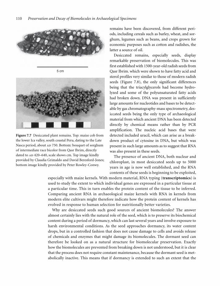
In some parts of the world, plant remains are found as desiccated specimens that have undergone little visible change even though they may be several thousand years old (Figure 7.7). Maize cobs, with or without attached kernels, are often found in this form from cold dry sites at high altitude in the Andes, but perhaps the most spectacular preserva-tion is that found in the Old World, at Qasr Ibrim, on the upper Nile near the boundaries of modern Egypt and Sudan. Qasr Ibrim, which lies in an essentially rainless environment, was occupied from the 1st millennium bc to the 18th century ad, and has been excavated since the early 1960s by the Egyptian Exploration Society. A wide range of desiccated plant
c07.indd 109c07.indd 109 12/6/2010 6:44:25 AM12/6/2010 6:44:25 AM
110 Preservation and Decay of Biomolecules in Archaeological Specimens
remains have been discovered, from different peri-ods, including cereals such as barley, wheat, and sor-ghum, legumes such as beans, and crops grown for economic purposes such as cotton and radishes, the latter a source of oil.
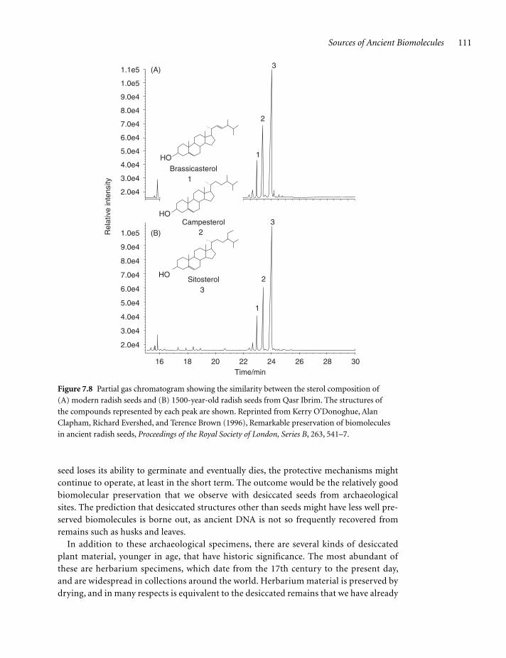
Desiccated remains, especially seeds, display remarkable preservation of biomolecules. This was first established with 1500-year-old radish seeds from Qasr Ibrim. which were shown to have fatty acid and sterol profiles very similar to those of modern radish seeds (Figure 7.8), the only significant differences being that the triacylglycerols had become hydro-lyzed and some of the polyunsaturated fatty acids had broken down. DNA was present in sufficiently large amounts for nucleotides and bases to be detect-able by gas chromatography-mass spectrometry, des-iccated seeds being the only type of archaeological material from which ancient DNA has been detected directly by chemical means rather than by PCR amplification. The nucleic acid bases that were detected included uracil, which can arise as a break-down product of cytosine in DNA, but which was present in such large amounts as to suggest that RNA was also present in these seeds.
The presence of ancient DNA, both nuclear and chloroplast, in most desiccated seeds up to 5000 years in age is now well established, and the RNA contents of these seeds is beginning to be exploited,
especially with maize kernels. With modern material, RNA typing (transcriptomics) is used to study the extent to which individual genes are expressed in a particular tissue at a particular time. This in turn enables the protein content of the tissue to be inferred. Comparing ancient RNA in archaeological maize kernels with RNA in kernels from modern elite cultivars might therefore indicate how the protein content of kernels has evolved in response to human selection for nutritionally better varieties.
Why are desiccated seeds such good sources of ancient biomolecules? The answer almost certainly lies with the natural role of the seed, which is to preserve its biochemical content during a period of dormancy, which can last several years and involve exposure to harsh environmental conditions. As the seed approaches dormancy, its water content drops, but in a controlled fashion that does not cause damage to cells and avoids release of chemicals and enzymes that might damage its biomolecules. The dormant seed can therefore be looked on as a natural structure for biomolecular preservation. Exactly how the biomolecules are prevented from breaking down is not understood, but it is clear that the process does not require constant maintenance, because the dormant seed is met-abolically inactive. This means that if dormancy is extended to such an extent that the
Figure 7.7 Desiccated plant remains. Top: maize cob from
the lower Ica valley, south coastal Peru, dating to the Late
Nasca period, about ad 750. Bottom: bouquet of sorghum
of intermediate race bicolor from Qasr Ibrim, directly
dated to ad 420–640, scale shows cm. Top image kindly
provided by Claudia Grimaldo and David Beresford-Jones;
bottom image kindly provided by Peter Rowley-Conwy.
5 cm
c07.indd 110c07.indd 110 12/6/2010 6:44:26 AM12/6/2010 6:44:26 AM
Sources of Ancient Biomolecules 111
seed loses its ability to germinate and eventually dies, the protective mechanisms might continue to operate, at least in the short term. The outcome would be the relatively good biomolecular preservation that we observe with desiccated seeds from archaeological sites. The prediction that desiccated structures other than seeds might have less well pre-served biomolecules is borne out, as ancient DNA is not so frequently recovered from remains such as husks and leaves.
In addition to these archaeological specimens, there are several kinds of desiccated plant material, younger in age, that have historic significance. The most abundant of these are herbarium specimens, which date from the 17th century to the present day, and are widespread in collections around the world. Herbarium material is preserved by drying, and in many respects is equivalent to the desiccated remains that we have already
HO
HO
HO
16 18 20
2.0e4
3.0e4
4.0e4
5.0e4
6.0e4
7.0e4
8.0e4
9.0e4
1.0e5Rel
ativ
e in
tens
ity
2.0e4
3.0e4
4.0e4
5.0e4
6.0e4
7.0e4
8.0e4
9.0e4
1.0e5
1.1e5
22Time/min
24 26 28 30
1
1
2
23
3
3
1
2
Brassicasterol
Campesterol
Sitosterol
(A)
(B)
Figure 7.8 Partial gas chromatogram showing the similarity between the sterol composition of
(A) modern radish seeds and (B) 1500-year-old radish seeds from Qasr Ibrim. The structures of
the compounds represented by each peak are shown. Reprinted from Kerry O’Donoghue, Alan
Clapham, Richard Evershed, and Terence Brown (1996), Remarkable preservation of biomolecules
in ancient radish seeds, Proceedings of the Royal Society of London, Series B, 263, 541–7.
c07.indd 111c07.indd 111 12/6/2010 6:44:26 AM12/6/2010 6:44:26 AM

112 Preservation and Decay of Biomolecules in Archaeological Specimens
considered. Nuclear and chloroplast DNA has been isolated from wheat grains from 19th century herbarium collections, and it seems likely that DNA is widespread in this type of material. Whether biomolecules other than DNA are present is not known. DNA has also been detected in seeds from corn dollies, decorative structures made of wheat, barley and other dried plants, some of which are several hundred years old (Figure 7.9). The cereal plants used to make some of the older corn dollies might come from varieties that are now extinct, hence the interest in studying them, but the dollies themselves are such valuable objects that destruction of even small pieces needs stringent justification. The roofs and walls of some old buildings also contain cereal plants used as thatch, insulation, and to add strength to mud bricks and daub. So far this type of material has not yielded ancient DNA, possibly because it has been exposed to the vicissitudes of climate and, although dried when first used, has subsequently gone through cycles of partial rehydration which might result in DNA degradation.
7.3.2 Some charred and waterlogged plant remains contain ancient DNA
Desiccated plant remains are only found in those parts of the world where the environmen-tal conditions are conducive for this type of preservation. The vast majority of archaeologi-cal plant specimens, including virtually all from Europe, have been preserved by burning, resulting in oxidized remains that archaeologists have traditionally referred to as carbon-ized, but which are more correctly called charred as it is clear that not all of the organic material has been completely converted into carbon. Charred seeds are typically black with a shiny “clinkered” surface, though some can be dark brown (Figure 7.10). Charred remains are frequently recovered as individual seeds, which probably became burnt by falling into the fire while being cooked. Occasionally much larger quantities are found in granaries or other seed stores that caught fire and were abandoned by their owners.
Ancient DNA was first detected in charred wheat seeds in the early 1990s. Initially the results appeared dubious as the total organic content of this type of material is very low and
Figure 7.9 A corn dolly
from the Museum of English
Rural Life, Reading. Image
courtesy of Diane Lister.
c07.indd 112c07.indd 112 12/6/2010 6:44:26 AM12/6/2010 6:44:26 AM

Sources of Ancient Biomolecules 113
other biomolecules are clearly not present, but similar detections have since been made with charred cereal grains of various kinds and with charred maize kernels and cobs. DNA from charred material typically has undergone a great deal of damage but short nucleotide sequences have been obtained, sufficient to identify short tandem repeat alleles in some instances. Many charred remains contain no detectable DNA at all, and even in a single assemblage there are often many seeds that contain no DNA and only a few that do.
Artificial charring experiments have been carried out to try to rationalize the presence of DNA in material that has been heated to temperatures sufficient to cause charring. This work has shown that if wheat seeds are heated above 225°C then all the DNA breaks down during the first 4 hours, but at lower temperatures the rate of DNA decay gradually slows to such an extent that some is still present after 5 hours. The rate of decay is slower when the amount of oxygen is limited, which might explain the presence of DNA in some grains recovered from burnt stores, such as at Assiros in Greece and Danebury, England. In these stores, grains were kept in sealed pots or clay lined pits, in which a small amount of germi-nation would take place, reducing the oxygen level to a point where further germination was inhibited and the seeds remained dormant over winter. Destruction of these stores by fire would therefore result in seed charring in the low oxygen conditions shown to reduce DNA degradation. Simulations in which grain stores have deliberately been burnt down have shown that although parts of the structure reach very high temperatures, in excess of 600°C, at a depth of 0.6 m immediately below the conflagration the temperature does not get above 200°C. The presence of DNA in charred grains from storage pits can therefore perhaps be understood, even if its survival in material from other sources remains unexplained.
Seeds and other plant remains can also be recovered from waterlogged environments, such as the sediment at the bottom of a lake, or simply from part of a site that is below the water table. Although many biomolecules, including DNA, are susceptible to hydrolysis, the anaerobic conditions associated with waterlogging might be expected to retard their degradation. In some cases, seeds obtained from waterlogged locations had previously been charred, examples being cereal grains from Swiss lake villages, where houses were built on stilts above the surface of the edge of a lake, and burnt grains were likely to fall
Figure 7.10 Charred grains
of spelt wheat from Assiros,
Greece, approximately 3000
years old. Image kindly
provided by Glynis Jones.
c07.indd 113c07.indd 113 12/6/2010 6:44:27 AM12/6/2010 6:44:27 AM
114 Preservation and Decay of Biomolecules in Archaeological Specimens
Further Reading
from the cooking area into the water. Ancient DNA has been found in several sets of such grains. Plant remains that have been preserved solely by waterlogging have not been stud-ied so extensively, but protein profiling of extracts of grape seeds from a waterlogged medieval site in York, England, suggests that partially degraded proteins may be present in some of these.
Brown, K.A., O’Donoghue, K. & Brown, T.A. (1995) DNA in
cremated bones from an Early Bronze Age cemetery cairn.
International Journal of Osteoarchaeology, 5, 181–7.
Bryant, V.M. & Dean, G.W. (2006) Archaeological coprolite
science: the legacy of Eric O. Callen (1912–70).
Palaeogeography, Palaeoclimatology, Palaeoecology, 237,
51–66.
David, A.R. & Tapp, E. (eds.) (1992) The Mummy’s Tale:
The Scientific and Medical Investigation of Natsef-Amun,
Priest in the Temple of Karnak. Michael O’Mara Books,
London.
Evershed, R.P. (1990) Lipids from samples of skin from
seven Dutch bog bodies: preliminary report.
Archaeometry, 32, 139–53.
Hedges, R.E.M., Millard, A.R. & Pike, A.W.G. (1995)
Measurements and relationships of diagenetic alteration
of bone from three archaeological sites. Journal of
Archaeological Science, 22, 201–9. [Describes the Oxford
Histological Index.]
Koon, H.E.C., O’Connor, T.P. & Collins, M.J. (2010) Sorting
the butchered from the boiled. Journal of Archaeological
Science, 37, 62–9. [Distinguishing between cooked and
burnt bone.]
Lister, D.L., Bower, M.A., Howe, C.J. & Jones, M.K. (2008)
Extraction and amplification of nuclear DNA from her-
barium specimens of emmer wheat: a method for assess-
ing DNA preservation by maximum amplicon length
recovery. Taxon, 57, 254–8.
Lynnerup, N. (2007) Mummies. Yearbook of Physical
Anthropology, 50, 162–90.
Nielsen-Marsh, C.M. & Hedges, R.E.M. (2007) Bone poros-
ity and the use of mercury intrusion porosimetry in
bone diagenesis studies. Archaeometry, 41, 165–74.
O’Donoghue, K., Clapham, A., Evershed, R.P. & Brown,
T.A. (1996) Remarkable preservation of biomolecules in
ancient radish seeds. Proceedings of the Royal Society of
London series B, 263, 541–7. [Preservation in desiccated
seeds from Qasr Ibrim.]
Pruvost, M., Schwarz, R. & Correia, V.B., et al. (2007)
Freshly excavated fossil bones are best for amplification
of ancient DNA. Proceedings of the National Academy of
Sciences USA, 104, 739–44.
Reiche, I., Vignaud, C. & Menu, M. (2002) The crystallinity
of ancient bone and dentine: new insights by transmis-
sion electron microscopy. Archaeometry, 44, 447–59.
Smith, C.I., Nielsen-Marsh, C.M., Jans, M.M.E. & Collins,
M.J. (2007) Bone diagenesis in the European Holocene I:
patterns and mechanisms. Journal of Archaeological
Science, 34, 1485–93.
Threadgold, J. & Brown, T.A. (2003) Degradation of DNA
in artificially charred wheat seeds. Journal of
Archaeological Science, 30, 1067–76.
Turner-Walker, G. (2008) The chemical and microbial deg-
radation of bones and teeth. In Advances in Human
Palaeopathology (ed. Pinhasi, R. & Mays, S.), 3–29. John
Wiley and Sons, Chichester.
Wilson, A.S., Taylor, T. & Ceruti, M.C., et al. (2007) Stable
isotope and DNA evidence for the ritual sequences in
Inca child sacrifice. Proceedings of the National
Acadamy of Sciences USA, 104, 16456–61. [Segmental
analysis of hair reveals a change in diet one year before
sacrifice.]
Wilson, J., Hardy, K. & Allen, R., et al. (2010) Automated
classification of starch granules using supervised pattern
recognition of morphological properties. Journal of
Archaeological Science, 37, 594–604.
c07.indd 114c07.indd 114 12/6/2010 6:44:27 AM12/6/2010 6:44:27 AM