biomems and biomedical nanotechnology filebiomems and biomedical nanotechnology mauro ferrari,...
TRANSCRIPT

BioMEMS and BiomedicalNanotechnology
Volume IIMicro/Nano Technology for Genomicsand Proteomics
BioMEMS and BiomedicalNanotechnologyMauro Ferrari, Ph.D., Editor-in-ChiefProfessor, Brown Institute of Molecular Medicine ChairmanDepartment of Biomedical EngineeringUniversity of Texas Health Science Center, Houston, TX
Professor of Experimental TherapeuticsUniversity of Texas M.D. Anderson Cancer Center, Houston, TX
Professor of BioengineeringRice University, Houston, TX
Professor of Biochemistry and Molecular BiologyUniversity of Texas Medical Branch, Galveston, TX
President, the Texas Alliance for NanoHealthHouston, TX
Volume IIMicro/Nano Technology for Genomics and Proteomics
Edited by
Mihrimah OzkanDept. of Electrical EngineeringUniversity of California, RiversideRiverside, California USA
Michael J. HellerDept. of BioengineeringUniversity of California, San DiegoLa Jolla, California USA

Mihrimah OzkanUniversity of California, RiversideRiverside, California
Michael HellerUniversity of California, RiversideRiverside, California
Mauro FerrariOhio State UniversityColumbus, Ohio
Library of Congress Cataloging-in-Publication Data
Volume IIISBN-10: 0-387-25564-8 e-ISBN 10: 0-387-25843-4 Printed on acid-free paper.ISBN-13: 978-0387-25564-4 e-ISBN-13: 978-0387-25843-0SetISBN-10: 0-387-25661-3 e-ISBN:10: 0-387-25749-7ISBN-13: 978-0387-25561-3 e-ISBN:13: 978-0387-25749-5
C© 2006 Springer Science+Business Media, LLCAll rights reserved. This work may not be translated or copied in whole or in part without the written permission ofthe publisher (Springer Science+Business Media LLC, 233 Spring Street, New York, NY 10013, USA), except forbrief excerpts in connection with reviews or scholarly analysis. Use in connection with any form of informationstorage and retrieval, electronic adaptation, computer software, or by similar or dissimilar methodology nowknown or hereafter developed is forbidden.The use in this publication of trade names, trademarks, service marks and similar terms, even if they are notidentified as such, is not to be taken as an expression of opinion as to whether or not they are subject toproprietary rights.
9 8 7 6 5 4 3 2 1 SPIN 11407157
springer.com

Dedicated to Richard Smalley (1943–2005), in Memoriam
To Rick,
father founder of nanotechnologyprime inspiration for its applications to medicinegracious mentor to its researchersour light—forever in the trenches with us
(Rick Smalley received the 1996 Chemistry Nobel Prizefor the co-discovery of carbon-60 buckeyballs)
Contents
List of Contributors . . . . . . . . . . . . . . . . . . . . . . . . . . . . . . . . . . . . . . . . . . . . . . . . . . . . . . . . . xvii
Foreword . . . . . . . . . . . . . . . . . . . . . . . . . . . . . . . . . . . . . . . . . . . . . . . . . . . . . . . . . . . . . . . . . . . xxi
Preface . . . . . . . . . . . . . . . . . . . . . . . . . . . . . . . . . . . . . . . . . . . . . . . . . . . . . . . . . . . . . . . . . . . . . xxiii
I. Application of Microarray Technologies . . . . . . . . . . . . . . . . . . . . . . . . . . . . . . . . . . 1
1. Electronic Microarray Technology and Applications in Genomicsand Proteomics . . . . . . . . . . . . . . . . . . . . . . . . . . . . . . . . . . . . . . . . . . . . . . . . . . . . . . . . . 3Ying Huang, Dalibor Hodko, Daniel Smolko, and Graham Lidgard1.1 Introduction . . . . . . . . . . . . . . . . . . . . . . . . . . . . . . . . . . . . . . . . . . . . . . . . . . . . . . . . . 31.2 Overview of Electronic Microarray Technology . . . . . . . . . . . . . . . . . . . . . . . . . 4
1.2.1 NanoChip r© Array and NanoChip r© Workstation . . . . . . . . . . . . . . . . . . 51.2.2 Capabilities of the NanoChip r© Electronic Microarrays . . . . . . . . . . . . . 7
1.3 Applications . . . . . . . . . . . . . . . . . . . . . . . . . . . . . . . . . . . . . . . . . . . . . . . . . . . . . . . . . 101.3.1 Single Nucleotide Polymorphisms (SNPs)—Based Diagnostics . . . . . 101.3.2 Forensic Detection . . . . . . . . . . . . . . . . . . . . . . . . . . . . . . . . . . . . . . . . . . . . . 101.3.3 Gene Expression Profiling . . . . . . . . . . . . . . . . . . . . . . . . . . . . . . . . . . . . . . 121.3.4 Cell Separation . . . . . . . . . . . . . . . . . . . . . . . . . . . . . . . . . . . . . . . . . . . . . . . . 121.3.5 Electronic Immunoassays . . . . . . . . . . . . . . . . . . . . . . . . . . . . . . . . . . . . . . . 141.3.6 Miniaturization of Electronic Microarray Technology
and Applications . . . . . . . . . . . . . . . . . . . . . . . . . . . . . . . . . . . . . . . . . . . . . . . 151.3.7 Applications in Proteomics . . . . . . . . . . . . . . . . . . . . . . . . . . . . . . . . . . . . . . 18
1.4 Summary and Outlook . . . . . . . . . . . . . . . . . . . . . . . . . . . . . . . . . . . . . . . . . . . . . . . . 19References................................................................................. 19
2. Gene Expression Profiling Utilizing Microarray Technologyand RT-PCR . . . . . . . . . . . . . . . . . . . . . . . . . . . . . . . . . . . . . . . . . . . . . . . . . . . . . . . . . . . 23Dominick Sinicropi, Maureen Cronin, and Mei-Lan Liu2.1 Introduction . . . . . . . . . . . . . . . . . . . . . . . . . . . . . . . . . . . . . . . . . . . . . . . . . . . . . . . . . 232.2 Real-Time PCR . . . . . . . . . . . . . . . . . . . . . . . . . . . . . . . . . . . . . . . . . . . . . . . . . . . . . . 25
2.2.1 Detection Systems.............................................................. 252.2.2 Real-Time RT-PCR Data Analysis .......................................... 312.2.3 Qualification of Gene Panels Using Real-Time RT-PCR................ 322.2.4 Real-Time RT-PCR Summary................................................ 34

viii CONTENTS
2.3 Microarrays . . . . . . . . . . . . . . . . . . . . . . . . . . . . . . . . . . . . . . . . . . . . . . . . . . . . . . . . . . 352.3.1 Technology Platforms ......................................................... 352.3.2 Target Amplification and Labeling .......................................... 372.3.3 Applications ..................................................................... 40
2.4 Comparison of Gene Expression Profiling Methods . . . . . . . . . . . . . . . . . . . . . 412.4.1 Comparison of cDNA Arrays with Other Gene Expression
Profiling Methods .............................................................. 412.4.2 Comparison of Oligonucleotide Arrays with Other Gene
Expression Profiling Methods................................................ 422.4.3 Comparison of cDNA and Oligonucleotide Microarray
Expression Profiles ............................................................. 442.5 Summary . . . . . . . . . . . . . . . . . . . . . . . . . . . . . . . . . . . . . . . . . . . . . . . . . . . . . . . . . . . 44
Acknowledgements .................................................................... 45References ............................................................................... 45
3. Microarray and Fluidic Chip for Extracellular Sensing . . . . . . . . . . . . . . . . . . . 47Mihrimah Ozkan, Cengiz S. Ozkan, Shalini Prasad, Mo Yang, and Xuan Zhang3.1 Introduction . . . . . . . . . . . . . . . . . . . . . . . . . . . . . . . . . . . . . . . . . . . . . . . . . . . . . . . . 473.2 Antibody Based Biosensors . . . . . . . . . . . . . . . . . . . . . . . . . . . . . . . . . . . . . . . . . . 503.3 Nucleic Acid Based Biosensors . . . . . . . . . . . . . . . . . . . . . . . . . . . . . . . . . . . . . . . 513.4 Ion Channel Biosensors . . . . . . . . . . . . . . . . . . . . . . . . . . . . . . . . . . . . . . . . . . . . . . 513.5 Enzyme Based Biosensors . . . . . . . . . . . . . . . . . . . . . . . . . . . . . . . . . . . . . . . . . . . 513.6 Cell Based Biosensors . . . . . . . . . . . . . . . . . . . . . . . . . . . . . . . . . . . . . . . . . . . . . . . 523.7 Cellular Microorganism Based Sensors . . . . . . . . . . . . . . . . . . . . . . . . . . . . . . . . 523.8 Fluorescence Based Cell Biosensors . . . . . . . . . . . . . . . . . . . . . . . . . . . . . . . . . . . 533.9 Cellular Metabolism Based Biosensors . . . . . . . . . . . . . . . . . . . . . . . . . . . . . . . . 55
3.10 Impedance Based Cellular Sensors . . . . . . . . . . . . . . . . . . . . . . . . . . . . . . . . . . . . 563.11 Intracellular Potential Based Biosensors . . . . . . . . . . . . . . . . . . . . . . . . . . . . . . . 573.12 Extracllular Potential Based Biosensors . . . . . . . . . . . . . . . . . . . . . . . . . . . . . . . 583.13 Cell Patterning Techniques . . . . . . . . . . . . . . . . . . . . . . . . . . . . . . . . . . . . . . . . . . . 603.14 Dielectrophoresis for Cell Patterning . . . . . . . . . . . . . . . . . . . . . . . . . . . . . . . . . . 613.15 Basis of Dielectrophoresis . . . . . . . . . . . . . . . . . . . . . . . . . . . . . . . . . . . . . . . . . . . . 623.16 Microelectrodes and Dielectrophoresis . . . . . . . . . . . . . . . . . . . . . . . . . . . . . . . . 633.17 Dielectric Properties of Cells . . . . . . . . . . . . . . . . . . . . . . . . . . . . . . . . . . . . . . . . . 643.18 Effect of Electric Fields on Cells . . . . . . . . . . . . . . . . . . . . . . . . . . . . . . . . . . . . . . 643.19 Cell Types and the Parameters for Dielectrophoretic Patterning . . . . . . . . . . 653.20 Biosensing System . . . . . . . . . . . . . . . . . . . . . . . . . . . . . . . . . . . . . . . . . . . . . . . . . . 663.21 Chip Assembly . . . . . . . . . . . . . . . . . . . . . . . . . . . . . . . . . . . . . . . . . . . . . . . . . . . . . 663.22 Environmental Chamber . . . . . . . . . . . . . . . . . . . . . . . . . . . . . . . . . . . . . . . . . . . . . 673.23 Experimental Measurement System . . . . . . . . . . . . . . . . . . . . . . . . . . . . . . . . . . . 673.24 Cell Culture . . . . . . . . . . . . . . . . . . . . . . . . . . . . . . . . . . . . . . . . . . . . . . . . . . . . . . . . 67
3.24.1 Neuron Culture ................................................................ 673.24.2 Primary Osteoblast Culture ................................................. 68
3.25 Signal Processing . . . . . . . . . . . . . . . . . . . . . . . . . . . . . . . . . . . . . . . . . . . . . . . . . . . 683.26 Selection of Chemical Agents . . . . . . . . . . . . . . . . . . . . . . . . . . . . . . . . . . . . . . . . 69
CONTENTS ix
3.26.1 Ethanol.......................................................................... 693.26.2 Hydrogen Peroxide ........................................................... 693.26.3 Pyrethroid ...................................................................... 703.26.4 Ethylene Diamene Tetra Acetic Acid (EDTA)........................... 70
3.27 Chemical Agent Sensing . . . . . . . . . . . . . . . . . . . . . . . . . . . . . . . . . . . . . . . . . . . . . 703.27.1 Signature Pattern for Control Experiments............................... 70
3.28 Electrical Sensing Cycle . . . . . . . . . . . . . . . . . . . . . . . . . . . . . . . . . . . . . . . . . . . . . 703.29 Ethanol Sensing . . . . . . . . . . . . . . . . . . . . . . . . . . . . . . . . . . . . . . . . . . . . . . . . . . . . . 71
3.29.1 Single Neuron Sensing....................................................... 713.29.2 Single Osteoblast Sensing................................................... 71
3.30 Hydrogen Peroxide Sensing . . . . . . . . . . . . . . . . . . . . . . . . . . . . . . . . . . . . . . . . . . 723.30.1 Single Neuron Sensing....................................................... 723.30.2 Single Osteoblast Sensing................................................... 73
3.31 Pyrethroid Sensing . . . . . . . . . . . . . . . . . . . . . . . . . . . . . . . . . . . . . . . . . . . . . . . . . . 743.31.1 Single Neuron Sensing....................................................... 743.31.2 Single Osteoblast Sensing................................................... 75
3.32 EDTA Sensing . . . . . . . . . . . . . . . . . . . . . . . . . . . . . . . . . . . . . . . . . . . . . . . . . . . . . . 763.32.1 Single Neuron Sensing....................................................... 763.32.2 Single Osteoblast Sensing................................................... 76
3.33 Immunohistochemistry . . . . . . . . . . . . . . . . . . . . . . . . . . . . . . . . . . . . . . . . . . . . . . 773.34 Visualization of Physiological Changes Due to the Effect
of the Chemical Analytes . . . . . . . . . . . . . . . . . . . . . . . . . . . . . . . . . . . . . . . . . . . . 803.34.1 Effect of Ethanol on Neurons............................................... 803.34.2 Effect of Ethanol on Osteoblasts ........................................... 803.34.3 Effect of Hydrogen Peroxide on Neurons ................................ 833.34.4 Effect of Hydrogen Peroxide on Osteoblasts ............................ 843.34.5 Effect of Pyrethroid on Neurons ........................................... 863.34.6 Effect of Pyrethroid on Osteoblasts........................................ 883.34.7 Effect of EDTA on Neurons................................................. 893.34.8 Effect of EDTA on Osteoblasts............................................. 91
3.35 Discussion and Conclusions . . . . . . . . . . . . . . . . . . . . . . . . . . . . . . . . . . . . . . . . . . 93References ............................................................................... 98
4. Cell Physiometry Tools based on Dielectrophoresis . . . . . . . . . . . . . . . . . . . . . . . 103Ronald Pethig4.1 Introduction . . . . . . . . . . . . . . . . . . . . . . . . . . . . . . . . . . . . . . . . . . . . . . . . . . . . . . . . . 1034.2 Dielectrophoresis . . . . . . . . . . . . . . . . . . . . . . . . . . . . . . . . . . . . . . . . . . . . . . . . . . . . . 1044.3 Dielectric Polarizability of Bioparticles . . . . . . . . . . . . . . . . . . . . . . . . . . . . . . . . . 1074.4 Dynamics of Interfacial Polarization. . . . . . . . . . . . . . . . . . . . . . . . . . . . . . . . . . . . 1074.5 Surface Charge Effects . . . . . . . . . . . . . . . . . . . . . . . . . . . . . . . . . . . . . . . . . . . . . . . . 1134.6 Other Physiometric Effects . . . . . . . . . . . . . . . . . . . . . . . . . . . . . . . . . . . . . . . . . . . . 1164.7 Traveling Wave Dielectrophoresis . . . . . . . . . . . . . . . . . . . . . . . . . . . . . . . . . . . . . . 1184.8 Controlling Possible DEP-Induced Damage to Cells . . . . . . . . . . . . . . . . . . . . . 120
Concluding Comments.................................................................. 123References................................................................................. 124

x CONTENTS
5. Hitting the Spot: The Promise of Protein Microarrays . . . . . . . . . . . . . . . . . . . . 127Joanna S. Albala5.1 Introduction . . . . . . . . . . . . . . . . . . . . . . . . . . . . . . . . . . . . . . . . . . . . . . . . . . . . . . . . . 1275.2 Generation of Protein Microarrays . . . . . . . . . . . . . . . . . . . . . . . . . . . . . . . . . . . . . 128
5.2.1 Content............................................................................. 1285.2.2 Surface Chemistry ............................................................... 1295.2.3 Microarray Production .......................................................... 1295.2.4 Detection........................................................................... 130
5.3 Protein Arrays for Analysis of Proteins Involved in Recombination& DNA Repair . . . . . . . . . . . . . . . . . . . . . . . . . . . . . . . . . . . . . . . . . . . . . . . . . . . . . . . 1305.3.1 Protein Expression Microarrays ............................................... 1305.3.2 Protein Interaction Arrays ...................................................... 132
5.4 Summary: Protein arrays-Hope or hype? . . . . . . . . . . . . . . . . . . . . . . . . . . . . . . . . 133Acknowledgements...................................................................... 133References................................................................................. 133
6. Use of Electric Field Array Devices for Assisted Assembly of DNANanocomponents and Other Nanofabrication Applications . . . . . . . . . . . . . . . 137Michael J. Heller, Cengiz S. Ozkan, and Mihrimah Ozkan6.1 Introduction . . . . . . . . . . . . . . . . . . . . . . . . . . . . . . . . . . . . . . . . . . . . . . . . . . . . . . . . . 1386.2 Active Microelectronic Array Hybridization Technology . . . . . . . . . . . . . . . . . 1416.3 Electric Field Assisted Nanofabrication Process . . . . . . . . . . . . . . . . . . . . . . . . . 1466.4 Integration of Optical Tweezers for Manupilation of Live Cells . . . . . . . . . . . 153
Conclusions ............................................................................... 156Abbreviations ............................................................................. 156Acknowledgements...................................................................... 157References................................................................................. 157
7. Peptide Arrays in Proteomics and Drug Discovery . . . . . . . . . . . . . . . . . . . . . . . 161Ulrich Reineke, Jens Schneider-Mergener, and Mike Schutkowski7.1 Introduction . . . . . . . . . . . . . . . . . . . . . . . . . . . . . . . . . . . . . . . . . . . . . . . . . . . . . . . . . 1617.2 Generation of Peptide Arrays . . . . . . . . . . . . . . . . . . . . . . . . . . . . . . . . . . . . . . . . . . 162
7.2.1 Coherent Surfaces and Surface Modification ............................... 1637.2.2 Generation of Micro-Structured Surfaces ................................... 1737.2.3 Peptide Array Preparation ...................................................... 1827.2.4 Techniques for Array Production with Pre-Synthesized Peptides....... 200
7.3 Library Types . . . . . . . . . . . . . . . . . . . . . . . . . . . . . . . . . . . . . . . . . . . . . . . . . . . . . . . . 2037.3.1 Protein Sequence-Derived Libraries.......................................... 2047.3.2 De Novo Approaches............................................................ 210
7.4 Assays for Peptide Arrays . . . . . . . . . . . . . . . . . . . . . . . . . . . . . . . . . . . . . . . . . . . . . 2147.4.1 Screening .......................................................................... 2157.4.2 Read-Out........................................................................... 219
7.5 Applications of Peptide Arrays . . . . . . . . . . . . . . . . . . . . . . . . . . . . . . . . . . . . . . . . 2217.5.1 Antibodies ......................................................................... 2227.5.2 Protein-Protein Interactions .................................................... 2247.5.3 Enzyme-Substrate and Enzyme-Inhibitor Interactions .................... 226
CONTENTS xi
7.5.4 Application of Peptide Arrays: Miscellaneous ............................. 2287.5.5 Peptidomimetics.................................................................. 231
7.6 Bibliography . . . . . . . . . . . . . . . . . . . . . . . . . . . . . . . . . . . . . . . . . . . . . . . . . . . . . . . . . 231References................................................................................. 265
8. From One-Bead One-Compound Combinatorial Librariesto Chemical Microarrays . . . . . . . . . . . . . . . . . . . . . . . . . . . . . . . . . . . . . . . . . . . . . . . 283Kit S. Lam, Ruiwu Liu, Jan Marik, and Pappanaicken R. Kumaresan8.1 Introduction . . . . . . . . . . . . . . . . . . . . . . . . . . . . . . . . . . . . . . . . . . . . . . . . . . . . . . . . . 2838.2 OBOC Peptide Libraries . . . . . . . . . . . . . . . . . . . . . . . . . . . . . . . . . . . . . . . . . . . . . . 2848.3 Encoded OBOC Small Molecule Combinatorial Libraries . . . . . . . . . . . . . . . . 2878.4 Peptide and Chemical Microarrays . . . . . . . . . . . . . . . . . . . . . . . . . . . . . . . . . . . . . 289
8.4.1 Immobilization Methods for Pre-Synthesized Libraries .................. 2898.4.2 In Situ Synthesis of Microarrays .............................................. 2928.4.3 CD, Microfluidics, Fiber Optic Microarray, Multiplex Beads ........... 295
8.5 Detection Methods in Chemical Microarrays . . . . . . . . . . . . . . . . . . . . . . . . . . . . 2968.5.1 Identification and Characterization of Bound Proteins.................... 2968.5.2 Detection Methods to Identify Post-Translational Modification
of Proteins or to Quantitate Enzyme Activity in Analytes................ 2978.6 Application of Chemical Microarray . . . . . . . . . . . . . . . . . . . . . . . . . . . . . . . . . . . 297
8.6.1 Protein Binding Studies......................................................... 2988.6.2 Post-Translational Modification, Enzyme-Substrate
and Inhibitor Studies ............................................................ 2998.6.3 Cell-Binding Studies ............................................................ 3008.6.4 Drug Discovery and Cell Signaling .......................................... 3008.6.5 Diagnostic Studies ............................................................... 3018.6.6 Non-Biological Applications .................................................. 301
8.7 Future Directions . . . . . . . . . . . . . . . . . . . . . . . . . . . . . . . . . . . . . . . . . . . . . . . . . . . . . 302Acknowledgements...................................................................... 303Abbreviations ............................................................................. 303References................................................................................. 304
II. Advanced Microfluidic Devices and Human Genome Project . . . . . . . . . . . . . . . . 309
9. Plastic Microfluidic Devices for DNA and Protein Analyses . . . . . . . . . . . . . . . 311Z. Hugh Fan and Antonio J. Ricco9.1 Introduction . . . . . . . . . . . . . . . . . . . . . . . . . . . . . . . . . . . . . . . . . . . . . . . . . . . . . . . . . 311
9.1.1 Detection........................................................................... 3119.1.2 Materials ........................................................................... 312
9.2 Electrokinetic Pumping . . . . . . . . . . . . . . . . . . . . . . . . . . . . . . . . . . . . . . . . . . . . . . . 3129.3 Plastic Devices . . . . . . . . . . . . . . . . . . . . . . . . . . . . . . . . . . . . . . . . . . . . . . . . . . . . . . . 314
9.3.1 Pumping and Detection ......................................................... 3159.3.2 Device Fabrication ............................................................... 316
9.4 DNA Analyses . . . . . . . . . . . . . . . . . . . . . . . . . . . . . . . . . . . . . . . . . . . . . . . . . . . . . . . 3189.4.1 Integrating PCR and DNA Fragment Separations ......................... 318

xii CONTENTS
9.4.2 DNA Sequencing ............................................................... 3209.4.3 DNA Sample Purification ..................................................... 321
9.5 Protein Analyses . . . . . . . . . . . . . . . . . . . . . . . . . . . . . . . . . . . . . . . . . . . . . . . . . . . . 3229.5.1 Isoelectric Focusing for Studying Protein Interactions .................. 3239.5.2 Enzymatic Digestion for Protein Mapping................................. 324Concluding Remarks................................................................... 326Acknowledgements .................................................................... 326References ............................................................................... 326
10. Centrifuge Based Fluidic Platforms . . . . . . . . . . . . . . . . . . . . . . . . . . . . . . . . . . . . . . 329Jim V. Zoval and M.J. Madou10.1 Introduction . . . . . . . . . . . . . . . . . . . . . . . . . . . . . . . . . . . . . . . . . . . . . . . . . . . . . . . . 32910.2 Why Centrifuge as Fluid Propulsion Force? . . . . . . . . . . . . . . . . . . . . . . . . . . . 33010.3 Compact Disc or Micro-Centrifuge Fluidics . . . . . . . . . . . . . . . . . . . . . . . . . . . 333
10.3.1 How it Works .................................................................. 33310.4 Some Simple Fluidic Function Demonstrated on a CD . . . . . . . . . . . . . . . . . . 334
10.4.1 Mixing of Fluid ............................................................... 33410.4.2 Valving ......................................................................... 33510.4.3 Volume Definition (Metering) and Common
Distribution Channels ........................................................ 33810.4.4 Packed Columns .............................................................. 339
10.5 CD Applications . . . . . . . . . . . . . . . . . . . . . . . . . . . . . . . . . . . . . . . . . . . . . . . . . . . . 33910.5.1 Two-Point Calibration of an Optode-Based Detection System ...... 33910.5.2 CD Platform for Enzyme-Linked Immunosorbant
Assays (ELISA) ............................................................... 34010.5.3 Multiple Parallel Assays ..................................................... 34110.5.4 Cellular Based Assays on CD Platform................................... 34210.5.5 Automated Cell Lysis on a CD ............................................. 34410.5.6 Integrated Nucleic Acid Sample Preparation and
PCR Amplification ........................................................... 35610.5.7 Sample Preparation for MALDI MS Analysis .......................... 35810.5.8 Modified Commercial CD/DVD Drives in
Analytical Measurements ................................................... 359Conclusion............................................................................... 361Acknowledgements .................................................................... 362References ............................................................................... 362
11. Sequencing the Human Genome: A Historical PerspectiveOn Challenges For Systems Integration . . . . . . . . . . . . . . . . . . . . . . . . . . . . . . . . . . 365Lee Rowen11.1 Overview . . . . . . . . . . . . . . . . . . . . . . . . . . . . . . . . . . . . . . . . . . . . . . . . . . . . . . . . . . . 36511.2 Approaches Used to Sequence the Human Genome . . . . . . . . . . . . . . . . . . . . . 366
11.2.1 Overview........................................................................ 36611.2.2 Strategy Used for Sequencing Source Clones............................ 36811.2.3 Construction of the Chromosome Tiling Paths .......................... 37911.2.4 Data Sharing ................................................................... 379

CONTENTS xiii
11.3 Challenges for Systems Integration . . . . . . . . . . . . . . . . . . . . . . . . . . . . . . . . . . . . 38011.3.1 Methodological Challenges for Sequencing Source
Clones: 1990–1997 ........................................................... 38111.3.2 Challenges for Sequencing the Entire Human
Genome: 1998–2003.......................................................... 38611.4 Are there Lessons to be Learned from the Human Genome Project? . . . . . . 395
Acknowledgements .................................................................... 397References ............................................................................... 398
III. Nanoprobes for Imaging, Sensing and Therapy . . . . . . . . . . . . . . . . . . . . . . . . . . . . 401
12. Hairpin Nanoprobes for Gene Detection . . . . . . . . . . . . . . . . . . . . . . . . . . . . . . . . . 403Philip Santangelo, Nitin Nitin, Leslie LaConte, and Gang Bao12.1 Introduction . . . . . . . . . . . . . . . . . . . . . . . . . . . . . . . . . . . . . . . . . . . . . . . . . . . . . . . . 40312.2 Nanoprobe Design Issues for Homogeneous Assays . . . . . . . . . . . . . . . . . . . . 40512.3 In Vitro Gene Detection . . . . . . . . . . . . . . . . . . . . . . . . . . . . . . . . . . . . . . . . . . . . . . 408
12.3.1 Pathogen Detection ........................................................... 40912.3.2 Mutation Detection and Allele Discrimination .......................... 409
12.4 Intracellular RNA Targets . . . . . . . . . . . . . . . . . . . . . . . . . . . . . . . . . . . . . . . . . . . . 41112.4.1 Cytoplasmic and Nuclear RNA............................................. 41112.4.2 RNA Secondary Structure ................................................... 418
12.5 Living Cell RNA Detection . . . . . . . . . . . . . . . . . . . . . . . . . . . . . . . . . . . . . . . . . . 41812.5.1 Cellular Delivery of Probes.................................................. 41912.5.2 Intracellular Probe Stability ................................................. 42412.5.3 Intracellular mRNA Detection .............................................. 428
12.6 Opportunities and Challenges . . . . . . . . . . . . . . . . . . . . . . . . . . . . . . . . . . . . . . . . 431Acknowledgements .................................................................... 433References ............................................................................... 433
13. Fluorescent Lanthanide Labels with Time-Resolved FluorometryIn DNA Analysis . . . . . . . . . . . . . . . . . . . . . . . . . . . . . . . . . . . . . . . . . . . . . . . . . . . . . . . 437Takuya Nishioka, Jingli Yuan, and Kazuko Matsumoto13.1 Introduction . . . . . . . . . . . . . . . . . . . . . . . . . . . . . . . . . . . . . . . . . . . . . . . . . . . . . . . . 43713.2 Lanthanide Fluorescent Complexes and Labels . . . . . . . . . . . . . . . . . . . . . . . . . 43813.3 Time-Resolved Fluorometry of Lanthanide Complexes . . . . . . . . . . . . . . . . . 44113.4 DNA Hybridization Assay . . . . . . . . . . . . . . . . . . . . . . . . . . . . . . . . . . . . . . . . . . . 442
Conclusion............................................................................... 445References ............................................................................... 445
14. Role of SNPs and Haplotypes in Human Disease and Drug Development . . 447Barkur S. Shastry14.1 Introduction . . . . . . . . . . . . . . . . . . . . . . . . . . . . . . . . . . . . . . . . . . . . . . . . . . . . . . . . 44714.2 SNP Discovery. . . . . . . . . . . . . . . . . . . . . . . . . . . . . . . . . . . . . . . . . . . . . . . . . . . . . . 44814.3 Detection of Genetic Variation . . . . . . . . . . . . . . . . . . . . . . . . . . . . . . . . . . . . . . . . 44914.4 Disease Gene Mapping . . . . . . . . . . . . . . . . . . . . . . . . . . . . . . . . . . . . . . . . . . . . . . 450

xiv CONTENTS
14.5 Evolution . . . . . . . . . . . . . . . . . . . . . . . . . . . . . . . . . . . . . . . . . . . . . . . . . . . . . . . . . . . 45014.6 Haplotypes . . . . . . . . . . . . . . . . . . . . . . . . . . . . . . . . . . . . . . . . . . . . . . . . . . . . . . . . . 45214.7 Drug Development . . . . . . . . . . . . . . . . . . . . . . . . . . . . . . . . . . . . . . . . . . . . . . . . . . 452
Concluding Remarks................................................................... 454References ............................................................................... 454
15. Control of Biomolecular Activity by Nanoparticle Antennas . . . . . . . . . . . . . . 459Kimberly Hamad-Schifferli15.1 Background and Motivation . . . . . . . . . . . . . . . . . . . . . . . . . . . . . . . . . . . . . . . . . . 459
15.1.1 ATP Synthase as a Molecular Motor ...................................... 45915.1.2 Biological Self Assembly of Complex Hybrid Structures ............ 46115.1.3 DNA as a Medium for Computation ...................................... 46315.1.4 Light Powered Nanomechanical Devices ................................. 463
15.2 Nanoparticles as Antennas for Controlling Biomolecules . . . . . . . . . . . . . . . . 46515.2.1 Technical Approach........................................................... 46815.2.2 Dehybridization of a DNA Oligonucleotide Reversibly by
RFMF Heating of Nanoparticles ........................................... 46915.2.3 Determination of Effective Temperature by RFMF
Heating of Nanoparticles .................................................... 46915.2.4 Selective Dehybridization of DNA Oligos by RFMF
Heating of Nanoparticles .................................................... 471Conclusions and Future Work ....................................................... 473References ............................................................................... 474
16. Sequence Matters: The Influence of Basepair Sequenceon DNA-protein Interactions . . . . . . . . . . . . . . . . . . . . . . . . . . . . . . . . . . . . . . . . . . . . 477Yan Mei Wang, Shirley S. Chan, and Robert H. Austin16.1 Introduction . . . . . . . . . . . . . . . . . . . . . . . . . . . . . . . . . . . . . . . . . . . . . . . . . . . . . . . . 47716.2 Generalized Deformations of Objects . . . . . . . . . . . . . . . . . . . . . . . . . . . . . . . . . . 48116.3 Sequence Dependent Aspects to the Double Helix Elastic Constants . . . . . . 48416.4 Sequence Dependent Bending of the Double Helix and the
Structure Atlas of DNA . . . . . . . . . . . . . . . . . . . . . . . . . . . . . . . . . . . . . . . . . . . . . . 48516.5 Some Experimental Consequences of Sequence Dependent Elasticity . . . . . 486
16.5.1 Phage 434 Binding Specificity and DNase I Cutting Rates ........... 48616.5.2 Nucleosome Formation: Sequence and Temperature Dependence... 491Conclusions.............................................................................. 494References ............................................................................... 494
17. Engineered Ribozymes: Efficient Tools for MolecularGene Therapy and Gene Discovery . . . . . . . . . . . . . . . . . . . . . . . . . . . . . . . . . . . . . . 497Maki Shiota, Makoto Miyagishi, and Kazunari Taira17.1 Introduction . . . . . . . . . . . . . . . . . . . . . . . . . . . . . . . . . . . . . . . . . . . . . . . . . . . . . . . . 49717.2 Methods for the Introduction of Ribozymes into Cells . . . . . . . . . . . . . . . . . . . 49817.3 Ribozyme Expression Systems . . . . . . . . . . . . . . . . . . . . . . . . . . . . . . . . . . . . . . . 499
17.3.1 The Pol III System ............................................................ 499

CONTENTS xv
17.3.2 Relationship Between the Higher-Order Structure ofRibozymes and their Activity .............................................. 500
17.3.3 Subcellular Localization and Efficacy of Ribozymes................... 50117.3.4 Mechanism of the Export of tRNA-Ribozymes from the
Nucleus to the Cytoplasm ................................................... 50417.4 RNA-Protein Hybrid Ribozymes . . . . . . . . . . . . . . . . . . . . . . . . . . . . . . . . . . . . . . 505
17.4.1 Accessibility to Ribozymes of their Target mRNAs .................... 50517.4.2 Hybrid Ribozymes that Efficiently Cleave their Target mRNAs,
Regardless of Secondary Structure ........................................ 50517.5 Maxizymes: Allosterically Controllable Ribozymes . . . . . . . . . . . . . . . . . . . . . 508
17.5.1 Shortened Hammerhead Ribozymes that Function as Dimers ........ 50817.5.2 Design of an Allosterically Controllable Maxizyme.................... 50917.5.3 Inactivation of an Oncogene in a Mouse Model ......................... 51217.5.4 Generality of the Maxizyme Technology ................................ 512
17.6 Identification of Genes Using Hybrid Ribozymes . . . . . . . . . . . . . . . . . . . . . . . 51317.7 Summary and Prospects . . . . . . . . . . . . . . . . . . . . . . . . . . . . . . . . . . . . . . . . . . . . . 515
References ............................................................................... 516
About the Editors . . . . . . . . . . . . . . . . . . . . . . . . . . . . . . . . . . . . . . . . . . . . . . . . . . . . . . . . . . . 519Index . . . . . . . . . . . . . . . . . . . . . . . . . . . . . . . . . . . . . . . . . . . . . . . . . . . . . . . . . . . . . . . . . . . . . . . 521
List of Contributors
VOLUME II
Joanna S. Albala, Biology & Biotechnology Research Program, Lawerence LivermoreNational Laboratory, Livermore, California USA
Robert H. Austin, Dept. of Physics, Princeton University, Princeton, New Jersey USA
Gang Bao, Dept. of Biomedical Engineering, Georgia Institute of Technology and EmoryUniversity, Atlanta, Georgia USA
Shirley S. Chan, Dept. of Physics, Princeton University, Princeton, New Jersey USA
Maureen Cronin, Genomic Health, Inc., Redwood City, California USA
Z. Hugh Fan, Dept. of Mechanical and Aerospace Engineering, University of Florida,Gainesville, Florida USA
Mauro Ferrari, Ph.D., Professor, Brown Institute of Molecular Medicine Chairman, De-partment of Biomedical Engineering, University of Texas Health Science Center, Houston,TX; Professor of Experimental Therapeutics, University of Texas M.D. Anderson CancerCenter, Houston, TX; Professor of Bioengineering, Rice University, Houston, TX; Professorof Biochemistry and Molecular Biology, University of Texas Medical Branch, Galveston,TX; President, the Texas Alliance for NanoHealth, Houston, TX
Kimberly Hamad-Schifferli, Dept. of Mechanical Engineering, Massachusetts Instituteof Technology, Cambridge, Massachusetts USA
Michael J. Heller, Dept. of Bioengineering, University of California, San Diego, La Jolla,California USA
Dalibor Hodko, Nanogen Inc., San Diego, California USA
Dr. Ying Huang, Nanogen Inc., San Diego, California USA
Pappanaicken R. Kumaresan, Division of Hematology/Oncology & Internal Medicine,University of California Davis, Sacramento, California USA

xviii LIST OF CONTRIBUTORS
Leslie LaConte, Dept. of Biomedical Engineering, Georgia Institute of Technology andEmory University, Atlanta, Georgia USA
Kit S. Lam, Division of Hematology/Oncology & Internal Medicine, University ofCalifornia Davis, Sacramento, California USA
Graham Lidgard, Nanogen Inc., San Diego, California USA
Mei-Lan Liu, Genomic Health, Inc., Redwood City, California USA
Ruiwu Liu, Division of Hematology/Oncology & Internal Medicine, University ofCalifornia Davis, Sacramento, California USA
M.J. Madou, Dept. of Mechanical and Aerospace Engineering, University of California,Irvine, Irvine, California USA
Jan Marik, Division of Hematology/Oncology & Internal Medicine, University ofCalifornia Davis, Sacramento, California USA
Kazuko Matsumoto, Dept. of Chemistry and Advanced Research Institute for Science &Engineering, Waseda University, Tokyo, Japan
Makoto Miyagishi, Dept. of Chemistry and Biotechnology, The University of Tokyo,Tokyo, Japan
Takuya Nishioka, Dept. of Chemistry and Advanced Research Institute for Science &Engineering, Waseda University, Tokyo, Japan
Nitin Nitin, Dept. of Biomedical Engineering, Georgia Institute of Technology and EmoryUniversity, Atlanta, Georgia USA
Cengiz S. Ozkan, Dept. of Mechanical Engineering, University of California, Riverside,Riverside, California USA
Prof. Mihrimah Ozkan, Dept. of Electrical Engineering, University of California,Riverside, Riverside, California USA
Prof. Ronald Pethig, School of Informatics, University of Wales, Bangor, Gwynedd,United Kingdom
Shalini Prasad, Dept. of Electrical Engineering, University of California, Riverside,Riverside, California USA
Ulrich Reineke, Jerini AG, Berlin, Germany
Antonio J. Ricco, NASA Ames Research Center, Mountain View, California USA
Lee Rowen, Institute for Systems Biology, Seattle, Washington USA

LIST OF CONTRIBUTORS xix
Philip Santangelo, Dept. of Biomedical Engineering, Georgia Institute of Technology andEmory University, Atlanta, Georgia USA
Jens Schneider-Mergener, Institut fur Medizinische Immunologie, UniversitatsklinikumCharite, Berlin, Germany
Mike Schutkowski, Jerini AG, Berlin, Germany
Barkur S. Shastry, Dept. of Biological Sciences, Oakland University, Rochester, MichiganUSA
Maki Shiota, Dept. of Chemistry and Biotechnology, The University of Tokyo, Tokyo,Japan
Dominick Sinicropi, Genomic Health, Inc., Redwood City, California USA
Daniel Smolko, Nanogen Inc., San Diego, California USA
Kazunari Taira, Dept. of Chemistry and Biotechnology, The University of Tokyo, Tokyo,Japan
Yan Mei Wang, Dept. of Physics, Princeton University, Princeton, New Jersey USA
Mo Yang, Dept. of Mechanical Engineering, University of California, Riverside, Riverside,California USA
Jingli Yuan, Dept. of Chemistry and Advanced Research Institute for Science & Engineer-ing, Waseda University, Tokyo, Japan
Xuan Zhang, Dept of Mechanical Engineering, University of California, Riverside,Riverside, California USA
Jim V. Zoval, Dept. of Mechanical and Aerospace Engineering, University of California,Irvine, Irvine, California USA

Foreword
Less than twenty years ago photolithography and medicine were total strangers to oneanother. They had not yet met, and not even looking each other up in the classifieds. Andthen, nucleic acid chips, microfluidics and microarrays entered the scene, and rapidly thesestrangers became indispensable partners in biomedicine.
As recently as ten years ago the notion of applying nanotechnology to the fight against dis-ease was dominantly the province of the fiction writers. Thoughts of nanoparticle-vehicleddelivery of therapeuticals to diseased sites were an exercise in scientific solitude, and groundsfor questioning one’s ability to think “like an established scientist”. And today we havenanoparticulate paclitaxel as the prime option against metastatic breast cancer, proteomicprofiling diagnostic tools based on target surface nanotexturing, nanoparticle contrast agentsfor all radiological modalities, nanotechnologies embedded in high-distribution laboratoryequipment, and no less than 152 novel nanomedical entities in the regulatory pipeline inthe US alone.
This is a transforming impact, by any measure, with clear evidence of further acceleration,supported by very vigorous investments by the public and private sectors throughout theworld. Even joining the dots in a most conservative, linear fashion, it is easy to envisionscenarios of personalized medicine such as the following:
� patient-specific prevention supplanting gross, faceless intervention strategies;� early detection protocols identifying signs of developing disease at the time when
the disease is most easily subdued;� personally tailored intervention strategies that are so routinely and inexpensively
realized, that access to them can be secured by everyone;� technologies allowing for long lives in the company of disease, as good neighbors,
without impairment of the quality of life itself.
These visions will become reality. The contributions from the worlds of small-scale tech-nologies are required to realize them. Invaluable progress towards them was recordedby the very scientists that have joined forces to accomplish the effort presented in this4-volume collection. It has been a great privilege for me to be at their service, andat the service of the readership, in aiding with its assembly. May I take this opportu-nity to express my gratitude to all of the contributing Chapter Authors, for their in-spired and thorough work. For many of them, writing about the history of their spe-cialty fields of BioMEMS and Biomedical Nanotechnology has really been reporting abouttheir personal, individual adventures through scientific discovery and innovation—a sort

xxii FOREWORD
of family album, with equations, diagrams, bibliographies and charts replacing Holidaypictures . . . .
It has been a particular privilege to work with our Volume Editors: Sangeeta Bhatia,Rashid Bashir, Tejal Desai, Michael Heller, Abraham Lee, Jim Lee, Mihri Ozkan, andSteve Werely. They have been nothing short of outstanding in their dedication, scientificvision, and generosity. My gratitude goes to our Publisher, and in particular to Greg Franklinfor his constant support and leadership, and to Angela De Pina for her assistance.
Most importantly, I wish to express my public gratitude in these pages to Paola, for herleadership, professional assistance throughout this effort, her support and her patience. Toher, and our children Giacomo, Chiara, Kim, Ilaria and Federica, I dedicate my contributionto BioMEMS and Biomedical Nanotechnology.
With my very best wishes
Mauro Ferrari, Ph.D.Professor, Brown Institute of Molecular Medicine Chairman
Department of Biomedical EngineeringUniversity of Texas Health Science Center, Houston, TX
Professor of Experimental TherapeuticsUniversity of Texas M.D. Anderson Cancer Center, Houston, TX
Professor of BioengineeringRice University, Houston, TX
Professor of Biochemistry and Molecular BiologyUniversity of Texas Medical Branch, Galveston, TX
President, the Texas Alliance for NanoHealthHouston, TX

Preface
Numerous miniaturized DNA microarray, DNA chip, Lab on a Chip and biosensor deviceshave been developed and commercialized. Such devices are improving the way many impor-tant genomic and proteomic analyses are performed in both research and clinical diagnosticlaboratories. The development of these technologies was enabled by a synergistic combina-tion of disciplines that include microfabrication, microfluidics, MEMS, organic chemistryand molecular biology. Some of these new devices and technologies utilize sophisticated mi-crofabrication processes developed by the semiconductor industry. Microarrays with largenumbers of test sites have been developed which employ photolithography combinatorialsynthesis techniques or ink jet type printing deposition methods to produce high-densityDNA microarrays. Other microarray technologies have incorporated microelectrodes toproduce electric fields which are able to affect the transport and hybridization of DNAmolecules on the surface of the device. As remarkable as this generation of devices andtechnological appears, the advent of new nanoscience and nanofabrication techniques willlead to even further miniaturization, higher integration and another generation of deviceswith higher performance properties. Thus, in some sense these devices and systems willfollow a similar evolution as did microelectronics in going from 8 bit, to 16 bit to 32 bittechnology. Where feature sizes for integrated components of microelectronic devices isnow well into the submicron scale, nanoscale biodevices will soon follow. Likewise, the po-tential applications for this new generation of micro/nanoarray, lab on a chip and nanosensordevices is also broadening into areas of whole genome sequencing, biowarfare agent detec-tion, and remote environmental sensing and monitoring. Today the possibility of makinghighly sophisticated smart micro/nano scale in-vivo diagnostic and therapeutic deliverydevices is being seriously considered.
Nevertheless, considerable problems do exist. Unfortunately, many applications forthese bioresearch or biomedically related devices do not have the large consumer marketsthat will drive and fund their development. The economic forces which drive the devel-opment of high volume retail consumer microelectronic and optoelectonic devices (suchas computers, cell phones, digital cameras, and fiber optic communications), are not therefor most bioresearch or biomedical devices. Thus, it is very common to see so-called“good” technologies in the bioresearch and biomedical device area fail somewhere alongthe arduous path to commercialization. This is particularly true for any biomedical deviceor system which has to go through the regulatory process. Frequently, the problem relates tothe inability to economically manufacture a viable device for commercialization as opposedto a working prototype device. Thus, a key aspect for achieving final success for our new

xxiv PREFACE
generation of bioresearch and biomedical micro/nano biodevices will be the correspond-ing development of both viable and efficient nanofabrication and micro/nano integrationprocesses.
The Volume II: Micro/Nano Technologies for Genomics and Proteomics presents awide range of exciting new science and technology, and includes key sections on DNAmicro/nanoarrays which additional chapters on peptide arrays for proteomics and drugdiscovery, new dielectrophoretic cell separation systems and new nanofabrication and in-tegration processes; advanced microfluidic devices for the human genome project (wholegenome sequencing); and final section on nanoprobes for imaging and sensing. Overall thisvolume should be of considerable value for a wide range of multidisciplinary scientistsand engineers who are either working in or interested in bionanotechnology and the nextgeneration of micro/nano biomedical and clinical diagnostic devices.
Mihrimah OzkanDept. of Electrical Engineering,
University of California, Riverside,Riverside, California USA
Michael J. HellerDept. of Bioengineering,
University of California, San Diego,La Jolla, California USA
Mauro FerrariProfessor, Brown Institute of Molecular Medicine Chairman
Department of Biomedical EngineeringUniversity of Texas Health Science Center, Houston, TX
Professor of Experimental TherapeuticsUniversity of Texas M.D. Anderson Cancer Center, Houston, TX
Professor of Bioengineering, Rice University, Houston, TX
Professor of Biochemistry and Molecular BiologyUniversity of Texas Medical Branch, Galveston, TX
President, the Texas Alliance for NanoHealth, Houston, TX

IApplication of MicroarrayTechnologies

1Electronic Microarray Technologyand Applications in Genomicsand Proteomics
Ying Huang, Dalibor Hodko, Daniel Smolko, and Graham LidgardNanogen Inc., 10398 Pacific Center Court, San Diego, CA 92121, USA.
Keywords: Electronic microarray/ Miniaturization/ Single nucleotide polymorphism/ Geneexpression profiling/ Cell separation/ Protein kinase/ Forensic detection/ Biological warfare
Electronic microarrays that contain planar arrays of microelectrodes have been de-veloped to provide unique features of speed, accuracy and multiplexing for genomic andproteomic applications through utilizing electric field control to facilitate analytes concen-tration, DNA hybridization, stringency and multiplexing. An overview of electronic mi-croarray technology is presented followed by its variety applications in genomic researchand DNA diagnostics, forensic detection, biologic warfare, and proteomics.
1.1. INTRODUCTION
DNA microarrays have provided a new and powerful tool to perform important molec-ular biology and clinical diagnostic assays. The basic idea behind DNA microarray technol-ogy has been to immobilize known DNA sequences referred to as probes in micrometer-sizedspots on a solid surface (microarray) and specifically hybridize a complementary sequenceof the analyte DNA or a target. A fluorescently labeled reporter facilitates fluorescent de-tection of the presence or absence of a particular target or gene in the sample. By usinglaser-scanning and fluorescence detection devices such as CCD cameras, different targethybridization patterns can be read on the microarray and the results quantitatively ana-lyzed. This chapter describes a specific microarray technology where an electric field and
4 YING HUANG ET AL.
phenomena induced by the application of the electric field are used to direct and concentratethe DNA molecules through permeation layer [25] on the array.
Whereas, in the past, different technologies have been used to immobilize DNA probesincluding physical deposition [13, 40], photolithographic synthesis [Fodor et al., 1993,7], and utilization of electric field [25]. Accordingly, several substrates have been used togenerate different DNA microarrays ranging from glass slides, membrane to silicon. Highdensity microarrays have been used to identify disease outcomes through relevant RNAexpression patterns on thousands of genes [1] and for gene sequencing [33]. However,focused arrays, which often consists of 100–1,000 test sites, are better suited to detect apanel of genes for applications in point of care diagnostics, detection of infectious diseases,as well as identification of biological warfare agents. In these particular applications, speed,accuracy and multiplexing are basic requirements. Electronic microarrays, one type ofthe focused arrays, can meet all these requirements through utilizing electric field controlto facilitate analytes concentration, DNA hybridization, stringency and multiplexing [17,23–25, 45]. In this chapter, an overview of electronic microarray technology is presentedfollowed by its applications in genomics and proteomics.
1.2. OVERVIEW OF ELECTRONIC MICROARRAY TECHNOLOGY
Nanogen, Inc. has developed an electronic micro-array based technology (NanoChip r©
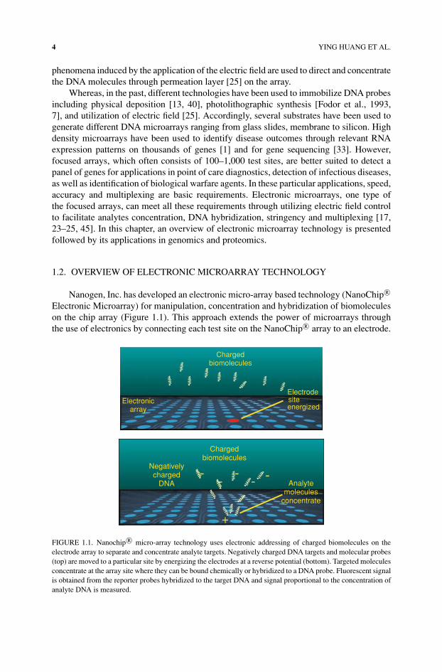
Electronic Microarray) for manipulation, concentration and hybridization of biomoleculeson the chip array (Figure 1.1). This approach extends the power of microarrays throughthe use of electronics by connecting each test site on the NanoChip r© array to an electrode.
Chargedbiomolecules
Analytemolecules
concentrate
Negativelycharged
DNA
+
Electronic
Chargedbiomolecules
Electrodesiteenergizedarray
FIGURE 1.1. Nanochip r© micro-array technology uses electronic addressing of charged biomolecules on theelectrode array to separate and concentrate analyte targets. Negatively charged DNA targets and molecular probes(top) are moved to a particular site by energizing the electrodes at a reverse potential (bottom). Targeted moleculesconcentrate at the array site where they can be bound chemically or hybridized to a DNA probe. Fluorescent signalis obtained from the reporter probes hybridized to the target DNA and signal proportional to the concentration ofanalyte DNA is measured.

ELECTRONIC MICROARRAY TECHNOLOGY AND APPLICATIONS 5
Most biological molecules have a natural positive or negative charge. When biologicalmolecules are exposed to an electric field (Figure 1.1), molecules with a positive chargemove to electrodes with a negative potential, and molecules with a negative charge moveto electrodes with a positive potential. Current and voltages are applied to the test sitesvia individual electrode activation to facilitate the rapid and controlled transport of chargedmolecules to any test sites. Additional advantages of electrically facilitated transport include(1) the ability to produce reconfigurable electric fields on the microarray surface that allowsthe rapid and controlled transport of charged molecules to any test sites [22, 25]; (2) theability to carry out site selective DNA or oligonucleotide addressing and hybridization [11];(3) the ability to significantly increase DNA hybridization rate by concentration of targetat the test sites (Kassegne et al., 2003); and (4) the ability to use electronic stringency toimprove hybridization specificity (Sosnowski, et al., 1997).
1.2.1. NanoChip r© Array and NanoChip r© Workstation
1.2.1.1. Fabrication Electronic microarrays consist of an array of electrodes thathave been fabricated on silicon with array sizes ranging from 5 to 10,000 individualelectrodes or test sites [24, 25, 42]. Figure 1.2 shows a number of electronic microarrayswith arrays ranging from 4 to 100 electrodes or test sites. These arrays have been designed
FIGURE 1.2. A series of designs of Nanogen’s silicon microarray chips. Chip sizes shown range from 4–100 sites.These include different electrode geometries as well as chips designed for particles and or biomolecular microsep-arations (last column of chips).

6 YING HUANG ET AL.
SiliconBase
PlatinumMicroelectrode
Hydrogel Permeation LayerContaining Streptavidin
80 µm diameter test site
FIGURE 1.3. Cross-section of a single platinum micro-electrode pad on the NanoChip r© microarray. A hydro-gel permeation layer loaded with streptavidin covers the electrode array and serves for capturing electronicallyaddressed molecules.
for both microassays and microseparations. The current commercialized NanoChip r© arraycomprising 100 platinum microelectrodes with additional 20 outer microelectrodes actingas counter-electrodes.
The electrodes on the NanoChip r© array are fabricated on a silicon substrate usingstandard photolithography and deposition processes. Each electrode is 80 µm in diameterwith 200 µm center-to-center space between and is connected to the outside edge of thechip by a platinum wire trace. Figure 1.3 shows a cross section of a single electrode padon the NanoChip r© array. The base structure of the array consists of silicon over which aninsulating layer of silicon dioxide is applied. Platinum is deposited and selectively held inplace to form electrodes and accompanying electrical traces. These wire traces terminate atthe edges of the chip forming electrical contact pads. Additional layers of silicon dioxideand silicon nitride are deposited to electrically insulate the platinum electrical traces, leavingthe central array of 80 µm diameter microelectrodes, outer microelectrodes and contactspads exposed. The chips are flip-chip bonded to a ceramic substrate which contains contactsto pogo-pins.
1.2.1.2. Permeation Layer Typically, on the surface of the array, a 10 µm thick hy-drogel permeation layer (Figure 1.3). containing co-polymerized streptavidin is depositedby microreaction molding. This permeation layer serves two main functions [24]. Firstit protects the sensitive analytes from the adverse electrochemical effects at the platinumelectrode surface during active operation. These electrochemical products include the gen-eration of hydrogen ions (H+) and oxygen at the positively biased (anode) microelectrodesand hydroxyl ions (OH−) and hydrogen at the negatively biased electrodes (cathode), aswell as various free radical entities. Secondly, the permeation layer also serves as a matrixfor the attachment of biotinylated molecules e.g., analytes capture oligos, antibodies, andprimers through biotin and streptavidin binding [24, 25]. Figure 1.4 is a close-up photographof the 100-site array covered with the permeation layer.

ELECTRONIC MICROARRAY TECHNOLOGY AND APPLICATIONS 7
FIGURE 1.4. Photograph of the 100-site NanoChip r© microarray. A hydrogel permeation layer covers the elec-trode array including working and counter electrodes. The hydrogel layer is visible as a circle surrounding counterelectrodes (yellow pads).
1.2.1.3. NanoChip r© Cartridge The 100-site array is assembled into a completeNanoChip r© cartridge (Figure 1.5a) by ultrasonically welding two molded polymethylmethacrylate (PMMA) cartridge bodies that contain fluidic channels and inlet and out-let ports. The cartridge eliminates sample evaporation, prevents sample contamination andprovides a fluidic interface to the NanoChip r© Workstation.
1.2.1.4. NanoChip r© Workstation The NanoChip r© electronic microarrays are oper-ated through a fully integrated and automated NanoChip r© Workstation (Figure 1.5b). Thesystem consists of three major subsystems: (1) the NanoChip r© Loader for loading pa-tient samples on one to four NanoChip r© Cartridges, (2) the NanoChip r© Reader, a highlysensitive, laser-based fluorescence scanner for detection of assay results and (3) computerhardware and software which automates import, analysis and export of sample informationmaking data analysis simple.
1.2.2. Capabilities of the NanoChip r© Electronic Microarrays
Using electric field, Nanochip r© electronic microarrays have provided many uniquefeatures over other passive microarrays (Table 1.1). Electronic addressing allows usersto quickly customize arrays in their own laboratory. Electronic hybridization provides anextremely accurate and specific hybridization process by creating optimal electric and pHconditions at the hybridization sites [11, 25]. Electronic stringency, in combination withthermal control, enables researchers to remove unbound and nonspecifically-bound DNAquickly and easily after hybridization at the microarrays, achieving rapid determination ofsingle base mismatch mutations in DNA hybrids [44].

8 YING HUANG ET AL.
Nanochip®Loader
Nanochip®Reader
(a)
(b)
NanoChip*ElectronicMicroarray
NanoChip*Cartridge
FIGURE 1.5. Photograph of the Nanochip r© cartridge containing the electronic microarray, and b) Nanogen’sNanochip r© Workstation which allows fully automated processing of 4 cartridges simultaneously in the loaderand fluorescent detection in the reader.
1.2.2.1. Assay Formats on NanoChip r© Electronic Microarrays The open architec-ture of electronic microarray enables flexibility in assay design. Since each individual elec-trode can be selectively activated, different assays can be generated depending on differentanalytes to be addressed (Figure 1.6). For example, a dot blot assay [19] is conducted whenbiotinylated PCR amplicon is addressed to the selected electrodes and remained embeddedthrough interaction with strepavidin in the permeation layer. The DNA at each electrode isthen hybridized to mixtures of allelespecific oligonucleotides (discriminators) and fluores-cently labeled oligonucleotides. The thermal or electronic stringency is used to discriminate
TABLE 1.1. Comparison between NanoChip r© microarray active hybridization technology andpassive hybridization technologies.
Concentration StringencyHybridization time Concentration of targets factor at a site control
NanoChip r© activehybridization
10–100 seconds Directed and localized at thearray sites; Ability tocontrol individual sites
>1000 times Electronic,ThermalChemical
Passivehybridization
1–2 hours Non-directed; Sites cannot becontrolled independently
Low, diffusiondependent
Thermal,Chemical

ELECTRONIC MICROARRAY TECHNOLOGY AND APPLICATIONS 9
Red
•
•
•
• SDA
•
•
•
•
•
•
•
•
•
•
•
Electronic addressing of biotinylated analytes
Electronic hybridization
Green
Report and stringency
•
•
•
•
•
•
•
•
•
•
•
•
•
•
•
• Denatured PCR product
• Capture oligos
• Capture oligos
• primer
• Antibody
• Helper oligos
• Denatured PCR product
• RNA (or cDNA)
• Denatured genomic DNA
• Antigen
• Dot blot SNP
• Sandwiched SNP
• Gene expression profiling
• Anchored SDA
• Immunoassay
FIGURE 1.6. The open platform of the electronic microarray enables flexibility in assay design. Depending on thenature of the biotinylated analytes (denatured PCR products, capture oligos, SDA primers or antibody) in electronicaddressing and on the targets (helper oligos, denatured PCR products, RNA, denatured genomic DNA or antigen),different assays (dot blot SNP, sandwiched SNP, gene expression profiling, anchored SDA or immunoassay) canbe performed on the electronic microarrays.
the SNP. In this assay format, multiple samples can be analyzed at different electrodes ona single array.
A “sandwich” assay is created when biotinylated capture probes are addressed to se-lective electrodes. The capture probes can be sequence specific oligos or antibodies. Theembedded capture probes are then electronically hybridized to the targets. Depending onthe specific targets (PCR amplicons, or RNA, or antigens), the assays can be SNP analysis,gene expression profiling, or immunoassay.
Most recently, a special assay, anchored strand displacement amplification (aSDA),which integrates the amplification and discrimination, is demonstrated on electronic mi-croarrays [12, 29, 50, 51]. In this assay format, biotinylated SDA primers were addressedand anchored on selective electrodes. These anchored primers were then electronically hy-bridized to the denatured genomic DNA. Target DNA was amplified over the electrodeswhen an enzyme mix containing restriction endonuclease and DNA polymerase and dNTPswas introduced to the array. After amplification the final discrimination was determinedusing same base-stacking principle [37]. Using aSDA, multiple genes from one sample orone gene from multiple samples can be simultaneously amplified and detected on a singleelectronic microarray.
1.2.2.2. Electronic Multiplexing By electronically controlling each test site, the elec-tronic microarrays also provide a platform to perform different types of multiplexed assays:
(1) single array multiplexing where multiple genes from one sample can be analyzed;(2) single array multiplexing where multiple samples with one gene of interest can be
analyzed;(3) single array multiplexing where multiple samples with multiple genes of interest
can be analyzed;

10 YING HUANG ET AL.
(4) single site multiplexing where several targets are discriminated on the same siteusing different fluorescent probes;
(5) single site multiplexing where several targets are addressed, different discriminatoroligonucleotides hybridized and reporter addressed—this method allows the use ofa universal reporter for different targets.
The ability to electronically control individual test sites permits biochemically unrelatedmolecules to be used simultaneously on the same microchip. In contrast, sites on a conven-tional DNA array cannot be controlled separately, and all process steps must be performedon an entire array. Nanogen’s electronic microarray technology delivers increased versa-tility over such conventional methods. This is particularly important in applications suchas biological warfare and infectious disease detection since the accurate identification ofbiological agents requires determination of two or more (often five) characteristic genes ofa particular agent.
1.3. APPLICATIONS
1.3.1. Single Nucleotide Polymorphisms (SNPs)—Based Diagnostics
Given the advances in genomic studies, more and more single nucleotide polymophisms(SNPs) are found to be contributory factors for human disease and can be used as geneticmarkers for molecular diagnostics. The speed, accuracy and flexibility provided by elec-tronic microarrays have received great interest from clinical diagnostic researchers for avariety of genotyping applications (Table 1.2). Using the Nanochip r© system, researchers atAmerican Medical Laboratories analyzed 635 clinical samples for the G1691A mutation onthe factor V Leiden, associated with thrombosis with 100% accurate in characterizing wild-type, heterozygous, and homozygous samples [15]. Researchers at ARUP Laboratories haveevaluated 3 thrombosis associated SNPs, factor V (Leiden), factor II (prothrombin), andmethylenetetrhydrofolate reductase (MTHFR), on 225 samples with 100% accuracy [14].Schrijver et al at Stanford University Medical Center found that the SNP analysis basedon Nanogen electronic microarray for factor V (Ledein) and factor II (prothrombin) on800 samples were comparable with other SNP analysis methods such as restriction enzymedigestion (RFLP) and the Roche LightCycler [41]. Researchers at Children’s NantionalMedical Center, Washington DC, genotyped 8 common MeCP2 mutations associated toRett syndrome on 362 samples with 100% specificity [47]. Using electronic microarray,researchers at Mayo Clinic Cancer Center have developed genotyping assays for 5 differentcytokine polymorphisms [43]. Moreover, 8 SNPs distributed within a highly variable regionof the polC gene from six isolates of Staphylococcus aureus were analyzed on electronicmicroarrays [9].
1.3.2. Forensic Detection
Short tandem repeats (STRs) represent another type of polymorphism with importantapplications in forensic DNA identification. In 1990, the FBI created a combined DNAindex system (CODIS), which consist of 13 polymorphic STR loci, to provide a databaseof forensic DNA profile for nearly all forensic laboratories in the United States [5, 6]. The

ELECTRONIC MICROARRAY TECHNOLOGY AND APPLICATIONS 11
TABLE 1.2. Examples of Nanochip r© technology application in genomics.
Relevance Test Ref
Cystic Fibrosis CFTR http://www.nanogen.com/products/cystic fibrosis.htmThrombosis Factor V Leiden
Factor II (prothrombin)MTHFRFactor V/Prothrombin
[41][15][14]http://www.nanogen.com/products/Factor vII.htm
HereditaryHemochromatosis
HFE http://www.nanogen.com/products/HH.htm
Alzheimer’s Disease ApoE http://www.nanogen.com/products/apoe.htmhttp://biz.yahoo.com/prnews/040106/latu005 1.html
β-thalassemia Factor VII [39]Asthma and chronic
obstructivepulmonary disease
B(2)-adrenergic receptor [53]
Ulcerative colitis N-acetyltransferase 1 (NAT 1)N-acetyltransferase 2 (NAT 2)
[38]
Cancer p53 [3]Rett syndrome MeCP2 [47]Immunologic defect Mannose binding protein
(MBP)[19]
Parkinson’s discease CYP1A2/CYP2E1 [10]Cytokine Tumor necrosis factor-α
(TNF-α)IL-4Interferon-γ (CA)n repeatsIL-1 RN VNTRCCR5
[43]
Bacterial ID Staphylococcus aureus pol C [9]Escherichia coli gyrASalmonella gyrACampylobacter gyrAE. coli parCStaphylococcus mecA
[50]
typical STR loci are selected groups of four nucleotide repeats that are represented in thehuman population by 4-15 alleles distinguished by a different number of repeat units [8].Unlike SNPs, STRs are more difficult to analyze by conversional passive hybridizationtechniques [20, 34]. However, electronic hybridization techniques have been proven toovercome these problems and allow STR analysis to be performed on electronic microarrayin a rapid and high fidelity fashion [37]. Multiplex hybridization analysis of three STRloci (CSF1PO, TH01 and TPOX) was achieved for 12 individuals, 100% concordant withgenotyping results of an accredited forensic laboratory.
Given the recent discovery of abundance SNPs and the ease of automation and minia-turization of detection techniques, SNP assays have started to be implemented in DNAforensic analysis. According to Chakraboty et al. [6], somewhere in the range of 30-60SNP loci would be needed to equal the power of the 13 STR loci with regard to genotypicmatch probability and/or paternity exclusion. The flexibility of electronic microarray willpermit accelerated development of SNPs for DNA forensic analysis by allowing an easy