biology practical-2 semester - ibn · pdf filebiology practical-2nd semester 1 ... they are...
TRANSCRIPT

Biology practical-2nd Semester 1. Microscope2. Tissuesa) Epithelial tissuesb) Connective tissuec) Muscular tissued) Nervous tissue3. Cell divisiona) Mitosisb) Meiosis4. Blood groups5. Rabbit dissectiona) Digestive systemb) Respiratory systemc) Urinogenital systemd) Circulatory system


CLO No
P. LO No
Lecture – Intended Learning Outcomes
1.1 P1.1 List the parts of microscope and its functions
1.1 P1.2 Write the basic characteristics of plant and animal cells after the preparation of wet mount of cheek epithelium and onion cells.
At the end of this lesson you will be able to:

1.Compound Microscope2. How to use Microscope?
3. How to prepare wet mounts?
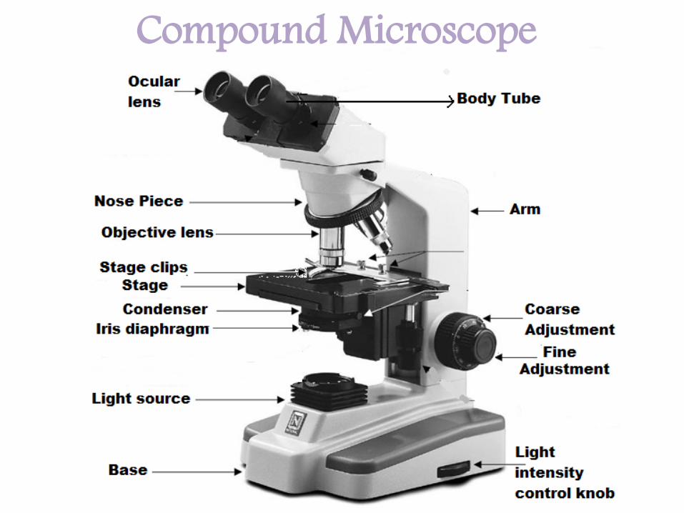
Compound Microscope

Parts of Microscope Functions1. Arm Support body tube and help to carrying
microscope2. Base support3. Ocular lens Magnify 5, 10, 15X4. Objective lens Magnify 4, 10, 40, 100X5. Nose Piece Revolving part to which objective lenses are
attached6. Body Tube Joins nose piece with ocular lens7. Mechanical Stage Supports the Slide8. Coarse adjustment Moves the stage up and down to focus the object.
9.Fine adjustment Fine Focusing10. Light Source (illumination) Bulb from below to light the specimen
11. Iris Diaphragm Regulates the amount of light passing through Specimen
12. Condenser Help in concentrate the light before entering the specimen

Steps for using the Microscope• 1. Carry the microscope with two hands• 2. Plug the microscope in and switch it on .• 3. Click the lowest power objective (scanner4X) into position . • 4. Use-coarse adjustment to lower the stage • 5.Place the slide onto the stage.• 6. look through the ocular and adjust the condenser for
brightness.• 7.Using the coarse adjustment to raise the stage until touch
the slide. • 8.Look into the microscope and adjust until object comes into
focus .
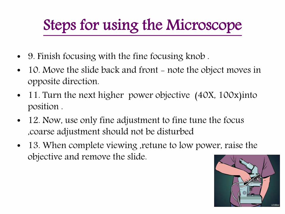
Steps for using the Microscope
• 9. Finish focusing with the fine focusing knob . • 10. Move the slide back and front - note the object moves in
opposite direction.• 11. Turn the next higher power objective (40X, 100x)into
position .• 12. Now, use only fine adjustment to fine tune the focus
,coarse adjustment should not be disturbed• 13. When complete viewing ,retune to low power, raise the
objective and remove the slide.

Wet Mount - preparationTools & Materials• Microscope • 2 Flat slides • 2 Cover slides• Water • Stain (safranin-methylene blue)• Paper towel (Tissues)• Toothpick• Scissors-forceps • Onion • Plastic dropper

Wet Mount - preparation
Mount specimen as shown in the diagram to avoid air bubbles under the cover slip
1.Place the
specimen in the centre + stain
2. Place the cover glass as shown in the figure
3. Observe
under microscope

11
In the Laboratory!• Carefully observe each type of epithelium under the microscope.• Use only the fine focus to adjust to your comfort.• Do not disturb the stage or position of the slide.• Draw the tissue image as you see under the microscope.

Onion cell

Human Cheek Epithelial Cells

Plant vs. Human Cell Lab• We did a Lab , comparing plant and animal cells. We looked at cheek cells
and onion cells.
• Plant cells have cell walls that protect and support the cell, which makes the cells look so geometric.
• Cheek cells are different; they don’t have a cell wall to support the structure, so they are not as geometrical as and rounder than the onion cells.
• Plant and animal cells also have many similarities, like both being eukaryotic. They are more complex than prokaryotic cells.
• Both types of cell have a nucleus, a cell membrane and cytoplasm.

Animal cell & plant cell


At the end of this lesson you will be able to answer:
1. List out the different parts of microscope and its functions

At the end of this lesson you will be able to answer:
2. Give 3 differences between animal and plant cells
Note: ILO is achieved if you can answer the above questions:
S.NO Plant Cell Animal Cell1.
2.
3.


CLO No
P. LO No
Lecture – Intended Learning Outcomes
1.1 P 2.1 Name the types of epithelial tissues with respect to its characters, function and location.
At the end of this lesson you will be able to:

• What is Histology?

Human Tissues
Epithelial Tissue Connective Tissue Muscular Tissue Nervous Tissue

One Cell thick More than one layer
1. Simple Squamous2. Simple Cuboidal3. Simple Columnar4. Pseudo stratified
1. Keratinised2. Non keratinised3. Transitional
Classified into two depending on the number of layers

FunctionLocation CharactersName
Protection1. Cheek epithelium
2. lining of blood vessel
3. air sacs of lungs
1. Single layer of Flat cells
2. Bulged nucleiSimple Squamous
TissueEpithelial( النسيج الطالئي الحرشفي البسيط(

FunctionLocation CharactersNameAbsorption1.Lining of kidney tubules
2.Ducts of salivary gland
and mammary glands
1.Single layer of cubical cells
2. Large, central & round nuclei
CuboidalSimple TissueEpithelial
( النسيج الطالئي المكعب البسيط(
Secretion

FunctionLocation CharactersNameProtectionAbsorption
Lining of small intestine1.Single layer of columnarcells
2. Oval and basal nuclei
Simple ColumnarTissueEpithelial
( النسيج الطالئي العمادي البسيط(
Secretion

FunctionLocation CharactersNameProtection1.Trachea
2. Ducts of many glands
1.Single layer of columnar cells of different height
2. Nuclei at different levels giving false appearance of many layers
stratified-PseudoTissueEpithelial
)النسيج طالئي طبقي كاذب (
Secretion

Types Simple EpitheliumTypes Character Location function
Simple Squamous
1. Single layer of Flat cells2. Bulged nuclei
1. Cheek epithelium2. lining of blood vessel 3. air sacs of lungs
protection
Simple Cuboidal
1. Single layer of cubical cells2. Large, central & round nuclei
1. Lining of kidney tubules
2. Ducts of salivary gland and mammary glands
SecretionAbsorption
Simple Columnar
1. Single layer of columnar cells2. Oval and basal nuclei
Lining of small intestine ProtectionSecretionAbsorption
Pseudostratified
1. Single layer of columnar cells of different height2. Nuclei at different levels giving false appearance of many layers
1. Trachea2. Ducts of many glands
ProtectionSecretion

Simple Squamous Epithelium

Simple Cuboidal Epithelium
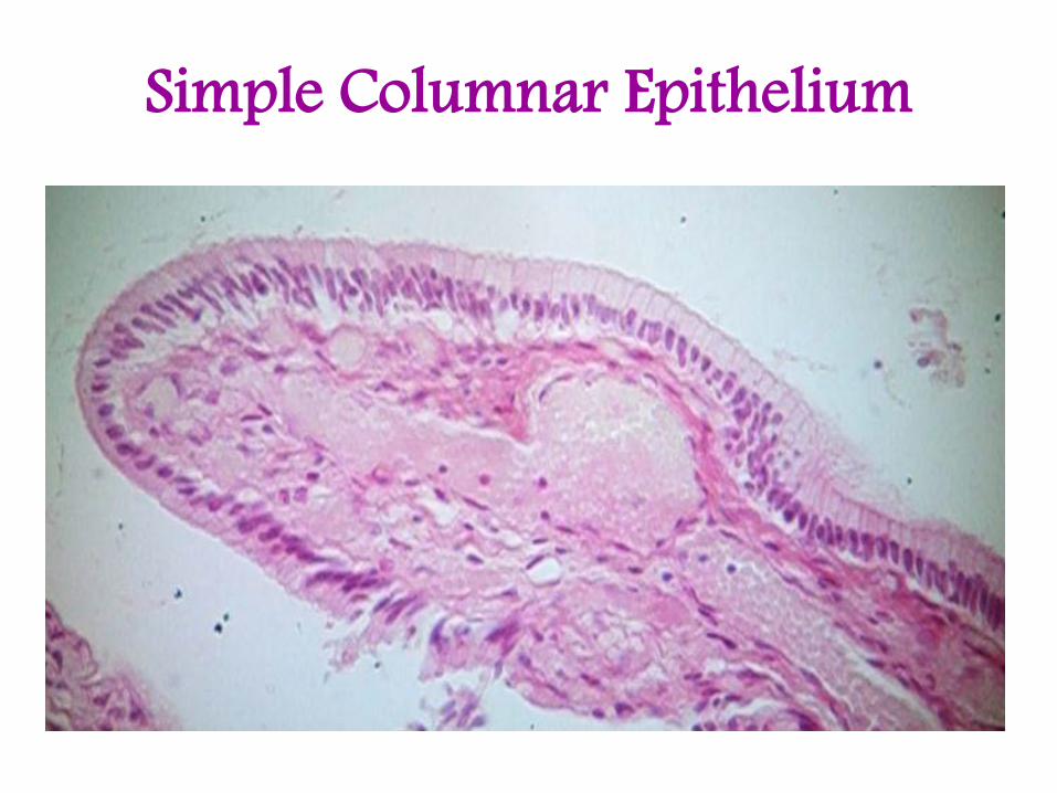
Simple Columnar Epithelium

Pseudo -Stratified Epithelium

Simple EpitheliumSquamous Cuboidal Columnar Pseudo stratified

FunctionLocation CharactersNameProtectionMouth
Buccal cavity
Vagina
esophagus
1.Many layers2. basal cells –cubical or columnar and surface cells - Squamous
Non-keratinizedStratified Epithelial
)النسيج طالئي طبقي غيرالمقترن(Covering

FunctionLocation CharactersNameProtectionSkin1.Many layers
2. Top layer cells – dead (keratinized)
keratinizedStratified Epithelial
)النسيج طالئي طبقي المقترن(Covering

FunctionLocation CharactersNameexpansionUrinary bladder1.It is 4-8 layers
2. basal cells - columnar Intermediate cells – cubicalWhen bladder expands the cells - Squamous (flat)
Transitional Epithelial
)النسيج طالئي طبقي اإلنتقالي(

Types of stratified epitheliumTypes Character Location function
Non-keratinized
1. Many layers2. basal cells - cubical or columnar and surface cells - squamous
MouthBuccal cavityVaginaesophagus
CoveringProtection
Keratinized 1. Many layers2. Top layer cells – dead (keratinized)
Skin Protection
Transitional 1. It is 4-8 layers2. basal cells - columnar Intermediate cells – cubicalWhen bladder expands the cells -squamous (flat)
Urinary bladder expansion

Non keratinized epithelium

Keratinized epithelium

Transitional epithelium

Stratified epithelium
Non keratinized Keratinized Transitional

At the end of this lesson you will be able to answer:
1. Name the types of epithelial tissues

At the end of this lesson you will be able to answer:
2. Tabulate the characters, function and location of epithelial tissues .
Note: ILO is achieved if you can answer the above questions:
Simple epithelial tissue
Character function Location
1.
2.
3.
4.
Stratified epithelial tissue
Character function Location
1.
2.
3.

The End of Lab -3-
Lab - 4Connective Tissue
At the end of this lesson you will be able to:
CLO No
P. LO No
Lecture – Intended Learning Outcomes
1.1 P 3.1 Name the types of loose and dense connective tissues with respect to its characters, function and location.
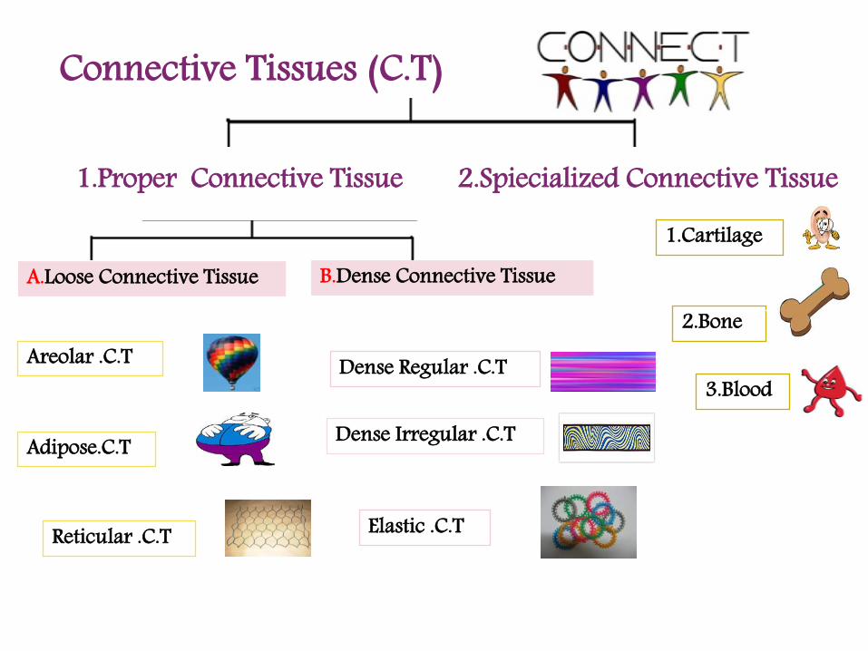
A.Loose Connective Tissue B.Dense Connective Tissue
Areolar .C.T
Adipose.C.T
Reticular .C.T
Dense Regular .C.T
Dense Irregular .C.T
Elastic .C.T
1.Cartilage
2.Bone
3.Blood
Connective Tissues (C.T)
2.Spiecialized Connective Tissue1.Proper Connective Tissue

Types Character Location functionAreolar connective tissue
Areolar tissue
Fibroblasts Collagen fibers Elastic fibers ( Strength & flexibility ) ( stretch )
1. Below skin in most internal organs
Support
Protection

Types Character Location functionAdipose connective tissue
Loose connective tissue containing fibroblast in between the adipocytes (Fat cells).
1. Below skin,around the kidneys & heart
Stores energy,
Insulating layer,
Cushion.
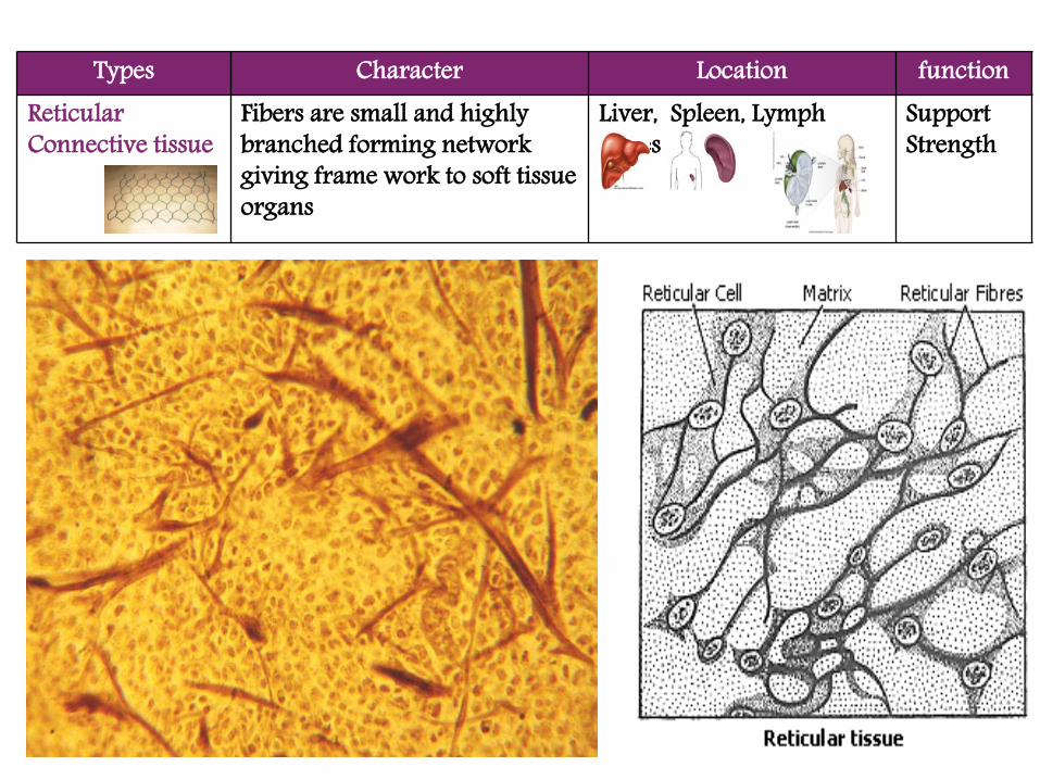
Types Character Location functionReticularConnective tissue
Fibers are small and highly branched forming network giving frame work to soft tissue organs
Liver, Spleen, Lymph nodes
SupportStrength

Areolar tissue –fibroblasts, collagen & Elastic fibresBelow skinSupport , protection
containing fibroblast in between the adipocytes (Fat cells).Below skinStores energy
Fibers are highly branched forming network.Liver, Spleen, Lymph nodesSupport

Connective tissue properTypes Character Location function
Dense regular connective tissue
Collagen fibers – Dense and regularly packed (one direction)
1. Tendon (muscle-bone)
2. Ligament (bone- bone)
Support
Ligament
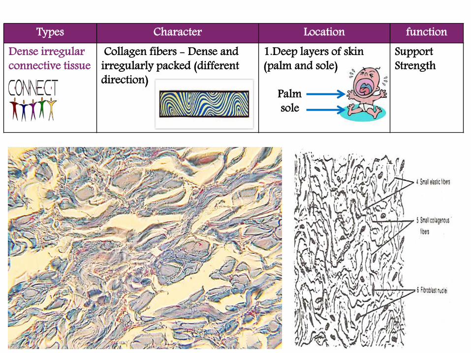
Types Character Location functionDense irregular connective tissue
Collagen fibers - Dense and irregularly packed (different direction)
1.Deep layers of skin(palm and sole)
Palm sole
SupportStrength

Types Character Location function
Elastic Connective Tissue
Contain more elastic fibers than collagen fibers
Large arteries, lung tissue
elasticity

Collagen fibers – Dense & regularly packed (one direction)1. Tendon (muscle-bone)2. Ligament (bone- bone)Support
Collagen fibers - Dense & irregularly packed (different direction) Deep layers of skin (palm & sole)Support, Strength
Contains more elastic fibers.Large arteries, lung tissueElasticity

Areolar connective tissue
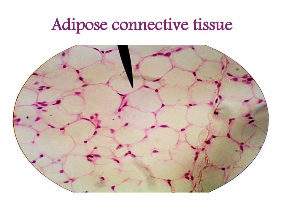
Adipose connective tissue

Reticular Connective tissue

Dense regular connective tissue

Dense irregular connective tissue

Elastic Connective Tissue

At the end of this lesson you will be able to answer:
1. Give the classification of Connective tissues

At the end of this lesson you will be able to answer:
2. Tabulate the types of loose and dense connective tissues with respect to its characters, function and location.
Note: ILO is achieved if you can answer the above questions:
Loose connective tissue
Character function Location
1.
2.
3.
Dense Connective tissue
Character function Location
1.
2.
3.
Blood tissue Character function Location
1.

The End of Lab -4-

Specialized Connective Tissue

CLO No
P. LO No
Lecture – Intended Learning Outcomes
1.1 P 4.1 Name the types of cartilage, bone and vascular (fluid) tissues with respect to its characters, function and location.
At the end of this lesson you will be able to:

Types Character Location functionHyaline cartilage
1. It is glossy (halos= gloss) whitish appearance2. The matrix is clear homogeneouscontaining binucleated chondrocytesThe outer membrane is perichondrium
Trachea
Larynx
BronchiEnd of the long bones
Support
Perichondrium
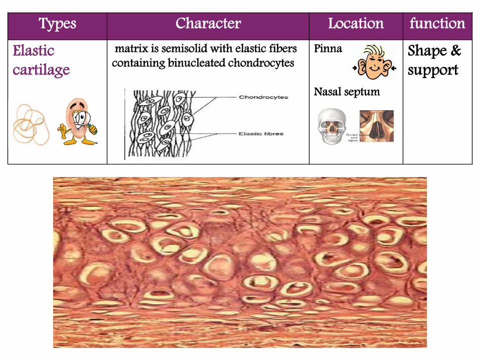
Types Character Location functionElasticcartilage
matrix is semisolid with elastic fibers containing binucleated chondrocytes
Pinna
Nasal septum
Shape & support

Types Character Location function
Compact Bone
The bone shows haversian system in which osteocytes are arranged in concentric rings around haversian canal
bones Structural frame work, Support , protection

Types Character Location function
Spongy /cancellousBone
It consist of long slender bony trabeculae forming a net work enclosing irregular marrow cavities
Epiphysis of long bone
Blood cells are formed
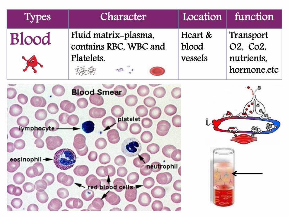
Types Character Location function
Blood Fluid matrix-plasma, contains RBC, WBC and Platelets.
Heart & blood vessels
Transport O2, Co2, nutrients, hormone.etc

Hyaline cartilage
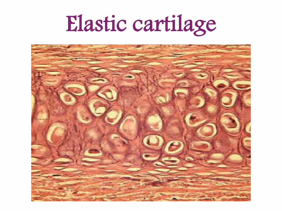
Elastic cartilage

Compact Bone

Spongy Bone

Blood

At the end of this lesson you will be able to answer:
1. Tabulate the types of cartilage, bone and blood tissues with respect to its characters, function and location.
Note: ILO is achieved if you can answer the above questions:
Cartilage tissue Character function Location
1.
2.
Bone tissue Character function Location
1.
2.
Blood tissue Character function Location
1.
The End of Lab -5-


CLO No
P. LO No
Lecture – Intended Learning Outcomes
1.1 5.1 Name the types of muscle, nerve and reproductive tissues with respect to its characters, function and location
At the end of this lesson you will be able to:

(1)Endocrine System
Reproductive Tissues

Testis

Types Character Location Function
Testis Testis shows semeniferous tubule lined by germinal epithelium give rise to sperm which are seen in various stages of development.
-Between semniferous tubules there are interstitial cells(Leyding cells)which produce testosterone.
Testis produce Sperms and testosterone
Germinal epithelium
Franz Leydig

Germinal epithelium
Primary spermatocyte
Secondary spermatocyte
Sprmatid
Sperm
Interstitial cell )(Leyding cell


Ovary

Types Character Location function
Ovary -The ovary lined by germinal epithelium gives rise to egg which is seen in various stages of follicular development.-Primary follicle, growing follicle, mature Graffian follicle, corpus luteum, corpus albicans.Mature follicle releases the ovum and undergoes changes to form corpus luteum
ovary produce ovum estrogen
& progesterone
Germinal epithelium
Corpus albicans
1.Primary follicle
2.Growing follicle
Ovulation
3. Mature Graffian Follicle
Corpus luteum
ovum


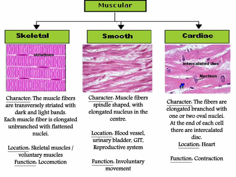
(2)Muscular Tissues

Types Character Location function1.Skeletal Muscles
1.The muscle fibers are transversely striated with dark and light bands.2.Each muscle fiber is elongated unbranched with flattened nuclei
Voluntary muscles attached to the skeleton
Locomotion

Types Character Location function
2.CardiacMuscles
1.The fibers are elongated branched with one or two oval nuclei.2.At the end of each cell there are intercalated disk help in uniform distribution of nutrients and energy.
Heart Contraction to pump theblood

Types Character Location function
3.SmoothMuscle
1. The fibers are elongated, spindle shaped cells2.with elongated nucleus in the centre
Blood vessel,
urinary bladder
gastrointestinal canalReproductive system
Involuntary movement of blood ,food & fluids

: The muscle fibers Characterare transversely striated with
dark and light bands.Each muscle fiber is elongated
unbranched with flattened nuclei.
: Skeletal muscles / Locationvoluntary muscles
: LocomotionFunction
: Muscle fibers Characterspindle shaped, with
elongated nucleus in the centre.
: Blood vessel, Locationurinary bladder, GIT, Reproductive system
Function: Involuntary
movement
The fibers are Character:elongated branched with one or two oval nuclei.At the end of each cell there are intercalated
disc.: HeartLocation
: ContractionFunction

Skeletal Muscles

Smooth Muscle
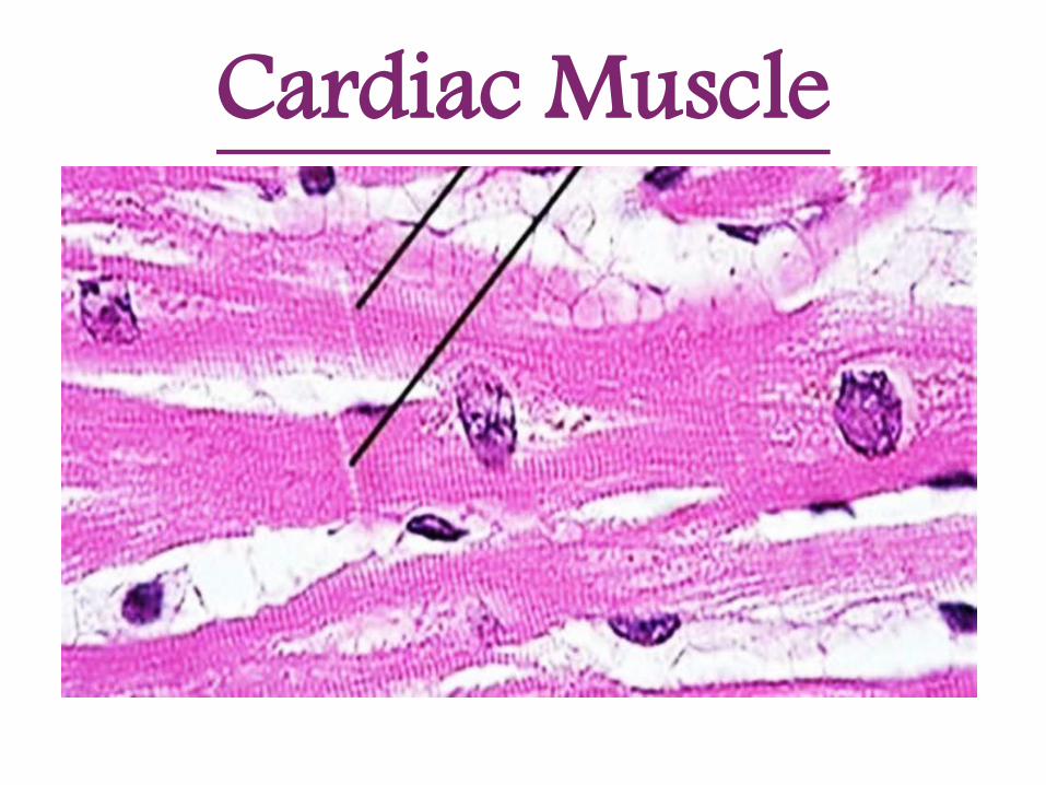
Cardiac Muscle
(3)Nervous Tissues

Types Character Location functionNervousTissue
1.The cell body with prominent nucleus. 2.The cell body has cytoplasmic processes - long axon and many short –dendrites
The central nervous system; Brain, spinal cord
Conductnerveimpulse

Types Character Location functionMylinated nerve
fiber1.Myelin sheath appears as a thick dark band on the periphery of the nerve fiber .2.Dips at regular intervals called nodes of Ranvier.
Nerve axons Protection
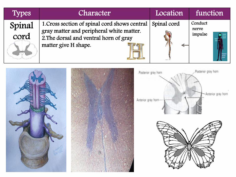
Types Character Location functionSpinal cord
1.Cross section of spinal cord shows central gray matter and peripheral white matter.2.The dorsal and ventral horn of gray matter give H shape.
Spinal cord Conductnerveimpulse

The cell body Character:with prominent nucleus. The cell body has cytoplasmic processes -long axon and many short –dendrites
Brain, spinal cord: LocationConduct nerve : Function
impulse
Myelin sheath Character:appears as a thick dark band on the periphery of the nerve fiber and dips at regular intervals called nodes of Ranvier.
Myelinated axons: LocationAccelerate : Function
conduction of nerve impulse
Cross section of Character:spinal cord shows central gray matter and peripheral white matter.The dorsal and ventral horn of gray matter appears as H shape.
Spinal cord: LocationConduct nerve : Function
impulse

Nervous Tissue

Myelinated nerve fiber

Spinal Cord

At the end of this lesson you will be able to answer:
1. Tabulate the types of muscle and nerve tissues with respect to its characters, function and location
Note: ILO is achieved if you can answer the above questions:
Muscular tissue Character function Location
1.
2.
3.
Nervous tissue Character function Location
1.
2.
3.
Reproductive tissue Character function Location
1.
2.


CLO No
P. LO No
Lecture – Intended Learning Outcomes
2.2 P 6.1 Explain the stages of mitotic cell division.
At the end of this lesson you will be able to:

CELL DIVISION
MITOSIS MEIOSIS (somatic cells) (germ cells)
GROWTH SEXUAL REPRODUCTION
REPAIR
ASEXUAL REPRODUCTION

prophase• Chromosomes condense and become
visible – this prevents tangling with other chromosomes.
• Due to DNA replication during interphase, each chromosome consists of two identical sister chromatids connected at the centromere.
• Centrioles move to opposite poles of cell.
• Nucleolus disappears. • Phase ends with the breakdown of the
nuclear membrane.

Metaphase• Spindle fibers
(microtubules) connect centrioles to chromosomes.
• Chromosomes align along equator of cell and attaches to a spindle fiber by its centromere.
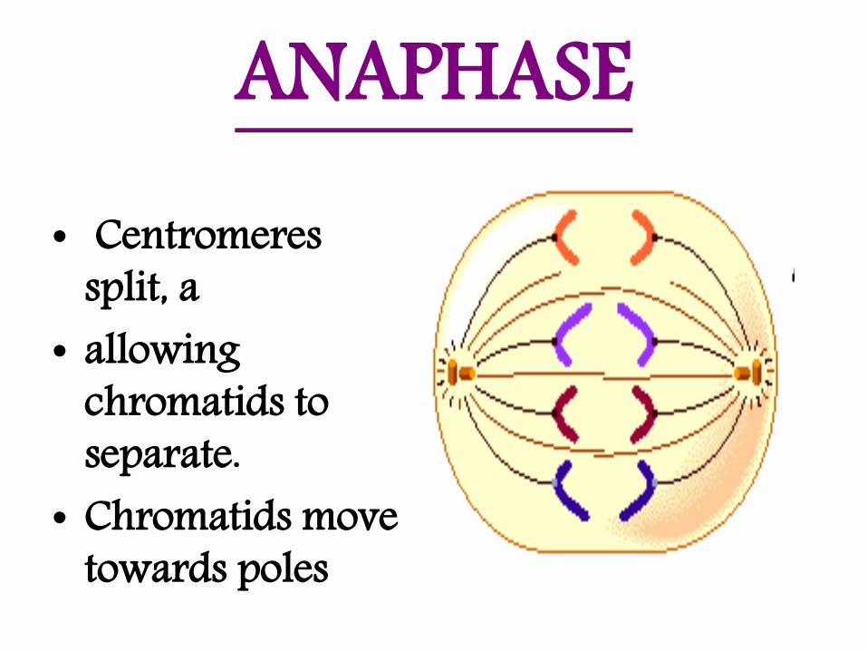
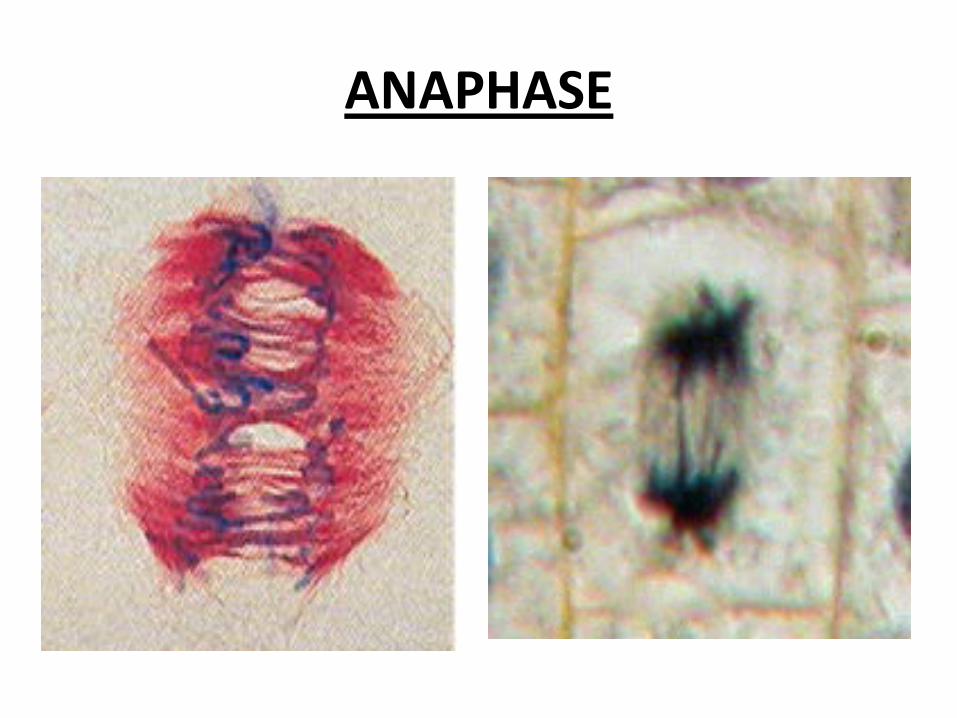
ANAPHASE• Centromeres
split, a • allowing
chromatids to separate.
• Chromatids move towards poles

TELOPHASE
• Spindle fibers disperse.
• Nuclear membranes form around each set of chromatids.
• Nucleoli form.

CYTOKINESIS
2 DAUGHTER CELL
- In Animal cells a ring of actin filaments forms round the equator of the cell, and then tightens to form a cleavage furrow, which splits the cell in two.

Phase
Character
Location
Function
PROPHASE METAPHASE ANAPHASE TELOPHASE
CHROMSOMES BECOME THICK AND
SHORT.
EACH CHROMOSOME ATTACH TO THE SPINDLE FIBRE WITH
THE CENTROMER.
THE CENTROMER
E DIVIDES AND THE SISTER
CHROMATIDS SEPARATE.
NUCLEOLUS REAPPEARS.
(somatic cells) (somatic cells) (somatic cells)
GROWTH GROWTH GROWTH GROWTH
(somatic cells)

PROPHASE

METAPHASE
ANAPHASE

TELOPHASE


ExperimentRoot of onion


At the end of this lesson you will be able to answer:
1. Tabulate the different stages of meiosis.
Note: ILO is achieved if you can answer the above questions:
Stages of Mitosis Character function Location
1.
2.
3.
4.

The End of Lab

CLO No
P. LO No
Lecture – Intended Learning Outcomes
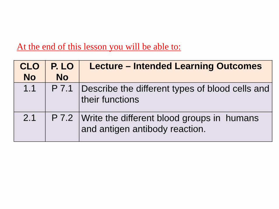
1.1 P 7.1 Describe the different types of blood cells andtheir functions
2.1 P 7.2 Write the different blood groups in humans and antigen antibody reaction.
At the end of this lesson you will be able to:

BLOOD CELL


Types Character FunctionI. R.B.C / erythrocytes
Male – 4.2 – 5.4 m/µl Female - 3.6 – 5.0 m/µl
Non- nucleated biconcave cells.Transport of O2 , Co2
nutrients and hormones
II. W.B.C / Leucocytes Nucleated and classified into 2 types Defense
1. Granulocytes 3 types
a) Neutrophils 60-65% The nucleus is multilobed (3-5) Phagocytosis
b) Eosinophils 1- 3 % Nucleus is bilobedParticipate in Allergic reaction
c) Basophils 0.5 – 1 % Nucleus is ‘U’ or ‘S’ shaped Participate in Allergic and inflammatory reaction
2. Agranulocytes types
a) Lymphocytes 25-35% Large nucleus fills most of the cell Immune response
b) Monocytes 5-6% Kidney shaped or U shaped nucleus phagocytosis
III. Platelets
(thrombocytes) Round to oval flattened cells Clotting

Neutrophil Eosinophil Basophil
Lymphocyte Monocyte

R.B.C

ABO BLOOD GROUP SYSTEM

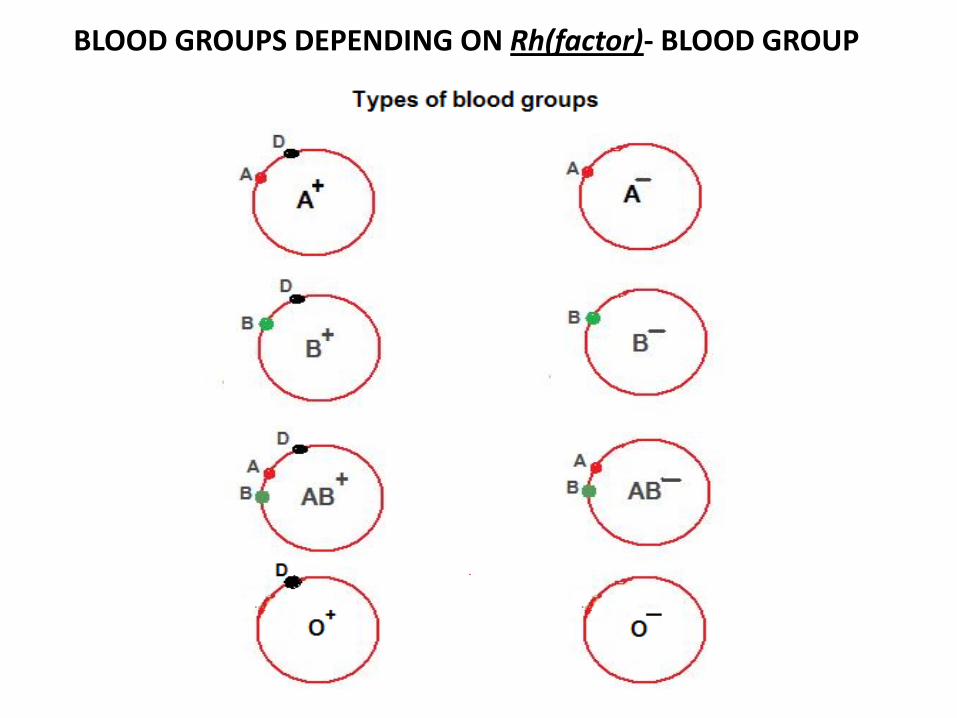
BLOOD GROUPS DEPENDING ON Rh(factor)- BLOOD GROUP


Blood Groups Antisera - A Antisera-B Antisera - D
A+ Agglutination No-agglutination Agglutination
B+ Agglutination
AB+
O+
A-
B-
AB-
O-
BLOOD GROUP LABORATORY REPORT
Agglutination
Agglutination
Agglutination
Agglutination
Agglutination
Agglutination
Agglutination
Agglutination
Agglutination
No-agglutination
No-agglutination
No-agglutination
No-agglutination
No-agglutination No-agglutination
No-agglutination
No-agglutination No-agglutination
No-agglutination
No-agglutination

RECIPIENT DONOR
O - O + A - A + B - B + AB - AB +
O -
O +
A -
A +
B -
B +
AB -
AB +
WHICH RECEPIENT CAN RECEIVE BLOOD FROM WHICH DONORMARK IN THE APPROPRIATE BOX

The End of Lab 7