biology of tooth movement
TRANSCRIPT
1. Introduction
2. Physiologic tooth movement
3. Histology of tooth movement
4. Optimum orthodontic force
5. Hyalinization
6. Phases of tooth movement
7. Theories of tooth movement
8. Bone deposition
9. Bone resorption
Orthodontic treatment is made possible by the fact
that teeth can be moved through the alveolar bone by
applying appropriate forces . orthodontic tooth
movement is a unique process where a solid object
(tooth) is made to move through a solid medium
(bone).
Orthodontic treatment is possible due to the fact
whenever a prolonged forces is applied on a tooth ,
bone remodeling occurs around the tooth resulting in
its movement. Bone subject to pressure as a result of
compression of periodontal ligament resorbs while
bone forms under tensile as a result of stretching of
the periodontal ligament.
Physiologic tooth movements are naturally occurring tooth movements that take place during and after tooth eruption .
It includes:
1) Tooth eruption
2) Migration or drift of tooth
3) Changes in tooth position during mastication
It is the axial movement of the tooth from its developmental position in the jaw to its final position in the oral cavity.
Following are some of the theories:
A) Blood pressure theory: The tissue around the developing end of the root is highly vascular. This pressure is believed to cause the axial movement of the teeth
B) Root growth: The apical growth of roots results in an axially directed force that brings about the eruption of the teeth.
C ) Hammock ligament: A band of fibrous tissue exists below the root apex spanning from one side of the alveolar wall to the other. The fibrous tissue appears to form a network below the developing root and is rich in fluid droplets.
D) Periodontal ligament traction: It states that the periodontal ligament is rich in fibroblast that contains contractile tissue. The contraction of these periodontal fibers results in axial movement of the tooth.
It refers to the minor changes in tooth position observed after eruption. Human dentition shows a natural tendency to move in a mesial and occlusal direction.
This is largely true in case of maxillary dentition. As the teeth undergo occlusaland proximal wear, they move in a mesialand occlusal direction to maintain inter-proximal and occlusal contact.
During mastication, the teeth and periodontal structures are subjected to intermittent heavy forces which occur in cycles of one second or less and may range from 1-50 kilograms based on the type of food being mastication.
When a tooth is subjected to such heavy forces, the tissue fluid present in the periodontal space, being incompressalbe, prevents major displacement of the tooth within the socket.
Whenever the forces of mastication are sustained and are more than the usual one second cycle, the periodontal fluid is squeezed out and a pain is felt as the tooth is displaced into periodontal ligament space.

When a tooth is moved due to application of orthodontic force , there is bone resorption on the pressure side and new bone formation on the tension side . the histologic changes seen during tooth movement can be studied under 2 headings :
Changes following application of mild force
Changes following application of extreme force
When a force is applied to a tooth, areas of pressure and tension are produced.
a. Changes on the pressure side: - the periodontal ligament in the direction of the tooth movement get compressed to almost 1/3rd of its original thickness and vascularity is increased which helps in the mobilization of cells such as fibroblast and osteoclasts. Osteoclasts are bone resorbing cells that line up the socket wall on the pressure side. When continued forces are applied a change in orientation of bony tabeculae is seen.
Trabeculae are usually parallel to the long axis to the tooth and become horizontally oriented i.e. parallel to the direction of orthodontic force. The osteoclasts start resorbing bone when the force are applied within physiologic limits, the resorption is seen in the alveolar plate immediately adjacent to the ligament this is known as frontal resorption.
b. Changes on the tension side: The area of tooth opposite to the direction of force is called the tension side. On application of force the periodontal membrane on the tension side gets stretched, the distance between alveolar process and tooth is widened. Vascularity is increased which causes mobilization of the cells such as fibroblast and osteoblast in response osteod is led down by osteoblast in the periodontal ligament.
c. Secondary modeling changes:
When force is applied the adjacent bone shows osteoclastic and osteoblastic activity on the pressure and tension side. Bony changes also take place elsewhere to maintain the width or thickness of the alveolar bone. these changes are called as secondary remodeling.
Whenever extreme forces are applied to the teeth , it results in crushing or total compression of the periodontal ligament. On the pressure side the root closely approximates the lamina Dura, compresses the periodontal ligament and leads to occlusion of the blood vessels. The ligament is deprived of nutritional supply leading to regressive changes called hyalinization. Bone resorption occurs in the adjacent marrow spaces and in the alveolar plate below, behind and above the hyalinized zones this kind of resorptionis called undermining or rearward resorption.
On the tension side, the periodontal ligament gets over-stretched leading to tearing of the blood vessels and ischemia.
Thus when the extreme force is applied there is net increase in osteoclastic activity as compare to bone formation with the result that tooth become loosened in its socket. Pain and hyperemia of the gingiva may occur due to application of extreme forces during orthodontic tooth movement.
It is one which moves teeth most rapidly in the desired direction, with the least possible damage to tissue and with minimum patient discomfort.
Equivalent to capillary pulse pressure which is 20-25 gm/sq. cm
It has following characteristics:
Produces rapid tooth movement
Minimal patient discomfort
The lag phase of tooth movement is minimal
No marked mobility of the teeth being moved.
From a histologic point of view the use of optimum force has the following characteristics:
The vitality of the tooth and supporting periodontal ligament is maintained
Initiates maximum cellular response
Produces direct or frontal resorption
It is a form of tissue degeneration characterized by formation of a clear, eosinophilic homogenous substance. This hyalinization occurs in organs such as kidneys,lungs,etc.Hyalinization of the periodontal ligament denotes a compressed and locally degenerated periodontal ligament.the convention pathologic process of hyalinization presents irreversible one, however , hyalinization is a reversible process in PDL.
The change observed during formation of hyalinizedbone are as follows:
There is gradual shrinkage of the periodontal fibers The cellular structures become indistinct. Some nuclei
becomes smaller(pycnotic ) while some nuclei disappear.
The compressed collagenous fibres gradually unite into a more or less cell free mass
Certain changes also occur in the ground substance There is a breakdown of the blood vessel walls leading
to spilling of their contents Osteoclasts are formed in marrow spaces and
adjacent are of the inner bony surface offer aperiodof 20 to 30 hrs .
The presence of hyalinized zone indicate that the ligament is non functional and therefore bone resorption cannot occur .
The elimination of hyalinized tissue occurs by 2 mechanisms :
Resorption of the alveolar bone by osteoclasts differentiating in the peripheral intact periodontal membrane and adjacent marrow spaces
Invasion of the cells and blood vessels from the periphery of the compressed zone by which the necrotic tissue is removed . the invading cells penetrate the hyalinised tissue and eliminate the unwanted fibrous tissue by enzymatic action and phagocytosis.
Greater the force wider is the area hyalinization. If the lighter forces are used the hyalinisedzones are smaller and the larger area of functioning ligament is available. Frontal resorption predominates in case where lighter forces are used.
The location and extent of hyalinized tissue largely depends upon the nature of tooth movement. In case of tipping tooth movement the hyalinization would be closed to alveolar crestwhile in case of bodily tooth movement it would be closure to the middle portion of the root. Whenever exxesive forces are applied during tipping tooth ,movement , it can result In 2 areas of hyalinization , 1 in the apical region and other ion the marginal area.
Studies have shown that tooth movement through 3 stages.
Burstone categorises the stages as:
Initial stage
Lag phase
Post lag phase
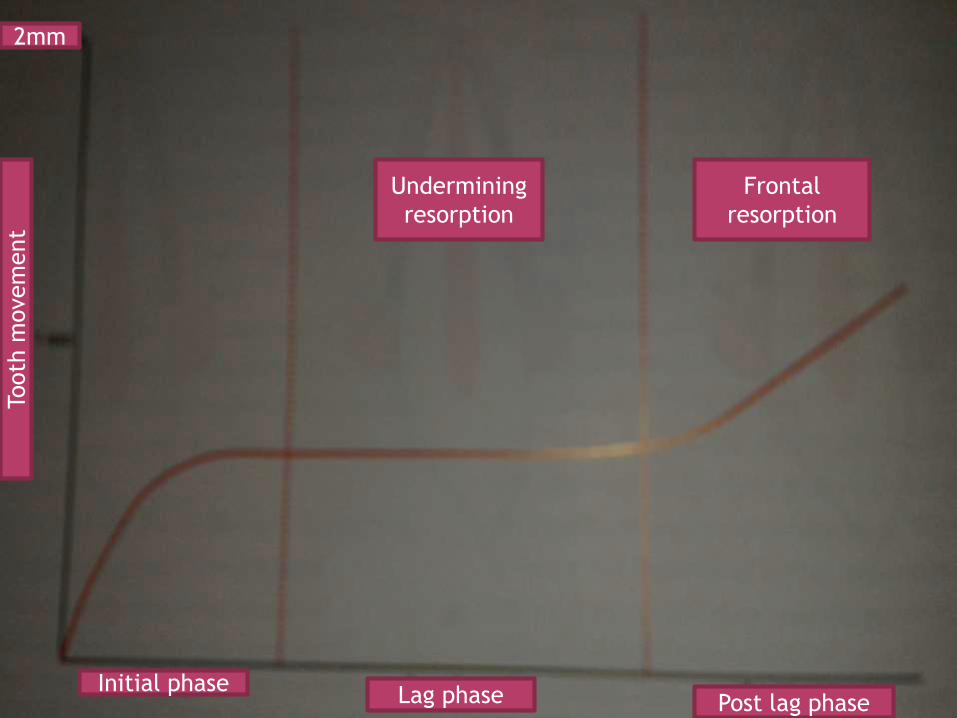
Initial phaseLag phase Post lag phase
Undermining
resorption
Frontal
resorption
Tooth
movem
ent
2mm
During this phase very rapid tooth movement is observed over a short distance which then stops. This movements represents displacement of the tooth in the periodontal membrane space and probably behind of alveolar bone to a certain extent. The tooth movement in the initial phase is between 0.4 to 0.9 mm and usually occurs in a week’s time.
little or no tooth movement occurs during this phase. this phase is characterized by formation of hyalinaised tissue in the periodontal ligament. The duration of the lag phase depends on the amount of force used to move the tooth if light forces are used the area of hyalinization is small and frontal resorption occurs . If heavy forces are used the are of hyalinization is large . the la phase usually extends for 2 to 3 weeks but at times be as long as 10 weeks the duration of lag phase depends on a varied no of factors including the density of alveolar bone , age of the patient an the extent of the hyalinized tissue.
After the lag phase tooth movement progresses rapidly as the hyalinized zone is removed and bone undergoes resorption. During this post lag period osteoclasts are found over a large surface area resulting in direct resorption of bony surface facing the periodontal ligament.
The mechanism of movement of a tooth by an orthodontic force is a subject of ongoing
research for decades . numerous theoris have been put forward to explain the same . the theories that have been accepted and have
stood the test of time are :
PRESSURE TENSION THEORY by Schwarz:
BLOOD FLOW THEORY by bien:
BONE BENDING PIEZO ELECTRIC THEORY by Farrar
Oppenheim in 1911 was the first person to study the tissue changes in the bone incident to orthodontic tooth movement . Schwarz (1932) is said to be the author of this theory according to him when a tooth is subjected to in orthodontic force is results in areas of pressure and tension. The area of periodontium in the direction of tooth movement is under pressure while the area of periodontium opposite the tooth movement is under tension . according to him the areas of pressure show bone resorption while areas of tension show bone deposition.
This theory Is also called as the blood flow theory as proposed by bien . According to this theory tooth movement occurs as a result of alteration in fluid dynamics in the periodontal ligament . The periodontal ligament occupies the periodontal space which is confined between two hard tissues namely the tooth and the alveolar socket . The periodontal space contains the fluid system made up of interstitial fluid cellular elements blood vessels and viscous ground substance in addition to the periodontal fibers.
It is a confined space and the passage of fluid in an out of the space is limited . The contents of the periodontal ligament thus creates a unique hydrodynamic condition resembling a hydrolic mechanism and a shock absorber when the force is removed the fluid is replenished by diffusion from capillary walls and recirculation of the interstitial fluid.
when the force applied is of short duration such as during mastication the fluid in the periodontal space is replenished as soon a the force is removed. But when a force of greater magnitude and duration is applied such a during orthodontic tooth movement the interstitial fluid in the periodontal space gets squeezed out and move toward the apex and cervical margins and results in decreased tooth movement . this is called the squeeze film effect by bien .
Farrar (1876) first noted deformation or bending of interseptal alveolar walls. He was the first to suggest that BONE BENDING may be a possible mechanism for bringing about tooth movement.
Piezoelectricity is a phenomenon observed in many crystalline materials in which a deformation of the crystal structure produces a flow of electric current as a result of displacement of electrons from one part of the crystal lattice to the other.
The possible sources of electric current are:-
Collagen
Hydorxyapatite
Collagen-hydoxyapatite interface
Mucopolysaccharide fraction of the ground substance
When a crystal structure is deformed, electrons migrate from one location to another resulting in an electric charge. As long as the forces is maintained, the crystal structure is stable and no further electric effect is observed. When the force is released the crystals return to their original shape and a reverse flow of electrons is observed. This rhythmic activity produces a constant interplay of electric signals where as occasional application and release of force produces occasional electric signals.
Piezoelectric signals have 2 unusual characteristics:- Quick decay rate: when a force is applied, a
piezoelectric signal is produced. This electric charge quickly dies away to zero even though the force is maintained.
When the force is released, electron flow in a opposite direction is seen.
On application of force on tooth,adjacent
bone bends.
Area of concavity :negative charged evoke
bone deposition
Area of convexity :positive charge evoke
bone resorption
Bone formative changes are observed on the tension side.In the process of bone deposition there is increase in the number of osteoblasts which are bone forming cells. they are ovoid cells with basophilic cytoplasm and have an oval nucleus. They lie against the bone surface where active bone formation is in progress i.e. periosteum/endosteum and help in the formation of the organic matrix and also control the deposition of mineral salts.
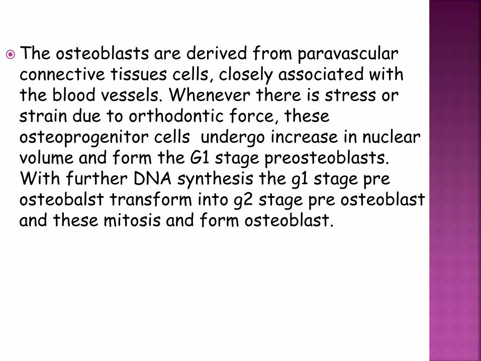
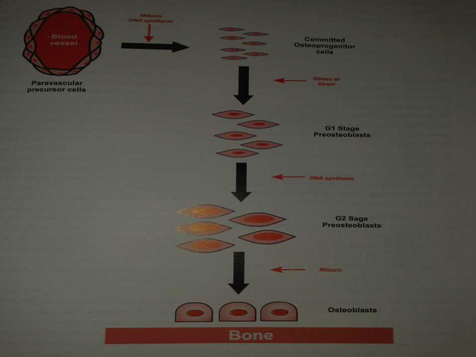
The osteoblasts are derived from paravascular connective tissues cells, closely associated with the blood vessels. Whenever there is stress or strain due to orthodontic force, these osteoprogenitor cells undergo increase in nuclear volume and form the G1 stage preosteoblasts. With further DNA synthesis the g1 stage pre osteobalst transform into g2 stage pre osteoblast and these mitosis and form osteoblast.
The bone formed passes through 3 stages they are THE OSTEOID, BUNDLE BONE and THE LAMELLATED BONE.
The bone formed by the osteoblast is the osteoid which is tightly calcified with further calcification it forms bundle bone . the fibres of the periodontal apparatus that attached to the bundle bone. When the bundle bone reaches a certain maturity part of it get re organized into mature lamellated bone.

It is brought about by cells called osteoclasts . they are multinucleated giant cells and may have 12 or more nuclei . they are irregularly oval or club shaped with branching processes . they occur in a bay like depressions in bone called howships lacunae and have prominent mitochondria, lisosomes and vacuoles. Each of their nuclei has a single nucleolus. The part of osteoclasts in contact with the resorbing bone has a ruffeled border
The osteoclasts derived from :
Activation of previously present inactive osteoclasts
Migration from adjacent bone
Formation of new osteoclasts from local macrophages of periodontal ligament
Influx of monocytes from blood vessels
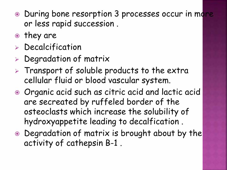
During bone resorption 3 processes occur in more or less rapid succession .
they are
Decalcification
Degradation of matrix
Transport of soluble products to the extra cellular fluid or blood vascular system.
Organic acid such as citric acid and lactic acid are secreated by ruffeled border of the osteoclasts which increase the solubility of hydroxyappetite leading to decalfication .
Degradation of matrix is brought about by the activity of cathepsin B-1 .
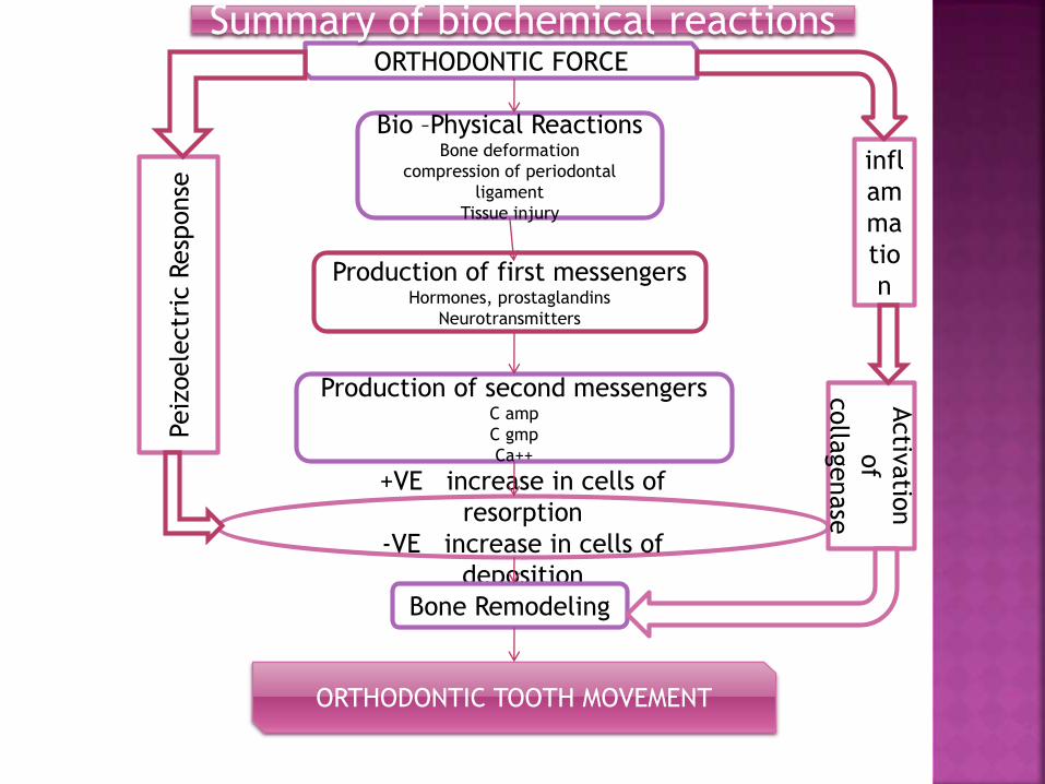
Bio –Physical ReactionsBone deformation
compression of periodontal
ligament
Tissue injury
Production of first messengersHormones, prostaglandins
Neurotransmitters
Production of second messengersC amp
C gmp
Ca++
+VE increase in cells of
resorption
-VE increase in cells of
deposition
Bone Remodeling
ORTHODONTIC TOOTH MOVEMENT
ORTHODONTIC FORCE
infl
am
ma
tio
n
Activ
atio
n
of
colla
genase
Peiz
oele
ctr
icResp
onse
Summary of biochemical reactions